retro orbital injections
DESCRIPTION
ProtocolTRANSCRIPT

Retro-orbital injections in mice
Tal Yardeni, MSc1,2, Michael Eckhaus, VMD, DACVP3, H. Douglas Morris, PhD, DABMP4,Marjan Huizing, PhD1, and Shelley Hoogstraten-Miller, DVM, MS, DACLAM5
1Medical Genetics Branch, National Human Genome Research Institute, National Institutes ofHealth, Bethesda, MD.2Graduate Partner Program, Sackler School of Medicine, Tel Aviv University, Tel Aviv, Israel.3Division of Veterinary Resources, Office of Research Services, National Institutes of Health,Bethesda, MD.4Mouse Imaging Facility, National Institute of Neurological Disorders and Stroke, NationalInstitutes of Health, Bethesda, MD.5Office of Laboratory Animal Medicine, National Human Genome Research Institute, NationalInstitutes of Health, Bethesda, MD.
AbstractIntravenous vascular access is technically challenging in the adult mouse and even morechallenging in neonatal mice. The authors describe the technique of retro-orbital injection of thevenous sinus in the adult and neonatal mouse. This technique is a useful alternative to tail veininjection for the administration of non-tumorigenic compounds. The authors report that they haveroutinely used this technique in the adult mouse to administer volumes up to 150 µl withoutincident. Administration of retro-orbital injections is more challenging in neonatal mice but canreliably deliver volumes up to 10 µl.
For intravascular delivery of agents to the adult mouse, personnel typically use the lateraltail veins1. This procedure is technically challenging and often has a high rate of failure. Inpreparation for tail vein venipuncture, the mouse is often placed under a heat lamp topromote peripheral vasodilation and is then mechanically restrained1. In our opinion, thisapproach can cause distress in the animals, especially if the initial venipuncture isunsuccessful and repeated attempts are made.
Intravenous injections in the neonatal mouse are even more challenging. Percutaneousinjection of the superficial temporal vein has been described2,3, but this method has severaldisadvantages. First, it requires two people3. Second, owing to increased pigmentation andskin thickness in older neonates, it might not be useful for mice that are older than 4 d (ref.3). Third, it is technically challenging.
Retro-orbital injections may seem aesthetically distasteful, but we argue that this easilymastered technique is ultimately more humane than alternative intravascular injectiontechniques in mice. We routinely use this method for bone marrow transplantation4,leukemia induction5, administration of experimental compounds6 and gene therapy7.Although researchers have reported using this technique on both neonatal and adult
Correspondence should be addressed to S.H.-M. ([email protected]).COMPETING FINANCIAL INTERESTSThe authors declare no competing financial interests.
NIH Public AccessAuthor ManuscriptLab Anim (NY). Author manuscript; available in PMC 2011 August 19.
Published in final edited form as:Lab Anim (NY). 2011 May ; 40(5): 155–160. doi:10.1038/laban0511-155.
NIH
-PA Author Manuscript
NIH
-PA Author Manuscript
NIH
-PA Author Manuscript

mice8–11, we have not found a thorough description of the use of this technique in adults andneonates in the literature. Here we describe our technique for retro-orbital injections in adultand neonatal mice.
The procedures described below were approved by the National Human Genome ResearchInstitute (NHGRI) ACUC. These procedures were carried out in a facility accredited by theAssociation for Assessment and Accreditation of Laboratory Animal Care International inaccordance with the Guide for the Care and Use of Laboratory Animals12.
ADULT MOUSE INJECTION TECHNIQUEWe prepare the syringes first. We prefer to use a 27.5-gauge (or smaller), 0.5-in insulinneedle and syringe (e.g., Terumo U-100, Terumo Medical Corporation, Elkton, MD). Atuberculin syringe with a 27-gauge (or smaller), 0.5-in hypodermic needle can also be used.When using a tuberculin syringe, a large volume (50–70 µl; ref. 13) of the injectate is lost inthe dead space of the syringe hub and needle, which is important to keep in mind if theinjectate is valuable. We recommend that injectate volumes not exceed 150 µl.
Because the needle is being placed in the retro-bulbar space (the region behind the globe ofthe eye) the mouse should be anesthetized so that it remains still during the procedure. Weprefer to use an inhalant anesthetic, because of its rapid induction and recovery times. Weplace the mouse in a plexiglas chamber and use isoflurane to induce anesthesia. The size ofthe plexiglas chamber we use varies depending on the procedure space being used. Wesometimes use a plexiglas chamber that is 10 in wide × 4 in high × 4.5 in deep (HarvardApparatus, Holliston, MA).
We then place a funnel-shaped nose cone connected to a non-rebreathing apparatus(Surgivet, Dublin, OH) on the anesthetized mouse. We use a down-draft table or a charcoalscavenging device to manage waste anesthetic gases and decrease exposure of personnel tothe gases. A down-draft table pulls the inhalant gas away, whereas a charcoal scavengingdevice absorbs the gas. To decrease the likelihood of the mouse developing hypothermia, themouse can be placed on a warming device, such as a disposable hand warmer (GrabberWarmer, Grabber Inc., Grand Rapids, MI; Fig. 1). Because the surface temperature of thehand warmer can approach 130–150 °F, the warming device should be covered with aprotective layer of paper toweling or gauze to prevent thermal injury to the mouse.
A right-handed operator will probably find it easiest to administer the injection into the rightretro-orbital sinus of the mouse. The mouse is placed in left lateral recumbency with its headfacing to the right. The operator then partially protrudes the mouse’s right eyeball from theeye socket by applying gentle pressure to the skin dorsal and ventral to the eye (Fig. 2). Boththe trachea and the ventral cervical (neck) vessels (the jugular veins and carotid arteries) runalong the ventral cervical area. The veins drain the head area and the arteries supply thisarea. Care must be taken not to apply excessive pressure to the ventral cervical vessels,because this could impede blood flow and impede the injection. It is also important not toapply pressure to the trachea, because this could cause the trachea to collapse, therebystopping the mouse from being able to breathe.
In addition to using the inhalant anesthetic, we also place a drop of ophthalmic anesthetic(0.5% proparacaine hydrochloride ophthalmic solution, Alcon Laboratories, Inc., FortWorth, TX) on the eye that will receive the injection (Fig. 3). This provides additionalprocedural and post-procedural analgesia. While being careful not to scratch the cornea, theoperator can remove excess ophthalmic anesthetic by holding an absorbent gauze pad to themedial canthus. The needle is carefully introduced, bevel down, at an angle ofapproximately 30°, into the medial canthus (Fig. 4). Most injections are carried out with the
Yardeni et al. Page 2
Lab Anim (NY). Author manuscript; available in PMC 2011 August 19.
NIH
-PA Author Manuscript
NIH
-PA Author Manuscript
NIH
-PA Author Manuscript
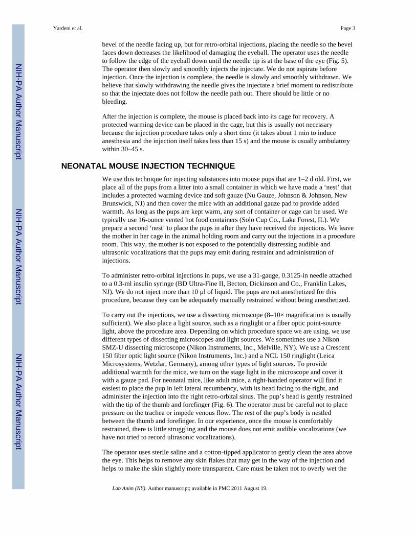
bevel of the needle facing up, but for retro-orbital injections, placing the needle so the bevelfaces down decreases the likelihood of damaging the eyeball. The operator uses the needleto follow the edge of the eyeball down until the needle tip is at the base of the eye (Fig. 5).The operator then slowly and smoothly injects the injectate. We do not aspirate beforeinjection. Once the injection is complete, the needle is slowly and smoothly withdrawn. Webelieve that slowly withdrawing the needle gives the injectate a brief moment to redistributeso that the injectate does not follow the needle path out. There should be little or nobleeding.
After the injection is complete, the mouse is placed back into its cage for recovery. Aprotected warming device can be placed in the cage, but this is usually not necessarybecause the injection procedure takes only a short time (it takes about 1 min to induceanesthesia and the injection itself takes less than 15 s) and the mouse is usually ambulatorywithin 30–45 s.
NEONATAL MOUSE INJECTION TECHNIQUEWe use this technique for injecting substances into mouse pups that are 1–2 d old. First, weplace all of the pups from a litter into a small container in which we have made a ‘nest’ thatincludes a protected warming device and soft gauze (Nu Gauze, Johnson & Johnson, NewBrunswick, NJ) and then cover the mice with an additional gauze pad to provide addedwarmth. As long as the pups are kept warm, any sort of container or cage can be used. Wetypically use 16-ounce vented hot food containers (Solo Cup Co., Lake Forest, IL). Weprepare a second ‘nest’ to place the pups in after they have received the injections. We leavethe mother in her cage in the animal holding room and carry out the injections in a procedureroom. This way, the mother is not exposed to the potentially distressing audible andultrasonic vocalizations that the pups may emit during restraint and administration ofinjections.
To administer retro-orbital injections in pups, we use a 31-gauge, 0.3125-in needle attachedto a 0.3-ml insulin syringe (BD Ultra-Fine II, Becton, Dickinson and Co., Franklin Lakes,NJ). We do not inject more than 10 µl of liquid. The pups are not anesthetized for thisprocedure, because they can be adequately manually restrained without being anesthetized.
To carry out the injections, we use a dissecting microscope (8–10× magnification is usuallysufficient). We also place a light source, such as a ringlight or a fiber optic point-sourcelight, above the procedure area. Depending on which procedure space we are using, we usedifferent types of dissecting microscopes and light sources. We sometimes use a NikonSMZ-U dissecting microscope (Nikon Instruments, Inc., Melville, NY). We use a Crescent150 fiber optic light source (Nikon Instruments, Inc.) and a NCL 150 ringlight (LeicaMicrosystems, Wetzlar, Germany), among other types of light sources. To provideadditional warmth for the mice, we turn on the stage light in the microscope and cover itwith a gauze pad. For neonatal mice, like adult mice, a right-handed operator will find iteasiest to place the pup in left lateral recumbency, with its head facing to the right, andadminister the injection into the right retro-orbital sinus. The pup’s head is gently restrainedwith the tip of the thumb and forefinger (Fig. 6). The operator must be careful not to placepressure on the trachea or impede venous flow. The rest of the pup’s body is nestledbetween the thumb and forefinger. In our experience, once the mouse is comfortablyrestrained, there is little struggling and the mouse does not emit audible vocalizations (wehave not tried to record ultrasonic vocalizations).
The operator uses sterile saline and a cotton-tipped applicator to gently clean the area abovethe eye. This helps to remove any skin flakes that may get in the way of the injection andhelps to make the skin slightly more transparent. Care must be taken not to overly wet the
Yardeni et al. Page 3
Lab Anim (NY). Author manuscript; available in PMC 2011 August 19.
NIH
-PA Author Manuscript
NIH
-PA Author Manuscript
NIH
-PA Author Manuscript
pup, because this could increase the risk of hypothermia. We do not use alcohol or a topicalophthalmic anesthetic. The ophthalmic anesthetic will not penetrate the skin, and we thinkthat alcohol might irritate the pup’s facial skin. The operator then inserts the needle, beveldown, at the ‘3 o’clock’ position into the eye socket (the area that will become the medialcanthus) at an angle of approximately 30° (Fig. 7). The operator mentally visualizes the backof the socket and advances the needle to the area of the retro-orbital sinus. The injection ismade in a gentle, smooth, fluid motion. If the injection is successful, the operator mightobserve blanching of the superficial temporal vein, but this does not always occur.Regardless of whether blanching is noted, we have seen the injectate in the target organs7.The needle should be withdrawn slowly, allowing a brief moment for the injectate toredistribute. We sometimes see a small drop of blood at the injection site, which can begently cleaned with a cotton-tipped applicator. The operator then places the pup in thesecond prepared nest. When all the pups in a group have received injections, the pups arechecked for any additional bleeding and cleaned, if necessary. We then return the pups totheir mother in the home cage.
RETRO-ORBITAL SINUS MORPHOLOGYAn exact description of the retro-orbital sinus of the mouse seems to be lacking in theliterature, but it may be best described as a confluence, pool or sinus of several vessels,likely including the supraorbital vein, inferior palpebral vein, dorsal nasal vein and thesuperficial temporal veins (Fig. 8; refs. 14,15). We have been able to visualize the retro-orbital sinus area as a large area with a substantial amount of blood flow (Fig. 9) by using X-ray computed tomography (CT; SkyScan 1172 MicroCT; SkyScan, Kontich, Belgium) andan intravascular X-ray CT contrast agent (Fenestra VC; ART Advanced ResearchTechnologies, Inc., Montreal, Canada). We acquired the CT data with an acceleratingvoltage of 51 kV, a 0.5-mm aluminum filter and a true three-dimensional image resolutionof 6.5 µm.
To assess the accuracy of the injections, we have collected post-mortem histological samplesfrom neonates (Fig. 10) and adults (Fig. 11) at 30 min and 7 d after these mice receivedretro-orbital injections. We have observed some leakage of the injectate from and around thesinus. However, the various experiments in which we use this technique have reported 100%success in adults and neonates. This indicates that the injectate reached its target site 100%of the time. We say that we have achieved 100% success because we see the expectedoutcome (for example, leukemia develops when we inject leukemic cells) 100% of the time.
DISCUSSIONResearchers have published studies comparing the effectiveness of retro-orbital injectionsand tail vein injections in adult mice10,11. These reviews have shown that the two routes canbe used interchangeably and that both routes are equally effective10,11. Retro-orbitalinjections in neonates have also been mentioned8,9 but, to our knowledge, not described indetail.
We think that retro-orbital injection is an easy and reliable method for intravascular deliveryof many agents. We have used this method to inject substances into hundreds of adult mice,without incident. We have also used this method to administer intra-orbital injections inmore than 50 litters (more than 400 pups (1–2 d old)) with complications rarely observed.Occasionally, we have noted a temporary paleness or cyanosis in a pup that has received aninjection, which we attribute to a combination of the restraint and the injection itself. Theseside effects could perhaps be lessened or alleviated by warming the injectate to bodytemperature instead of administering the injectate at room temperature. We have never
Yardeni et al. Page 4
Lab Anim (NY). Author manuscript; available in PMC 2011 August 19.
NIH
-PA Author Manuscript
NIH
-PA Author Manuscript
NIH
-PA Author Manuscript

observed this phenomenon in adults, in which we routinely inject compounds at roomtemperature. We also occasionally inject compounds into adult mice that must be maintainedon ice until just before the injection. We allow these to warm up only briefly before weinject them. We have not experienced any problems with maternal rejection of any pup thathas received a retro-orbital injection. This could be due to the precautions we take inseparating the mothers from their pups before the injections or to the good maternal abilitiesof the particular dams.
As with any intravenous route, injection volume is limited. Although researchers havedescribed administering intravenous injection volumes of 200–300 µl for adults16,17, wethink that injectate volumes should not exceed 150 µl for adults. In an ‘average’ 30-gmouse, the estimated blood volume is approximately 2.2 ml (7.2% of the body weight16).An injection volume of 200–300 µl, even if given slowly, may result in temporary vascularoverload. A newborn mouse pup weighs approximately 1–1.5 g. Even though neonates haveslightly higher blood volume per unit of body weight than do adult mice18, the circulatingblood volume in a neonatal mouse probably does not exceed 0.08 ml (80 µl). In light of thisand the very small retro-orbital space in a pup, we feel that a maximum injection volume of10 µl in the neonate is reasonable.
Post-mortem histological samples from neonates (Fig. 10) and adults (Fig. 11) that receivedretro-orbital injections showed normal morphology of the injected sinus and surroundingtissue. Because there can be leakage of the injectate from and around the sinus, retro-orbitalinjection might not be an acceptable route by which to carry out solid tumor transplantation.If the retro-orbital route is used for injecting cell suspensions, only single-cell suspensionscan be used. Personnel can make a one-cell suspension by using a 70-µm cell strainer (BDFalcon, BD Biosciences, Bedford, MA) to filter the suspension before using it. Failure tofilter cell suspensions could lead to obstruction of vessels supplying critical organs andsubsequent death of the animal.
Although we believe that retro-orbital injection is an easy and reliable method forintravascular delivery of various compounds in mice, a novice operator must receivesufficient training on cadavers and terminally anesthetized mice before using the techniquein live mice. For initial training, we recommend administering injections of dyes (e.g., Indiaink, new methylene blue) or microbeads (6-µm Polybead, Polysciences Inc., Warrington,PA) that are approximately the size of a mouse red blood cell, because these allow thenovice to see the location of the injection.
In conclusion, retro-orbital sinus injection of both neonatal and adult mice is a usefulmethod for intravascular delivery of many agents. In the hands of a skilled operator, theincidence of complications is rare and distress to the animal is minimized. We believe this isa useful alternative method for intravascular administration.
AcknowledgmentsWe thank Brenda Klaunberg and Danielle Donahue from the NIH Mouse Imaging Facility and Darryl Leja from theNHGRI Intramural Publication Support Office for their assistance with this project. This work was carried out inpartial fulfillment of the requirements for a PhD degree for T.Y. (Sackler Faculty of Medicine, Tel Aviv University,Tel Aviv, Israel). This research was supported in part by the Intramural Research Program of the NHGRI, theNational Institutes of Health.
References1. Suckow, MA.; Danneman, P.; Brayton, C. The Laboratory Mouse. Suckow, MA., editor. Roca
Raton, FL: CRC; 2001.
Yardeni et al. Page 5
Lab Anim (NY). Author manuscript; available in PMC 2011 August 19.
NIH
-PA Author Manuscript
NIH
-PA Author Manuscript
NIH
-PA Author Manuscript
2. Billingham RE, Brent L. Acquired tolerance of foreign cells in newborn animals. Proc. R. Soc.Lond. B. Biol. Sci. 1956; 146:78–90. [PubMed: 13379354]
3. Sands MS, Barker JE. Percutaneous intravenous injection in neonatal mice. Lab. Anim. Sci. 1999;49:328–330. [PubMed: 10403452]
4. Cannons JL, et al. Optimal germinal center responses require a multistage T cell:B cell adhesionprocess involving integrins, SLAM-associated protein, and CD84. Immunity. 2010; 32:253–265.[PubMed: 20153220]
5. Hyde RK, et al. Cbfb/Runx1 repression-independent blockage of differentiation and accumulationof Csf2rb-expressing cells by Cbfb-MYH11. Blood. 2010; 115:1433–1443. [PubMed: 20007544]
6. Song H, et al. Mammalian Mst1 and Mst2 kinases play essential roles in organ size control andtumor suppression. Proc. Natl. Acad. Sci. USA. 2010; 107:1431–1436. [PubMed: 20080598]
7. Yardeni, T., et al. A non-viral, GNE-Lipoplex treatment to correct sialylation defects in Gne-mutant(M712T) mice American Society of Gene Cell Therapeutics 2010 Annual Meeting; Washington,DC. Abstract #144
8. Jerbtsova M, Liu XH, Ye X, Ray PE. Adenovirus-mediated gene transfer to glomerular cells innewborn mice. Pediatr. Nephrol. 2005; 20:1395–1400. [PubMed: 16133067]
9. Jerbtsova M, Ye X, Ray PE. A simple technique to establish a long-term adenovirus mediated genetransfer to the heart of newborn mice. Cardiovasc. Hematol. Disord. Drug Targets. 2009; 9:136–140. [PubMed: 19519372]
10. Price JE, Barth RF, Johnson CW, Staubus AE. Injection of cells and monoclonal antibodies intomice: comparison of tail vein and retroorbital routes. Proc. Soc. Exp. Biol. Med. 1984; 177:347–353. [PubMed: 6091149]
11. Steel CW, Stephens AL, Hahto SM, Singletary SJ, Ciavarra RP. Comparison of the lateral tail veinand the retro-orbital venous sinus as routes of intravenous drug delivery in a transgenic mousemodel. Lab Anim. (NY). 2008; 37:26–32. [PubMed: 18094699]
12. Institute of Laboratory Animal Resources. Washington, DC: National Academy Press; 1996. Guidefor the Care and Use of Laboratory Animals.
13. Bhambhani V, Beri RS, Puliyel JM. Inadvertent overdosing of neonates as a result of the deadspace of the syringe hub and needle. Arch. Dis. Child Fetal Neonatal. Ed. 2005; 90:F444–F445.[PubMed: 16113158]
14. Cook, MJ. The Anatomy of the Laboratory Mouse. New York: Academic; 1965. p. 96-98.15. Timm KI. Orbital venous anatomy of the rat. Lab. Anim. Sci. 1979; 29:636–638. [PubMed:
513633]16. Fox, JG., et al., editors. The Mouse in Biomedical Research. 2nd edn.. Vol. vol. III. Oxford, UK:
Elsevier; 2007.17. Bauck, L.; Bihun, C. Basic anatomy, physiology, husbandry, and clinical techniques. In: Hillyer,
EV.; Quesenberry, KE., editors. Ferrets, Rabbits, and Rodents: Clinical Medicine and Surgery.Philadelphia, PA: W.B. Saunders; 1997. p. 303
18. Jain, NC., editor. Veterinary Hematology. 4th edn.. Philadelphia, PA: Lee & Febiger; 1986.
Yardeni et al. Page 6
Lab Anim (NY). Author manuscript; available in PMC 2011 August 19.
NIH
-PA Author Manuscript
NIH
-PA Author Manuscript
NIH
-PA Author Manuscript
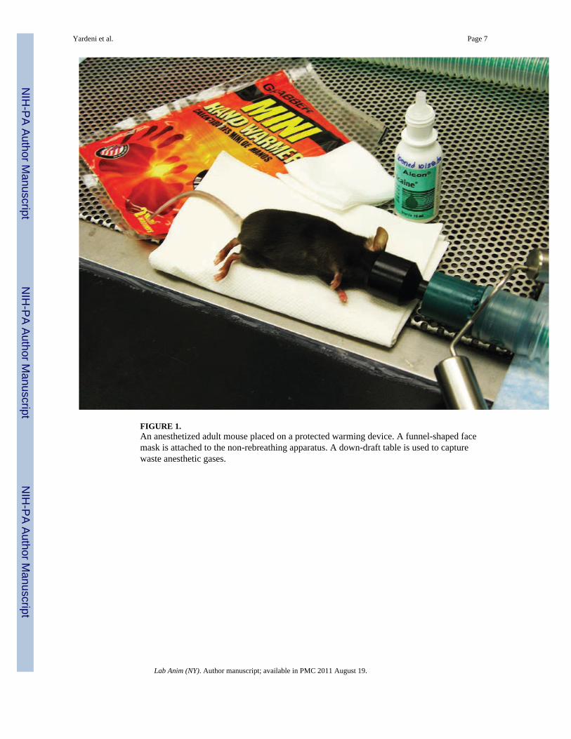
FIGURE 1.An anesthetized adult mouse placed on a protected warming device. A funnel-shaped facemask is attached to the non-rebreathing apparatus. A down-draft table is used to capturewaste anesthetic gases.
Yardeni et al. Page 7
Lab Anim (NY). Author manuscript; available in PMC 2011 August 19.
NIH
-PA Author Manuscript
NIH
-PA Author Manuscript
NIH
-PA Author Manuscript
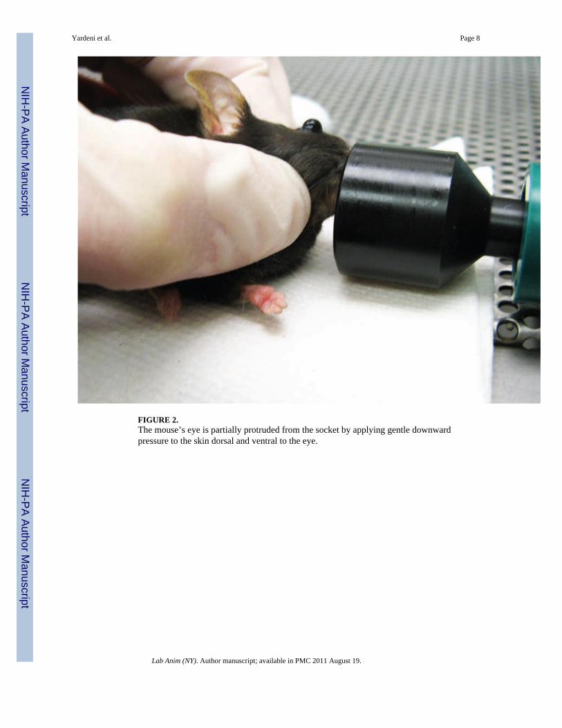
FIGURE 2.The mouse’s eye is partially protruded from the socket by applying gentle downwardpressure to the skin dorsal and ventral to the eye.
Yardeni et al. Page 8
Lab Anim (NY). Author manuscript; available in PMC 2011 August 19.
NIH
-PA Author Manuscript
NIH
-PA Author Manuscript
NIH
-PA Author Manuscript

FIGURE 3.The operator places a drop of topical ophthalmic anesthetic on the eye of the mouse beforecarrying out the injection.
Yardeni et al. Page 9
Lab Anim (NY). Author manuscript; available in PMC 2011 August 19.
NIH
-PA Author Manuscript
NIH
-PA Author Manuscript
NIH
-PA Author Manuscript
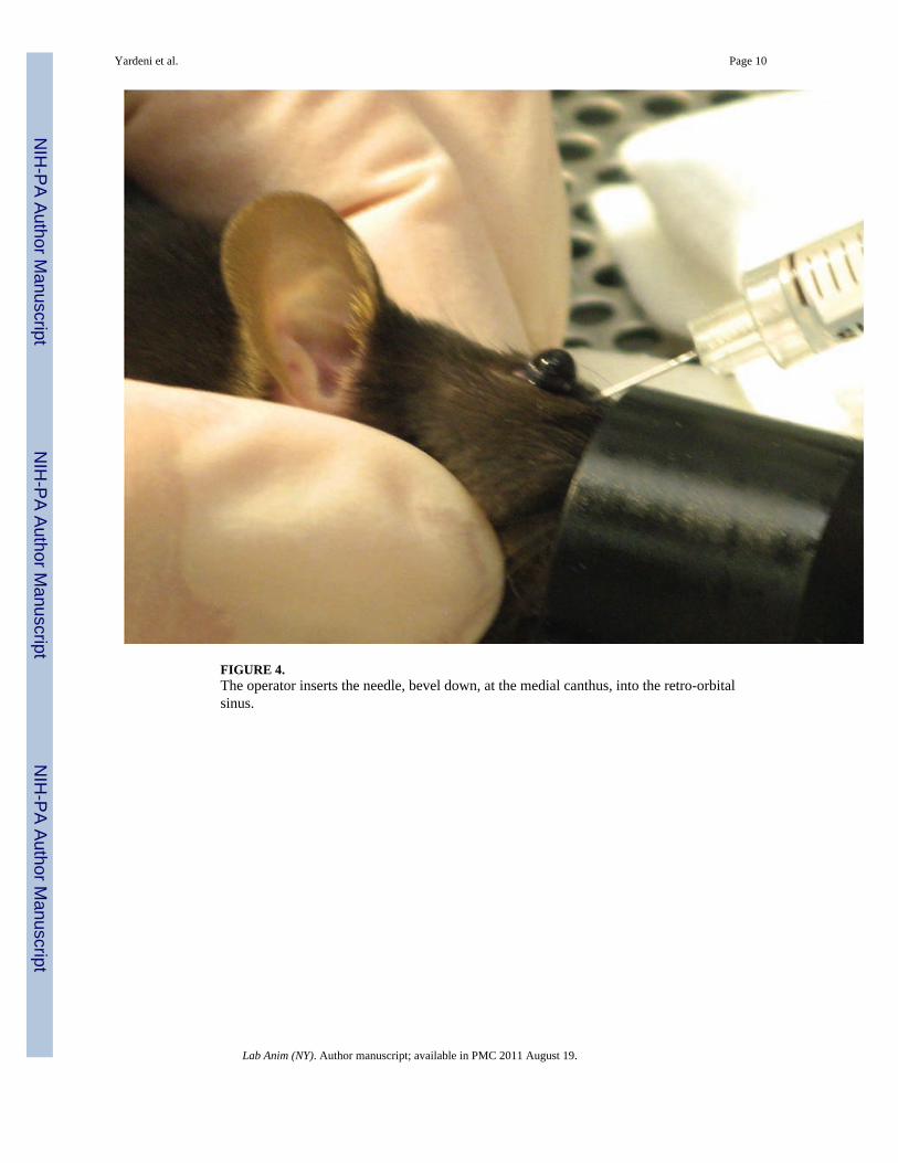
FIGURE 4.The operator inserts the needle, bevel down, at the medial canthus, into the retro-orbitalsinus.
Yardeni et al. Page 10
Lab Anim (NY). Author manuscript; available in PMC 2011 August 19.
NIH
-PA Author Manuscript
NIH
-PA Author Manuscript
NIH
-PA Author Manuscript

FIGURE 5.Correct placement of the needle relative to the retro-orbital sinus, the eyeball and the back ofthe orbit. Illustration by Darryl Leja.
Yardeni et al. Page 11
Lab Anim (NY). Author manuscript; available in PMC 2011 August 19.
NIH
-PA Author Manuscript
NIH
-PA Author Manuscript
NIH
-PA Author Manuscript
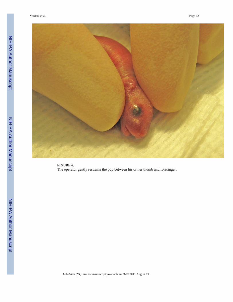
FIGURE 6.The operator gently restrains the pup between his or her thumb and forefinger.
Yardeni et al. Page 12
Lab Anim (NY). Author manuscript; available in PMC 2011 August 19.
NIH
-PA Author Manuscript
NIH
-PA Author Manuscript
NIH
-PA Author Manuscript

FIGURE 7.The operator inserts the needle, bevel down, at a 30° angle in the area that will become themedial canthus.
Yardeni et al. Page 13
Lab Anim (NY). Author manuscript; available in PMC 2011 August 19.
NIH
-PA Author Manuscript
NIH
-PA Author Manuscript
NIH
-PA Author Manuscript

FIGURE 8.The blood vessels contributing to the retro-orbital sinus of the mouse. Illustration by DarrylLeja.
Yardeni et al. Page 14
Lab Anim (NY). Author manuscript; available in PMC 2011 August 19.
NIH
-PA Author Manuscript
NIH
-PA Author Manuscript
NIH
-PA Author Manuscript
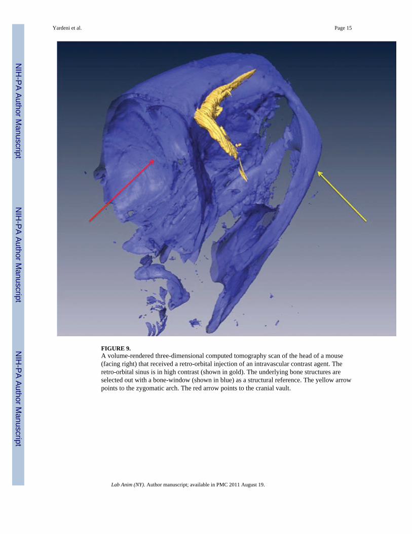
FIGURE 9.A volume-rendered three-dimensional computed tomography scan of the head of a mouse(facing right) that received a retro-orbital injection of an intravascular contrast agent. Theretro-orbital sinus is in high contrast (shown in gold). The underlying bone structures areselected out with a bone-window (shown in blue) as a structural reference. The yellow arrowpoints to the zygomatic arch. The red arrow points to the cranial vault.
Yardeni et al. Page 15
Lab Anim (NY). Author manuscript; available in PMC 2011 August 19.
NIH
-PA Author Manuscript
NIH
-PA Author Manuscript
NIH
-PA Author Manuscript

FIGURE 10.Section through the right eye and surrounding tissue of a 1-d-old mouse pup 30 min afteradministration of a retro-orbital injection (12.5× magnification; hematoxylin and eosinstaining). The inset shows the injected retro-orbital sinus (100× magnification). The retro-orbital sinus (black arrows) and surrounding tissue appear normal. Red arrows point to theHarderian gland. Blue arrow points to the corneal surface of the eyeball. Yellow arrowpoints to the fused eyelids.
Yardeni et al. Page 16
Lab Anim (NY). Author manuscript; available in PMC 2011 August 19.
NIH
-PA Author Manuscript
NIH
-PA Author Manuscript
NIH
-PA Author Manuscript
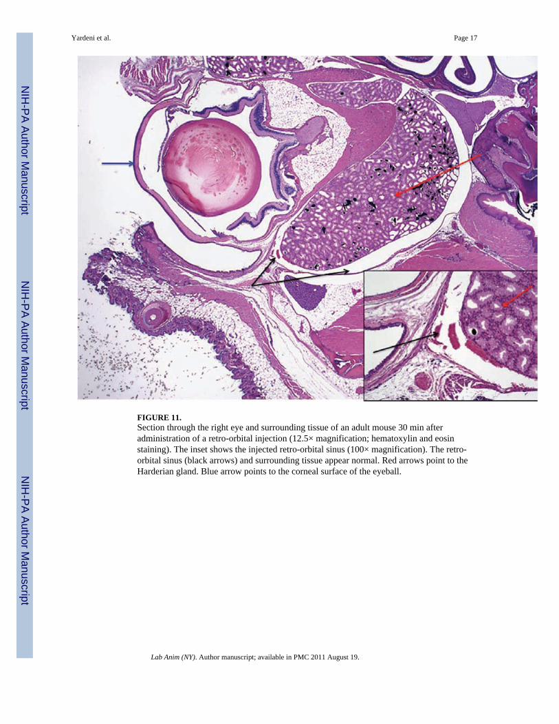
FIGURE 11.Section through the right eye and surrounding tissue of an adult mouse 30 min afteradministration of a retro-orbital injection (12.5× magnification; hematoxylin and eosinstaining). The inset shows the injected retro-orbital sinus (100× magnification). The retro-orbital sinus (black arrows) and surrounding tissue appear normal. Red arrows point to theHarderian gland. Blue arrow points to the corneal surface of the eyeball.
Yardeni et al. Page 17
Lab Anim (NY). Author manuscript; available in PMC 2011 August 19.
NIH
-PA Author Manuscript
NIH
-PA Author Manuscript
NIH
-PA Author Manuscript