retracted: one-step synthesis of au–ag alloy nanoparticles by a convenient electrochemical method
TRANSCRIPT

ARTICLE IN PRESS
1386-9477/$ - se
doi:10.1016/j.ph
�Correspondsity, Jinan 2501
fax: +86531 88
E-mail addr
Physica E 33 (2006) 28–34
www.elsevier.com/locate/physe
One-step synthesis of Au–Ag alloy nanoparticles by a convenientelectrochemical method
Min Zhoua, Shenhao Chena,b,�, Shiyong Zhaoa, Houyi Maa
aDepartment of Chemistry, Shandong University, Jinan 250100, PR ChinabState Key Laboratory for Corrosion and Protection, Shenyang 110016, PR China
Received 9 September 2005; accepted 14 October 2005
Available online 20 March 2006
Abstract
Gold–silver alloy nanoparticles with various mole ratios were synthesized in aqueous solution by an electrochemical co-reduction of
chloroauric acid (HAuCl4) and silver nitrate (AgNO3) in the presence of poly(vinyl-pyrrolidone) (PVP) used as a protecting agent. The
UV–visible absorption spectra of the bimetallic systems and the physical mixtures of individual metallic nanoparticles suggested the
formation of alloy nanoparticles. The absorption maximum of the plasmon band shifts linearly to the red with increasing Au content.
The size distribution of the alloy nanoparticles was examined using transmission electron microscopy. The EDX analysis on individual
particles confirmed the presence of two metals in a particle. The X-ray diffraction also confirmed the formation of homogeneous
gold–silver nanostructure. The effects of the preparation conditions including the concentration of polymer stabilizer (PVP), the molar
ratio of HAuCl4 and AgNO3 and reaction time were investigated. Also, the formation process for bimetallic nanoparticles was discussed.
r 2006 Elsevier B.V. All rights reserved.
PACS: 61.46.+w; 82.45.Aa
Keywords: Electrochemical synthesis; Alloy nanoparticles; Polymer stabilizer
ACTED
R
1. IntroductionNanoparticles have drawn considerable interest invarious fields of science and engineering because of theirunique physical and chemical properties, leading topotential applications in electronics, for optical andmagnetic devices [1–6]. Metal nanoparticles have beenobtained by thermal [7], photochemical [8], radiolytic [9],sonochemical [10] or electrochemical [11] methods byusing various reagents. It has been confirmed thatelectrochemical methods have some advantages overtradition methods in synthesis of metal nanoparticles[12,13]. Reetz and Helbig [11] developed a sacrificial anodemethod to prepare size-selective metal particles in an
RET
e front matter r 2006 Elsevier B.V. All rights reserved.
yse.2005.10.012
ing author. Department of Chemistry, Shadong Univer-
00, PR China. Tel.: +86 531 88364959;
565167.
ess: [email protected] (S. Chen).
organic phase. In this method, tetra-alkylammonium saltsserved as the supporting electrolyte and stabilizer for themetal nanoclusters. The salient features of this methodincluded ease of operation, high yield, and the absence ofundesired side products: these features are especially goodfor the wide application of this method to the electro-chemical synthesis of metallic nanostructured materials.For example, Yu et al. [14] and Mohamed et al. [15]synthesized gold nanorods in aqueous solution via thiselectrochemical method by introducing a shape-inducingcosurfactant; Rodrigues-Sanchez et al. [13] prepared silvernanoparticles, ranging in size from 2 to 7 nm, in acetonitrilein an analogous manner. Small metal particles are the highpurity of the particles and the possibility of a preciseparticle size control can be achieved by adjusting currentdensity or applied potential. Recently [16,17], we synthe-sized size-controlled silver, gold and platinum nanoparti-cles by using a novel rotating cathode electrochemicaltechnique.

ARTICLE IN PRESSM. Zhou et al. / Physica E 33 (2006) 28–34 29
A
D
Bimetallic nanoparticles have drawn considerable inter-est in various fields of science and engineering because oftheir catalytic, electronic, and optical properties distinctfrom those of corresponding monometallic nanoparticles[18–21]. Depending on the synthesis method, the resultingbimetallic particles can exhibit alloy behavior or core–shellsystem behavior. The alloy nanoparticles are homoge-neously distributed over the whole volume on an atomicscale; however, the core–shell metal nanoparticles consti-tute the core of the structure, and the other one the externalshell. Generally, bimetallic nanoparticles can be preparedby simultaneous reduction or by successive reduction oftwo metal ions in the presence of suitable stabilizationstrategy such as steric hindrance and static-electronicrepulsive force. The former reduction methods mayobtained a particle structure of homogeneous alloy[22–30], while the latter are the purpose for the productionof core–shell structure particles [31–33]. Au and Ag havevery similar lattice constants and are completely miscibleover the entire composition range [34]. Hence, single-phasealloys can be achieved with any desired composition.Several reports have appeared describing the synthesis ofAu–Ag alloy nanoparticles. For example, Sampath et al.prepared mercaptopropionic acid (MPA) capped Au–Agalloy nanoparticles consisting of various mole fractions ofgold and silver in aqueous medium using a citratereduction method [22]. Hostetler and co-workers describeda synthesis of alkanethiolate monolayer-protected gold–silver alloy nanoparticles that can be isolated in solvent-free forms and redissolved without change [23]. Shi and co-workers have reported the formation of Au–Ag alloynanoparticles in monolithic mesoporous silica where aminogroups are used to stabilize the alloy particles [24]. Esumiand co-workers have prepared Au–Ag alloy phases inlaponite suspensions [25]. Hartland et al. [26] preparedgold–silver nanoparticles with a core–shell structure byradiation chemistry, and laser-induced heating is used tomelt the AucoreAgshell particles into homogeneous alloyednanoparticles. Chen and Yeh [27] synthesized gold–silveralloy nanoparticles by laser irradiation of mixturesconsisting of gold and silver nanoparticles. Murphysynthesized gold–silver alloy nanoparticles via reductionof varying mole ratio of HAuCl4 and AgNO3 by sodiumborohydride in the presence of sodium citrate as a cappingagent, in water [28]. Liu et al. [29] reported that Au–Agalloy nanoparticles in HCl aqueous solutions without theaddition of any stabilizer had been synthesized bysonoelectrochemical method. Izgaliev et al. [30] describedAu–Ag alloy formation under laser irradiation of a mixtureof the monometallic colloidal solutions.
In this paper, we report that the Au–Ag alloynanoparticles were prepared by the electrochemical reduc-tion of the two metal ions, and the nanoparticles wereformed in aqueous solutions in the presence of polymerstabilization. In this process, we present UV–visiblespectroscopy, transmission electron microscopy, energy-dispersive X-ray analysis (EDX) and X-ray diffraction
RETR
(XRD) evidence for the electrochemical formation of thealloy nanoparticles stabilized by PVP. The influence ofpreparation parameters on the particle size and sizedistribution is also discussed.
2. Experimental section
2.1. Materials
PVP [Poly(vinyl-pyrrolidone)] K30 [weight-average mo-lecular weight (MW),30000] was supplied from BASF Co.Germany. Chloroauric acid (HAuCl4), silver nitrate(AgNO3) and potassium nitrate (KNO3) was purchasedfrom Chinese Shanghai Chemical Company. All chemicalswere of analytical grade and used without further purifica-tion. Double-distilled water was used throughout theexperiment.
2.2. Preparation of Au–Ag alloy nanoparticles
The electrochemical synthesis of bimetallic nanoparticleswas performed in a simple two-electrode cell. Two1.0 cm� 1.0 cm platinum sheets (Aldrich, 99.9%) being5 cm apart were employed to prepare the electrolysiselectrodes. The electrolysis was carried out in the potentio-static manner by using an EG&G M173 potentiostat/galvanostat at room temperature (�20 1C) under stirring.Typically, 5ml of 5� 10�3mol/dm3 of HAuCl4 solution,
5ml of 5� 10�3mol/dm3 of AgNO3 solution, 30ml of5 g/dm3 PVP solution and 5ml of 0.1mol/dm3 KNO3
solution was added while stirring. All the solution werepurged by nitrogen. The electrolysis was carried out in thepotentiostatic manner at ambient temperature undermechanical stirring. The KNO3 serves as the supportingelectrolyte, and PVP serves as the stabilizer for nanopar-ticles to prevent their further growth.
CTE
2.3. Characterization with UV–visible spectrophotometer,
TEM and XRD
The solution right after reduction was measured at roomtemperature by a Hitachi 4100 UV–visible spectrophot-ometer using a 1 cm path-length quartz cell. Transmissionelectron micrographs were taken with a Hitachi H-800transmission electron microscope (TEM) operated at100 kV accelerating voltage. Samples were prepared byadding ethanol to a fraction of the alloy nanoparticlessynthesized, and droplet of it was dropped on a carbon-coated copper grid. EDX was performed with energy-dispersive X-ray analyzer (Phoenix). The alloy nanoparti-cles was identified by XRD (Rigaku D/Max 2200PCdiffractometer with Cu-K radiation and graphite mono-chromator) with a scanning rate of 8 1C/min in the 2 rangefrom 20 to 70 1C.

ARTICLE IN PRESSM. Zhou et al. / Physica E 33 (2006) 28–3430
A
3. Results and discussion
3.1. Formation of Au–Ag alloy nanoparticles
The as-prepared Au–Ag alloy nanoparticles were firstconfirmed by the UV–visible absorption spectra. Twosurface plasmon peaks which came from the two differentcompositions would be appeared in the UV–visibleabsorption spectra when synthesized monometallic nano-particles solution was physical mixture. In our experiments,UV–visible absorption spectra of monometallic Au and Agnanoparticles showed one absorption peak (519 and418 nm, respectively) in Fig. 1 (curve a,b). The case of1:1molar ratio alloy nanoparticles demonstrated only onesingle peak at 469 nm (curve c). However, when the twomonometallic nanoparticles were physical mixture, twodistinctive absorption peaks were observed (curve d). Itwas obvious that the two absorption peaks came from theircorresponding monometallic metal nanoparticles. There-fore, this UV–visible spectral feature suggested that the co-reduced solution consists of alloy nanoparticles and not amixture of Ag nanoparticles and Au nanoparticles.
In the present work, we demonstrated that sphericalAu–Ag alloy nanoparticles were synthesized by directelectro-reduction of AuCl4
� and Ag+ ions in PVP-contain-ing aqueous electrolytes. PVP was added to the electrolytein order to stabilize the alloy nanoparticles by preventingthem from aggregating. Samples of as-prepared alloynanoparticles (molar ratio 1:1) were also characterized byTEM. Representative micrographs and the size distributionhistograms were presented for alloy nanoparticles in Fig. 2.It can be seen in Fig. 2a that the alloy particlesdemonstrated dominatingly regular spherical particlesand fairly even dispersion. The particles size distributionwas shown as a histogram in Fig. 2b, from which it wasestimated that the mean diameter of the particles was12 nm. The above nanoparticles stored in an air-sealed
TR
800700600500Wavelength (nm)
4003000.0
0.5
1.0
1.5
2.0
Abs
orba
nce
Mixture of Au and AgAu
alloy
Agabcd
Fig. 1. Absorption spectra of nanoparticles: (a) monometallic Ag
nanoparticles; (b) monometallic Au nanoparticles; (c) 1:1 (v/v) mixture
of (a) and (b); (d) alloy nanoparticles.
RE
bottle at ambient condition were found very stable, sinceno apparent change was observed by UV–visible absorp-tion spectra even after about 3 months.The formation process of PVP-protected alloy nanopar-
ticles synthesized in aqueous solution was presentedas a representative example in Fig. 3 at a voltage of 10Vwith electrolysis time. The molar ratio of silver and goldions was 1:1. At the beginning of reaction, the solution waspale yellow and showed no peak in its UV–visiblespectrum, indicated no particles in the solution. It can beobserved that a plasmon absorption band was appeared at519 nm after 5min. This band was characteristic absorp-tion of gold nanoparticles. After 10min of electrolysis,while the peak position was blue shift slightly withabsorbance peak from 519 to 509 nm. At the same time,the color of the solution changed into pale red. Whenthe time of electrolysis was 15min, the absorption peakat 492 nm was appeared. And the color of the solutionturned to reddish brown. The increase in the intensity ofthe adsorption band with electrolysis time was due to theincrease in concentration of alloy nanoparticles. Theconcentration of alloy nanoparticles became higher andhigher as the electrolysis proceeded. The changes in colorof the solution also contributed to the formation ofmore and more alloy nanoparticles. But in the presentexperimental condition longer electrolysis time did notaffect the size of the particles because all the metal ionswere utilized for atom formation and the atoms ultimatelyformed the colloidal bimetallic particles, in good agreementwith the UV–visible results. Increasing further the electro-lysis time to 30min, generated a unique peak 469 nm,which attested that Ag and Au particles have formed analloy. Further electrolysis at the solution used in thisexperiment does not produce a significant change in thespectrum after 30min.
3.2. Influence of Au–Ag alloy nanoparticles by various
experimental condition
PVP seemed to be a good stabilizer for alloy nanopar-ticles since particle size and particle size distribution weresmall and narrow. Compared with alloy particle which wasnot protected by PVP (Fig. 4), the average particlediameters were much small and there were no conglom-eration in the presence of PVP. The higher concentration ofPVP, the particle size is smaller than lower, as showed inFig. 2. The PVP molecules are adsorbed on the surface ofnanoparticles generated by electrolysis, hence preventingtheir agglomeration. However, the higher PVP concentra-tion (45 g/l) could slow down the process of alloyformation and even stopped it, since that would make theelectrolyte more viscous and therefore slow the reductionrate of metal ions. The above results show that the presenceof PVP strong influences the size of particles.Fig. 5 showed UV–visible spectra of the Au–Ag alloy
nanoparticles with different molar ratio of Au and Ag. Themonometallic Ag and Au nanoparticles showed one
CTED

ARTICLE IN PRESS
252015105Particles diameter (nm)
00
5
10
15
20
25
30
35
40
Abu
ndan
ce (
%)
100 nm
(b)
(a)
Fig. 2. (a) TEM image and (b) particle size distribution of AuAg (1:1) alloy nanoparticles electrochemical synthesized in an aqueous solution of
5� 10�3mol/dm3 HAuCl4+5� 10�3mol/dm3 AgNO3+0.1mol/dm3 KNO3+5g/dm3 PVP, where PVP was the stabilizer for the alloy clusters.
800700600500400
Wavelength (nm)
0.2
0.4
0.6
0.8
1.0
1.2
1.4
Abs
orba
nce
60 min
30 min
20 min
10 min
5 min
Fig. 3. Evolution of absorption spectra of alloy nanoparticles for
electrolysis time: (a) 5min; (b)10min; (c)15min; (d) 30min; (e) 60min.
Fig. 4. TEM image of alloy particles synthesized in the absence of PVP.
M. Zhou et al. / Physica E 33 (2006) 28–34 31
RETRACTED
characteristic peak at 418 nm (labeled as 1:0) and 519 nm(labeled as 0:1), respectively, both consistent with previousresults [25]. The UV–visible spectra of AuAg2, AuAg, andAu2Ag nanoparticles labeled as 1:2, 1:1, 2:1 were alsoshowed in Fig. 5. Their corresponding absorption peakswere demonstrated clearly at 446, 469, and 482 nm,respectively. All these peaks were located at intermediatewavelengths between the absorption peaks of Au nano-particles and Ag nanoparticles. Fig. 6 displayed therelation between the UV–visible absorption peak positionsto the amount of Au in the prepared alloy nanoparticles. InFig. 6, it can be seen that the absorption peak position ofalloy nanoparticles with different Au and Ag ratio
occurred in increased in a linear fashion, when the molarfractions of Au increased. According to the Murray’sreport [23], this phenomenon was considered formation ofsilver–gold alloy nanoparticles.The color change of the products caused by reduction of
metal salts was found to be dependent on the concentra-tions of AgNO3 and HAuCl4 in solution. The pure Agsolution turned from colorless to light yellow afterelectrochemical reduction. In contrast, the AuAg2 solution

ARTICLE IN PRESS
800700600500400300
Wavelength (nm)
0.0
0.5
1.0
1.5
Abs
orba
nce
AuAu2AgAgAuAuAg2Ag
Fig. 5. UV–visible absorption spectra of the alloy nanoparticles. Change
in the absorption spectra of the alloy nanoparticles with various molar
ratios of Au and Ag.
100806040200Au %
420
440
460
480
500
520
λ max
(nm
)
Fig. 6. Position of absorption band plotted with respect to molar ratio of
Au and Ag in alloy nanoparticles.
700600500Wavelength (nm)
4003000.0
0.5
1.0
1.5
2.0
Abs
orba
nce
60 °C40 °C20 °C
Fig. 7. TEM images of alloy nanoparticles for various temperatures.
M. Zhou et al. / Physica E 33 (2006) 28–3432
ETRA
D
turned yellowish red. As the Au/Ag molar ratio increasedthe intensity of the reddish color increased.
The silver–gold alloy peak at different temperature for aconstant molar ratio of Ag:Au ¼ (1:1) is shown in Fig. 7. Itcan be seen that absorption intensity of the alloynanoparticles increased slightly with increase of reactiontemperature, however the position of absorption peakhardly changed.
3.3. Morphology and structure characterization of Au–Ag
alloy nanoparticles
In order to determine the size and shape of the particles,TEM studies were carried out. Fig. 8 revealed a typical setof TEM photographs and size distribution of nanoparticlesobtained at various molar ratios of Au and Ag. For
R
instance, Fig. 8a showed a TEM image of gold–silver alloynanoparticles with a gold molar ratio of 1:2. Their averagesize is determined to be 11 nm. The average sizes of theother particles are 12 nm (1:1), 13 nm (2:1), 18 nm (pureAg), and 13 nm (pure gold). The small variation in sizeresults from the fact that the reduction with electrochemi-cal reduction produces a homogeneous size distribution ofnanoparticles but with a slightly different mean size forevery preparation.In order to examine the composition of the particles
formed, EDX analysis of Au–Ag alloy nanoparticles wasperformed. By focusing the electron beam on the individualparticle, EDX of individual particles was carried out toestimate their composition. A typical EDX spectrum,which shows an Au–Ag alloy composition of a nanopar-ticle, is presented in Fig. 9. Both Au and Ag were observedwith strong intensities. It was found that the EDXcomposition of a sample with a nominal element composi-tion of 1:1 in various part of the nanoparticles isabout 51.0:49.0, which is very close to its nominal value.The result of EDX spectroscopy analysis is a powerfulevidence for the homogeneous alloy structure of thenanoparticles.In order to confirm the crystalline structures and to
determine the size of particles formed, XRD analysis of thesamples containing alloy nanoparticles of Au and Ag wasperformed. The measured diffraction patterns for Au–Agalloy nanoparticles having molar ratios of Au:Ag ¼ 2:1,1:1 and 1:2 were shown in Fig. 10. In all the cases thegeneral behavior of the diffraction pattern was the same,and four peaks are obtained. The peaks corresponding to2y ¼ 38:11, 44.29, 64.45 and 77.47 may be attributed togold–silver alloys. These X-ray diffraction patterns wereused for the calculation of the crystallite size using theScherrer equation. The most intense peak corresponding togold–silver alloy nanoparticles, which was centered at
CTE
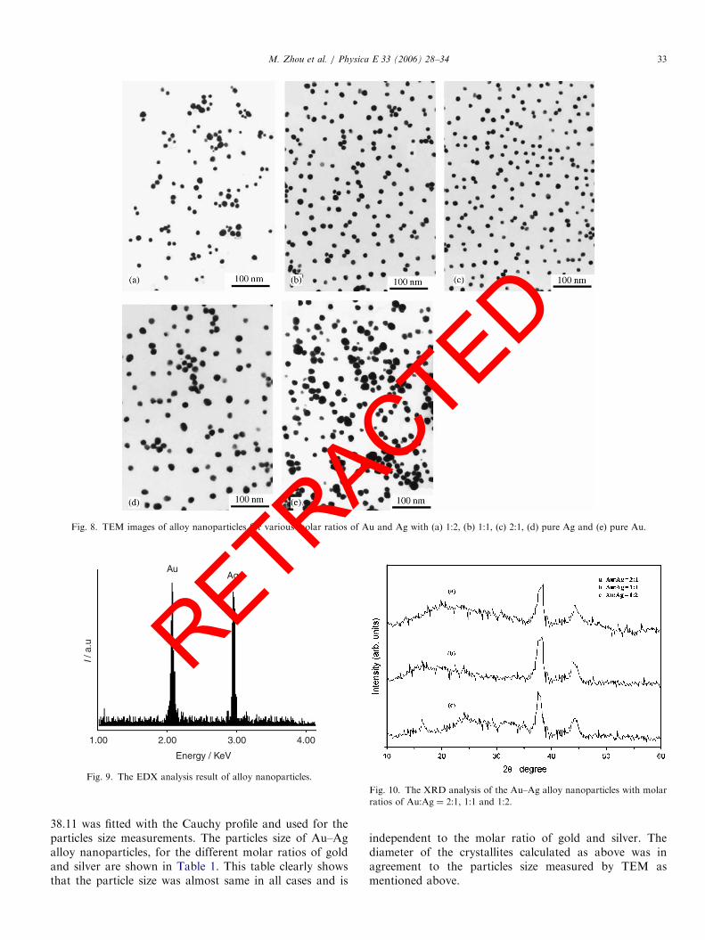
ARTICLE IN PRESS
Fig. 8. TEM images of alloy nanoparticles for various molar ratios of Au and Ag with (a) 1:2, (b) 1:1, (c) 2:1, (d) pure Ag and (e) pure Au.
4.003.002.001.00
Energy / KeV
I / a
.u
AuAg
Fig. 9. The EDX analysis result of alloy nanoparticles.
Fig. 10. The XRD analysis of the Au–Ag alloy nanoparticles with molar
ratios of Au:Ag ¼ 2:1, 1:1 and 1:2.
M. Zhou et al. / Physica E 33 (2006) 28–34 33
RETRACTED
38.11 was fitted with the Cauchy profile and used for theparticles size measurements. The particles size of Au–Agalloy nanoparticles, for the different molar ratios of goldand silver are shown in Table 1. This table clearly showsthat the particle size was almost same in all cases and is
independent to the molar ratio of gold and silver. Thediameter of the crystallites calculated as above was inagreement to the particles size measured by TEM asmentioned above.

ARTICLE IN PRESS
Table 1
The particles size of the Au–Ag alloy nanoparticles with molar ratios of
Au:Ag ¼ 2:1, 1:1 and 1:2
Molar ratios Au/Ag 2:1 1:1 1:2
Average particles size (nm) 12:7 12:0 12.9
M. Zhou et al. / Physica E 33 (2006) 28–3434
A
4. Conclusions
In summary, an electrochemical reduction approach forthe synthesis of Au–Ag alloy nanoparticles has beendeveloped successfully. Structural and compositional ana-lysis of Au–Ag alloy nanoparticles was performed bymeans of UV–visible spectroscopy, TEM, EDX and XRD.The Au–Ag alloy nanoparticles showed only one absorp-tion peak the position of which easily be controlled bychanging the molar ratios of Au to Ag.
Acknowledgements
The authors gratefully acknowledge the financial sup-port by the Funds for the Chinese National Science Fund(20373038).
References
[1] M. Daniel, D. Astruc, Chem. Rev. 104 (2004) 293.
[2] Y. Sun, Y. Xia, Science 298 (2002) 2176.
[3] R. Shenhar, V. Rotello, Acc. Chem. Res. 36 (2003) 549.
[4] G. Schmid, Adv. Mater. 10 (1998) 515.
[5] S.A. Empedocles, R. Neuhauser, M.G. Bawendi, Nature 399 (1999)
126.
[6] P. Zhang, T.K. Sham, Phys. Rev. Lett. 90 (2003) 245502.
[7] T. Shimizu, T. Teranishi, S. Hasegawa, M. Miyake, J. Phys. Chem. B
107 (2003) 2719.
[8] S. Meltzer, R. Resch, B.E. Koel, M.E. Thompson, A. Madhukar,
A.G. Requicha, P. Will, Langmuir 17 (2001) 1713.TR
RE[9] A. Dawson, P.V. Kamat, J. Phys. Chem. B 104 (2000) 11842.
[10] V.G. Pol, A. Gedanken, Chem. Mater. 15 (2003) 1111.
[11] M.T. Reetz, W. Helbig, J. Am. Chem. Soc. 116 (1994) 7401.
[12] M. T Reetz, M. Winter, R. Breinbauer, T. Thomas, V. Walter, Chem.
Eur. J. 7 (2001) 1084.
[13] L. Rodrigues-Sanchez, M.C. Blanco, M.A. Lopez-Quintela, J. Phys.
Chem. B 104 (2000) 9683.
[14] Y.Y. Yu, S.S. Chang, C.L. Lee, C.R.C. Wang, J. Phys. Chem. B 101
(1997) 6661.
[15] M.B. Mohamed, K.Z. Ismail, S. Link, M.A. El-Sayed, J. Phys. Chem.
B 102 (1998) 9370.
[16] S. Yin, H. Ma, S. Wang, S. Chen, J. Phys. Chem. B 107 (2003) 8898.
[17] S. Yin, H. Ma, S. Wang, Y. Jiao, W. Pan, S. Chen, S. Huang,
F. Meng, Chemphyschem 5 (2004) 68.
[18] F. Baletto, C. Mottet, R. Ferrando, Phys. Rev. Lett. 90 (2003)
135501–135504.
[19] J. H Liu, A.Q. Wang, Y.S. Chi, H.P. Lin, C.Y. Mou, J. Phys. Chem.
B 109 (2005) 40.
[20] S. Link, C. Burda, Z.L. Wang, M.A. El-Sayed, J. Chem. Phys. 111
(1999) 1255.
[21] M. Liu, P. Guyot-Sionnest, J. Phys. Chem. B 108 (2004) 5882.
[22] S. Devarajan, B. Vimalan, S. Sampath, J. Colloid. Interface. Sci. 278
(2004) 126.
[23] M.J. Hostetler, C. Zhong, B.K.H. Yen, J. Anderegg, S.M. Gross,
N.D. Evans, M. Porter, R.W. Murray, J. Am. Chem. Soc. 120 (1998)
9396.
[24] H.Z. Shi, L.D. Zhang, W.P. Cai, J. Appl. Phys. 87 (2000) 1572.
[25] N. Aihara, K. Torigoe, K. Esumi, Langmuir 14 (1998) 4945.
[26] J.H. Hodak, A. Henglein, M. Giersig, G.V. Hartland, J. Phys. Chem.
B 104 (2000) 11708.
[27] Y. Chen, C. Yeh, Chem. Commun. (2001) 371.
[28] M.P. Mallin, C.J. Murphy, Nano Lett. 2 (2002) 1235.
[29] Y. Liu, H. Lee, H. Peng, Chem. Phys. Lett. 400 (2004) 436.
[30] A.T. Izgaliev, A.V. Simakin, G.A. Shafeev, F. Bozon-Verduraz,
Chem. Phys. Lett. 390 (2004) 467.
[31] P. Selvakannan, A. Swami, D. Srisathiyanarayanan, P.S. Shirude,
R. Pasricha, A.B. Mandale, M. Sastry, Langmuir 20 (2004)
7825.
[32] Y. Cao, R. Jin, C.A. Mirkin, J. Am. Chem. Soc. 123 (2001) 7961.
[33] S. Mandal, P. Selvakannan, R. Pasricha, M. Sastry, J. Am. Chem.
Soc. 125 (2003) 8440.
[34] H. Okamoto, T.B. Massalski, in: Phase Diagrams of Binary Gold
Alloys, ASM International, Metals Park, 1987.
CTED