restriction endonuclease-mediated selective polymerase chain reaction: a novel assay for the...
TRANSCRIPT
Technical AdvanceRestriction Endonuclease-Mediated SelectivePolymerase Chain Reaction
A Novel Assay for the Detection of K-ras Mutations inClinical Samples
Robyn Ward,* Nicholas Hawkins,†Roslynn O’Grady,* Catherine Sheehan,*Terence O’Connor,‡ Helen Impey,§Natalie Roberts,§ Caroline Fuery,§ andAlison Todd§
From the Departments of Medical Oncology,* and Colorectal
Surgery,‡ St. Vincent’s Hospital; School of Pathology,† University
of New South Wales; and Johnson and Johnson Research Pty
Limited,§ Sydney, New South Wales, Australia
The enriched polymerase chain reaction (PCR) assayhas been used extensively in the detection of ras genemutations in many types of human malignancies. Al-though it is very sensitive, it has a number of featuresthat limit its use in the routine diagnostic laboratory.The aim of this study was to develop a novel enrichedPCR strategy, in which the concurrent activity of therestriction enzyme BstNI and Taq polymerase al-lowed the amplification of mutant K-ras while inhib-iting the formation of wild-type product. This restric-tion endonuclease-mediated selective PCR assay usesthree sets of primers, together with BstNI, in thereaction mix, and the amplification products are an-alyzed by gel electrophoresis. The reliability of therestriction endonuclease-mediated selective PCR as-say to detect activated K-ras was determined in avariety of clinical samples, including 139 fresh colo-rectal carcinomas and 113 paraffin-embedded blocksfrom 80 separate tumors of the colon and rectum,pancreas, breast, or kidney. Codon 12 mutations ofthe K-ras oncogene were identified in DNA from bothfresh and paraffin-embedded tumors in a rapid, sen-sitive, and reproducible manner. Mutations were de-tected in 33 (24%) of the fresh colorectal cancers and16 (20%) of the paraffin-embedded tumors. These re-sults were 97% concordant in cases in which paraffinblocks and fresh specimens from the same tumor
were available for analysis. We conclude that restric-tion endonuclease-mediated selective PCR is a sensi-tive, rapid, and robust assay for the detection of pointmutations in a variety of clinical samples. Impor-tantly, there is no need for manipulation of the sam-ple once the PCR has been set up, and therefore, thechance of contamination is significantly reduced. Incontrast to previous assays, restriction endonuclease-mediated selective PCR is not labor intensive, and itsformat is suitable for use in routine diagnostic labo-ratory. (Am J Pathol 1998, 153:373–379)
Activating point mutations at codons 12, 13, or 61 of theras proto-oncogenes occur frequently in human tumors.1
In colon and pancreatic cancer, more than 90% of thesemutations occur in the K-ras gene, and most of these arefound in codon 12.2,3 Many studies have suggested thatdetection of activated ras may have diagnostic or prog-nostic importance. Because these mutations are ac-quired early in tumor development,4 the detection of ac-tivated K-ras in DNA from the stools of patients withcolorectal cancer may allow diagnosis at a stage at whichcurative surgery is still possible.5–7 Other studies havedemonstrated that the presence of mutant K-ras in theregional lymph nodes or peripheral blood of patients withcolorectal cancer identifies those individuals most likelyto relapse.8,9 Moreover, the recent development of spe-cific treatments targeting the activated ras genes pro-vides a further impetus for the development of new strat-egies for the detection of mutant ras in clinical samples.
Supported by the Leo and Jenny Leukaemia and Cancer Foundation andthe St. Vincent’s Clinic Foundation.
Accepted for publication May 2, 1998.
Address reprint requests to Dr. Robyn Ward, Department of MedicalOncology, St. Vincent’s Hospital, Victoria Street, Darlinghurst, 2010 NewSouth Wales, Australia. E-mail: [email protected].
American Journal of Pathology, Vol. 153, No. 2, August 1998
Copyright © American Society for Investigative Pathology
373

A number of assays have been used for the detectionof activated ras, and these protocols vary in their sensi-tivity and complexity. Typically, they rely on amplificationof the ras gene by the polymerase chain reaction (PCR),followed by detection of the mutant product by electro-phoresis, colorimetric analysis, or other means. Many ofthese PCR protocols have used mismatched bases withinprimer sequences, allowing the identification of mutantamplicons by the creation of restriction enzyme sites.10
The sensitivity of this type of protocol has been signifi-cantly improved with the development of the enrichedPCR assay,11,12 which is based on an initial round ofamplification followed by restriction enzyme digestion tocleave wild-type amplicons. Because only mutant ampli-cons remain as templates, a further round of amplificationresults in the “enrichment” of mutant ras product. Thisassay has been applied extensively to the analysis of rasgene mutations in many types of cancer, including colo-rectal tumors.12,13 However, the assay remains a rela-tively long and labor-intensive procedure, with substan-tial risk of contamination.
We describe a novel enriched PCR strategy, knownas restriction endonuclease-mediated selective PCR(REMS-PCR), in which the restriction enzyme BstNI isincorporated as part of a conventional PCR reaction. Thisapproach exploits both the thermostability of the enzymeand its compatibility with standard PCR buffers. The con-current activity of these enzymes in the one reactionallows simultaneous amplification of mutant signal andinhibition of amplification of wild-type K-ras. Further, wedemonstrate the reliability and sensitivity of this assay forthe detection of activated K-ras in a variety of clinical sam-ples and its suitability for use in diagnostic laboratories.
Materials and Methods
Patient Samples
After informed consent was obtained, 137 individualsundergoing surgical resection of adenocarcinoma of thecolon or rectum at St. Vincent’s Hospital (Sydney, NSW,Australia) were enrolled in this prospective study from1993 to 1997. Fresh representative samples (500 �g) ofall tumors were immediately frozen at �70°C. A total of139 fresh tumor specimens were assayed from 80 malesand 59 females, with ages ranging from 29 to 94 (mean,67.3 � 12.1). Of these tumors, 21% were modified Dukes’stage A, 33% were stage B, 38% were stage C, and 7%were stage D.14,15 In addition, paraffin-embedded blocksof 51 colorectal, 12 breast, 11 renal, and 6 pancreatictumors were obtained from the Department of AnatomicalPathology, St. Vincent’s Hospital, after routine process-ing. The colorectal paraffin blocks were collected from 21females and 30 males, ranging in age from 49 to 95years. Of these tumors, 14% were Dukes’ stage A, 38%were stage B, 40% were stage C, and 8% were stage D.To determine the effect of paraffin block age on assayreliability, we selected tumors that had been processedat various time points over the last 10 years.
For all tumors, the histopathological diagnosis, stageand tumor size were determined independently by ahistopathologist within the Department of Anatomical Pa-thology, St. Vincent’s Hospital.
DNA Preparation from Fresh Tissues andParaffin Sections
For preparation of DNA from fresh tissues, the frozentissue was macerated in a 500-�l solution of 10 mmol/LTris-HCl, 1 mmol/L ethylenediamine tetra-acetic acid, 100mmol/L NaCl, 1% sodium dodecyl sulfate, and 500 �g/mlproteinase K, using a sterile Eppendorf homogenizer at4°C. The DNA was extracted with phenol/chloroform afterincubation overnight with shaking at 50°C and precipi-tated with ethanol. It was then resuspended in water, andthe concentration was determined by spectrophotometry.
For the analysis of paraffin-embedded tissues, threeconsecutive 10-�m sections were cut from paraffinblocks, and each section was placed in a separate sterile2.0-ml screw-capped tube. To prevent cross contamina-tion from tissue with flakes of paraffin, the blade wascleaned with a jet of compressed air after each sectionwas cut. A sham tissue block, which did not containtumor tissue, was cut after every 10 blocks. DNA was alsoextracted from this sample and used in subsequent PCRanalysis. For each tumor block, an adjacent section wasstained with hematoxylin and eosin and examined bylight microscopy to determine the amount of tumorpresent.
For the extraction of DNA from paraffin-embeddedtissues, each section was immersed in 300 �l of lysisbuffer (50 mmol/L KCl, 3 mmol/L CaCl2, 0.4% Triton-X,and 10 mmol/L Tris-HCl, pH 8.0), together with 3 U ofPreTaq (1 U/�l Boehringer Mannheim). The tubes wereboiled for 5 minutes and then centrifuged at 14,000 rpmfor 2 minutes. The supernatants were then transferred totubes containing 100 �l of 0.25 mol/L ACES buffer (N-(2-acetamido)-2-aminoethanesulfonic acid, 0.125 mol/LNaOH, and 0.5% Tween 20, pH 6.8) and 25 �l of acationic polymer (2.4% aqueous, pH 5, Ortho Diagnos-tics, Raritan, NJ), and the mixture was vortexed and thenmicrofuged at 14,000 rpm for 2 minutes. The pellet wasresuspended in 100 �l of 20 mmol/L NaOH and incu-bated at room temperature for 30 minutes. To detach thepolymer, samples were boiled for 5 minutes, and thesolution containing the DNA was stored at �20°C.
REMS-PCR Mutational Analysis
Mutations at the first and second bases of codon 12 ofthe K-ras gene were detected using REMS-PCR. EachPCR reaction contained three sets of primers (Table 1,Figure 1).
The diagnostic primers 5BKIT and 3K2 induce a cleav-age site for BstNI in wild-type K-ras amplicons. BecauseBstNI is included in the REMS-PCR reaction, the amplifi-cation of such a wild-type template is prevented. How-ever, when a template also contains K-ras gene withmutations at bases 1 and 2 of codon 12, no BstNI cleav-
374 Ward et alAJP August 1998, Vol. 153, No. 2
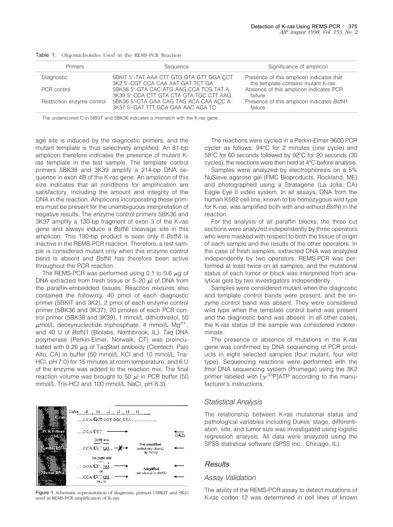
age site is induced by the diagnostic primers, and themutant template is thus selectively amplified. An 81-bpamplicon therefore indicates the presence of mutant K-ras template in the test sample. The template controlprimers 5BK38 and 3K39 amplify a 214-bp DNA se-quence in exon 4B of the K-ras gene. An amplicon of thissize indicates that all conditions for amplification aresatisfactory, including the amount and integrity of theDNA in the reaction. Amplicons incorporating these prim-ers must be present for the unambiguous interpretation ofnegative results. The enzyme control primers 5BK36 and3K37 amplify a 130-bp fragment of exon 3 of the K-rasgene and always induce a BstNI cleavage site in thisamplicon. This 130-bp product is seen only if BstNI isinactive in the REMS-PCR reaction. Therefore, a test sam-ple is considered mutant only when this enzyme controlband is absent and BstNI has therefore been activethroughout the PCR reaction.
The REMS-PCR was performed using 0.1 to 0.6 �g ofDNA extracted from fresh tissue or 5–20 �l of DNA fromthe paraffin-embedded tissues. Reaction mixtures alsocontained the following: 40 pmol of each diagnosticprimer (5BKIT and 3K2), 2 pmol of each enzyme controlprimer (5BK36 and 3K37), 20 pmoles of each PCR con-trol primer (5BK38 and 3K39), 1 mmol/L dithiothreitol, 50�mol/L deoxynucleotide triphosphate, 4 mmol/L Mg2�,and 40 U of BstN1 (Biolabs, Northbrook, IL). Taq DNApolymerase (Perkin-Elmer, Norwalk, CT) was preincu-bated with 0.26 �g of TaqStart antibody (Clontech, PaloAlto, CA) in buffer (50 mmol/L KCl and 10 mmol/L Tris-HCl, pH 7.0) for 15 minutes at room temperature, and 6 Uof the enzyme was added to the reaction mix. The finalreaction volume was brought to 50 �l in PCR buffer (50mmol/L Tris-HCl and 100 mmol/L NaCl, pH 8.3).
The reactions were cycled in a Perkin-Elmer 9600 PCRcycler as follows: 94°C for 2 minutes (one cycle) and58°C for 60 seconds followed by 92°C for 20 seconds (30cycles); the reactions were then held at 4°C before analysis.
Samples were analyzed by electrophoresis on a 5%NuSieve agarose gel (FMC Bioproducts, Rockland, ME)and photographed using a Stratagene (La Jolla, CA)Eagle Eye II video system. In all assays, DNA from thehuman K562 cell line, known to be homozygous wild typefor K-ras, was amplified both with and without BstNI in thereaction.
For the analysis of all paraffin blocks, the three cutsections were analyzed independently by three operatorswho were masked with respect to both the tissue of originof each sample and the results of the other operators. Inthe case of fresh samples, extracted DNA was analyzedindependently by two operators. REMS-PCR was per-formed at least twice on all samples, and the mutationalstatus of each tumor or block was interpreted from ana-lytical gels by two investigators independently.
Samples were considered mutant when the diagnosticand template control bands were present, and the en-zyme control band was absent. They were consideredwild type when the template control band was presentand the diagnostic band was absent. In all other cases,the K-ras status of the sample was considered indeter-minate.
The presence or absence of mutations in the K-rasgene was confirmed by DNA sequencing of PCR prod-ucts in eight selected samples (four mutant, four wildtype). Sequencing reactions were performed with thefmol DNA sequencing system (Promega) using the 3K2primer labeled with [�-33P]ATP according to the manu-facturer’s instructions.
Statistical Analysis
The relationship between K-ras mutational status andpathological variables including Dukes’ stage, differenti-ation, site, and tumor size was investigated using logisticregression analysis. All data were analyzed using theSPSS statistical software (SPSS Inc., Chicago, IL).
Results
Assay Validation
The ability of the REMS-PCR assay to detect mutations ofK-ras codon 12 was determined in cell lines of known
Table 1. Oligonucleotides Used in the REMS-PCR Reaction
Primers Sequence Significance of amplicon
Diagnostic
PCR control
Restriction enzyme control
5BKIT 5�-TAT AAA CTT GTG GTA GTT GGA CCT3K2 5�-CGT CCA CAA AAT GAT TCT GA5BK38 5�-GTA CAC ATG AAG CCA TCG TAT A3K39 5�-CCA CTT GTA CTA GTA TGC CTT AAG5BK36 5�-CTA GAA CAG TAG ACA CAA ACC A3K37 5�-GAT TTT GCA GAA AAC AGA TC
Presence of this amplicon indicates thatthe template contains mutant K-ras
Absence of this amplicon indicates PCRfailure
Presence of this amplicon indicates BstN1failure
The underscored C in 5BKIT and 5BK36 indicates a mismatch with the K-ras gene.
Figure 1. Schematic representation of diagnostic primers (5BKIT and 3K2)used in REMS-PCR amplification of K-ras.
Detection of K-ras Using REMS-PCR 375AJP August 1998, Vol. 153, No. 2
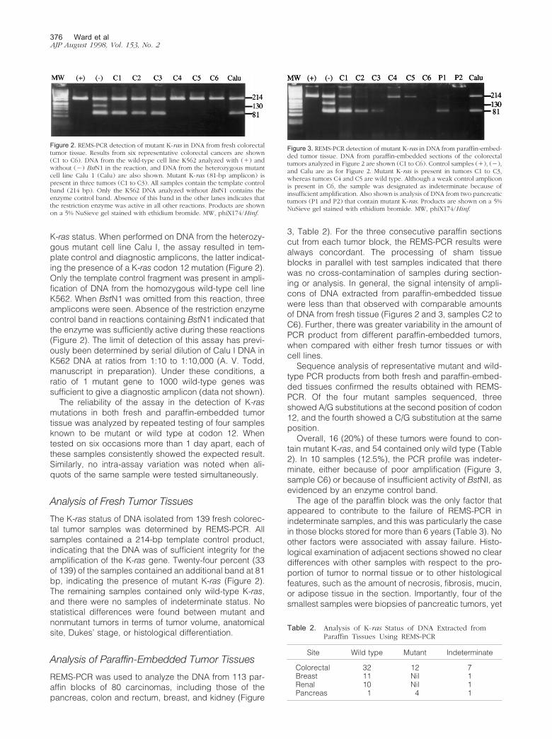
K-ras status. When performed on DNA from the heterozy-gous mutant cell line Calu I, the assay resulted in tem-plate control and diagnostic amplicons, the latter indicat-ing the presence of a K-ras codon 12 mutation (Figure 2).Only the template control fragment was present in ampli-fication of DNA from the homozygous wild-type cell lineK562. When BstN1 was omitted from this reaction, threeamplicons were seen. Absence of the restriction enzymecontrol band in reactions containing BstN1 indicated thatthe enzyme was sufficiently active during these reactions(Figure 2). The limit of detection of this assay has previ-ously been determined by serial dilution of Calu I DNA inK562 DNA at ratios from 1:10 to 1:10,000 (A. V. Todd,manuscript in preparation). Under these conditions, aratio of 1 mutant gene to 1000 wild-type genes wassufficient to give a diagnostic amplicon (data not shown).
The reliability of the assay in the detection of K-rasmutations in both fresh and paraffin-embedded tumortissue was analyzed by repeated testing of four samplesknown to be mutant or wild type at codon 12. Whentested on six occasions more than 1 day apart, each ofthese samples consistently showed the expected result.Similarly, no intra-assay variation was noted when ali-quots of the same sample were tested simultaneously.
Analysis of Fresh Tumor Tissues
The K-ras status of DNA isolated from 139 fresh colorec-tal tumor samples was determined by REMS-PCR. Allsamples contained a 214-bp template control product,indicating that the DNA was of sufficient integrity for theamplification of the K-ras gene. Twenty-four percent (33of 139) of the samples contained an additional band at 81bp, indicating the presence of mutant K-ras (Figure 2).The remaining samples contained only wild-type K-ras,and there were no samples of indeterminate status. Nostatistical differences were found between mutant andnonmutant tumors in terms of tumor volume, anatomicalsite, Dukes’ stage, or histological differentiation.
Analysis of Paraffin-Embedded Tumor Tissues
REMS-PCR was used to analyze the DNA from 113 par-affin blocks of 80 carcinomas, including those of thepancreas, colon and rectum, breast, and kidney (Figure
3, Table 2). For the three consecutive paraffin sectionscut from each tumor block, the REMS-PCR results werealways concordant. The processing of sham tissueblocks in parallel with test samples indicated that therewas no cross-contamination of samples during section-ing or analysis. In general, the signal intensity of ampli-cons of DNA extracted from paraffin-embedded tissuewere less than that observed with comparable amountsof DNA from fresh tissue (Figures 2 and 3, samples C2 toC6). Further, there was greater variability in the amount ofPCR product from different paraffin-embedded tumors,when compared with either fresh tumor tissues or withcell lines.
Sequence analysis of representative mutant and wild-type PCR products from both fresh and paraffin-embed-ded tissues confirmed the results obtained with REMS-PCR. Of the four mutant samples sequenced, threeshowed A/G substitutions at the second position of codon12, and the fourth showed a C/G substitution at the sameposition.
Overall, 16 (20%) of these tumors were found to con-tain mutant K-ras, and 54 contained only wild type (Table2). In 10 samples (12.5%), the PCR profile was indeter-minate, either because of poor amplification (Figure 3,sample C6) or because of insufficient activity of BstNI, asevidenced by an enzyme control band.
The age of the paraffin block was the only factor thatappeared to contribute to the failure of REMS-PCR inindeterminate samples, and this was particularly the casein those blocks stored for more than 6 years (Table 3). Noother factors were associated with assay failure. Histo-logical examination of adjacent sections showed no cleardifferences with other samples with respect to the pro-portion of tumor to normal tissue or to other histologicalfeatures, such as the amount of necrosis, fibrosis, mucin,or adipose tissue in the section. Importantly, four of thesmallest samples were biopsies of pancreatic tumors, yet
Figure 2. REMS-PCR detection of mutant K-ras in DNA from fresh colorectaltumor tissue. Results from six representative colorectal cancers are shown(C1 to C6). DNA from the wild-type cell line K562 analyzed with (�) andwithout (�) BstN1 in the reaction, and DNA from the heterozygous mutantcell line Calu 1 (Calu) are also shown. Mutant K-ras (81-bp amplicon) ispresent in three tumors (C1 to C3). All samples contain the template controlband (214 bp). Only the K562 DNA analyzed without BstN1 contains theenzyme control band. Absence of this band in the other lanes indicates thatthe restriction enzyme was active in all other reactions. Products are shownon a 5% NuSieve gel stained with ethidium bromide. MW, phiX174/Hinf.
Figure 3. REMS-PCR detection of mutant K-ras in DNA from paraffin-embed-ded tumor tissue. DNA from paraffin-embedded sections of the colorectaltumors analyzed in Figure 2 are shown (C1 to C6). Control samples (�), (�),and Calu are as for Figure 2. Mutant K-ras is present in tumors C1 to C3,whereas tumors C4 and C5 are wild type. Although a weak control ampliconis present in C6, the sample was designated as indeterminate because ofinsufficient amplification. Also shown is analysis of DNA from two pancreatictumors (P1 and P2) that contain mutant K-ras. Products are shown on a 5%NuSieve gel stained with ethidium bromide. MW, phiX174/Hinf.
Table 2. Analysis of K-ras Status of DNA Extracted fromParaffin Tissues Using REMS-PCR
Site Wild type Mutant Indeterminate
Colorectal 32 12 7Breast 11 Nil 1Renal 10 Nil 1Pancreas 1 4 1
376 Ward et alAJP August 1998, Vol. 153, No. 2
mutations of K-ras were reproducibly detected from theseblocks. Assay failure was also not related to the amountor extent of degradation of DNA in the paraffin samples,as determined by gel electrophoresis. In fact, most sam-ples contained only high-molecular weight DNA (�4000bp), whereas REMS-PCR was often conclusive in thosesamples with clear evidence of some DNA degradation(data not shown).
Correlation with the Subset of Tissues withFresh Results
Of the 33 colorectal tumors in which both fresh andparaffin-embedded tissues were analyzed, 27 were wildtype in both assays, 5 were mutant, and in 1 case therewas a discordant result. In this case, the tumor wasmutant by analysis of the frozen tissue, but wild type inDNA from the paraffin sections, despite the fact that itwas shown by histological examination to contain tumor.DNA sequence analysis of this case confirmed theREMS-PCR results from fresh and paraffin-embeddedtissues.
To assess the possibility that tumors are heteroge-neous for K-ras mutations, we examined 4 to 6 tissueblocks taken from each of 10 colorectal tumors. In 2 ofthese 10 tumors, REMS-PCR showed mutations of K-rasin all 9 blocks examined. Likewise, 32 blocks from theother 8 tumors contained only wild-type K-ras. Theseresults show that there is no clear evidence to supporttumor heterogeneity for mutations of K-ras in colorectaltumors.
DiscussionThe REMS-PCR assay exploits the thermostable proper-ties of BstNI and Taq polymerase, as well as their abilityto function effectively in a common buffer system, therebyallowing simultaneous amplification of mutant signal andinhibition of amplification of wild-type K-ras. This strategyrepresents a significant advance over existing enrichedPCR approaches, producing a robust assay suitable foruse in the diagnostic laboratory. The principal advantageof the REMS procedure comes from the greatly reducedhandling of amplified product. Unlike conventional en-riched PCR, in which the two rounds of PCR are sepa-rated by an enzyme digestion step, all reactions in the
REMS-PCR occur concurrently in the one tube. This re-duced handling has two important implications for theapplicability of the assay as a diagnostic procedure.Firstly, there is greatly reduced risk of contamination byPCR product, a major problem in most PCR assays and acommon cause of false positive results. Secondly, re-duced handling means considerable savings in both timeand labor costs and makes the process more amenableto automation.
A further significant advantage of the REMS-PCR as-say over current enriched PCR methods is that eachreaction includes internal controls for both template in-tegrity and BstN1 enzyme activity. The control for tem-plate integrity allows discrimination between those reac-tions that are negative because of complete digestion ofwild-type product and those in which amplification ofeither wild-type or mutant product has failed for anyreason, including insufficient quantity or quality of thetemplate. This proved to be particularly important in theanalysis of the paraffin-embedded samples. The tem-plate control primers have been designed to amplify alonger DNA template than the diagnostic primers. Thus, areduction in the integrity of the template is readily appar-ent in the proportionally greater loss of the template con-trol product.
Incomplete BstNI digestion is a potential problem inREMS-PCR, particularly because restriction enzyme ac-tivity is influenced by a number of factors including for-malin fixation of the tissue.16 The inclusion of a restrictionenzyme control primer set within the PCR reaction allowsready identification of this problem. If BstNI has remainedactive throughout the reaction, the enzyme control ampli-con will not be seen after REMS-PCR.
By incorporating these features into the REMS-PCRprocedure, we were able to detect mutations in codon 12of K-ras, with minimal manipulation of samples, in a timeof less than 3 hours, including DNA extraction and gelelectrophoresis. Despite this, the sensitivity of the assayremained at a level of 1 mutant per 1000 wild-type genes,a level comparable with that achieved in previous en-riched PCR strategies.17
To be applicable in routine laboratories, it is importantthat the assays be reliable as well as rapid and sensitive.The reliability of the REMS-PCR assay was evaluated in arigorous manner, with assessment of coded samples bytwo independent operators on several occasions. Therewas no inter- or intra-assay variation when the technique
Table 3. Effect of Tumor Age on REMS-PCR Reliability
Tumor age
Paraffin Fresh
Wild type Mutant Indeterminate Wild type Mutant Indeterminate
�1 year 11 0 0 27 7 01–3 years 22 4 2 60 15 03–6 years 18 5 1 19 11 06–10 years 1 7 3 NA NA NA�10 years 2 0 4 NA NA NA
Results of REMS-PCR analysis are shown for both paraffin-embedded and fresh (frozen) tissue, according to the age of the tumor at the time ofanalysis. Paraffin sections were from colorectal, pancreas, renal, and breast cancers, whereas fresh tissues are from only colorectal tumors. Numbersrepresent the number of tumors in each category.
NA, not available.
Detection of K-ras Using REMS-PCR 377AJP August 1998, Vol. 153, No. 2
was applied to fresh specimens. Moreover, the assayallowed the rapid, unambiguous, and reproducible de-termination of K-ras status in all 139 fresh colorectalspecimens examined.
When assayed with methods of appropriate sensitivity,the frequency of K-ras codon 12 mutations in colorectalcancer has been reported to vary between 20 and50%.18,19 In part, this may represent geographical varia-tion in the frequency of mutations.20,21 Two recent studiesin the Australian population have reported that, in sam-ples of 233 and 103 individuals, respectively, 24% ofcolorectal tumors contained mutations at codon 12 of theK-ras gene.22,23 The results with REMS-PCR are clearly inagreement with these reports and suggest that the assayreflects the true incidence in this population.
Large archives of frozen tissues are rarely available forretrospective analysis, and it can be difficult to confirmthe presence of tumor cells within these tissues. Wetherefore also sought to evaluate the utility of REMS-PCRin the analysis of K-ras mutations in paraffin-embeddedtumor blocks. A number of factors are known to influencethe success of PCR analysis of paraffin-embedded tis-sues, including the type and amount of tumor and themethods used for fixation and embedding.16,24–26 Theeffect of variation in these factors was of particular con-cern in the REMS-PCR assay, in which simultaneousactivity of both Taq polymerase and restriction enzymesis required. We therefore used the assay to analyze ran-domly selected blocks from a variety of tumors collectedat different time points over the previous 10 years. TheREMS-PCR assay proved robust in the analysis of DNAfrom paraffin tissue collected up to 6 years previouslyand proved effective on a range of tissues containingvarying amounts of tumor. The analysis of paraffin tissuedid not appear to generate false positive results, becauseno amplicons were seen in sham blocks, no diagnosticamplicons were seen in multiple blocks from wild-typetumors, and there was a 97% concordance with the as-say on fresh colorectal tumors. Furthermore, activatedK-ras was not detected in either the breast or renal can-cers, tumors that have rarely been reported to containmutations in this gene.27,28 Although REMS-PCR wasoften less efficient using paraffin blocks, the K-ras statusof the tumor could still be determined in more than 90% ofcases in which the block age was less than 6 years.
The factors responsible for the suboptimal amplifica-tion in some tissues were not easily identified. Neither thefragment size, the amount of DNA extracted from paraffinblocks, nor the method of extraction, appeared to influ-ence the success of REMS-PCR. It is possible that thefixation process itself may have contributed to the failureof amplification or inactivation of BstN1 seen in someparaffin samples. Structural modifications induced inDNA by formaldehyde may not only reduce the effectiveamount of PCR template, but may also interfere with theactivity of restriction enzymes.16 This possibility is sup-ported by our observation that the inclusion of dithiothre-itol in the assay markedly reduced the number of inde-terminate results.
There are conflicting data concerning the effect ofblock age on successful PCR amplification.29,30 Our
study indicated that, in the case of REMS-PCR, the fre-quency of indeterminate results increased with increas-ing block age, and we would therefore not recommend itsuse for blocks collected more than 6 years previously.
In conclusion, REMS-PCR provides a sensitive, fast,and robust assay for the detection of point mutations in avariety of clinical samples. Importantly, there is no needfor manipulation of the sample once the PCR has beenset up, and thus the chance of contamination is signifi-cantly reduced. In contrast to previous assays, theREMS-PCR is not labor intensive, and its format would besuitable for use in a routine diagnostic laboratory.
References
1. Barbacid M: ras genes. Annu Rev Biochem 1987, 56:779–8272. Almoguera C, Shibata D, Forrester K, Martin J, Arnheim N, Perucho
M: Most human carcinomas of the exocrine pancreas contain mutantc-K-ras genes. Cell 1988, 53:549–554
3. Bos JL: ras oncogenes in human cancer: a review. Cancer Res 1989,49:4682–4689
4. Vogelstein B, Fearon ER, Hamilton SR, Kern SE, Preisinger AC, Lep-pert M, Nakamura Y, White R, Smits AMM, Bos JI: Genetic alterationsduring colorectal-tumor development. N Engl J Med 1988, 319:525–532
5. Sidransky D, Tokino Y, Hamilton SR, Kinzler KW, Levin B, Frost P,Vogelstein B: Identification of ras oncogene mutations in the stool ofpatients with curable colorectal tumors. Science 1992, 256:102–105
6. Losi L, Benhattar J, Costa J: Stability of K-ras mutations throughoutthe natural history of human colorectal cancer. Eur J Cancer 1992,28A:1115–1120
7. Ratto C, Flamini G, Sofo L, Nucera P, Ippoliti M, Curigliano G, FerrettiG, Sgambato A, Merico M, Doglietto GB, Cittadini A, Crucitti F:Detection of oncogene mutation from neoplastic colonic cells exfoli-ated in feces. Dis Colon Rectum 1996, 39:1238–1244
8. Hayashi N, Ito I, Yanagisawa A, Kato Y, Nakamori S, Imaoka S,Watanabe H, Ogawa M, Nakamura Y: Genetic diagnosis of lymph-node metastasis in colorectal cancer. Lancet 1995, 345:1257–1259
9. Hardingham JE, Kotasek D, Sage RE, Eaton MC, Pascoe VH, Do-brovic A: Detection of circulating tumor cells in colorectal cancer byimmunobead-PCR is a sensitive prognostic marker for relapse ofdisease. Mol Med 1995, 1:789–794
10. Cohen JB, Levinson AD: A point mutation in the last intron responsiblefor increased expression and transforming activity of the c-Ha-rasoncogene. Nature 1988, 334:119–124
11. Todd AV, Iland HJ: Allele-specific enrichment: a method for thedetection of low level N-ras gene mutations in acute myeloid leuke-mia. Leukemia 1991, 5:160–161
12. Levi S, Urbano-Ispizua A, Gill R, Thomas DM, Gilbertson J, Foster C,Marshall CJ: Multiple K-ras codon 12 mutations in cholangiocarcino-mas demonstrated with a sensitive polymerase chain reaction tech-nique. Cancer Res 1991, 51:3497–3502
13. Singh J, Rao CV, Kulkarni N, Simi B, Reddy BS: Molecular markers asintermediate end-points in chemoprevention of colon cancer: modu-lation of ras activation by sulindac and phenylhexylisothiocyanateduring colon carcinogenesis. Int J Oncol 1994, 5:1009–1018
14. Dukes CE, Bussey HJR: The spread of rectal cancer and its effect onprognosis. Br J Cancer 1958, 12:309–320
15. Lineweaver W: Staging of colon cancer. Contemp Surg 1984, 25:1916. Hamazaki S, Koshiba M, Habuchi T, Takahashi R, Sugiyama T: The
effect of formalin fixation on restriction endonuclease digestion ofDNA and PCR amplification. Pathol Res Pract 1993, 189:553–557
17. Ward RL, Santiago F, Hawkins NJ, Coomber D, Oconnor T, Todd AV:A rapid PCR ELISA for the detection of activated K-ras in colorectalcancer. J Clin Pathol 1995, 48:M273–M277
18. Hasegawa H, Ueda M, Watanabe M, Teramoto T, Mukai M, KitajimaM: K-ras gene mutations in early colorectal cancer: flat elevated vs.polyp-forming cancer. Oncogene 1995, 10:1413–1416
19. Minamoto T, Ronai Z, Yamashita N, Ochiai A, Sugimura T, Mai M,Esumi H: Detection of Ki-ras mutation in non-neoplastic mucosa of
378 Ward et alAJP August 1998, Vol. 153, No. 2

Japanese patients with colorectal cancers. Int J Oncol 1994, 4:397–401
20. Sasaki H, Nishii H, Takahashi H, Tada A, Furusato M, Terashima Y,Siegal GP, Parker SL, Kohler MF, Berchuck A: Mutation of the Ki-rasprotooncogene in human endometrial hyperplasia and carcinoma.Cancer Res 1993, 53:1906–1910
21. Scarpa A, Capelli P, Villanueva A, Zamboni G, Lluı̀s F, Accolla R,Mariuzzi G, Capella G: Pancreatic cancer in Europe: Ki-ras genemutation pattern shows geographical differences. Int J Cancer 1994,57:167–171
22. Fung C, Bragg T, Newland R, Dent O, Nicholson G, Bokey L, ChapuisP: K-ras mutation, and loss of heterozygosity of chromosome 17p,and survival in colorectal cancer. Aust NZ J Surg 1997, 67:239–244
23. Thomas RJS, Liu YS, St Clair F, Norris PM, Valentine R, Phillips WA:Frequency and clinico-pathological associations of ras mutations incolorectal cancer in the Victorian population. Aust NZ J Surg 1997,67:233–238
24. Greer CE, Wheeler CM, Manos MM: Sample preparation and PCRamplification from paraffin-embedded tissues. PCR Methods Appl1994, 3:S113–S122
25. Alaibac M, Filotico R, Giannella C, Paradiso A, Labriola A, Marzullo F:
The effect of fixation type on DNA extracted from paraffin-embeddedtissue for PCR studies in dermatopathology. Dermatology 1997, 195:105–107
26. Merkelbach S, Gehlen J, Handt S, Fuzesi L: Novel enzyme immuno-assay and optimized DNA extraction for the detection of polymerasechain reaction-amplified viral DNA from paraffin-embedded tissue.Am J Pathol 1997, 150:1537–1546
27. Koffa M, Malamou-Mitsi V, Agnantis NJ, Spandidos DA: Mutationalactivation of K-ras oncogene in human breast tumors. Int J Oncol1994, 4:573–576
28. Nagata Y, Abe M, Kobayashi K, Saiki S, Kotake T, Yoshikawa K, UedaR, Nakayama E, Shiku H: Point mutations of c-ras genes in humanbladder cancer and kidney cancer. Jpn J Cancer Res 1990, 81:22–27
29. Shibata D, Martin WJ, Arnheim N: Analysis of DNA sequences inforty-year-old paraffin-embedded thin-tissue sections: a bridge be-tween molecular biology and classical histology. Cancer Res 1988,48:4564–4566
30. Goelz SE, Hamilton SR, Vogelstein B: Purification of DNA from form-aldehyde fixed and paraffin embedded human tissue. Biochem Bio-phys Res Commun 1985, 130:118–126
Detection of K-ras Using REMS-PCR 379AJP August 1998, Vol. 153, No. 2