response assessment criteria for brain metastases ... - bm criteria... · medical oncology,...
TRANSCRIPT
www.thelancet.com/oncology Vol 16 June 2015 e270
Review
Lancet Oncology 2015; 16: e270–78
See Online for interview with Nancy Lin
*Contributed equally
Department of Medical Oncology (N U Lin MD, F S Hodi MD) and Center for Neuro-Oncology (E Q Lee MD, Prof P Y Wen MD), Dana-Farber Cancer Institute, Boston, MA, USA; Department of Radiology, Niigata University Graduate School of Medical and Dental Sciences, Chuo-ku, Niigata, Japan (Prof H Aoyama MD); Department of Radiation Oncology, University of California, San Francisco, CA, USA (I J Barani MD); Department of Radiology, Duke University Medical Center, Durham, NC, USA (Prof D P Barboriak MD); Department of Radiation-Oncology, MediClin Robert Janker Clinic & University of Bonn Medical Centre, Cooperation Unit Neuro-oncology, Bonn, Germany (B G Baumert MD); Department of Neuroradiology, University of Heidelberg, Heidelberg, Germany (Prof M Bendszus MD); Department of Radiation Oncology, The University of Texas MD Anderson Cancer Center, Houston, TX, USA (Prof P D Brown MD); Division of Medical Oncology, School of Medicine, University of Colorado Denver, Denver, CO, USA (D R Camidge MD); Department of Neurosurgery, University of California, San Francisco, CA, USA (Prof S M Chang MD); NCIC Clinical Trials Group, Ontario Institute for Cancer Research, Queen’s University, Kingston, ON, Canada (Prof J Dancey MD); Department of Medical Oncology, University Medical Center Groningen, University of Groningen, RB Groningen, Netherlands (Prof E G E de Vries MD); Department of Radiation Oncology, The Anschutz
Response assessment criteria for brain metastases: proposal from the RANO groupNancy U Lin*, Eudocia Q Lee*, Hidefumi Aoyama, Igor J Barani, Daniel P Barboriak, Brigitta G Baumert, Martin Bendszus, Paul D Brown, D Ross Camidge, Susan M Chang, Janet Dancey, Elisabeth G E de Vries, Laurie E Gaspar, Gordon J Harris, F Stephen Hodi, Steven N Kalkanis, Mark E Linskey, David R Macdonald, Kim Margolin, Minesh P Mehta, David Schiff , Riccardo Soffi etti, John H Suh, M artin J van den Bent, Michael A Vogelbaum, Patrick Y Wen, for the Response Assessment in Neuro-Oncology (RANO) group
CNS metastases are the most common cause of malignant brain tumours in adults. Historically, patients with brain metastases have been excluded from most clinical trials, but their inclusion is now becoming more common. The medical literature is diffi cult to interpret because of substantial variation in the response and progression criteria used across clinical trials. The Response Assessment in Neuro-Oncology Brain Metastases (RANO-BM) working group is an international, multidisciplinary eff ort to develop standard response and progression criteria for use in clinical trials of treatment for brain metastases. Previous eff orts have focused on aspects of trial design, such as patient population, variations in existing response and progression criteria, and challenges when incorporating neurological, neuro-cognitive, and quality-of-life endpoints into trials of patients with brain metastases. Here, we present our recommendations for standard response and progression criteria for the assessment of brain metastases in clinical trials. The proposed criteria will hopefully facilitate the development of novel approaches to this diffi cult problem by providing more uniformity in the assessment of CNS metastases across trials.
IntroductionBrain metastases are the most common cause of malignant brain tumours in adults. Of the nearly 1·5 million patients in the USA who received a primary diagnosis of cancer in 2007, about 70 000 of these primary diagnoses are estimated to eventually relapse in the brain.1,2 Despite the frequency of brain metastases, prospective trials in this patient population are limited, and the criteria used to assess response and progression in the CNS are heterogeneous.3 This heterogeneity largely stems from the recognition that existing criteria sets, such as RECIST,4,5 WHO,6 or Macdonald Criteria,7 are themselves distinct and have gaps and limitations in their ability to address issues specifi c to the assessment of patients with brain metastases (table 1).5 Key issues in the imaging of CNS metastases include the modality and frequency of assessment, the method of measurement (linear, bidimensional, volumetric), the magnitude of change that defi nes response or progression, dif-ferentiation between tumour-related and treatment-related changes, the inclusion (or exclusion) of cortico steroid use and clinical signs and symptoms with imaging defi nitions of progression and response, and the inclusion (or exclusion) of systemic disease status into the defi nition of CNS response and progression.
Scope and purpose of the proposed RANO-BM criteriaProspective clinical trials to assess new treatments for patients with active brain metastases are becoming increasingly common. Additionally, we welcome the trend away from automatic exclusion of patients with brain metastases from clinical trials of novel therapies. The concurrent proliferation of response criteria for assessment of CNS metastases has made interpretation of trial results challenging. The Response Assessment in
Neuro-Oncology Brain Metastases (RANO-BM) working group fi rst convened in 2011 to review the medical literature and propose new standard criteria for the radiological assessment of brain metastases in clinical trials. As reported in a previous review,9 the group acknowledges that objective response or progression-free survival, or both, might not always be the most relevant primary study endpoints, depending on the patient population, the treatment being assessed, and question being asked and that neuro-cognition and quality-of-life might be of greater importance in some settings. However, if an investigator chooses to include objective response or progression as key endpoints, we believe the trial community would be best served if the endpoints are assessed and defi ned more uniformly than they are at present. The criteria we propose are relevant for the assessment of parenchymal brain metastases only and do not cover leptomeningeal metastases, which are generally not radiographically measurable in a reliable and reproducible manner. Response criteria for lepto-meningeal metastases will be assessed by a diff erent RANO group. The proposed criteria for brain metastases also do not cover dural metastases or skull metastases invading the brain.
Process of RANO-BM criteria developmentThe RANO-BM is an international group of experts in medical oncology, neuro-oncology, radiation oncology, neurosurgery, neuroradiology, neuropsychology, bio-statistics, and drug development who, in collaboration with government and industry partners, are working towards the development of more streamlined and broadly acceptable criteria for assessment of brain metastases. After completion of a literature review and critique, the group convened a series of meetings and regular teleconferences to formulate the following proposal for
e271 www.thelancet.com/oncology Vol 16 June 2015
Review
Medical Campus, University of Colorado Denver, Aurora, CO,
USA (Prof L E Gaspar MD); MGH 3D Imaging Lab,
Massachusetts General Hospital, Boston, MA, USA
(Prof G J Harris PhD); Department of Neurosurgery,
Henry Ford Health System, Detroit, MI, USA
(S N Kalkanis MD); Department of Neurological Surgery, University of California,
San Francisco, CA, USA (Prof M E Linskey MD); UC Irvine
Medical Center, Orange, CA, USA (Prof M E Linskey);
Department of Oncology, London Regional Cancer
Program, London Health Sciences Centre, University of
Western Ontario, London, ON, Canada (D R Macdonald MD);
Division of Oncology, Stanford University, Stanford, CA, USA
(Prof K Margolin MD); Maryland Proton Treatment Center,
University of Maryland School of Medicine, Baltimore, MA,
USA (Prof M P Mehta MD); Division of Neuro-Oncology,
University of Virginia, Charlottesville, VA, USA
(Prof D Schiff MD); Department of Neurology/Neuro-Oncology, University of Turin, Turin, Italy
(Prof R Soffi etti MD); Department of Radiation
Oncology/T28 (Prof J H Suh MD) and Department of
Neurosurgery/ND40 (Prof M A Vogelbaum MD), Rose
Ella Burkhardt Brain Tumor and Neuro-Oncology Center,
Cleveland Clinic, Cleveland, OH,
response criteria in brain metastases from solid tumours. We selected RECIST 1.14 and the RANO response assessment criteria for high-grade gliomas (HGG)8 as the starting point. We identifi ed gaps in the existing RECIST and RANO-HGG criteria applicable to patients with solid tumour brain metastasis and, when possible, resolved areas of controversy with an evidence-based approach and through expert opinion and consensus. We have presented our proposed criteria to the US Food and Drug Administration (FDA) and the RECIST group for feedback. We fully recognise that this is a work in progress and that the criteria are subject to revision on the basis of new data.
Proposed RANO-BM criteriaSimilar to RECIST 1.1, defi nitions for radiographical res ponse will be based on unidimensional measurements.
Defi nitionsMeasurable disease is defi ned as a contrast-enhancing lesion that can be accurately measured in at least one dimension, with a minimum size of 10 mm, and is visible on two or more axial slices that are preferably 5 mm or less apart with 0 mm skip (and ideally ≤1·5 mm apart with 0 mm skip). Additionally, although the longest diameter in the plane of measurement is to be recorded, the diameter perpendicular to the longest diameter in the plane of measurement should be at least 5 mm for the lesion to be considered measurable. If the MRI is performed with thicker slices, the size of the measurable lesion at baseline should be at least double the slice thickness. Interslice gaps, if present, should also be considered in the determination of the minimum size of measurable lesions at baseline. Measurement of a tumour around a cyst or surgical cavity is a particularly diffi cult challenge. Generally, such lesions should be considered non-measurable unless there is a nodular
component that measures 10 mm or more in longest diameter and 5 mm or more in the perpendicular plane. The cystic or surgical cavity should not be measured for the determination of a response (fi gure 1).
Non-measurable disease includes all other lesions, including lesions with longest dimension less than 10 mm, lesions with borders that cannot be reproducibly measured, dural metastases, bony skull metastases, cystic-only lesions, and leptomeningeal disease.
We recognise that many patients with brain metastases present with small sub-centimetre lesions and that some centres routinely perform MRI imaging with 3 mm slice thickness or less. We have discussed whether the lower size limit of a measurable lesion could be reduced to 5 mm or even less. However, in view of concerns about reproducibility and interpretation of changes in small lesions, the overall consensus was to maintain consistency with RECIST 1.1. Patients with non-measurable disease can still be included in trials where response is not the primary endpoint (eg, in trials with progression-free survival, overall survival, or other primary endpoints). For studies in which CNS objective response is the primary endpoint, we generally recommend a cutoff of 10 mm to limit the study to measurable disease.
For investigators who choose to lower the minimum size limit of measurable disease to 5 mm, we strongly recommend MRI imaging with 1·5 mm slice thickness or less. Complete response and unequivocal progressive disease can probably be interpreted even with lesions as small as 5 mm. However, measurement of small changes, such as the minimum 20% increase in longest diameter to determine progressive disease or the minimum 30% decrease in longest diameter to determine partial response, might not be robust or reproducible. With the intrinsic uncertainty of measurements of small lesions, any lesion less than 10 mm in longest diameter should be
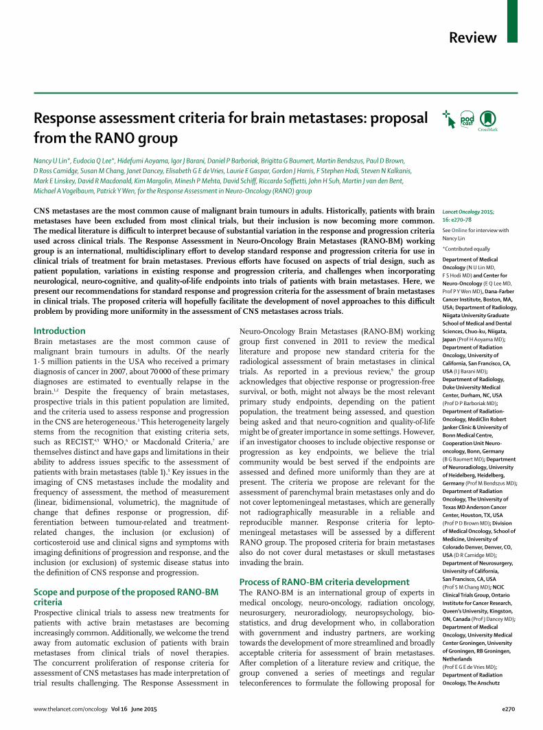
Imaging modality
Target lesion Maximum number of CNS target lesions
Measurement technique
Shrinkage required for partial response
Confi rmatory scans Steroids Neurological symptoms
Extracranial disease
RECIST 1.05 CT or MRI Longest diameter ≥10 mm
Five Unidimensional ≥30% Required in non-randomised trials where response in the primary endpoint
Not included Not included Included
RECIST 1.14 CT or MRI Longest diameter ≥10 mm
Two Unidimensional ≥30% Required in non-randomised trials where response in the primary endpoint
Not included Not included Included
Macdonald7 CT or MRI Minimum size not specifi ed
Not specifi ed Bidimensional ≥50% Required at least 1 month apart
Stable or decreased Stable to improved Not applicable
WHO6 Not specifi ed
Minimum size not specifi ed
All lesions Bidimensional ≥50% Required at least 4 weeks apart
Not included Not included Included
RANO (high-grade glioma)8
CT or MRI Contrast-enhancing lesions with two perpendiculardiameters ≥10 mm
At least two lesions, and up to fi ve lesions in patients with multiple lesions*
Bidimensional ≥50% Required at least 4 weeks apart
Stable or decreased compared with time of baseline scan
Stable to improved clinically
Not applicable
*For patients with multiple lesions, of which only one or two are increasing in size, the enlarging lesions should be considered the target lesions and other lesions will be considered non-target lesions.
Table 1: Comparison of standard response criteria

www.thelancet.com/oncology Vol 16 June 2015 e272
Review
USA; and Neuro-Oncology Unit, Daniel den Hoed Cancer Center, Erasmus University Medical Center, Rotterdam, Netherlands (Prof M J van den Bent MD)
Correspondence to:Dr Nancy U Lin, Department of Medical Oncology, Dana-Farber Cancer Institute, Boston, MA 02215, [email protected]
regarded as unchanged from baseline unless there is a minimum 3 mm change in the measured longest diameter.
The decision to include patients with multiple lesions with a sum diameter of 10 mm or more but of which the largest lesion measures less than 10 mm should be taken with caution if objective response is the primary endpoint. If such patients are included, response should be assessed using the sum of the longest diameters of the lesions, and the response criteria should be clearly delineated in the protocol. Thin-section MRI imaging with 1·5 mm or thinner slice thickness would be necessary in this setting (appendix).
Methods of measurementThe same method of assessment and the same technique should be used to characterise each identifi ed and reported lesion at baseline and during follow-up. Consistent use of imaging techniques across all imaging timepoints is important to ensure that the assessment of interval appearance, disappearance of lesions, or change in size is not aff ected by scan parameters such as slice thickness. Use of thin section imaging (appendix) is particularly important for the assessment of lesions less than 10 mm in longest diameter or small changes in lesion size, or both.
Gadolinium-enhanced MRI is the most sensitive and reproducible method available to measure CNS lesions selected for response assessment.10,11 Suggested brain MRI specifi cations are detailed in the appendix. MRI is strongly encouraged as the default standard imaging technique, although CT with and without contrast could be considered in specifi c circumstances (eg, countries with limited medical resources or contraindication for MRI).
Tumour-response assessmentOnly patients with measurable CNS disease at baseline should be included in protocols where objective CNS tumour response is the primary endpoint. For studies in which objective response is not the primary endpoint, the protocol must specify prospectively whether entry is restricted to those with measurable disease or if patients with non-measurable disease are also eligible. Assignment of CNS response is independent of systemic disease response. CNS lesions are to be assessed according to RANO-BM criteria, whereas non-CNS lesions would most typically be assessed according to RECIST 1.1 criteria. Generally, CNS lesions should initially be re-assessed by MRI at protocol-specifi ed intervals 6–12 weeks apart, although there might be specifi c circumstances in which longer (or shorter) intervals are desirable. For patients who remain stable for extended periods of time, a longer interval between scans might be appropriate.
All baseline assessments should be done as close as possible to the treatment start and no more than 4 weeks before the beginning of treatment. For previously treated lesions, we recommend documentation of how each
lesion was previously treated (eg, stereotactic radio-surgery, whole brain radiotherapy, surgical resection). When more than one measurable lesion in the CNS is present at baseline, all lesions up to a maximum of fi ve CNS lesions should be identifi ed as target lesions and will be recorded and measured at baseline. All measurements should be recorded in metric notation. Target lesions should be selected on the basis of their size (longest diameter) and as those that can be measured reproducibly. For patients with recurrent disease who have multiple lesions, of which only one or two are increasing in size, the enlarging lesions should be prioritised as target lesions for the response assessment. Lesions with prior local treatment (ie, stereotactic radiosurgery or surgical resection) can be considered measurable if progression has occurred since the time of local treatment. However, careful consideration should be given to lesions previously treated with stereotactic radiosurgery, in view of the possibility of treatment eff ect, which we discuss below. Whether such lesions can be considered measurable should be specifi ed prospectively in the clinical protocol. If lesions not previously treated with local therapies are present, these are preferred for selection as target lesions. A sum of the diameters for all target lesions will be calculated and reported as the baseline sum of longest diameters. All other CNS lesions should be identifi ed as non-target lesions and should also be recorded at baseline. Measurements are not required and these lesions should be classifi ed as present, absent, or unequivocal pro-gression, and followed up.
Figure 1: Axial contrast-enhanced T1-weighted MRI of a brain metastasis from breast carcinoma with a partial solid and cystic componentOnly the solid component is used for measurement of the longest diameter.
See Online for appendix

e273 www.thelancet.com/oncology Vol 16 June 2015
Review
Defi nition of best overall CNS responseBest overall CNS response is a composite of radiographical CNS target and non-target lesion responses (panel 1), corticosteroid use, and clinical status. For non-randomised trials in which CNS response is the primary endpoint, confi rmation of partial response or complete response at least 4 weeks later is necessary to deem either one the best overall response.
At each protocol-specifi ed timepoint, a response assess-ment should occur and CNS assessments should be coincident with extra-CNS assessment. Table 2 shows the additional corticosteroid and clinical status requirements to deem a partial response or complete response.
Assessment of target and non-target CNS lesionsWhile on study, all CNS target lesions should have their actual measurement recorded, even if very small (eg, 2 mm). If the lesion disappears, the value should be recorded as 0 mm. However, if the lesion is suffi ciently
small (but still present) to be assigned an exact measure, a default value of 5 mm should be recorded on the case report form.
Lesions might coalesce during treatment. As lesions coalesce, a plane between them may be maintained that would aid in obtaining maximum longest diameter of each individual lesion. If the lesions have truly coalesced such that they are no longer separable, the vector of the longest diameter in this instance should be the maximum longest diameter for the coalesced lesion.
New lesions can appear during treatment. The fi nding of a new CNS lesion should be unequivocal and not due to technical or slice variation. A new lesion is one that was not present on prior scans. If the MRI is obtained with slice thickness of 1∙5 mm or less, the new lesion should also be visible in axial, coronal, and sagittal reconstructions of 1·5 mm or thinner projections. If a new lesion is equivocal, for example because of its small size (ie, ≤5 mm), continued therapy can be considered, and a follow-up assessment will clarify if it really is new disease. If repeated scans confi rm a new lesion, progression should be declared using the date of the initial scan showing the new lesion. In the case of immunotherapy, however, new lesions alone cannot constitute progressive disease.
Unequivocal progression of non-target lesions can merit discontinuation of therapy. When a patient also has measurable disease, to be deemed as having unequivocal progression on the basis of non-target disease alone there must also be an overall substantial worsening in non-target disease such that, even in the presence of stable disease or partial response in target disease, the overall tumour burden has increased suffi ciently to merit discontinuation of therapy. When the patient has only non-measurable disease, there must be an overall level of substantial worsening to merit discontinuation of therapy.
The RANO-BM group acknowledges the case of patients who have been treated with stereotactic radiosurgery12 or immunotherapy-based approaches, for whom there has been radiographical evidence of enlargement of target and non-target lesions, which do not necessarily represent tumour progression. If radiographical evidence of progression exists, but clinical evidence indicates that the radiological changes are due to treatment eff ect (and not to progression of cancer), additional evidence is needed to distinguish between true progression and treatment eff ect, in which case standard MRI alone is insuffi cient. The methods used to distinguish between true progression and treatment eff ect should be specifi ed prospectively in the clinical protocol. Patients can be continued on protocol therapy pending further investi-gation with one or more of the following options.
The scan can be repeated at the next protocol-scheduled assessment or sooner, and generally within about 6 weeks. An investigator can choose a shorter time interval if progressive symptoms or other clinical concerns arise. Continued tumour growth might be consistent with
Panel 1: Response assessment of target and non-target lesions
Target lesionsComplete responseDisappearance of all CNS target lesions sustained for at least 4 weeks; with no new lesions, no use of corticosteroids, and patient is stable or improved clinically.
Partial responseAt least a 30% decrease in the sum longest diameter of CNS target lesions, taking as reference the baseline sum longest diameter sustained for at least 4 weeks; no new lesions; stable to decreased corticosteroid dose; stable or improved clinically.
Progressive diseaseAt least a 20% increase in the sum longest diameter of CNS target lesions, taking as reference the smallest sum on study (this includes the baseline sum if that is the smallest on study). In addition to the relative increase of 20%, at least one lesion must increase by an absolute value of 5 mm or more to be considered progression.
Stable diseaseNeither suffi cient shrinkage to qualify for partial response nor suffi cient increase to qualify for progressive disease, taking as reference the smallest sum longest diameter while on study.
Non-target lesionsNon-target lesions should be assessed qualitatively at each of the timepoints specifi ed in the protocol.
Complete responseRequires all of the following: disappearance of all enhancing CNS non-target lesions, no new CNS lesions.
Non-complete response or non-progressive diseasePersistence of one or more non-target CNS lesion or lesions.
Progressive diseaseAny of the following: unequivocal progression of existing enhancing non-target CNS lesions, new lesion(s) (except while on immunotherapy-based treatment), or unequivocal progression of existing tumour-related non-enhancing (T2/FLAIR) CNS lesions. In the case of immunotherapy-based treatment, new lesions alone may not constitute progressive disease.
www.thelancet.com/oncology Vol 16 June 2015 e274
Review
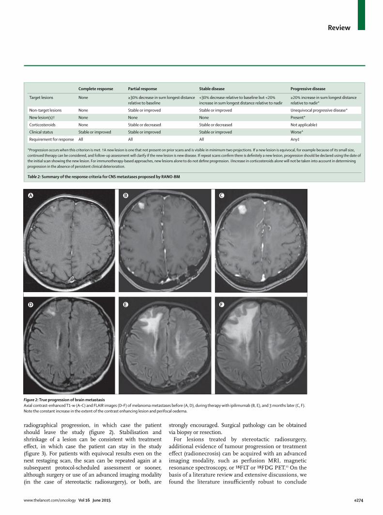
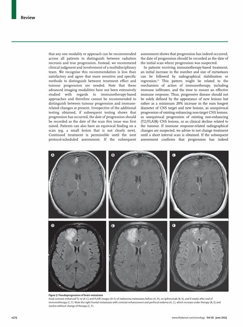
radiographical progression, in which case the patient should leave the study (fi gure 2). Stabilisation and shrinkage of a lesion can be consistent with treatment eff ect, in which case the patient can stay in the study (fi gure 3). For patients with equivocal results even on the next restaging scan, the scan can be repeated again at a subsequent protocol-scheduled assessment or sooner, although surgery or use of an advanced imaging modality (in the case of stereotactic radiosurgery), or both, are
strongly encouraged. Surgical pathology can be obtained via biopsy or resection.
For lesions treated by stereotactic radiosurgery, additional evidence of tumour progression or treatment eff ect (radionecrosis) can be acquired with an advanced imaging modality, such as perfusion MRI, magnetic resonance spectroscopy, or ¹⁸FLT or ¹⁸FDG PET.13 On the basis of a literature review and extensive discussions, we found the literature insuffi ciently robust to conclude
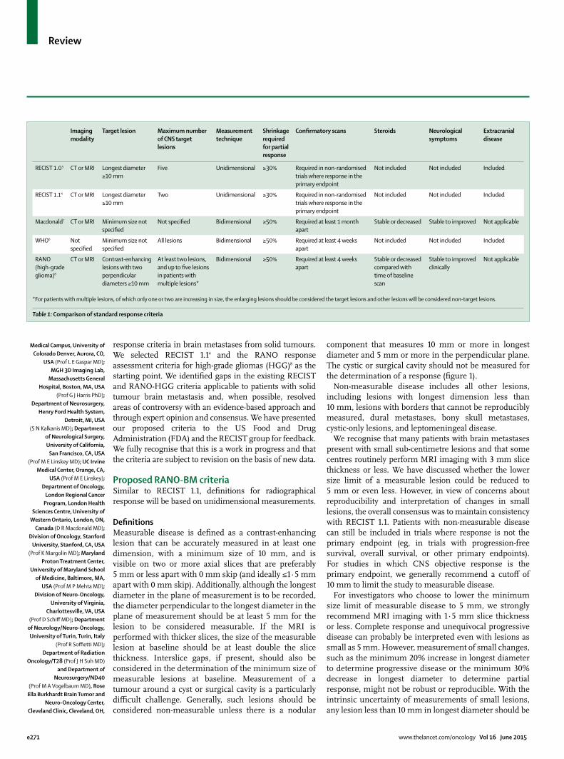
Complete response Partial response Stable disease Progressive disease
Target lesions None ≥30% decrease in sum longest distance relative to baseline
<30% decrease relative to baseline but <20% increase in sum longest distance relative to nadir
≥20% increase in sum longest distance relative to nadir*
Non-target lesions None Stable or improved Stable or improved Unequivocal progressive disease*
New lesion(s)† None None None Present*
Corticosteroids None Stable or decreased Stable or decreased Not applicable‡
Clinical status Stable or improved Stable or improved Stable or improved Worse*
Requirement for response All All All Any‡
*Progression occurs when this criterion is met. †A new lesion is one that not present on prior scans and is visible in minimum two projections. If a new lesion is equivocal, for example because of its small size, continued therapy can be considered, and follow-up assessment will clarify if the new lesion is new disease. If repeat scans confi rm there is defi nitely a new lesion, progression should be declared using the date of the initial scan showing the new lesion. For immunotherapy-based approaches, new lesions alone to do not defi ne progression. ‡Increase in corticosteroids alone will not be taken into account in determining progression in the absence of persistent clinical deterioration.
Table 2: Summary of the response criteria for CNS metastases proposed by RANO-BM
Figure 2: True progression of brain metastasisAxial contrast-enhanced T1-w (A–C) and FLAIR images (D–F) of melanoma metastases before (A, D), during therapy with ipilimumab (B, E), and 3 months later (C, F). Note the constant increase in the extent of the contrast enhancing lesion and perifocal oedema.
A C
E
B
D F
e275 www.thelancet.com/oncology Vol 16 June 2015
Review
that any one modality or approach can be recommended across all patients to distinguish between radiation necrosis and true progression. Instead, we recommend clinical judgment and involve ment of a multidisciplinary team. We recognise this recommendation is less than satisfactory and agree that more sensitive and specifi c methods to distinguish between treatment eff ect and tumour progression are needed. Note that these advanced imaging modalities have not been extensively studied with regards to immunotherapy-based approaches and therefore cannot be recommended to distinguish between tumour progression and immune-related changes at present. Irrespective of the additional testing obtained, if subsequent testing shows that progression has occurred, the date of progression should be recorded as the date of the scan this issue was fi rst raised. Patients can also have an equivocal fi nding on a scan (eg, a small lesion that is not clearly new). Continued treatment is permissible until the next protocol-scheduled assessment. If the subsequent
assessment shows that progression has indeed occurred, the date of progression should be recorded as the date of the initial scan where progression was suspected.
In patients receiving immunotherapy-based treat ment, an initial increase in the number and size of metastases can be followed by radiographical stabilisation or regression.14 This pattern might be related to the mechanism of action of immunotherapy, including immune infi ltrates, and the time to mount an eff ective immune response. Thus, progressive disease should not be solely defi ned by the appearance of new lesions but rather as a minimum 20% increase in the sum longest diameter of CNS target and new lesions, as unequivocal progression of existing enhancing non-target CNS lesions, as unequivocal progression of existing non-enhancing (T2/FLAIR) CNS lesions, or as clinical decline related to the tumour. If immune response-related radiographical changes are suspected, we advise to not change treatment until a short interval scan is obtained. If the subsequent assessment confi rms that progression has indeed
Figure 3: Pseudoprogression of brain metastasisAxial contrast-enhanced T1-w (A–C) and FLAIR images (D–F) of melanoma metastases before (A, D), on ipilimumab (B, E), and 6 weeks after end of immunotherapy (C, F). Note the right frontal metastases with contrast enhancement and perifocal oedema (A, C), which increase under therapy (B, E) and resolve without change of therapy (C, F).
A C
E
B
D F

www.thelancet.com/oncology Vol 16 June 2015 e276
Review
occurred, the date of progression should be recorded as the date of the initial scan where progression was suspected.
Corticosteroid use and clinical deteriorationIn the absence of clinical deterioration related to the tumour, an increase in corticosteroid dose alone should not be used as a sole determinant of progression. Patients with stable imaging results and whose corticosteroid dose has increased for reasons other than clinical deterioration related to the tumour do not qualify as having stable disease or progression. These patients should be observed closely, and if their corticosteroid dose can be reduced back to baseline, they will be considered as having stable disease, but if further clinical deterioration related to the tumour becomes apparent, they will be considered as having progression.
The defi nition of clinical deterioration is left to the discretion of the treating physician, but it is recommended that patients who have a decrease in score on the Karnofsky performances scale from 100 or 90 to 70 points or less, a decrease of minimum 20 points from 80 or less, or a decrease from any baseline to 50 points or less, for at least 7 days, be considered as having neurological deterioration, unless this functional impairment is attributable to comorbid events, treatment-related toxicity, or changes in corticosteroid dose.
Volumetric criteriaResearch of the value of volumetric versus unidimensional measurements for the assessment of CNS lesion response is ongoing.15–18 Volumetric measurement was the topic of much discussion and debate within the RANO-BM group. The RANO-BM group judges that the existing data are not yet strong enough to justify the universal requirement of volumetric response criteria in clinical trials of patients with brain metastases. Volumetric analyses in real-time adds cost and complexity and is not available at all centres.
Yet, RANO-BM also believes that the assessment and reporting of volumetric response in clinical trials (in addition to the unidimensional RANO-BM criteria) will add to the knowledge base, either justify or negate the need for volumetric measurements in future trials, and encourage its inclusion as a secondary endpoint when feasible.
The appropriate cutoff to defi ne a partial response on the basis of volumetric measurements was another topic of debate. If a tumour forms a perfect sphere, a 30% unidimensional reduction corresponds to about a 65% volumetric reduction, and there are data showing concordance of response assessments with these cut-off s in patients with brain metastasis.17 Also, volumetric changes of minimum 20% appear to be reproducible between readers,19,20 and results of one study21 showed that 20% or greater volumetric reduction was associated with improvements in neurological signs and symptoms.
The RANO-BM group believes that use of the same criteria and cutoff s across trials will allow trial results to be interpreted in their proper context. Thus, for investigators who choose to report volumetric response data, we propose the following. First, partial volumetric response should be defi ned as a 65% or greater decrease in the sum volume of CNS target lesions, in addition to the corticosteroid and clinical status criteria as outlined previously. Second, volumetric response should be reported as a waterfall plot to provide a global sense of potential effi cacy. Third, in the absence of high quality data across multiple studies to show a clear correlation between lower volumetric thresholds and some measure of patient benefi t, such as quality of life, neuro-cognitive function, or overall survival, it is premature to formally defi ne a category of minor response or to lower the threshold at which to consider a volumetric response. However, we encourage digital archiving of trial images and accompanying linked clinical outcome data to allow
CNS (RANO-BM) Non-CNS (RECIST 1.1) Response
Complete response, partial response, or stable disease
Complete response, partial response, or stable disease
Log as CNS and non-CNS complete response, partial response, or stable diseases
Complete response, partial response, or stable disease
Progressive disease Log as CNS complete response, partial response, or stable disease; log as non-CNS progressive disease
Progressive disease Complete response, partial response, or stable disease
Log as CNS progressive disease; log as non-CNS complete response, partial response, or stable disease
Progressive disease Progressive disease Log as both CNS and non-CNS progressive disease
Table 3: CNS and non-CNS response assessment
CNS (RANO-BM) Non-CNS (RECIST 1·1) Bi-compartmental PFS Note
Complete response, partial response, or stable disease
Progressive disease Log as a progression-free survival event Log as non-CNS progressive disease
Progressive disease Complete response, partial response, or stable disease Log as a progression-free survival event Log as CNS progressive disease
Progressive disease Progressive disease Log as a progression-free survival event Log as both CNS and non-CNS progressive disease
Table 4: Bi-compartmental progression-free survival
e277 www.thelancet.com/oncology Vol 16 June 2015
Review
for studies to be pooled to determine whether diff erent cutpoints could be justifi ed in the future.
Treatment of non-CNS (extracranial) diseasePreclinical and clinical data sometimes show a diff erential response in intracranial versus extracranial locations, which could be related to inadequate drug penetration, diff erences in tumour microenvironment, or tumour heterogeneity between organ sites, among other possibilities. Many systemic agents are not expected to have CNS activity, primarily because of poor drug penetration. Local CNS therapies, such as whole-brain radiotherapy, stereotactic radiosurgery, or surgery, are not expected to aff ect extracranial sites at all.
Traditionally, RECIST has used a summation of representative target lesions across all organ sites. Historically, patients with brain metastases have been excluded from systemic therapy trials. Even when included, patients with brain metastases often had to have stable, treated CNS lesions on study entry, and CNS lesions were rarely chosen as target lesions. The Macdonald and RANO-HGG criteria do not provide guidance about the treatment of extracranial disease, since extracranial disease is not relevant in most patients with primary brain tumours. The consequences have been an absence of fl exibility to continue protocol therapy in the setting of discordant CNS versus non-CNS response or progression, a disincentive to image the brain as part of clinical trials, and the use of diff erent defi nitions of response and progression endpoints in local therapy trials and systemic therapy trials.
We propose that CNS and non-CNS should be assessed as separate compartments (table 3). As such, CNS response
will be scored irrespectively of extra cranial response and vice versa. For progression, CNS and non-CNS will be scored according to RANO-BM and RECIST 1.1 criteria, respectively (table 4). If progression occurs in either or both compartments, the criteria for bi-compartmental progression-free survival will have been met. Protocols can also prospectively specify CNS progression-free survival and non-CNS progression-free survival as endpoints. Protocols should specify the plan for patients who progress in one compartment only. For example, a patient who develops isolated CNS progression in a systemic therapy trial can be given the option to have their CNS disease treated with whole-brain radiotherapy, stereotactic radiosurgery, or surgery and remain on protocol therapy until the time of non-CNS disease progression, unacceptable toxicity, or death. The date of non-CNS progressive disease should be recorded when it occurs.
Additional endpoints for localised therapy trialsPatients with brain metastases frequently undergo focal treatments such as surgical resection and stereotactic radiosurgery. With these modalities, the technical success of the treatment is appropriately measured by assessment of the site of localised therapy and not distant sites. For example, outcomes after stereotactic radio-surgery are commonly reported as local control (ie, control of the treated lesion) and distant brain failure (ie, the appearance of new or progressive lesions outside the treated fi eld). This situation is analogous to breast cancer, in which trials of locoregional therapy will commonly report endpoints such as ipsilateral invasive breast cancer recurrence or regional invasive breast cancer recurrence.22 Panel 2 outlines the RANO-BM-proposed defi nitions of bicompartmental progression-free survival, CNS progression-free survival, non-CNS progression-free survival, and local CNS progression-free survival, which account for the variety of trial endpoints that might be chosen depending on the clinical situation, treatment modality, and overall study goal.
ConclusionWe recognise that our proposal adds complexity to the assessment of patients with brain metastases enrolled in clinical trials. However, limitations of the existing response criteria have led to frequent, but inconsistent, modifi cations by investigators. Additionally, because brain metastases can be treated using multiple modalities, which might or might not have eff ects outside of the treated fi eld or outside the brain, endpoints in trials have also been defi ned diff erently according to the modality. Whereas the choice of primary and secondary endpoints will naturally vary according to the treatment modality, overall study goal, and study type (eg, proof of concept, technical validation, phase 3 registration study), we believe the defi nition of the endpoints should ideally remain constant. Frequently asked questions are listed and answered in the appendix.
Panel 2: Sites of inclusion for assessment of bi-compartmental progression-free survival, CNS progression-free survival, non-CNS progression-free survival, and CNSlocal progression-free survival
Bi-compartmental progression-free survivalInclude local CNS lesions, distant CNS lesions, and non-CNS lesions
CNS progression-free survivalInclude local CNS lesions and distant CNS lesions
Non-CNS progression-free survivalInclude non-CNS lesions only
CNSlocal progression-free survivalInclude local CNS lesions only
Search strategy and selection criteria
We searched Medline, PubMed, and the references of relevant articles using the following search terms: “brain metastases”, “breast cancer”, “lung cancer”, “melanoma”, “whole brain radiotherapy”, “stereotactic radiosurgery”, and “radiation necrosis”. Additional cross-referenced search terms were added for specifi c topics such as “volumetric”, “perfusion MRI”, “positron emission tomography”, and “immunotherapy”. We included only articles published in English between Jan 1, 1980, and Oct 1, 2014.
www.thelancet.com/oncology Vol 16 June 2015 e278
Review
Future plans include collaborations with RECIST investigators to analyse historical datasets and to solicit feedback from other investigators to refi ne the proposed criteria in future iterations. However, we should note that any retrospective analysis of historical datasets will be limited by the quality and nature of the recorded data. For example, because very few studies simultaneously collect unidimensional, bidimensional, and volumetric measurements, retrospective studies of large datasets are unlikely to provide answers to all of the questions raised above unless there is a large-scale eff ort to collect archival images and conduct central radiology review. In addition, because information for corticosteroid use, functional status, neurological symptoms, neuro-cognitive functioning, and quality of life were also variably collected and assessed, associations between response and functional outcomes will be challenging to validate. We would encourage investigators interested in the specialty of brain metastasis to strategise together on how best to gather the necessary common data elements across trials to allow such analyses in the future.
ContributorsAll authors contributed to the literature search and writing of the report.
MB prepared the fi gures.
Declaration of interestsDPB has a leadership position in ECOG-ACRIN and declares non-
fi nancial support from GE Medical Systems, outside the submitted work.
BGB declares personal fees and non-fi nancial support from Roche
Pharma AG, academic institutions, and Bayer Pharma AG, outside the
submitted work. MB declares grants and personal fees from Guerbet,
Codman, Bayer, personal fees from Novartis and Vascular Dynamics,
grants from Siemens, personal fees from Roche, outside the submitted
work. FSH declares grants and non-fi nancial support from Bristol-Myers
Squibb, personal fees from Merck, non-fi nancial support from
Genentech, personal fees and non-fi nancial support from Novartis,
outside the submitted work; in addition, FSH has a patent immune
therapy target pending. NUL declares grants from Breast Cancer Research
Foundation, during the conduct of the study, grants to support clinical
trials from Array Biopharma, Genentech, GlaxoSmithKline, Novartis, and
Kadmon. DRM declares personal fees from Roche Canada and personal
fees and non-fi nancial support from Merck Canada, outside the submitted
work. MPM declares consulting relationships with AbbVie, BMS, Celldex,
Electa, Genentech, Merck, Novelos, Novocure, and Philips and has served
on the Board of Directors of Pharmacyclis with stock options, unrelated to
the submitted work. MJvdB declares grants and personal fees from Roche
and AbbVie, personal fees from Amgen, MSD, Actelion, and Merck,
outside the submitted work. MAV declares personal fees and non-
fi nancial support from Merck, personal fees from Neuralstem and
Pharmacokinesis, personal fees and other from Infuseon Therapeutics,
and grants and non-fi nancial support from National Cancer Institute,
outside the submitted work. PYW declares research support from AbbVie,
Agios, Anigochem, AstraZeneca, Exelixis, Genentech/Roche, GlaxoSmith
Kline, Karyopham, Merck, Novartis, Sanofi -Aventis, Vascular Biogenics,
Celldex, SigmaTau, Midatech, Momenta, outside the submitted work. HA,
IJB, PDB, DRC, SMC, JD, EGEdV, LEG, GJH, SNK, EQL, MEL, KM, DS,
RS, and JHS declare no competing interests.
AcknowledgmentsWe thank Patricia Cortazar, Suzanne Demko, Ruthann Giusti,
Patricia Keegan, and Richard Pazdur from the US Food and Drug
Administration for their feedback regarding these proposed radiographic
criteria. This work was supported in part by the Breast Cancer Research
Foundation (grant to NUL), but this support did not directly infl uence the
writing of this manuscript or the decision to submit it for publication.
References1 Davis FG, Dolecek TA, McCarthy BJ, Villano JL. Toward
determining the lifetime occurrence of metastatic brain tumors estimated from 2007 United States cancer incidence data. Neuro Oncol 2012; 14: 1171–77.
2 American Cancer Society. Cancer Facts & Figures. Atlanta, GA: American Cancer Society; 2007.
3 Lin NU, Lee EQ, Aoyama H, et al. Challenges relating to solid tumour brain metastases in clinical trials, part 1: patient population, response, and progression. A report from the RANO group. Lancet Oncol 2013; 14: e396–406.
4 Eisenhauer EA, Therasse P, Bogaerts J, et al. New response evaluation criteria in solid tumours: revised RECIST guideline (version 1.1). Eur J Cancer 2009; 45: 228–47.
5 Therasse P, Arbuck SG, Eisenhauer EA, et al. New guidelines to evaluate the response to treatment in solid tumors. European Organization for Research and Treatment of Cancer, National Cancer Institute of the United States, National Cancer Institute of Canada. J Natl Cancer Inst 2000; 92: 205–16.
6 Miller AB, Hoogstraten B, Staquet M, Winkler A. Reporting results of cancer treatment. Cancer 1981; 47: 207–14.
7 Macdonald DR, Cascino TL, Schold SC Jr, Cairncross JG. Response criteria for phase II studies of supratentorial malignant glioma. J Clin Oncol 1990; 8: 1277–80.
8 Wen PY, Macdonald DR, Reardon DA, et al. Updated response assessment criteria for high-grade gliomas: response assessment in neuro-oncology working group. J Clin Oncol 2010; 28: 1963–72.
9 Lin NU, Wefel JS, Lee EQ, et al. Challenges relating to solid tumour brain metastases in clinical trials, part 2: neurocognitive, neurological, and quality-of-life outcomes. A report from the RANO group. Lancet Oncol 2013; 14: e407–16.
10 Schellinger PD, Meinck HM, Thron A. Diagnostic accuracy of MRI compared to CCT in patients with brain metastases. J Neurooncol 1999; 44: 275–81.
11 Sze G, Milano E, Johnson C, Heier L. Detection of brain metastases: comparison of contrast-enhanced MR with unenhanced MR and enhanced CT. AJNR Am J Neuroradiol 1990; 11: 785–91.
12 Patel TR, McHugh BJ, Bi WL, Minja FJ, Knisely JP, Chiang VL. A comprehensive review of MR imaging changes following radiosurgery to 500 brain metastases. AJNR Am J Neuroradiol 2011; 32: 1885–92.
13 Shah R, Vattoth S, Jacob R, et al. Radiation necrosis in the brain: imaging features and diff erentiation from tumor recurrence. Radiographic 2012; 32: 1343–59.
14 Wolchok JD, Hoos A, O’Day S, et al. Guidelines for the evaluation of immune therapy activity in solid tumors: immune-related response criteria. Clin Cancer Res 2009; 15: 7412–20.
15 Bauknecht HC, Romano VC, Rogalla P, et al. Intra- and interobserver variability of linear and volumetric measurements of brain metastases using contrast-enhanced magnetic resonance imaging. Invest Radiol 2010; 45: 49–56.
16 Harris GJ, Plotkin SR, Maccollin M, et al. Three-dimensional volumetrics for tracking vestibular schwannoma growth in neurofi bromatosis type II. Neurosurgery 2008; 62: 1314–20.
17 Sze G, Mehta M, Schultz C, et al. Radiologic response evaluation of brain metastases: uni-dimensional (1D), W.H.O. RECIST vs bi-dimensional (2D) or 3-dimensional (3D) criteria. Proc Am Soc Clin Oncol 2001; 20 (suppl): abstr 234.
18 van der Weide H, de Kunder S, Houben R, Bosmans G, Lambin P, Baumert BG. Infl uence of metastatic volume on local control and survival assessed by 3D volumetric measurements after radiosurgery in patients with brain metastases and planned observation. Strahlenther Onkol 2011; 187: 523–24 (abstr).
19 Yang DY, Sheehan J, Liu YS, et al. Analysis of factors associated with volumetric data errors in gamma knife radiosurgery. Stereotact Funct Neurosurg 2009; 87: 1–7.
20 Pan HC, Cheng FC, Sun MH, Chen CC, Sheehan J. Prediction of volumetric data errors in patients treated with gamma knife radiosurgery. Stereotact Funct Neurosurg 2007; 85: 184–91.
21 Lin NU, Dieras V, Paul D, et al. Multicenter phase II study of lapatinib in patients with brain metastases from HER2-positive breast cancer. Clin Cancer Res 2009; 15: 1452–59.
22 Hudis CA, Barlow WE, Costantino JP, et al. Proposal for standardized defi nitions for effi cacy end points in adjuvant breast cancer trials: the STEEP system. J Clin Oncol 2007; 25: 2127–32.