respiratory system trachea bronchus (bronchi) bronchioles
TRANSCRIPT


Respiratory SystemRespiratory System
trachea
bronchus (bronchi)
bronchioles
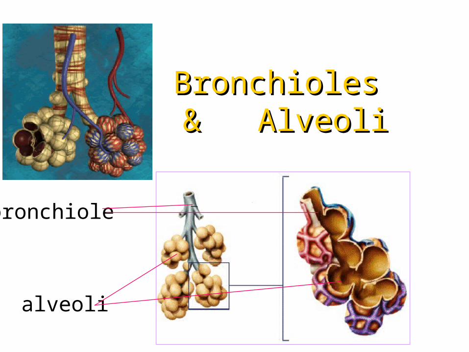
Bronchioles Bronchioles & Alveoli& Alveoli
bronchiole
alveoli

Nasal CavityNasal Cavity• Nasal cavity possess hairs for
trapping large dirt particles
• wall of nasal cavity lined with ciliated
epithelium and mucus-secreting cells
to trap dirt and bacteria
• near surface are numerous blood vessels
so incoming air are warmed, moistened
& filtered before entering lungs
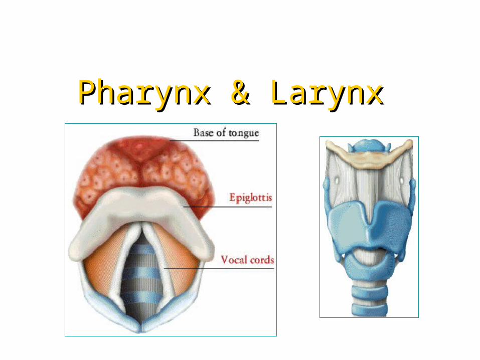
Pharynx & LarynxPharynx & Larynx
• pharynx belongs to both
respiratory & digestive system
• glottis is the opening of larynx and it is
covered by epiglottis during swallowing
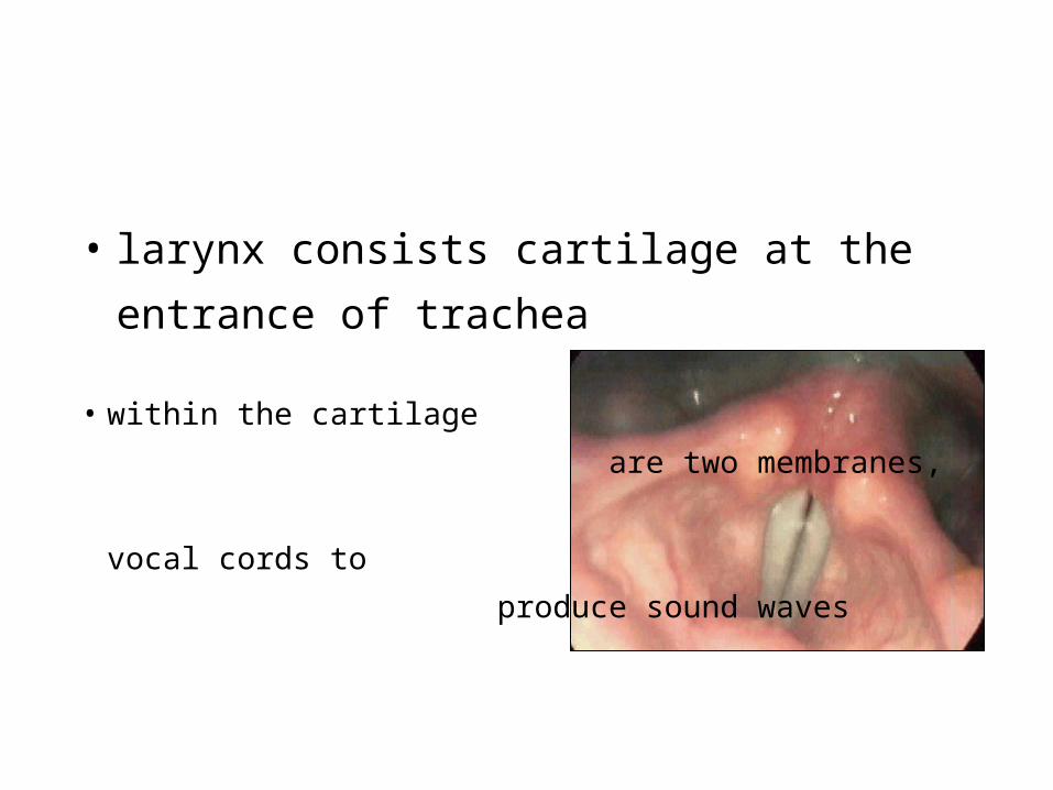
• larynx consists cartilage at the
entrance of trachea
• within the cartilage
are two membranes,
vocal cords to
produce sound waves
Trachea (Windpipe) Trachea (Windpipe) and Bronchiand Bronchi
• trachea lies in front of oesophagus
and extended into thoracic cavity
• at the lower end of trachea is
divided into two bronchi which
subdivides into many bronchioles
• each bronchiole terminates in
hollow, lobed air sacs called alveoli
• inner lining of the trachea
produces mucus and possess cilia
• mucus are used to trap the dirt &
germ while cilia are used to waft the
mucus towards the throat, it is then
either coughed out or swallowed.
Those coughed out are called phlegm
• wall of trachea strengthened by C-
shaped cartilages to keep trachea
open
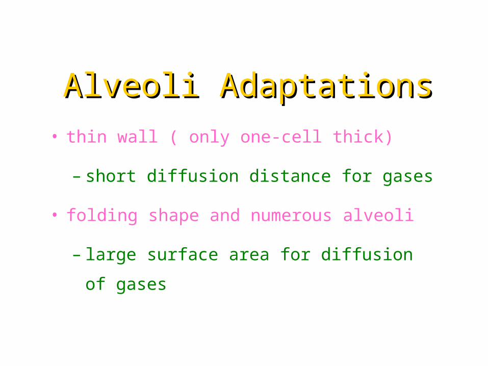
Alveoli AdaptationsAlveoli Adaptations
• thin wall ( only one-cell thick)
– short diffusion distance for gases
• folding shape and numerous alveoli
– large surface area for diffusion of
gases
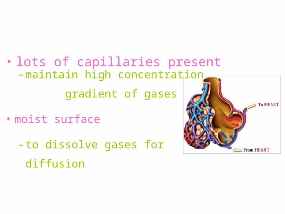
• lots of capillaries present– maintain high concentration
gradient of gases
• moist surface
– to dissolve gases for diffusion

Structure of LungStructure of Lung• lungs are protected by the thoracic
basket which is made up of
vertebrae, ribs and sternum
• each lung is surrounded by two
pleural membranes
• the inner membrane is in contact with the
lungs and the outer membrane lines against
the walls of the thorax and diaphragm
• between the two membranes is
pleural cavity which contains a pleural
fluid secreted by the membranes

• pleural fluid
lubricates the pleura
so to reduce friction
as the pleural
membranes rub
against each other
during breathing
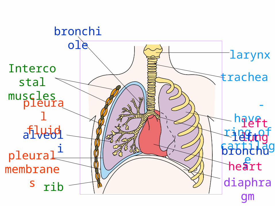
rib
pleural membran
es
alveoli
pleural fluid
Intercostal
muscles
bronchiole
larynx
trachea -
have ring of
cartilageleft lungleft
bronchusheart
diaphragm
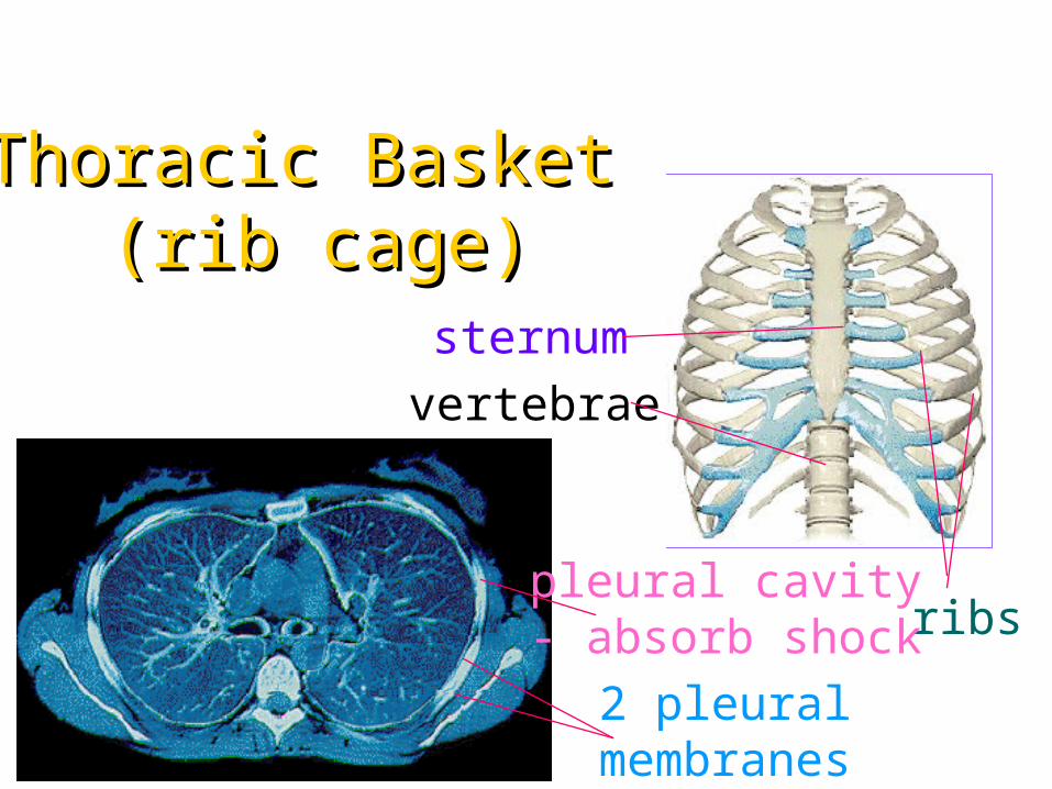
Thoracic Basket Thoracic Basket (rib cage)(rib cage)
vertebraesternum
ribspleural cavity- absorb shock
2 pleural membranes

Gaseous Gaseous Exchange in Exchange in
AlveoliAlveoli
tissue
Lungs(alveoli)CO2 (by
plasma and in form of
HCO3- )
O2 + haemoglobin
atmosphere
CO2O2
oxyhaemoglobin
(by red blood cell)
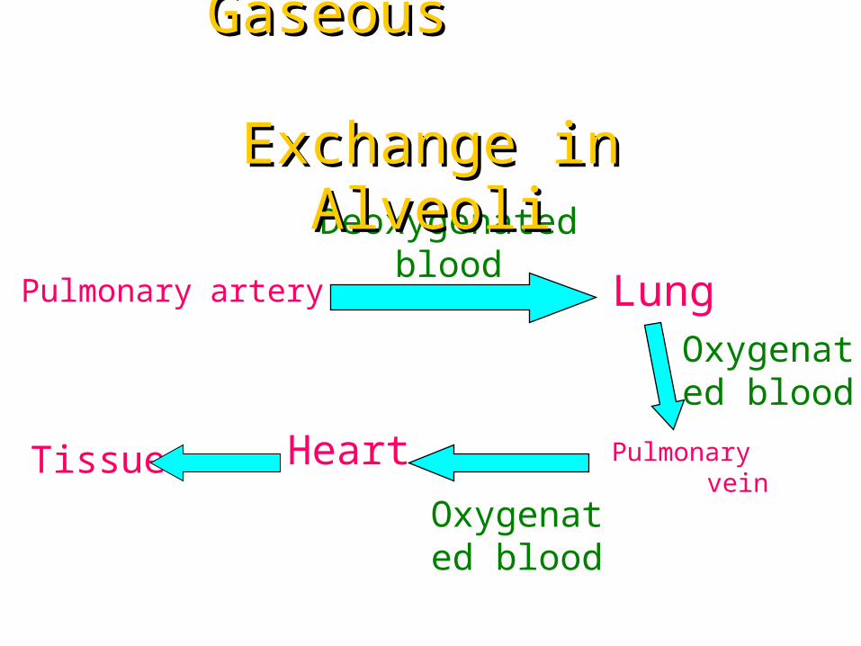
Heart
Deoxygenated blood
Oxygenated blood
Oxygenated blood
Lung Pulmonary artery
Pulmonary vein
Tissue
Gaseous Gaseous Exchange in Exchange in
AlveoliAlveoli
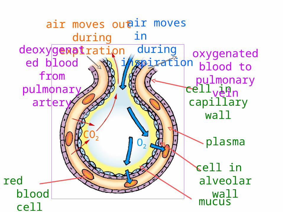
air moves out during
expiration
air moves in during
inspiration
O2
CO2
cell in capillary wall
plasma
cell in alveolar
wallmucus
red blood cell
deoxygenated blood
from pulmonary
artery
oxygenated blood to
pulmonary vein

Mechanism of BreathingMechanism of Breathing
• brought about by the action of diaphragm & intercostal muscles
• divided into two processes : inhalation (inspiration) & exhalation (expiration)

Inspiration (Inhalation)Inspiration (Inhalation)Diaphragm muscle- contract
Intercostal muscle - contract
Diaphragm- flattened
ribs & sternum - move upward
& outward

thoracic cavity expands (volume
increases)
air pressure in lung is lower than atmospheric pressure so air rushes
in
lungs inflated (expand)
Inspiration Inspiration (Inhalation)(Inhalation)

Expiration (Exhalation)Expiration (Exhalation)
intercostal muscles relax
diaphragm muscles relax
diaphragm becomes dome-shaped
ribs and sternum move downwards & inwards
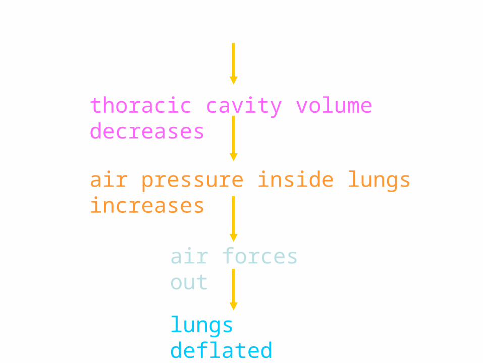
thoracic cavity volume decreases
air pressure inside lungs increases
air forces out
lungs deflated

Walking & RunningWalking & Running
time (seconds)
205 10 150
1000
2000
3000
lun
g v
olu
me (
cm
)
3
at rest
lun
g v
olu
me (
cm
)
time (seconds)
5 10 150
1000
2000
30003
20
during exercise

Calculations of the Rate Calculations of the Rate and Depth of Breathingand Depth of Breathing
• From the graphs the volume of
air he breathed in per minute
at rest and during exercise can
be measured :
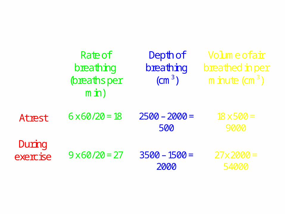
Rate ofbreathing
(breaths permin)
Depth ofbreathing
(cm3)
Volume of airbreathed in perminute (cm3)
At rest 6 x 60/20 = 18 2500 – 2000 =500
18 x 500 =9000
Duringexercise 9 x 60/20 = 27 3500 – 1500 =
2000
27 x 2000 =
54000
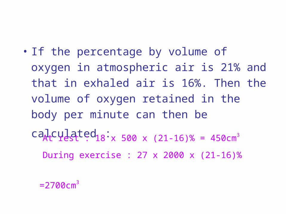
• If the percentage by volume of oxygen in atmospheric air is 21% and that in exhaled air is 16%. Then the volume of oxygen retained in the body per
minute can then be calculated : At rest : 18 x 500 x (21-16)% = 450cm3
During exercise : 27 x 2000 x (21-16)%
=2700cm3

COCO22 remains Constant remains Constant during Exerciseduring Exercise
CO2 concentration in blood remains
CONSTANT
muscles release CO2
ventilation rate increases
+
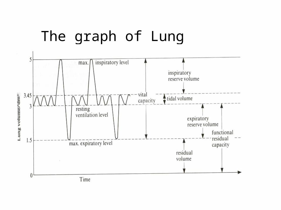
The graph of Lung capacities

Tidal Volume Tidal Volume
– amount of air entering & leaving the
lungs during normal breathing
– during exercise → can increase
volume

– during exercise → cannot increase
the capacity
– increase only after prolong training
– maximum air exhaled after taking
the deepest inhalation
Vital CapacityVital Capacity

– volume of air remaining in the lungs
which cannot be expelled even after
forced exhalation
Residual VolumeResidual Volume

Smoking & HealthSmoking & Health
• Effects on health :
(I) Tar :
(i) carcinogenic ( producing cancer )
(II) Nicotine :
(i) cause heart diseases
carbon monoxide
• Composition of a cigarette :
= cigarette+ tar+ nicotine
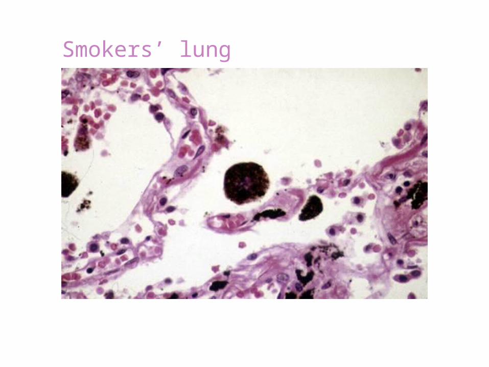
Smokers’ lung

(ii) dependence
(iii) retards growth of foetus
(III) Carbon Monoxide :
(i) combines irreversibly with haemoglobin and
prevents it from carrying oxygen
+ COhaemoglobin
(ii) decrease in physical fitness
(iii) cause air pollution
carboxyhaemoglobin

Smoking and Health Hazards
1.Lung Cancer
Smoking increase the risk of lung cancer
2.Heart disease
Nicotine increases the workload of heart
increase the rate of heart attack
Smoking and Health Hazards
3.Chronic bronchitis Smoking causes inflammation of trachea and
bronchitis The severe chronic bronchitis can cause death
4.Emphysema Causes of "smoker's cough" Smoker cough causes damage of alveolar wall
in lungs
Passive SmokingPassive Smoking• process of breathing in smoke from
cigarette smokers
• causes nose, throat & eye irritations
Warning
DO NOT SMOKE !!!!!