respiratory pathogens of military importance… respiratory pathogens of military importance: a...
TRANSCRIPT

NAVAL HEALTH RESEARCH CENTER
RESPIRATORY PATHOGENS OF MILITARY IMPORTANCE:
A REVIEW
G. C. Gray
Report No. 96-17
Approved (or public »teas*: distribution unllmiMd.
NAVAL HEALTH RESEARCH CENTER P.O. BOX85122
SAN DIEGO, CAUFORNIA 92186-5122
NAVAL MEDICAL RESEARCH AND DEVELOPMENT COMMAND BETHESDA, MARYLAND
V, "-J ~««i
Executive Summary
Problem. Respiratory pathogens have been and continue to be, a major cause of morbidity among military populations.
Objective. This manuscript was designed as a review for military medical officers. It describes the epidemiology, clinical manifestations and control strategies for the most commonly encountered bacterial and viral pathogens.
Approach. After a historical review of respiratory pathogens and the US Department of Defense's efforts to contain them, the text offers a pathogen-specific analysis of the most common etiologic agents. Streptococcus pyogenes, Streptococcus pneumoniae, Mycoplasma pneumoniae, Chlamydia pneumoniae, Influenza, and adenoviruses are reviewed.
Results. The Department of Defense has learned much regarding respiratory pathogens and their control. As these pathogens change and new threats emerge, military public health officials must adapt to meet their challenge.
Conclusions. The modern military medical officer will benefit from a knowledge of the epidemiology of respiratory pathogens. These and other bacterial and viral agents will continue to cause morbidity among military populations.
19 March 1997
Respiratory Pathogens of Military Importance: A Review*
CDR Gregory C. Gray, MC, USN Naval Health Research Center Emerging Illness Team P.O. Box 85122 San Diego, California 92186-5122
Respiratory pathogens have been, and continue to be, a major cause of morbidity among military populations. This review describes the epidemiology, clinical manifestations and control strategies for the most commonly encountered bacterial and viral pathogens. Streptococcus pyogenes, Streptococcus pneumoniae, Mycoplasma pneumoniae, Chlamydia pneumoniae, influenza, and adenoviruses are examined. As these pathogens change and new threats emerge, military public health policy must adapt to meet these challenges.
Respiratory pathogens have plagued military populations throughout U.S. history. Records from the War of 1812, the Spanish American War, and World War I document their devastation and the inability of public health officials to control them.1"3 Especially well documented are the epidemics that occurred during the mobilization for World War I.
In 1918, a 30-day epidemic of Streptococcus pneumoniae occurred at a military camp in Illinois, causing 2,349 hospital admissions for pneumonia, with a 50% mortality.4 During 1918, a 2-month military epidemic of influenza in Little Rock, Arkansas, affected 12,393 men, and led to 1,499 cases of pneumonia, 31% of whom died during treatment.5 Other reports record that hemolytic streptococci {Streptococcus pyogenes) were a continual cause of military epidemics of bronchopneumonia, empyema, and pharyngitis. In total,
*This document was prepared as Chapter 20, Diseases Spread by Close Personal Contact, of the book, Military Preventive Medicine: Mobilization and Deployment. Kelley PW, ed. Washington DC: Bordon Institute; 1997. It represents technical report no. 96-17, supported by the Naval Medical Research and Development Command, Bethesda, Maryland, Department of the Navy, under work unit no. 61102.0101.BKX-6425. The views expressed in this article are those of the author and do not reflect the official policy or position of the Department of the Navy, Department of Defense, or the U.S. Government.
it was estimated that more than 1.4 million U.S. Army personnel suffered from respiratory disease during World War I, accounting for more than 41% of all forms of disease and causing more than 77% or 45,000 of Army disease deaths.2
Before antimicrobials were widely available, strategies to prevent military epidemics of respiratory disease were not very successful. Generally, military public health officers could do little more than attempt to isolate and treat the afflicted with various therapies against streptococci, which included digitalis, whiskey, strychnine, and various horse serums.6"8 Following the success of researchers in South Africa, U.S. military officials attempted to control S. pneumoniae epidemics at select U.S. Army camps during World War I with a crude vaccine,910 but their efforts were unsuccessful.311
Commenting upon the trade-off between isolating the ill and compromising the mission, one military medical official of 1917 wrote that "Exposure to infection and hardships which will result in deaths from pneumonia may be just as necessary as going into action with resulting deaths from gunshot wounds."7
Despite the limited effective interventions available to preantibiotic-era public health officials, important observations were made regarding the types of acute respiratory diseases and their apparent bacterial causes. It was noted that crowding greatly contributed to respiratory epidemics,12,13 and more important than reduced floor space in sleeping quarters was the number
Respiratory Pathogens
of men placed in the same room.13 Southerners, blacks, and new military personnel from rural areas were thought to be at highest risk of developing pneumonia. Measles and influenza epidemics were observed to trigger epidemics of S. pneumoniae and S. pyogenes.
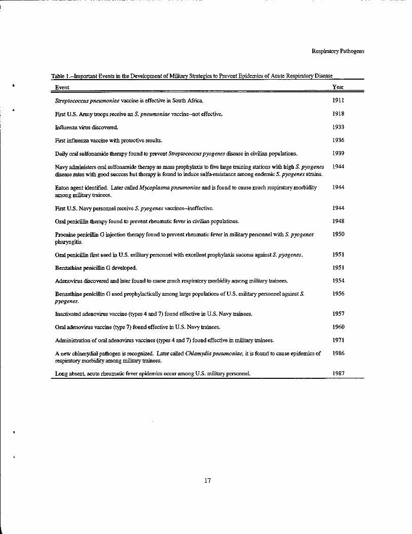
Military policy officials were determined to reduce respiratory disease during the mass mobilization of personnel for World War n. Beginning in 1941, the U.S. Department of War established a Board for the Investigation and Control of Influenza and Other Epidemic Diseases in the Army. Later, this board assembled various commissions of scientific experts and established numerous public health and research facilities to study and prevent respiratory disease.14 These joint military and university endeavors led to many of the antibiotic and vaccine prophylaxis interventions now used in military populations (Table 1).
As a result of these and subsequent research efforts, today most U.S. military personnel receive BPG or oral erythromycin prophylaxis, adenovirus vaccines, tuberculosis screening, and influenza vaccine during their first military training. This care is followed by annual influenza vaccines and periodic tuberculosis screening throughout their military careers. The pathogens recognized to cause respiratory disease among these young adults are similar to those causing community-acquired disease among the general U.S. adult population.15"18 Most frequently, they include S. pyogenes, Mycoplasma pneumoniae, S. pneumoniae, Chlamydia pneumoniae, adenoviruses, influenza viruses, and rhinoviruses. Less frequently, military personnel also suffer infections from Haemophilus influenzae, Haemophilus parainfluenzae, Legionella pneumophila, Moraxella catarrhalis, Bordetella pertussis, coxsackieviruses, respiratory syncytial virus, and parainfluenza viruses. The most significant pathogens to military populations are reviewed here in more detail.
Streptococcus pyogenes
S. pyogenes is the leading cause of bacterial respiratory morbidity among military personnel. New military trainees are particularly at high risk of clinically significant infection. During the late 1940s and the 1950s, dedicated scientific teams, sponsored by Army Board Commissions, worked at a number of Army, Navy, Air Force, and Marine Corps training centers and made much progress understanding and controlling this pathogen.
S. pyogenes is a Gram-positive coccus, a spherical bacteria, which, when grown on sheep blood agar, causes a clear zone of complete hemolysis (ß-hemolysis). It is distinguished from other ß-hemolytic streptococci by physiological and immunologic characteristics and by its implication as a cause of numerous acute clinical manifestations, including pharyngitis, peritonsillar abscess, pneumonia, empyema, scarlet fever, necrotizing fasciitis, myositis, bacteremia, and streptococcal toxic shock syndrome. S. pyogenes also causes the nonsuppurative manifestations of acute rheumatic fever and glomerulonephritis. The severity of 5. pyogenes infections has changed over time. In the late 1800s, epidemics of scarlet fever were common and associated with high mortality rates.19 Today, epidemics of scarlet fever are relatively rare.20 In a similar fashion, acute rheumatic fever was very common during the mobilization for World War n, with 21,000 cases recorded in the Navy alone, while until the 1980s, few cases were detected among military populations.21-22-23 Changes in disease rates have been attributed to changes in the prevalence of virulent strains of S. pyogenes.24 S. pyogenes strains are classified according to their capsular proteins, specifically T and M proteins. An excess of 90 unique M protein types have been commonly identified. M types 1,3, and 18 are associated with acute rheumatic fever, and M types 12, 1, 3, 4, and 25 often associated with glomerulonephritis.
The epidemiology of 5. pyogenes infection has been well described in military populations. Studies by military and university investigators in the 1950s demonstrated that, most often, transmission is by direct contact or large respiratory droplets, and not commonly by fomites. Geographically, the bacteria are thought to be endemic throughout the world, with perhaps some variation in the endemnicity of certain strains. Without prophylaxis, recent serologic evidence demonstrates that as much as 24% of military trainees maybe infected over an 11-week period.25 With prophylaxis, the recent serologic evidence demonstrates a reduction in attack rates to 11% to 13%.26-27
Risk factors for serologic evidence of S. pyogenes infection include being new to the military, crowding, lack of prophylaxis, close contact with an S. pyogenes carrier, and close contact with a nonprophylaxed trainee.27 A retrospective study of civilians with invasive S. pyogenes infections suggested that Native Americans, young persons, and persons with less underlying illness maybe at higher risk of severe S. pyogenes disease.28 Monthly
Respiratory Pathogens
BPG injections and twice-a-day oral erythromycin have a recently estimated efficacy of preventing serologic evidence of infection of 45% and 56%, respectively.26,29
S. pyogenes colonizes respiratory mucosal cells, causing pharyngitis and clinical manifestations, such as fever and leukocytosis in 36 to 72 hours. The complex interaction of cellular and extracellular S. pyogenes products with the host immune system is not well understood, however, it is recognized that immunity to S. pyogenes is type-specific. Some patients progress to more invasive forms of infection, including streptococcal toxic shock syndrome, bacteremia, and necrotizing fasciitis. S. pyogenes pyrogenic exotoxins A and B and other virulence factors have been implicated in severe infection. Numerous theories regarding the interaction of host immune response and S. pyogenes virulence factors have been postulated; however, it remains unclear why one subject is severely infected by a particular strain of S. pyogenes and another subject is merely colonized with the same S. pyogenes strain and suffers no symptoms.
Even in high prevalence situations such as epidemics, it is difficult to distinguish clinically S. pyogenes pharyngitis from that caused by other pathogens. Clinically, diagnosis is best made by culture or rapid antigen detection. Generally, a rapid antigen test is accepted if positive, but it should be confirmed by culture if negative. Since a high proportion of infected military persons may not seek medical attention despite symptoms, epidemiological studies of S. pyogenes generally are conducted by relying upon serologic tests, particularly the antistreptolysin 0 test. A two-dilution rise in antistreptolysin 0 titer is considered evidence of infection. Generally, such a rise may be detected in paired sera drawn 2 to 3 weeks apart. Other serologic tests for S. pyogenes infection include antideoxyribonuclease B, antihyaluronidase, and a hemagglutination test (Streptozyme). Some patients with glomerulonephritis or symptoms of acute rheumatic fever may not demonstrate a rise in antistreptolysin O titer, and should be studied further with antideoxyribonuclease B or antihyaluronidase tests before ruling out S. pyogenes as a cause.
Advances in S. pyogenes control were first made in civilian populations when it was discovered that continuous antimicrobial therapy prevented recurrence of rheumatic fever. Later it was learned that administering antibiotics to military personnel with pharyngitis could reduce the incidence of new acute rheumatic fever cases. Eventually, healthy military populations at high risk for S. pyogenes disease were studied with mass antimicrobial
prophylaxis against acute rheumatic fever and this intervention became standard practice for crowded training populations.
Sulfonamides were some of the first antibimicrobials available, and during the 1940s they were tested in various forms to prevent military epidemics of acute respiratory disease.21 Large field trials of daily 0.5 to 1.0 g of sulfadiazine among healthy Navy personnel resulted in an 85% reduction in the incidence of streptococcal infections and rheumatic fever;30 however, daily prophylactic use caused exfoliative dermatitis and granulocytopenia in a small proportion of recipients. Bacteriostatic sulfonamide prophylaxis also often failed to eradicate 5. pyogenes from the nasopharynx of military personnel, and as soon as the therapy was discontinued, epidemics recurred. However, the most serious drawback occurred when a Navy sulfonamide prophylaxis program reported that sulfonamide-resistant strains of S. pyogenes had become endemic after only one year of treating military personnel. After the failure of sulfonamides, other methods to reduce morbidity from S. pyogenes were attempted. Public health officials tried various environmental controls, including reductions in crowding,13 dust suppression,31 ultraviolet radiation,32 and disinfectant vapors,33 with varying degrees of success. Chlortetra-cycline also was tested as a mass prophylaxis agent, but it caused significant gastrointestinal side effects.34 Military and university scientists also unsuccessfully attempted to control S. pyogenes epidemics with inactivated, type-specific vaccines.35
Environmental controls, such as dust suppression by oiling floors and blankets, were largely abandoned when it was learned that penicillins were effective in the treatment and prevention of 5. pyogenes disease.21 Oral penicillin therapy was first noted as effective in preventing recurrent rheumatic fever in civilian populations in 1948.36
Later, both oral and procaine penicillin G were shown to be effective in preventing rheumatic fever among healthy high risk military personnel; however, their use in large military populations was logistically difficult due to the need for frequent dosing.343738 The development of benzathine penicillin G (BPG) in 1951 led to its 1956 successful, large-scale prophylactic testing in a military population.39 BPG's long-acting prophylactic effect, assurance of compliance, and few side effects soon won its acceptance as the standard prophylactic intervention for the U.S. Department of Defense (DoD) against S. pyogenes, and it remains an effective interventive tool today.21 BPG also has been used to combat epidemics of S. pneumoniaef0 and it seems to have a broader protective
Respiratory Pathogens
effect than that explained by just preventing S. pyogenes disease alone.41
Despite the availability of antibiotic prophylaxis and surveillance programs among high-risk populations, military epidemics of S. pyogenes continue to occur. In recent years, these epidemics have taken the form of pharyngitis and acute rheumatic fever.21'23-27 A 1989 epidemic of 5. pyogenes pharyngitis among Marine Corps trainees demonstrated that BPG prophylaxis for non- penicillin-allergic trainees alone might not be enough since unprotected penicillin-allergic recruits were shown to serve as an 5. pyogenes reservoir for reinfecting then- peers.27 This led to the Navy adoption of oral erythromycin prophylactic therapy for penicillin-allergic recruits.2"7
Currently military surveillance among trainees and preventive interventions vary between the services. BPG (1.2 million units intramuscularly, once monthly) and oral erythromycin (250 mg orally, twice a day) interventions have been very effective in controlling S. pyogenes epidemics. BPG remains effective for 2 to 4 weeks after injection. Oral erythromycin suffers from compliance problems due to twice-daily dosing and gastrointestinal side effects. Oral azithromycin (500 mg weekly) recently has been shown to have an 84% efficacy in preventing S. pyogenes infection and may be considered as alternate therapy when a agent with a broader spectrum is desired.29
Recent reports of erythromycin-resistant S. pyogenes isolates and epidemics due to penicillin-tolerant S. pyogenes strains are causes for concern. Fortunately, thus far no penicillin-resistant S. pyogenes isolates have been detected clinically, however, periodic surveillance of endemic strains among high-risk training populations should be conducted. This surveillance should contain antibiotic sensitivity testing of isolates as well as strain typing.
Ideally, the best hope for preventing 5. pyogenes disease among military populations lies with the development of vaccines. Several approaches are being considered, including various immunologic presentations of shared epitopes of various capsular M proteins; however, vaccines most likely will not be available for a number of years.
Streptococcus pneumoniae
A frequent cause of pneumonia in adults, S. pneumoniae (pneumococcus) causes significant morbidity among U.S. military populations. In the preantibiotic era,
S. pneumoniae infections could lead to large epidemics exceeding several hundred cases, particularly after influenza outbreaks. Today, S. pneumoniae epidemics occur less often, but they remain a threat. Most recently, an epidemic of 124 pneumonia hospitalizations was recorded among U.S. Marine trainees in Southern California, triggering mass BPG and pneumococcal vaccine injections.40
S. pneumoniae is an ovoid, Gram-positive coccus, which often forms distinctive pairs and chains. It grows well on sheep blood agar, causing partial hemolysis (a- hemolysis), and it is distinguished from other streptococci by chemical growth inhibition and immunologic reaction. S. pneumoniae causes various forms of pneumonia, meningitis, empyema, bacteremia, conjunctivitis, sinusitis, and arthritis. Eighty-four recognized strains or types are classified by distinct capsular polysaccharides.
S. pneumoniae is spread by respiratory droplets or person-to-person contact. It is thought not to have geographical limitations, but data are sparse regarding the geographical distribution of capsular types. The incidence of disease among military personnel has not been well studied. Navy data from 1981 to 1991 suggest that S. pneumoniae causes approximately 12% of military pneumonia hospitalizations, which occur at a rate of 9.5 per 100,000 person-years.42 Because the incidence of outpatient disease is unknown and there are diagnostic difficulties identifying this pathogen, these estimates greatly underestimate the impact of this pathogen. Personnel at increased risk include the immunocompromised, asplenic individuals, persons with sickle cell disease, renal disease, and diabetes mellitus. Military recruits are at high risk of S. pneumoniae infection.
S. pneumoniae is often found on the epithelium of healthy nasopharynx tissue and its pathogenesis is not well understood. Other respiratory pathogens, especially viruses, may serve as a cofactor for local S. pneumoniae tissue invasion, which if unchecked, may lead to clinical disease. Immunity is capsular type-specific and thought to last for years.
Because S. pneumoniae is considered normal, oral bacterial flora, it is difficult to confidently diagnose infection from the oral pharynx. The accepted clinical diagnostic gold standard is bacterial culture from a normally sterile site. Blood cultures from patients with S. pneumoniae pneumonia, if studied before antibiotic administration, should be positive 20% of the time. A clinically expedient and alternative diagnostic tool for S.
Respiratory Pathogens
pneumoniae pulmonary infection is a well-prepared sputum specimen. Gram-stained sputum specimens should contain few to no squamous cells per low-powered microscopic field. The numerous serologic techniques available to assess S. pneumoniae infection generally are confined to research institutions and involve detecting antibody to pneumococcal proteins or capsular polysaccharide. Generally, a rise in antibody titer from acute to convalescent sera is considered evidence of recent infection. A latex agglutination test for pneumococcal antigens in urine has been found to have some diagnostic value.
For militarypersonnel, the 23-valent polysaccharide pneumococcal vaccine is the best protection against 5. pneumoniae infection. In 1991, the Armed Forces Epidemiological Board recommended a single dose of this vaccine be given to asplenic individuals and military personnel at bases with high prevalence of pneumonia. It is used routinely in high-risk Marine Corps trainee populations during winter months. BPG, 1.2 million units intramuscularly, has been used to combat pneumococcal pneumonia epidemics, but it has never been evaluated for efficacy.40 A recent study of oral azithromycin, 500 mg weekly, demonstrated an 80% efficacy of preventing serologic evidence of pneumococcal infection.29
The recent rapid spread of clinically important, penicillin-resistant S. pneumoniae strains throughout the United States and other developed countries have frustrated clinicians. National U.S. surveillance has demonstrated increasing prevalence of penicillin- nonsusceptible strains and increasing numbers of strains and serotypes with multiple antibiotic resistance. National public health panels have called for increased surveillance for antibiotic resistance among S. pneumoniae isolates, careful use of antibiotics, and increased use of pneumococcal vaccine among high risk populations.
Mycoplasma pneumoniae
Long before microbiologists had distinguished agents causing acute respiratory disease etiologic agents, differences were noted in the clinical manifestations of pneumonias. Militarypersonnel frequently suffered from acute pneumonia, which was milder than lobar pneumonia. Although this atypical pneumonia demonstrated significant pulmonary involvement by chest radiograph, patients clinically lacked the high fever, pleuritic chest pain, and rigor associated with lobar pneumonia from S. pneumoniae. In some Army camps,
85% to 90% of pneumonias were of the atypical variety.43
The agent(s) causing this atypical pneumonia or primary atypical pneumonia (PAP) were a matter of some debate. Often, patients with atypical pneumonia were cold agglutinin positive. In 1944, Eaton described bis DoD- funded research, which demonstrated that a filterable agent taken from patients with atypical pneumonia could cause pulmonary lesions in rats. In 1961, Chanock described his DoD-funded research, which demonstrated that the Eaton agent was responsible for 68% of atypical pneumonias among Marine trainees and that as many as 44% of recruits had serologic evidence of infection over a 3-month training period.43 Later, the agent was renamed M. pneumoniae and confirmed to cause much acute respiratory disease among U.S. militarypersonnel.
Termed a pleuropneumonia-like organism (PPLO), M. pneumoniae lacks a rigid cell wall and is much smaller than other bacteria. It grows very slowly on special nutrient agar, and isolation techniques most often are performed by reference laboratories. M. pneumoniae grows on the surface of the epithelial cells, which line the respiratory tract. Generally, it is not considered an agent of the nasopharyngeal flora. However, due to its extracellular existence, it may be found in respiratory excretions weeks after clinical disease. M. pneumoniae infections are noted for their gradual onset of symptoms, dry cough, malaise, headache, and chills. While some infections may be asymptomatic, M. pneumoniae commonly causes a pharyngitis and may cause bronchopneumonia with patchy pulmonary infiltrates radiating from hilar areas. Occasionally, M. pneumoniae may cause severe pneumonia or severe disease of the central nervous system, including meningoencephalitis, aseptic meningitis, ascending paralysis, and transverse myelitis.44 M. pneumoniae also has been reported to cause various forms of cardiac disease, arthritis, and numerous dermatological conditions.44
M. pneumoniae is transmitted by respiratory droplet inhalation or person-to-person contact and has a worldwide geographical distribution. Among U.S. military populations infection risk increases in late summer,45,46
and females may be at higher risk of infection than males42,47,4S Certainly, crowding contributes to infection risk Antibody to this pathogen is common among young adults. A recent study demonstrated that upon service entry 57% of Marine trainees had evidence of previous M. pneumoniae infection.49 The prevalence and incidence of infection among U.S. military training populations as measured by serologic antibody titer change is high,
Respiratory Pathogens
especially during outbreaks. One study demonstrated seroconversion in as many as 57% of recruits over an 11- week period. Routine incidence in military training centers is more likely similar to the 6% to 8% detected among recent Marine Corps training populations over 3- month periods.29,49
Due to the low mortality and diagnostic difficulties, the pathogenesis of M. pneumoniae infections has not been well determined. The pathogen adheres to epithelial cell receptors, and after infection, antibodies to M. pneumoniae surface antigens are formed, which offer protection from further infection. Many M. pneumoniae infections evoke immunoglobulin M autoantibody, which agglutinates human erythrocytes (cold agglutinins) and, in some cases, may trigger an autoimmunogenic mycoplasma-receptor complex, which can lead to hemolytic anemias.32
Using special nutrient agar, M. pneumoniae may be isolated from the nasopharynx after several weeks of incubation. For the best yield, a pharyngeal culture should be inoculated immediately into nutrient agar broth for incubation. Reference laboratories will need several weeks to isolate and identify M. pneumoniae from clinical specimens. It is distinguished from other mycoplasmas by colony morphology, growth, and metabolic inhibition. Identification may be confirmed with serologic or molecular methods.
Clinically, the diagnosis of M. pneumoniae infection may be presumed if the symptom complex is consistent with disease and the patient has a positive cold agglutinin test (titer 11:32 in convalescent sera). A positive test is more common with severe pneumonia. However, this test lacks specificity in that approximately 50% of infected patients may have a negative cold agglutinin test. The cold agglutinin test is additionally problematic in that it maybe falsely positive in the presence of hematologic and hepatic diseases. Alternatively, acute M. pneumoniae infection may be diagnosed by detecting high IgM titers specific for M. pneumoniae50 or detecting M. pneumoniae-speci&c nucleic acid with PCR.51,52 Although several commercial diagnostic kits have been marketed for rapid diagnostic use in the clinical laboratory, their sensitivity and specificity have not approached that of reference laboratory serologic assays.
Both complement fixation and ELISA serologic assays have been used effectively to detect M. pneumoniae in epidemiological studies. Generally, these tests are performed by a reference laboratory, and a fourfold rise in antibody titer by either method (acute to 3- to 4- week
convalescent sera) is accepted as evidence of recent infection.50'53
The DoD conducts no specific surveillance for M. pneumoniae infection among military populations. The U.S. Army's surveillance program for acute respiratory disease41 would include morbidity from M. pneumoniae, but only in the aggregate with that from other pathogens. At present, the DoD has no sustained M. pneumoniae research program and no M. pneumoniae reference laboratory. Military investigators must rely upon other academic or federal laboratories for diagnostic assistance.
Few options are available for combating M. pneumoniae epidemics. Although several studies have demonstrated that preexisting antibody titers against M. pneumoniae may prevent infection,54,55 and vaccine candidates were tested in the 1960s and 1970s with mixed success,56"58 no vaccine is available. In 1965, a 10-day course of oxytetracycline was used prophylactically in an attempt to prevent disease in family members of patients.48
This 4-times-a-day regimen was reported to have a prophylactic effect. However, this result has never been validated. Recently, Navy researchers demonstrated that weekly oral azithromycin (500 mg) had a 64% protective serologic efficacy against M. pneumoniae among Marines, and this new strategy may hold some promise.29
Chlamydia pneumoniae
First accepted as a new species in 1989, C. pneumoniae has been found to be a frequent cause of
acute respiratory disease in military personnel. In Norway and Finland, C. pneumoniae has been shown to infect as many as 56% of military recruits.59,17 The agent is thought to cause approximately 8% of U.S. pneumonias.60
Like all chlamydia, C. pneumoniae is an obligate intracellular parasite depending upon its host cell for nutrients. It grows poorly on special media, and it is sensitive to freeze-thaw cycling. Many infections may be asymptomatic, and clinical manifestations are often insidious.61 C. pneumoniae has been implicated in pharyngitis (often with hoarseness),61 sinusitis, bronchitis, and lower respiratory tract infection. C. pneumoniae- infected patients often have no marked fever and no elevated white blood count. Some evidence shows that C. pneumoniae maybe involved in coronary heart disease,62
reactive airway disease,63,64 and chronic pharyngitis.65
Newly recognized and difficult to diagnose, C. pneumoniae has not been exhaustively studied. The pathogen is transmitted by person-to-person contact and
Respiratory Pathogens
respiratory droplets.66 Geographically, it has been found in many parts of the world and is thought to be both endemic and epidemic in some populations, with outbreaks lasting from 4 months to 3 years.67 No seasonal variation in risk is apparent.62 The prevalence of antibodies in adults is thought to average about 50%,68,69
with a higher proportion of men having antibodies than women.70 The pathogen is considered responsible for about 10% of pneumonias worldwide, with seroconversion peaking during teenage years at about 10% per year.71 Military training populations may suffer higher rates of infection. A 1989 study of U.S. Marine recruits demonstrated seroconversion in 3.9% over an 11-week training period.45 Another Marine trainee study conducted in 1994 found evidence of 8% seroconversions over a 63- day training period.29 Risk factors for C. pneumoniae infection are not well defined. Since military recruits seem to be at higher risk, crowding likely plays a role in transmission.
limited data are available regarding C. pneumoniae pathogenesis. The pathogen multiplies in macrophages, various connective tissues, and smooth muscle cells.72 A 1989 study of U.S. Marines suggested that a preexisting antibody is protective against serologic evidence of infection.49 However, evidence exists that humans maybe reinfected with C. pneumoniae. Generally, reinfection is milder, but among the elderly, reinfections may lead to severe disease.73,74
Difficulties in diagnosing C. pneumoniae infection are numerous. The pathogen is difficult to culture (sensitivity -50%),72 and due to evidence of an asymptomatic carriage,75'76 some authors argue that isolation apart from other evidence of infection may be misleading. Two serologic methods have been used to diagnose C. pneumoniae infection among young adults: complement fixation and microimmuno-fluorescence (IgM and IgG). Generally, a fourfold rise in liter from acute to convalescent sera is considered evidence of recent infection. A high acute microimmunofluorescence IgM titer also is accepted.77 Complement fixation is less sensitive than the microimmuno-fluorescence method, but the latter is technically more difficult and subject to significant reader error.72 Neither method is widely available, and investigators must rely upon reference laboratories for support. Both serologic methods may be confounded by Chlamydia trachomatis infections, which may cause cross reactions.72-78 Several different PCR diagnostic methods have been developed.79,80 Dacron
swabs are recommended, since other swab types may inhibit PCR technique.
Because effective diagnostic tests are not commercially available, the DoD does not conduct routine surveillance for C. pneumoniae. No vaccine is available, and the only evidence of an effective intervention has been the recent data suggesting that weekly oral azithromycin (500 mg) has a 58% efficacy in preventing serologic evidence of infection.29
Influenza
Before vaccines were available, influenza outbreaks could devastate a military population in a matter of weeks. A 1919 report of a 2-week outbreak of influenza in an Arkansas military camp recorded that the camp hospital received 188 to 486 influenza admissions per day, overwhelming hospital staff who themselves had a 25% incidence of disease.81 Despite the availability and annual use of influenza vaccine, epidemics still occur among U.S. military populations. During 1996, a U.S. Navy ship with a 600-person crew had a 50% attack rate, although 99% of the men had recently received influenza vaccine. Viral isolates indicated that the epidemic strain was not covered by that year's vaccine.
Some of the most studied viruses, influenza viruses are recognized for their antigenic uniqueness and classified into 3 types: A, B, and C. The viruses, especially types A and B, vary their antigenic presentation and cause cyclical pandemics. Type A influenza virus is classified by the antigenic presentation of its surface glycoproteins: hemagglutinin and neuraminidase. Mutations in the genes for these glycoproteins have caused pandemics throughout recent history (1889,1918,1957, and 1968). A major change in glycoproteins is known as an antigenic shift; minor changes are recognized as antigenic drift. Type B influenza virus also is recognized for antigenic drift, causing major epidemics, but the antigenic variation is less than that of type A. Type C influenza virus causes sporadic disease and varies its antigenic presentation to a lesser extent than do types A andB.
Clinically, influenza viral infection may range in manifestations from asymptomatic or cold-like symptoms, to severe pneumonia leading to death. The viruses may cause chills, fever, headache, myalgia, sore throat, backache, sneezing, anorexia, nausea, vomiting, and cough. Pneumonia is a serious complication and is thought to result from secondary bacterial infections about
Respiratory Pathogens
80% of the time, but it also may be due to an influenza virus.
The influenza viruses generally are transmitted by person-to-person contact or through droplet spread from sneezing or coughing. Influenza viruses vary geographically in their antigenic makeup, and surveillance for type A variants is conducted worldwide. Surveillance information is used to anticipate epidemics and select antigenic components for vaccine production.
Influenza epidemics often are explosive and result in high mortality. The 1918 to 1919 influenza pandemic resulted in an estimated 20 million deaths.82 During epidemics, more than 40% of a military population may be affected during a brief period.83 Persons at highest risk of experiencing severe clinical symptoms upon influenza infection are those with chronic cardiac, pulmonary, or renal conditions; those with diabetes mellitus or immunosuppression; pregnant women; and the elderly.
The influenza virus is thought to invade the host via the upper respiratory tract. Viral particles penetrate host respiratory epithelial cells, replicate, and infect neighboring cells. Peak viral loads are reached within 24 hours. Influenza virus is thought to remain largely in the respiratory tract Both secretory and serum antibodies are involved in host defense against influenza virus invasion. After natural infection, immunity is thought to wane after several years. Immunity is dependent upon the particular antigenic presentation of the infection virus.
Influenza is diagnosed by viral culture, antigen detection, or association with other laboratory-proven clinical cases. The diagnoses also may be made retrospectively in epidemiological studies by serologic assay. Numerous public health organizations conduct surveillance for influenza infections. The focus of such surveillance is the antigenic makeup of wild viruses.
Although vaccine work began shortly after the discovery of the influenza virus (Table 1), progress was slow due to viral antigenic variation. The first attempt at human influenza vaccination was published in 1936.84
Early influenza A vaccines contained contaminants from the embryonated egg culture and caused considerable reactions. Additionally, the varying antigenic makeup of influenza isolates was not well understood, and results from early vaccine studies were mixed.
In 1943, the DoD sponsored some of the first influenza vaccine testing among military students at several U.S. sites.85,86 One of the earliest combined influenza A and B vaccine trials among U.S. military students demonstrated a protective efficacy of 69% and
greatly encouraged more research.86 The DoD continued to test various types of influenza vaccines, both inactivated and attenuated,83,87,88 and these studies led to the present strategy of altering the antigenic makeup of this now very successful vaccine. Today, the military relies upon a whole cell, inactivated type of vaccine, which combines the antigenic makeup of type A and B viruses thought to be most threatening for the year ahead. This vaccine is given annually to U.S. military personnel. The DoD also sponsored research demonstrating the safety and effectiveness of using amantadine prophylactically to prevent influenza infection among close contacts.89,90
Today, both amantadine and rimantadine are still so used, but recent evidence suggests that resistant viruses may emerge to both drugs.91,92
Adenovirus
Soon after the discovery of adenoviruses in 1953,93,94
it was learned that these pathogens were an important cause of acute respiratory disease among military personnel, especially recruits.9S In 1958, it was estimated that adenoviruses caused 10% of military recruits to be hospitalized, and during winter months,they accounted for 90% of all recruit hospital admissions.96
Adenoviruses have been classified into 41 serotypes. Each serotype may have antigenically recognizable subtypes. Serotypes 4 and 7 account for most military respiratory epidemics. Types 3,12,14, and 21 also cause acute respiratory disease among military populations, but to a lesser extent. Adenovirus respiratory disease often is manifested by fever, cough, pharyngitis, and rhinitis. The infection may progress to a lower respiratory tract infection, which is generally milder than that caused by S. pneumoniae. Adenoviruses also cause gastrointestinal symptoms, epidemic keratoconjunctivitis, and epidemic pharyngo-conjunctival fever but respiratory disease is the most common presentation among military recruits.
Adenoviruses are transmitted through respiratory droplets and person-to-person contact. Geographically, adenoviruses are thought to have a worldwide distribution, and incidence rates among military trainees often have been high, especially during winter months. In 1958, Hilleman estimated that adenoviruses caused 10% of U.S. Army recruits to be hospitalized, and during winter months, explained 90% of all recruit hospital admissions.96"98 Hilleman also estimated that during the winter months, adenoviruses accounted for 72% of all respiratory disease.98 Most often, adenovirus infections occurred during the first 3 weeks of recruit training,99 and
Respiratory Pathogens
only the newest military personnel were affected.97
However, among Marine trainees, infection was often delayed until post-recruit training.98-100-101 In general, military trainees were found to be at much higher risk of infection than were similar civilian populations.
Studying adenoviruses is confounded by asymptomatic carriage and asymptomatic infection.102-103
It is not understood why some people suffer significant clinical disease upon infection and others remain asymptomatic. Some evidence indicates that adenovirus infection, when associated with infection from other respiratory pathogens, results in more severe disease.104
The virus is thought to invade respiratory tissues, and after a several-day incubation period to cause clinical disease and sometimes viremia. Some adenoviruses may cause prolonged infection, such as pharyngitis. Evidence also suggests that latent adenovirus infection may reactivate and cause clinical disease in the immunocompromised.
Today, adenoviruses are detected through culture and various antigen or nucleic acid detection techniques. Culture and identification of adenovirus is relatively easy, however, serotyping traditionally requires a reference laboratory to perform neutralization tests with specific horse or rabbit antisera. In patients with symptoms, the detection of adenovirus generally is accepted as evidence of infection. Epidemiological studies often rely upon serologic evidence of infection, which is gained through several methods, including complement fixation, neutralization tests, hemagglutination-inhibition antibody tests, and ELBA tests.105
The DoD developed a number of adenovirus vaccines.19-106-107 Early inactivated vaccines against serotypes 3,4, and 7 were effective, given separately or in combination, in greatly reducing military recruit respiratory morbidity.106 However, the inactivated vaccines suffered from production difficulties, and some seed virus cultures were contaminated with other viruses.108,109 Later, live vaccines were developed for serotypes 4,7, and 21. These vaccines caused excellent seroconversion and had few side effects when given orally via enteric coated tablets. The success of the serotypes 4 and 7 vaccines led to their adoption by the DoD as routine preventive therapy in the early 1970s, and they have remained very effective. Due to the infrequency of military epidemics fromserotype 21 virus, the serotype 21 vaccine was not developed further or employed. In addition to vaccine intervention, DoD researchers also explored administering serum immune globulin prophylactically against acute respiratory tract infection.
Results of these trials were mixed with some showing a protective effect, but not as protective as adenovirus vaccine.110"112
Emerging pathogens
With the myriad available antibiotic therapies and an assortment of effective vaccines, one might think that today's military preventive medicine personnel are well equipped to control most respiratory diseases. This might be true if pathogen-host relationships were not changing, but most certainly they have, and military populations continue to suffer from respiratory disease.
In the 1980s, along with more virulent S. pyogenes isolates came a newly recognized manifestation of infection, streptococcal toxic shock syndrome.113-28 This syndrome and other forms of invasive S. pyogenes infection, such as necrotizing fasciitis, have caused considerable alarm among military populations due to their rapid tissue destruction and high mortality rates. Risk factors for these rare invasive diseases have not been well identified but available data suggest that persons with HTV infection, diabetes, alcohol abuse, cancer and varicella infection may be at increased risk.
The success of various antibiotics in controlling S. pneumoniae and S. pyogenes infections may soon be overshadowed by the development of penicillin and erythromycin resistance among these pathogens. Already some DoD clinicians have changed empiric therapies, and the increasing prevalence of antimicrobial resistance promises to be a continual military problem.
Some successful childhood vaccines have caused unexpected adult pathology by postponing natural infection until the adult years. Such is the case with Bordetella pertussis, where recent studies have shown that childhood immunity induced by vaccine wanes in adulthood, and the proportion of U.S. adults susceptible to infection has increased with time.114 A recent study has shown that up to 26% of university students who report 6 or more days of cough may have evidence of acute pertussis.115 A similar study of coughing Marine Corps trainees in 1989 demonstrated that 17% were infected (GC Gray, personal communication). Since the yield of oral culture among B. pertussis adults is poor and no good rapid diagnostic techniques are available, recognizing such infections will be a problem for tomorrow's military clinician.
Respiratory Pathogens
Recommendations for outbreak control and intervention
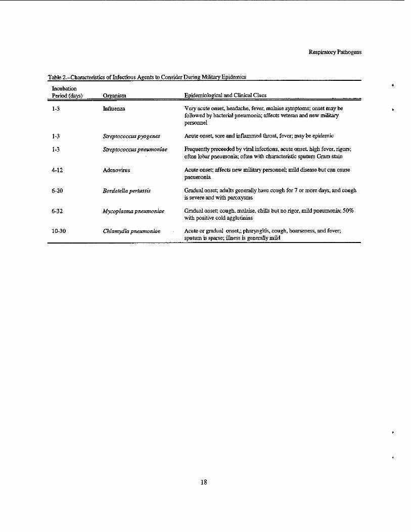
Military respiratory disease outbreaks often occur in an explosive fashion, and if the etiologic agent or agents are not easily recognized, the military preventive medicine officer may face a dilemma: wait for definitive diagnosis while the epidemic continues to build, or venture an empiric intervention that may later be judged inappropriate or expensive, and may have its own morbidity. In the epidemic setting, the preventive medicine officer is wise to collect basic descriptive epidemiological data and seek immediate expert scientific and logistic counsel. He or she should devise a working- case definition and plot out an epidemic curve. Examining the geographic distribution of cases, incubation periods, and the constellation of symptoms will enable the officer to eliminate some pathogens from a list of potential agents (Table 2). Next, it is important to review the availabilities and costs of advanced diagnostic techniques and chemotherapeutic agents.
With expert consultation, the preventive medicine officer should next report his or her findings to the appropriate commanding officer. It is wise to draft a
written report. After reviewing the case definition, consultations, and other background information, the preventive medicine officer should report likely etiologic agents and recommend appropriate intervention. Such interventions might include improved hygiene practices, environmental controls, special diagnostic support, and a vaccine or mass antibiotic prophylaxis, if indicated. Care should be taken to list costs and possible side effects of chemotherapeutic agents. Such a report, if acted upon quickly, may greatly reduce morbidity. Additionally, a written record of considerations will enable reviewers to better understand the decision-making process after the epidemic subsides.
10
Respiratory Pathogens
References
10
11
12
13
Duncan L.C. The days gone, by the medical service in the War of 1812. m. The pneumonia epidemic of 1812-13. Military Surgeon. 1933,72,48-56. Love A.G. A brief summary of the vital statistics of theU.S. Army during the world war. Military Surgeon. 1922,1,139-168. Finland M. Recent advances in the epidemiology of pneumococcal infections. Medicine. 1942,21,307-344. Hirsch EE., McKinney M. An epidemic of pneumococcus bronch-pneumonia. J. Infect. D«.,1919,24,594-617. Opie El., Freeman A.W., Blake F.G., Small J.C., Rivers TM. Pneumonia following influenza (at Camp Pike, Ark.). JAMA 1919,72,556-565. Park JH., dickering H.T. Type I pneumococcus lobar pneumonia among Puerto Rican laborers. JAMA 1919,73,183-186. Nichols HJ. The lobar pneumonia problem in the Army. New York Medical Journal. 1917,106,219-223. Tenney GF., Rivenburgh W.T. A group of sixty- eight cases of Type I pneumonia occurring in thirty days at Camp Upton. Arch. Intern. Med. 1919,24,545-552. Cecil RL, Austin JJH. Results of prophylactic inoculation against pneumococcus in 12,519 men. J. Exp. Med. 1918,28,19. Cecil RJL, Vaughan HP. Results of prophylactic vaccination against pneumonia at Camp Wheeler. J. Exp. Med. 1919,29,457. Denny F.W. The prophylaxis of streptococcal infections. In: Streptococcal Infections. New York, NY: Columbia University Press, 1954,176-196. Gorgas W.C. Recommendation as to sanitation concerning employees of the mines on The Rand made to the Transvaal chamber of mines. 7AMA,1914;62:1855-1865. Breese B.B., Stanbury J., Upham H, Calhoun AJ., Van Buren RL., Kennedy A.S. Influence of crowding on respiratory illness in a large naval training station. War Medicine. 1945,7,143.
14 Woodward TE. The Armed Forces Epidemiological Board. Its First Fifty Years 1940-1990. Washington, D.C.: Government Printing Office, 1990.
15 Amundson DE. Pneumonia in military re-cruits. Mil. Med. 1994,159,629-631.
16 Holmes K.K., Miller FJL, Edwards E.A., Johnson D.W. Etiology of pneumonia in nonrecruit military personnel. Am. J. Med. Sei. 1971,260,264-269.
17 Kleemola M., Saikku P., Visakorpi R., Wang S.P., Grayston J.T. Epidemics of pneumonia caused by TWAR, a new chlamydia organism, in military trainees in Finland. J. Infect. Dis. 1988,157,230-236.
18 LehtomaM K, Leinonen M., Takala A., Hovi T., Herva E., Koskela M. Etiological diagnosis of pneumonia in military conscripts by combined use of bacterial culture and serological methods. Eur. J. Gin. Microbiol. Infect. Dis. 1988,7,348- 354.
19 Rosenbaum MJ., Edwards E.A., Hoefler D.F., Mammen RE., Peckinpaugh R.O., Miller CM. Recent experiences with live adenovirus vaccines in Navy recruits. Mil. Med. 1975,140,251-257. Katz A.R., Morens DM. Severe streptococcal infections in historical perspective. Clin. Infect. Dis. 1992,14,298-307. Thomas RJ., Conwill DJE., Morton DE., Brooks TJ., Holmes C.K., Mahaffey W.B. Penicillin prophylaxis for streptococcal infections in the United States Navy and Marine Corps recruit camps, 1951-1985. Rev. Infect. Dis. 1988,10,125-130.
22 Wallace M.R., Garst PJD., Papadimos TJ., Oldfield E.C. The return of acute rheumatic fever in young adults. JAMA 1989, 262,2557- 2561.
23 Acute rheumatic fever among Army trainees- Fort Leonard Wood, Missouri. MMWR 1988, 37,519-522.
24 Schwartz B., Facklam R.R., Breiman RE. Changing epidemiology of group A streptococcal infection in the USA. Lancet. 1990,336,1167-1171.
20
21
11

Respiratory Pathogens
25 U.S Naval Medical Research Unit No. 4. The 34 prophylaxis and treatment of acute respiratory diseases with antihistimine drugs. J. Lab. Clin. Med. 1950,36,555-590.
26 Fujikawa J., Struewing J.P., Hyams K.C., Kaplan E.L., Tupponce A.K., Gray G.C. Streptococcal 35 prophylaxis for recruits: efficacy of oral erythromycin in prophylaxis of streptococcal infection for penicillin-allergic military recruits: a randomized double-blind study. J. Infect. Dis. 36 1992,166,162-165.
27 Gray G.C, Escamilla J., Hyams K.C., Struewing J.P., Kaplan EL, Tupponce A.K 37 Hyperendemic Streptococcus pyogenes infection despite prophylaxis with penicillin G benzathine. N. Engl. J. Med. 1991,325,92-97.
28 Hoge C.W., Schwartz B., Talkington DJF., 38 Breiman RJF., MacNeil EM., Englender SJ. The changing epidemiology of invasive group A streptococcal infections and the emergence of streptococcal toxic shock-like syndrome. JAMA 1993,269,384-389. 39
29 Gray G.C, McPhate D.C, Leinonen M., et al. Weekly oral azithromycin as prophylactic therapy against bacterial causes of acute respiratory disease. San Diego, CA: Naval 40 Health Research Center. Technical Report 96- 13.
30 Coburn A JF. The prevention of respiratory tract bacterial infections by sulfadiazine prophylaxis in the United States Navy. JAMA 1944,126,88.
31 Loosli CG., Lemon HM., Robertson OB.., Hamburger M. Transmission and control of 41 respiratory diseases in army barracks, IV: the effect of oiling procedures on the incidence of respiratory diseases and hemolytic streptococcal infections. 7. Infect. Dis. 1952,19,153.
32 Miller W.R., Jarrett E.T., Willmon TL., et al. 42 Evaluation of ultraviolet radiation and dust control measures in control of respiratory disease at a naval training center. J. Infect. Dis. 1948,82,86.
33 Personnel of the United States Naval Medical 43 Research Unit No. 4. The use of triethylene glycol vapor for control of acute respiratory diseases in Navy recruits, II: effect on acute respiratory diseases. Am. J. Hyg. 1952,55,215.
Naval Medical Research Unit No. 4. The Prophylaxis of Acute Respiratory Infections With Oral Penicillin or Chlortetracycline: Antibiotics Annual, 1953-54. New York, N.Y.: Medical Encyclopedia.1954,123-136. Epidemiology Unit No. 22, Failure of type specific Streptococcus pyogenes vaccine to prevent respiratory infections. Nov. Med. Bull. 1946,46,709. Milzer A., Kohn KJL, MacLean H. Oral prophylaxis of rheumatic fever with penicillin JAMA 1948,136,536. Stollerman GIL, Rusoff JH Prophylaxis against group A streptococcal infections in rheumatic fever patients: use of new repository penicillin preparations. JAMA 1952,150,1571-1575. Denny F.W., Wannamaker L.W., Brink W.R., Rammelkamp CH. Jr, Custer E.A. Prevention of rheumatic fever: treatment of the preceding streptococcal infection. JAMA 1950,143,151- 153. Frank Pi7. Streptococcal prophylaxis in Navy recruits with oral and benzathine penicillin. U.S. Armed Forces Medical Journal. 1958,4,543- 560. Reichler M., Reynolds R., Schwartz B., et al. Epidemic of pneumococcal pneumonia at a military training camp. 31st Interscience Conference on Antimicrobial Agents and Chemotherapy of the American Society for Microbiology, Chicago, IL, September 19- October 2,1991. Abstract 49. Gunzenhauser J£>., Brundage J.F., McNeil J.G., Miller R.N. Broad and persistent effects of benzathine penicillin G in the prevention of febrile, acute respiratory disease. J. Infect. Dis. 1992,166,365-373. Gray G.C, Mitchell B.S., Tueller JE., Cross E.R., Amundson DJE. Adult pneumonia hospitalizations in the U.S. Navy: rates and risk factors for 6,522 admissions, 1981-1991, Am. J. Epidemiol. 1994,139,793-802. Commission on Acute Respiratory Diseases, Fort Bragg, North Carolina. Epidemiology of atypical pneumonia and acute respiratory disease at Fort Bragg, North Carolina. Am. J. Pub. Health. 1944,34,335-346.
12
Respiratory Pathogens
44 Cassell GJH. Severe mycoplasma disease-rare 53 or underdiagnosed? West. J. Med. 1995,162,172-175.
45 Edwards E.A., Crawford YJE., Pierce WJE., Peckinpaugh R.O. A longitudinal study of Mycoplasma pneumoniae infections in Navy 54 recruits by isolation and seroepidemiology. Am. J. Epidemiol. 1976,104,556-562.
46 Mogabgab WJ. Mycoplasma pneumonia and adenovirus respiratory illnesses and university personnel, 1959-1966. Am. Rev. Respir. Dis. 55 1968,97,345-358.
47 Monto A.S., Bryna E.R., Rhodes LM. The Tecumseh study of respiratory illness, VII: Further observations on the occurrence of respiratory syncytial virus and Mycoplasma 56 pneumoniae infections. Am. J. Epidemiol. 1974,100,458-468.
48 Jensen KJ., Senterfit L.B., Scully WJE., Conway TJ., West RJF., Drummy W.W. Mycoplasma 57 pneumoniae infections in children, and epidemiologic appraisal in families with oxytetracycline. Am. J. Epidemiol. 1967,86,419- 432. 58
49 Gray GC, Hyams KC, Wang SP, Grayston JT. Mycoplasma pneumoniae and Chlamydia pneumoniae strain TWAR infections in U.S. 59 Marine Corps recruits. Mil. Med. 1994,159,292- 294.
50 Cassell GH., Drnec J., Waites K.B., et al. Efficacy of clarithromycin against Mycoplasma 60 pneumoniae. J. Antimicrob. Chemother. 1991, 27,47-59.
51 Williamson J., Marmion B.P., Worswick D.A., 61 et al. Laboratory diagnosis of Mycoplasma pneumoniae infection, 4: Antigen capture and PCR-gene amplification for detection of the mycoplasma: problems of clinical correlation. Epidemiol. Infect. 1992,109,519-537.
52 Ieven M., Ursi D., Bever H.V., Quint W., 62 Niesters H.G.M., Goossens H. Detection of Mycoplasma pneumoniae by two polymerase chain reactions and role of M. pneumoniae in acute respiratory tract infections in pediatric 63 patients. J. Infect. Dis. 1996,173,1445-1452.
Maletzky AJ., Cooney M.K., Luce R., et al. Epidemiology of viral and mycoplasma agents associated with childhood lower respiratory tract illness in a civilian population. J. Pediatr. 1971,78,407-414. McCormick D.P., Wenzel R.P., Senterfit L.B., Bean WJE. Relationship of pre-existing antibody to subsequent infection by Mycoplasma pneumoniae in adults. Infection and Immunity. 1974,9,53-59. Steinberg P., White RJ., Fuld SJL, Gutekunst R.R., Chanock R.M., Senterfit L.B. Ecology of Mycoplasma pneumoniae infections in Marine recruits at Parris Island, South Carolina. Am. J. Epidemiol. 1969,89,62-73. Wenzel RP., Craven RB., Davies J.A., Hendley J.O., Hamory B JH., Gwaltney JM. Field trial of an inactivated Mycoplasma pneumoniae vaccine. J. Infect. Dis. 1976,134,571-576. Mogabgab WJ. Protective effects of inactive Mycoplasma pneumoniae vaccine in military personnel, 1964-1966. Am. Rev. Respir. Dis. 1968,97,359-365. Smith C.B., Friedewald W.T., Chanock RJM. Inactivated Mycoplasma pneumoniae vaccine. JAMA 1967,199,353-358. Berdal B.P., Scheel 0gaard A.R., Hoel T., et al. Spread of subclinical Chlamydia pneumoniae infection in a closed community. Scand. J. Infect. Dis. 1992,24,431-436. Grayston J.T. Chlamydia pneumoniae strain TWAR pneumonia. Ann. Rev. Med. 1992,43,317-323. Thorn D.H., Grayston J.T., Wang S.P., Kuo C.C., Altman J. Chlamydia pneumoniae strain TWAR, Mycoplasma pneumoniae, and viral infections in acute respiratory disease in a university student health clinic population. Am. J. Epidemiol. 1990,132,248-256. Mendall MA, Carrington D., Strachan D., et al. Chlaymdia pneumoniae: risk factors for seropositivity and association with coronary heart disease. J. Infect. 1995,30,121-128. Emre U., Roblin P.M., Gelling M., et al. The association of Chlaymdia pneumoniae infection and reactive airway disease in children. Arch. Pediatr. Adolesc. Med. 1994,148,727-732.
13

Respiratory Pathogens
64 Emre U., Sokolovskay N., Roblin P.M., Schachter J., Hammerschlag M.R. Detection of zati-Chlaymdia pneumoniae IgE in children with reactive airway disease. J. Infect. Dis. 1995,172,265-267.
65 Falck G., Heyman L., Gnarpe J., Gnarpe H. Chlamydiapneumoniae and chronic pharyngitis. Scand. J. Infect. Dis. 1995,27,179-182.
66 Falsey A.R., Walsh EJE. Transmission of Chlamydia pneumoniae. J. Infect. Dis. 1993,168,493-496.
67 Grayston J.T. Chlamydia pneumoniae (TWAR) infections in children. Pediatr. Infect. Dis. J. 1994,13,675-685.
68 Grayston J.T., Campbell L.A., Kuo C.C., et al. A new respiratory tract pathogen: Chlamydia pneumoniae strain TWAR. J. Infect. Dis. 1990,161,618-625.
69 Einarsson S., Sigurdsson H.K., Magnusdottir SX)., Erlendsdottir H., Briem H., Gudmundsson S. Age specific prevalence of antibodies against Chlamydia pneumoniae in Iceland. Scand. J. Infect. Dis. 1994,26,393-397.
70 Freidan HM., Brauer D. Prevalence of antibodies to Chlamydia pneumoniae TWAR in a group of German medical students. J. Infect. 1993,27,89-93.
71 Cook PJ., Honeybourne D. Chlamydia pneumoniae. J. Antimicrob. Chemother. 1994,34,859-873.
72 Kauppinen M., Saikku P. Pneumonia due to Chlamydia pneumoniae: prevalence, clinical features, diagnosis, and treatment. Clin. Infect. Dis. 1995,21,S244-S252.
73 Fang GD., Fine M., Orloff J., et al. New and emerging etiologies for community-acquired pneumonia with implications for therapy: a prospective multicentre study of 359 cases. Medicine. 1990,69,307-316.
74 Ekman M.R., Grayston J.T., Visakorpi R., KleemolaM., Kuo C.C, Saikku P. An epidemic of infections due to Chlamydia pneumoniae in military conscripts. Clin. Infect. Dis. 1993,17,420-425.
75 Hyman CL., Augenbraun MM., Roblin P.M., Schachter J., Hammerschlag M.R. Asymptomatic respiratory tract infection with Chlamydia pneumonia TWAR. J. Clin. Microbiol. 1991,29,2082-2083.
76 Hyman CX., Roblin P.M., Gaydos CA., Quinn T.C., Schachter J., Hammerschlag M.R. Prevalence of asymptomatic nasopharyngeal carriage of Chlamydia pneumoniae in subjectively healthy adults: assessment of polymerase chain reaction enzyme immunoassay and culture. Clin. Infect. Dis. 1995,20,1174- 1178.
77 Grayston J.T. Infections caused by Chlamydia pneumoniae strain TWAR. Clin. Infect. Dis. 1992,15,757-763.
78 Kern D.G., Neill M.A., Schachter J. A seroepidemiologic study of Chlamydia pneumoniae in Rhode Island. Chest. 1993,104,208-213.
79 Campbell L.A., Melgosa M.P., Hamilton DJ., Kuo C.C., Grayston J.T. Detection of Chlamydia pneumoniae by polymerase chain reaction. J. Clin. Microbiol. 1992,30,434-439.
80 Gaydos CA., Eiden JJ., Oldach D., et al. Diagnosis of Chlamydia pneumoniae infection in patients with community acquired pneumonia by polymerase chain reaction enzyme immunoassay. Clin. Infect. Dis. 1994,19,157- 160.
81 Dwinell W.G.. Laboratory report on epidemic pneumonia. JAMA 1919,158,216-231.
82 Davenport FM. Influenza viruses. In: Evans AS, ed. Viral Infections in Humans. New York, NY: Plenum Publishing Corporation. 1984,373-396.
83 Kilbourne ED. The Epidemiology of Influenza. In: Influenza. New York, NY: Plenum Medical Book Company. 1987,255-289.
84 Chenoweth A., Waltz A.D., Stokes J. Jr., et al. Active immunization with the viruses of human and swine influenza Am. J. Dis. Child. 1936,52,757-758.
85 Woodward TJ5. TTte Armed Forces Epidemiological Board. The History of the Commissions. Washington D.C.: Office of the Surgeon General, 1994.
86 Members of the Commission on Influenza, Board for the Investigation and Control of Influenza and Other Epidemic Diseases in the Army. A clinical evaluation of vaccination against influenza. JAMA 1944,144,982-985.
14
Respiratory Pathogens
87 Brands T., Pearson HJE., Salk JJ3., Brown P.N. 97 Miller L.F., Tytel M., Pierce WJ5., Rosenbaum * Immunity in human subjects artificially infected M J. Epidemiology of nonbacterial pneumonia
with influenza virus type B. Am. J. Pub. Health. among naval recruits. JAMA 1963,185,92-99. 1944,34,317-334. 98 Hilleman M.R., Gauld RL., Butler RJL, et al.
88 Edwards EA, Mammen R.E., Rosenbaum M J., Appraisal of occurrence of adenovirus-caused Peckinpaugh R.O., Mitchell J.R., Maassab respiratory illness in military populations. Am. J. HEM. Live influenza vaccine studies in human Hygiene. 1957,66,29-41. volunteers. In: Perkins FT, Regamey RH, eds. 99 McNamra MJ., Pierce WJE., Crawford YJ3., International Symposium on Influenza Vaccines Miller LJ?. Patterns of adenovirus infection in for Men and Horses, 1972. London, England: the respiratory diseases of naval recruits. Am. 39th Symposium, Ser Immunobiol Standard. Rev. Respir. Dis. 1962,86,485-497.
89 Peckinpaugh R.O., Askin F.B., Pierce WJE., 100 Bloom HB., Forsyth B.R., Johnson KM., et al. Edwards EA., Johnson DJ*., Jackson G.G. Field Patterns of adenovirus infections in Marine studies with amantadine acceptability and Corps personnel. Am. J. Hygiene. 1964,80,328- protection. Ann. N. Y. Acad. Sei. 1970,173,62- 342. 73. 101 Wenzel R.P., McCormick D.P., Smith E.P.,
90 Dolin R., Reichman R.C., Madore H.P., Beam WJE. Acute respiratory disease: clinical Maynard R., Linton P.N., Webber-Jones J. A and epidemiologic observations of military controlled trial of amantadine and rimandtadine trainees. Mil. Med. 1971,136,873-880. in the prophylaxis of influenza A infection N. 102 Grayston J.T., Woolridge R.I., Loosli CG., EngLJ.Med. 1982,307,580-584. Gundelfinder BJF., Johnson P.B., Pierce WJ5.
91 Hayden F.G., Belshe R.B., Clover RD., Hay Adenovirus infections in naval recruits. J. Infect. AJ., Oakes M.G., Soo W. Emergence and Dis. 1959,104,61-70. apparent transmission of rimantadine-resistant 103 Foy HM. Adenoviruses. In: Evans A.S., ed. influenza A virus in families. N. Engt J. Med. Viral Infections of Humans, 3rd edition. New 1989,321,1696-1702. York, NY: Plenum Medical Book Company.
92 BelsheRB., Burk B., Newman F., Cerruti RJL, 1989,77-94. Sim I.S. Resistance of influenza A virus to 104 Stille W.T., Pierce W., Crawford YJ2. Multiple amantadine and rimantadine: results of one infections in acute respiratory illness. J. Infect. decade of surveillance. J. Infect. Dis. Dis. 1961,109,158-165. 1989,159,430-435. 105 Meurman O., Ruuskanen O., Sarkkinen H..
93 Hilleman M.R., Werner J JL Recovery of new Immunoassay diagnosis of adenovirus infections agent from patients with acute respiratory illness. in children. J. Clin. Microbiol. 1983,18,1190- Proc. Soc. Exp. Biol. Med. 1954,85,183-188. 1195.
94 Rowe WP., Huebner R J., Gilmore L.K., Parrott 106 Bell J.A., Hantover MJ., Heuner RJ., Looslie R.H., Ward T.G. Isolation of a cytopathogenic CG. Efficacy of trivalent adenovirus (APC) agent from human adenoids undergoing vaccine in naval recruits. JAMA 1956,161,1521- spontaneous degeneration in tissue culture. Proc. 1525. Soc. Exp. Biol. Med. 1953,84,570-573. 107 Dudding B A., Top FJL, Winter P.E., Buescher
95 Hilleman M.R., Werner JJEL, Adair C.V., EX., Lamson TJL, Leibovitz A. Acute Dreisbach A.R. Outbreak of acute respiratory respiratory disease in military trainees. The illness caused by RI-67 and influenza A viruses, adenovirus surveillance program, 1966-71. Am. Fort Leonard Wood, 1952-53. Am. J. Hygiene. J. Epidemiol. 1973,97,187-198. 1955,61,163-173. 108 Gaydos CA., Gaydos J.C. Adenovirus vaccines
96 Hilleman M.R. Efficacy of and indications for in U.S. military. Mil. Med. 1995,160,300-304. * use of adenovirus vaccine. Am. J. Pub. Health.
i
1958,48,153-158.
15
Respiratory Pathogens
109 Gurwith M J., Horwith G.S., Impellizzeri CA., Davis A.R., Lübeck MX)., Hung P.P. Current use and future directions of adenovirus vaccine. Seminars in Respiratory Infections. 1989,4,299-303.
110 Houser H. Gamma globulin prevention of severe respiratory illness caused by adenovirus types 4 and 7. Gin. Res. 1959,7,270.
111 Rytel M.W., Dowd JM., Edwards E.A., Pierce W.E., Pert JH. Prophylaxis of acute viral respiratory disease with gamma globulin. Dis. Chest. 1968,6,499-503.
112 Peckinpaugh R.O. Hyperimmune globulin in the control of acute respiratory disease. In: Uses of Immunoglobulins in Prevention and Therapy. NAMRU-4, Technical Report AD No. 726068. 1971,302-307.
113 Cone L.A., Woodard D.R., Schlievert P.M., Tomory G.S. Clinical and bacteriologic observations of a toxic shock-like syndrome due to Streptococcus pyogenes. N. Engl. J. Med. 1987,317,146-149.
114 Bass J.W., Stephenson S.R. The return of pertussis. Pediatr. Infect. Dis. J. 1987,6,141- 144.
115 Mink CM., Cherry J.D., Christenson P., et al. A search for Bordetella pertussis infection in university students. Clin. Infect. Dis. 1992,14,464-471.
16
Respiratory Pathogens
Table 1 .-Important Events in the Development of Military Strategies to Prevent Epidemics of Acute Respiratory Disease
Event Year
Streptococcus pneumoniae vaccine is effective in South Africa. 1911
First U.S. Army troops receive an S. pneumoniae vaccine-not effective. 1918
Influenza virus discovered. 1933
First influenza vaccine with protective results. 1936
Daily oral sulfonamide therapy found to prevent Streptococcus pyogenes disease in civilian populations. 1939
Navy administers oral sulfonamide therapy as mass prophylaxis to five large training stations with high S. pyogenes 1944 disease rates with good success but therapy is found to induce sulfa-resistance among endemic S. pyogenes strains.
Eaton agent identified. Later called Mycoplasma pneumoniae and is found to cause much respiratory morbidity 1944 among military trainees.
First U.S. Navy personnel receive S. pyogenes vaccines-ineffective. 1944
Oral penicillin therapy found to prevent rheumatic fever in civilian populations. 1948
Procaine penicillin G injection therapy found to prevent rheumatic fever in military personnel with S. pyogenes 1950 pharyngitis.
Oral penicillin first used in U.S. military personnel with excellent prophylaxis success against S. pyogenes. 1951
Benzathine penicillin G developed. 1951
Adenovirus discovered and later found to cause much respiratory morbidity among military trainees. 1954
Benzathine penicillin G used prophylactically among large populations of U.S. military personnel against 5. 1956 pyogenes.
Inactivated adenovirus vaccine (types 4 and 7) found effective in U.S. Navy trainees. 1957
Oral adenovirus vaccine (type 7) found effective in U.S. Navy trainees. 1960
Administration of oral adenoviru s vaccines (types 4 and 7) fou nd effective in military trainees. 1971
A new chlamydial pathogen is recognized. Later called Chlamydia pneumoniae, it is found to cause epidemics of 1986 respiratory morbidity among military trainees.
Long absent, acute rheumatic fever epidemics occur among U.S. military personnel. 1987
17
Respiratory Pathogens
Table 2-Characteristics of Infectious Agents to Consider During Military Epidemics
Incubation Period (days) Organism
1-3 Influenza
1-3 Streptococcus pyogenes
1-3 Streptococcus pneumoniae
4-12 Adenovims
6-20 Bordetella pertussis
Epidemiological and Clinical Clues
6-32
10-30
Mycoplasma pneumoniae
Chlamydia pneumoniae.
Very acute onset, headache, fever, malaise symptoms; onset may be followed by bacterial pneumonia; affects veteran and new military personnel
Acute onset, sore and inflammed throat, fever; may be epidemic
Frequently preceeded by viral infections, acute onset, high fever, rigors; often lobar pneumonia; often with characteristic sputum Gram stain
Acute onset; affects new military personnel; mild disease but can cause pneumonia
Gradual onset; adults generally have cough for 7 or more days, and cough is severe and with paroxysms
Gradual onset; cough, malaise, chills but no rigor, mild pneumonia; 50% with positive cold agglutinins
Acute or gradual onset,; pharyngitis, cough, hoarseness, and fever; sputum is sparse; illness is generally mild
18
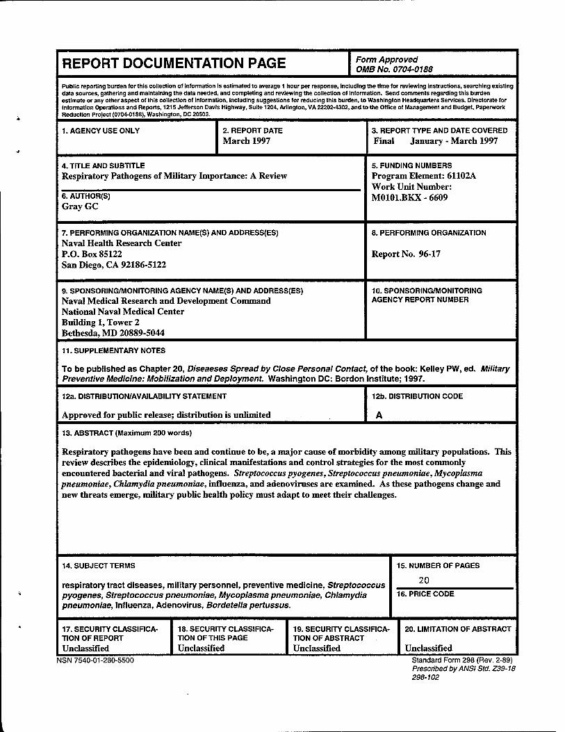
REPORT DOCUMENTATION PAGE Form Approved OMB No. 0704-0188
Public reporting burden for this collection of information is estimated to average 1 hour per response, including the time for reviewing instructions, searching existing data sources, gathering and maintaining the data needed, and completing and reviewing the collection of information. Send comments regarding this burden estimate or any other aspect of this collection of information, including suggestions for reducing this burden, to Washington Headquarters Services, Directorate for information Operations and Reports, 1215 Jefferson Davis Highway, Suite 1204, Arlington, VA 22202-4302, and to the Office of Management and Budget, Paperwork Reduction Project (0704-0188), Washington, DC 20503.
1. AGENCY USE ONLY 2. REPORT DATE March 1997
3. REPORT TYPE AND DATE COVERED Final January - March 1997
4. TITLE AND SUBTITLE Respiratory Pathogens of Military Importance: A Review
6. AUTHOR(S) Gray GC
5. FUNDING NUMBERS Program Element: 61102A Work Unit Number: M0101.BKX - 6609
7. PERFORMING ORGANIZATION NAME(S) AND ADDRESS(ES) Naval Health Research Center P.O. Box 85122 San Diego, CA 92186-5122
8. PERFORMING ORGANIZATION
Report No. 96-17
9. SPONSORING/MONITORING AGENCY NAME(S) AND ADDRESS(ES) Naval Medical Research and Development Command National Naval Medical Center Building 1, Tower 2 Bethesda, MD 20889-5044
10. SPONSORING/MONITORING AGENCY REPORT NUMBER
11. SUPPLEMENTARY NOTES
To be published as Chapter 20, Diseaeses Spread by Close Personal Contact, of the book: Kelley PW, ed. Military Preventive Medicine: Mobilization and Deployment. Washington DC: Bordon Institute; 1997. _^
12a. DISTRIBUTION/AVAILABILITY STATEMENT
Approved for public release; distribution is unlimited
12b. DISTRIBUTION CODE
13. ABSTRACT (Maximum 200 words)
Respiratory pathogens have been and continue to be, a major cause of morbidity among military populations. This review describes the epidemiology, clinical manifestations and control strategies for the most commonly encountered bacterial and viral pathogens. Streptococcus pyogenes, Streptococcus pneumoniae, Mycoplasma pneumoniae, Chlamydia pneumoniae, influenza, and adenoviruses are examined. As these pathogens change and new threats emerge, military public health policy must adapt to meet their challenges.
14. SUBJECT TERMS
respiratory tract diseases, military personnel, preventive medicine, Streptococcus pyogenes, Streptococcus pneumoniae, Mycoplasma pneumoniae, Chlamydia pneumoniae, Influenza, Adenovirus, Bordetella pertussus.
15. NUMBER OF PAGES
20 16. PRICE CODE
17. SECURITY CLASSIFICA- TION OF REPORT Unclassified
18. SECURITY CLASSIFICA- TION OF THIS PAGE Unclassified
19. SECURITY CLASSIFICA- TION OF ABSTRACT Unclassified
20. LIMITATION OF ABSTRACT
Unclassified
NSN 7540-01-280-5500 Standard Form 298 (Rev. 2-89) Prescribed by ANSI Std. Z39-18 298-102