respiratory infections in immuno-compromised hosts assist prof microbiology dr. syed yousaf kazmi
TRANSCRIPT

Respiratory Infections in Immuno-compromised
Hosts
Assist Prof MicrobiologyDr. Syed Yousaf Kazmi

LEARNING OBJECTIVES
1. Describe pathogenesis, clinical findings and lab diagnosis of rhino-cerebral mucormycosis
2. Describe pathogenesis, clinical findings & laboratory diagnosis of pneumocystis jiroveci pneumonia

RHINO-CEREBRAL MUCORMYCOSIS
IntroductionOpportunistic
mycosis(Fungus)Rhizopus oryzae 60% casesOthers-Rhizomucor, Absidia,
Mucor etc.Very lethal infectionsUsually not diagnosed until
deathRecent increase incidence
due to ?
Rhizopus oryzae

PATHOGENESIS• Saprophytic mould-
decaying organic matter• Widely present in
environment• Spores in air-Inhaled• Reach para-nasal sinuses• Acidosis esp Diabetic
ketoacidosis-very strong link
• Other conditions-leukemia, steroid therapy, burns, immunodeficiency, dialysis with iron chelator

PATHOGENESIS• Spores in sinuses germinate• Hyphae invade the blood
vessels• Thrombosis and infarction • Ischemic necrosis of part
distal to necrosis• Plane for invasion of fungus• Sinuses, bones, cartilage,
eye, brain tissue invaded• No body planes hinder its
spread
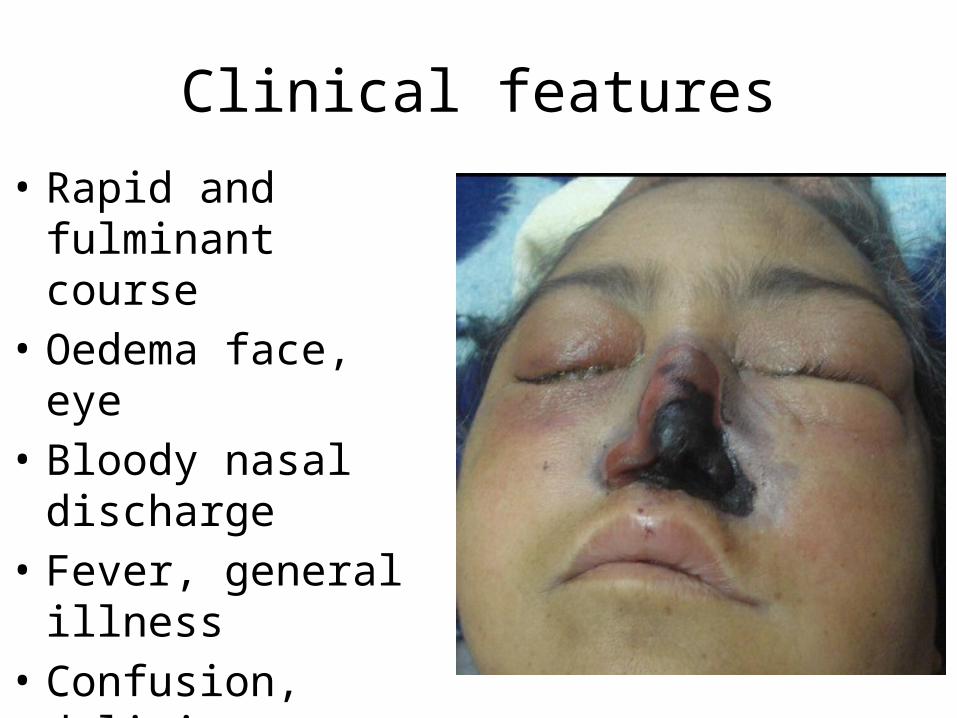
Clinical features
• Rapid and fulminant course
• Oedema face, eye• Bloody nasal
discharge• Fever, general
illness• Confusion,
delirium• Death in serious
cases

Laboratory diagnosis
CLINICAL SUSPICION IS UTMOST IN EARLY DIAGNOSIS & TREATMENTBlood Glucose- High in
DKAUrine ketone bodies-
+ve in DKABlood pH-usually lowBlood CP- usually high
TLC, Low Hb
Positive Urine ketone bodies

Laboratory diagnosisNasal swab/ fluid for
microscopy– Usually negative by Gram
stain– Special fungal stains required
Nasal swab/ fluid for fungal culture – Special fungal culture
medium e.g. Sabauraud Agar– Rapid growth– Identification by microscopy
Tissue for H/P• Stain for fungal hypha• Non septate hyphae

Pneumocystis jiroveci pneumonia
P. jiroveci –fungusPresent in environmentMany healthy people
harbour this fungus Opportunistic mycosisMost common cause of non-
bacterial pneumonia in AIDS patients
Cell mediated immunity-limits infection by this fungus

Pneumocystis jiroveci pneumonia
TransmissionPerson to person transmissionEnvironment to personOwn flora of throat?Predisposing conditionsHIV infectionMalnourishedSteroid therapyAntineoplastic treatmentOrgan transplant recipient

p. JIROVECI PNEUMONIA PATHOGENESIS
Cell mediated immunity is central in combating the Pneumocystis jiroveci pneumonia
Pneumocystis jiroveci pneumonia is strongly related to AIDS
The infection usually occurs when CD4 count drops below 400/uL

p. JIROVECI PNEUMONIA PATHOGENESIS
Cysts of Pneumocystis jiroveci are inhaled from environment which enter alveoli
Inflammatory response to cyst
Frothy exudate accumulates in alveoli that block gaseous exchange
Pneumonia develops due to fluid in lung-hinder gaseous exchange across alveolar membrane
Cyst form
Trophozoite form
Broncho-alveolar lavage fluid

p. Jiroveci pneumonia-clinical features
Progressive exertional dyspnoea (95%)
Fever (>80%)Non-productive cough
(95%)Chest discomfortWeight lossChillsHaemoptysis (rare)
diffuse bilateral infiltrates

Laboratory diagnosisSerum LDH(NV<95 IU/L)
Usually elevated in PNP(> 200 IU/L)
High sensitivity but low specificity
Bronchoalveolar lavage / lung biopsy for cyst stain. Methenamine silver, Giemsa,
Calcofluor white Gram stain not effective
Immuno-fluorescent staining On broncho-alveolar lavage,
lung biopsy specimen Sensitive test
Cysts of P. jiroveci in lung biopsy
Immuno-fluorescent staining of P. jiroveci cysts in BAL fluid

Laboratory diagnosis
PCR Rapid and sensitive test
Serology Not useful in acute
infection Used in establishing the
prevalence of P. jiroveci infection
Positive DNA PCR of P. jiroveci fungus