respiratory failure in children - nemours · respiratory failurerespiratory failure respiratory...
TRANSCRIPT

RESPIRATORY FAILURERESPIRATORY FAILURE
Respiratory Failurein Children
Respiratory Failurein Children
Diagnosis
and
ManagementDavid H. Corddry M.D.
Alfred I. duPont Institute of the Nemours Foundation

RESPIRATORY FAILURERESPIRATORY FAILURE
ObjectivesObjectivesDefine Respiratory Failure.
Review Physiology of Respiration.
Catagorize Respiratory Failure by Physiologic Mechanisms.
Develop an approach to Etiologic Diagnosis.
Outline Treatment Modalities based on Physiology and Etiology.
Discuss Exemples - i.e. Acute Upper Airway Obstruction.
RESPIRATORY FAILURERESPIRATORY FAILURE
DefinitionDefinition
Inability to meet one's need for tissue oxygenation and elimination of CO2, often but not always associated with distress.
Will focus on Pulmonary aspects of this process.
50% of pediatric ICU admissions.
Produced by a wide variety of diseases.

RESPIRATORY FAILURERESPIRATORY FAILURE
OrientationOrientation
Oxygenation and Ventilation are Essential to Living.
# Two simultaneous goals in management.
Diagnosis and treatment of underlying disease.
# Amelioration of pathophysiology producing ARF, independent of diagnosis.
Relative importance depends on degree of failure and rate of change.
# Focus on Physiologic Approach

RESPIRATORY FAILURERESPIRATORY FAILURE
Respiratory PhysiologyRespiratory Physiology
# Developmental aspects# Ventilation: Dead Space, Distribution, Lung
Volumes, FRC, Closing Capacity# Mechanics: Work, Compliance, Resistance, Time
Constants, Visco-Elastic Properties, Surfactant# Perfusion: Lung Zones, HPV, # V/Q matching: Shunt, Venous Admixture, Virtual
Shunt, Alveolar gas equation, A-a gradient.

RESPIRATORY FAILURERESPIRATORY FAILURE
Developmental PhysiologyDevelopmental Physiology
# Conducting Airways relatively smaller 1st 5 years.
# Cartilage spread to segmental bronchus, 12 w gest.
# Alveoli: fewer, smaller, less surface area /BSA
# Neonates, Premies: Pause, Apnea, Flat CO2 response, Decrease V to hypoxia
# Chest wall compliant: deforms, wastes effort.

RESPIRATORY FAILURERESPIRATORY FAILURE
Dead SpaceDead Space
# A. Anatomic = Conducting Airways, 2ml/kg
# B. Alveolar = non perfused alveoli (PE, hypo tension, excess PEEP, CC > FRC)
# Physiologic = A + B
# VD = (PaCO2 - PECO2) VE / PaCO2
# Normally VD / Vtidal = 0.3
# This increases in most disease states.
# More on this under V /Q matching.

RESPIRATORY FAILURERESPIRATORY FAILURE
Distribution of VentilationDistribution of Ventilation
P transpulmonary
Vbase
apex
# More ventilation to bases in healthy lungs due to less P-transpulm. at end expiration.
# Shift in pressure -volume relationship can change this dramatically.

RESPIRATORY FAILURERESPIRATORY FAILURE
Lung VolumesLung Volumes
TotalLung Capacity
VitalCapacity
ResidualVolume
TidalVolume
FuncionalResidualCapacityClosing
Capacity

RESPIRATORY FAILURERESPIRATORY FAILURE
FRCFRC
# Volume in Lung at end expiration. Balance between factors favoring collapse, and those favoring expansion.
# Represents gas volume available for exchange.
# Faster desaturation at lower FRC.
# Lower FRC favors atalectasis.

RESPIRATORY FAILURERESPIRATORY FAILURE
Closing CapacityClosing Capacity
# Volume at which small airways begin to collapse, preventing further gas exchange with those lung units.
# Noramlly well below FRC.
# Closer to FRC in Infants.
# When CC exceeds FRC, this happens during normal tidal breathing with resultant air trapping andmaldistribution of Ventilation.
RESPIRATORY FAILURERESPIRATORY FAILURE
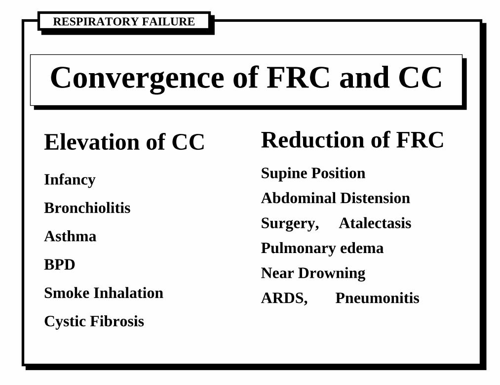
Convergence of FRC and CCConvergence of FRC and CC
Elevation of CCInfancy
Bronchiolitis
Asthma
BPD
Smoke Inhalation
Cystic Fibrosis
Reduction of FRCSupine PositionAbdominal DistensionSurgery, AtalectasisPulmonary edemaNear DrowningARDS, Pneumonitis

RESPIRATORY FAILURERESPIRATORY FAILURE
Work of BreathingWork of Breathing
# Done during inspiration.
# Overcome tissue visco-elastic resistance of lung and chest wall.
# Move air into lungs aginstresistance to flow.
#Tissue returns work to move air out.
Distending Pressure
T Vi od la ul m
e
FRC
inspiration
expiration

RESPIRATORY FAILURERESPIRATORY FAILURE
ComplianceCompliance
# Lung compliance
# Chest Wall compliance
# Total compliance
# Specific compliance is indexed to FRC.
V
Pressure
FRC

RESPIRATORY FAILURERESPIRATORY FAILURE
Decreased Total ComplianceDecreased Total ComplianceDecreased CL
Increased RecoilARDS, pneumonitis
edema, neart drowning
OverexpansionAsthma, Bronchiolitis
Toxic or Thermal Inhalation
Excess PEEP or CPAP
Volume lossAtalectasis Supine position
Decreased CW
Thoracic Trauma or Surgery
Abdominal Surgery
Diaphragmatic Loading
Abdominal Distension
PD, MAST
Pneumothorax
Pleural Effusion
Thoracic Bony deformities

RESPIRATORY FAILURERESPIRATORY FAILURE
ResistanceResistance# Pressure change needed to produce Flow.
# Laminar flow defined by Hagen-Poiseuille equation: Resistance = P / V = 8 h l / r
# Turbulent flow increases resisitance, and makes resistance flow dependent. such that P is proportional to V and density.
# 1 / Resisitance = Conductance.
# Specific Conductance = Conductance / Lung Volume. SImilar in infants and adults.
4
.
.
2

RESPIRATORY FAILURERESPIRATORY FAILURE
Sites of Increased AirwayResistance
Sites of Increased AirwayResistance
# In Adults -- Upper Airway, Nose.
# In Children -- Peripheral Airways.
# Dynamic Airway Compression: Increased Intrapleuralpressure during forced exhalation augments collapse ofintrathoracic airways.
# Worse with BPD, alpha-1-antitrypsin deficiency due to poor cartilege.
# Extrathoracic airway effected on inhalation.

RESPIRATORY FAILURERESPIRATORY FAILURE
Time ConstantsTime Constants# Time required for lung unit to fill to 63% of final volume.
# Time constant = Resistance x Compliance
# Those alveoli with shorter time constants fill faster.
# Local variation in resistance and compliance effect gas distribution.
# e.g. overall TC is increased in Asthma
RESPIRATORY FAILURERESPIRATORY FAILURE
SurfactantSurfactant
# LaPlace's Law P = 2 T / r
# This would predict that small alveoli would empty into large ones.
# However Surfactant allows a decrease in surface tension as the radius decreases.
# Therefore Pressure stays the same.
# Made by type II pneumocytes.
# Surfactant deficiency occurs in many disease states.
RESPIRATORY FAILURERESPIRATORY FAILURE
Pulmonary CirculationPulmonary Circulation# Development closely follows airway / alveolar
development.
# Limited in pulmonary hypoplasia (eg CDH)
# Muscular wall actively remodels during development.
# Smooth muscle gradually extends more distally, but may extend faster with ensuing Pulm. Ht'n.
# Pulm circulation recieves the entire C.O.

RESPIRATORY FAILURERESPIRATORY FAILURE
West Zones IWest Zones I
# Define by relationship of pressures affecting local pulmonary blood flow.
# Upstream pressure is PPA (pulmonary aretery)
# Downstream pressure is the greater of:
1. PPV (pulmonary veins) ~= (left atrium)
2. PA (alveolus)
# Note the latter increases with Positive Pressure Ventilation.

RESPIRATORY FAILURERESPIRATORY FAILURE
West Zones IIWest Zones II
PPA
PPA
PPA
PPA
PA
PA
PA
PA
PA
PA
PPV
PPV
PPV
PPV
>
>
>
>
>
>
1
2
3

RESPIRATORY FAILURERESPIRATORY FAILURE
HPVHPV# Alveolar Hypoxia leads to local pulmonary
vasoconsstriction.
# Usually useful to match perfusion to ventilation.
# With whole lung hypoxemia it produces pulmonary hypertension, and possible R to L shunt via PFO.
# Chronically leads to increased muscularity and chronic pulmonary hypertension

RESPIRATORY FAILURERESPIRATORY FAILURE
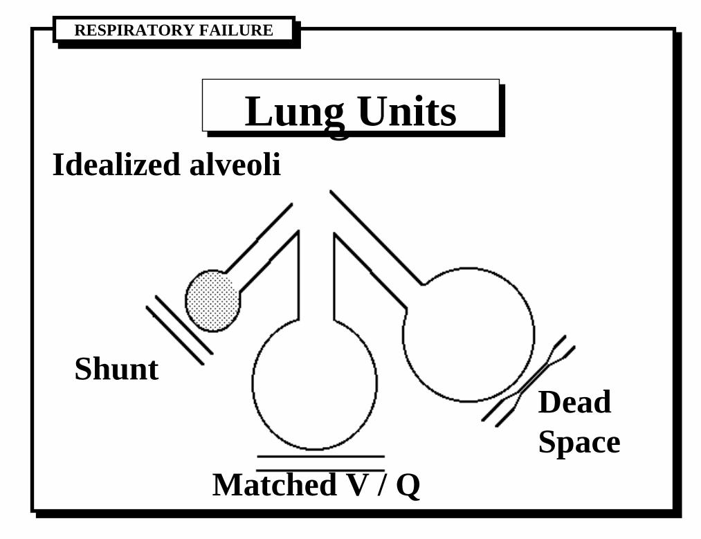
V / Q MatchingV / Q Matching..
# V / Q = 0.6 at bases; = 3 at apices
# True shunt is blood with no contact with aerated alveoli. (eg cardiac, atalectasis)
# Venous admixture (virtual shunt) amount of mixed venous blood to add to pulmonary end capillary blood to produce observed arterial O2 content.
# PAO2 = ( FiO2 ( PB - 47 ) ) - ( PaCO2 / R )
# Normally A-a DO2 is small due to obligate shunt.
RESPIRATORY FAILURERESPIRATORY FAILURE
Lung UnitsLung UnitsIdealized alveoli
Shunt
Matched V / Q
Dead Space

RESPIRATORY FAILURERESPIRATORY FAILURE
Virtual Shunt LinesVirtual Shunt Lines
0
100
200
300
400
20 30 40 50 60 70 80 90 100
ArterialPaO2
Inspired O2 concentration (%)
0 5% 10%
15%
20%
25%
30%
50%
Hb 10-14 g/dl
PaCO225-40 mmHg
a-v O2 difference5ml/100ml

RESPIRATORY FAILURERESPIRATORY FAILURE
Alveolar-Capillary MembraneAlveolar-Capillary Membrane# May contribute to "diffussion" block of O2 movement. But
this mechanism is rarely the sole cause of signifiganthypoxemia.
# However, transudation of fluid across the membrane is a major cause of respiratory failure.
# Function of 1. Pressure gradient. 2. Oncotic forces. 3. Filtration Coefficient.
# Leads to 1. Decreased Compliance 2. Alveolar collapse -> Shunt -> Increased Aa O2 gradient.

RESPIRATORY FAILURERESPIRATORY FAILURE
ExclusionsExclusions# Physiology review has focused on lung physiology.
# Also important, but not included in this review are:
1. CNS control of breathing.
2. Neuromuscular transmission.
3. Muscular function.
4. Toxicology
5. Cardiac Function and O2 delivery.

RESPIRATORY FAILURERESPIRATORY FAILURE
Sorting it Out 1Sorting it Out 1Won't Breath
Can't Breath
CNSToxic(lack of Drive)
(strength inadequatefor work required)
AirwaysLungsRespiratory Pump
# Remember, a child with chronic respiratory disease can present in acute failure due to an exacerbating process.

RESPIRATORY FAILURERESPIRATORY FAILURE
Sorting it Out 2Sorting it Out 2Airway
Lung
Pump
Extrathoracic large airwayIntrathoracic large airwaysmall airways
Increased closing capacityDecreased FRCDead SpaceShunt
IntrapleuralChest wallNeuromuscular

RESPIRATORY FAILURERESPIRATORY FAILURE
Sorting it Out 3Sorting it Out 3
X A/W ObsI A/W ObsSmall A/W
Inc CCDec FRCDead SpShunt
I PleuralChest WNeuroM
CNSToxic
Stridor Wheeze Rales BS Retract IncCO2 DecO2 CXR
I > EE > I
-
----
---
--
-+
++++
++++++-
---
--
---
-++-++
---
--
=<>=
=<>-<>
<><>=
==
+++++++
++++++
+++++-
--
+++++++++
++++
++++
++++++++++
++++++++
++++
++++++++++++
++++
++
-traptrap
traplow volinc vollow vol
shift+ / -bell
--

RESPIRATORY FAILURERESPIRATORY FAILURE
HypoxiaHypoxia# The four basic mechanisms which can produce hypoxia.
1. Inadequate FiO2.
2. Decreased Ventilation.
3. Shunt (pulmonary or cardiac).
4. Decreased Cardiac Output.

RESPIRATORY FAILURERESPIRATORY FAILURE
TreatmentTreatment# Provide supplemental Oxygen
# Judge severity, decide if immediate intervention needed.
# Monitors: Pulse oximeter, Respiratory rate, ABG
# Get Assistance if needed.
# Maintain Airway.
# Maintain Breathing
# Treat underlying cause and pathophysiology
# ECMO, Hyperbaric O2

RESPIRATORY FAILURERESPIRATORY FAILURE
OxygenOxygen# Simple masks, Nasal Cannula, impossible to know FiO2.
Better with Venturi mask.
# Non-Rebreather mask or Hood for infant provide known FiO2 from a mixer.
# High FiO2 may accelerate collapse of closed segments.
# O2 is toxic, Don't use high FiO2 for long periods unless necessary.
# O2 is life-saving, Always use high FiO2 in acute emergency.

RESPIRATORY FAILURERESPIRATORY FAILURE
Severity 1Severity 1The patients in trouble when:
1. Inadequate ventilation: PaCO2 > 50-55
2. Apnea, respiratory pauses (fatigue)
3. Rising PaCO2
4. Vital Capacity <15 ml/kg
5. Dead Space / Tidal Volume > 0.6
6. Change in Level of Consciousness

RESPIRATORY FAILURERESPIRATORY FAILURE
Severity 2Severity 27. Cyanosis or PaO2 < 70with FiO2 >0.6.
8. A-a DO2 >300 with FiO2 at 1.0.
9. Shunt Fraction > 15 - 20%.

RESPIRATORY FAILURERESPIRATORY FAILURE
AirwayAirway# Natural
# Supported: Jaw Lift, Suctioning, OPA, Nasal A/W,
#Artificial: ETT
Size: 3.0 Newborn
3.5 3-8 months
4.0 9-24 months
Size = ( Age / 4 ) + 4
Cuff adds half a size.

RESPIRATORY FAILURERESPIRATORY FAILURE
IntubationIntubation# Suction Available.
# Preoxygenate generously. Fill FRC with O2 may take a long time in diseased lungs.
# Monitoring
# Vascular access preferred.
# Sedative / hypnotic, and neuromuscular blockade.
# Cricoid pressure.
# Laryngoscopy and Intubation, Gently
# Confirm: BS, CO2, Chest movement, CXR
# SECURE IT.

RESPIRATORY FAILURERESPIRATORY FAILURE
Acute Upper Airway Obstructionin Children
Acute Upper Airway Obstructionin Children
Differential Diagnosis
# Epiglottitis
Croup (viral laryngotracheobronchitis)
Bacterial Tracheitis, Pharyngeal Abcess
Foreign Object, Thermal or Chemical Injury
Diphtheria
Angioneurotic Edema
Acute exacerbation of chronic obstruction

RESPIRATORY FAILURERESPIRATORY FAILURE
In Epiglottitis you need to secure
Any of these may require emergency airway management if severe.
the airway ASAP regardless of the patients current level of distress.

RESPIRATORY FAILURERESPIRATORY FAILURE
Children with StridorChildren with Stridor
9 4 %2 %4 %
CroupSevere CroupEpiglottitis
155 children presenting to the emergency room with acute stridor.
Mauro et al, Am. J. Dis. Child., 142(6):679-82, June 1988

RESPIRATORY FAILURERESPIRATORY FAILURE
SupraglottitisSupraglottitisAcute infection of the Epiglottis and Aryepiglotticfolds.
# Sudden onset of sore throat, dysphagia, often withstridor and shortness of breath.
May result in severe, rapidly progressive airway obstruction in 6 to 12 hours.
# Patients sit forward and drool, don't talk.
Usually with high fever and bacteremia.
Usually caused by Hemophilus influenzae type B.

RESPIRATORY FAILURERESPIRATORY FAILURE
Epiglottitis vs. CroupEpiglottitis vs. CroupEPIGLOTTITIS CROUP
Age All, peak 3-5 years Younger, peak 3 m-3 y
Etiology Bacterial (h. Flue B) Viral (parainfluenza)
Speed of Onset Rapid (<24 hours) Slow (1-4 days)
Appearance Anxious, toxic Frequently non-toxic
Position Upright, forward Variable
Temperature Usually high (>39) Normal to high
Resp. Distress Usually present Variable
Retractions Usually late Progressive
Voice/Cough Muffled or absent Hoarse/ "seal" bark
Stridor Yes, less with more obstruct. Yes
Mouth Open, forward, drooling Closed, nasal flaring
RESPIRATORY FAILURERESPIRATORY FAILURE
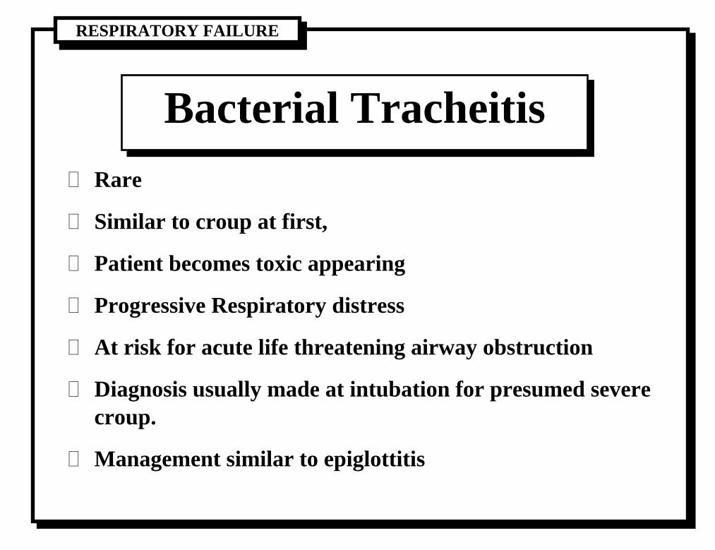
Bacterial TracheitisBacterial TracheitisRare
Similar to croup at first,
Patient becomes toxic appearing
Progressive Respiratory distress
At risk for acute life threatening airway obstruction
Diagnosis usually made at intubation for presumed severe croup.
Management similar to epiglottitis

RESPIRATORY FAILURERESPIRATORY FAILURE
Foreign objectForeign object# Should be considered in every child with acute upper
airway obstruction.
# History may or may not help
# Age, usually under 4 years, can be any age
# Stridor may or may not be present
# Wheezing may be present
# Fever not common early
# Usually not toxic appearing
# Radiograph may be definitive, only if positive
RESPIRATORY FAILURERESPIRATORY FAILURE
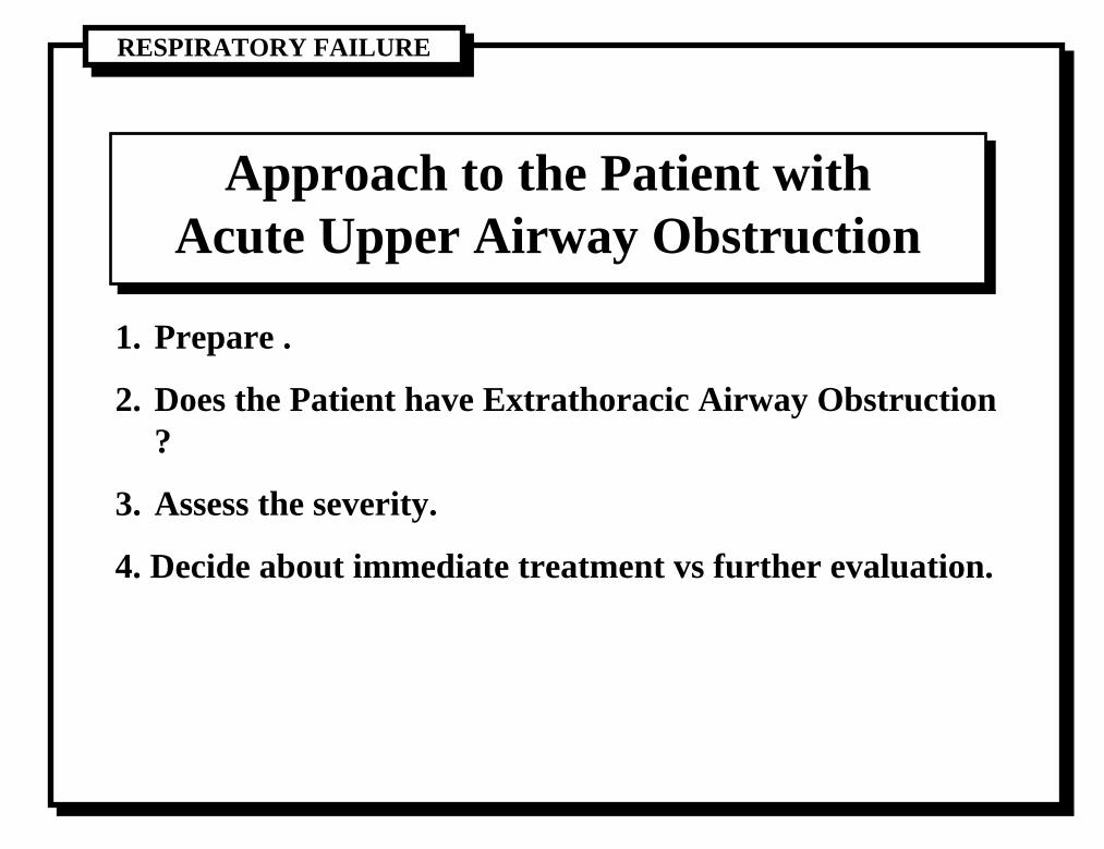
Approach to the Patient withAcute Upper Airway Obstruction
Approach to the Patient withAcute Upper Airway Obstruction
1. Prepare .
2. Does the Patient have Extrathoracic Airway Obstruction ?
3. Assess the severity.
4. Decide about immediate treatment vs further evaluation.

RESPIRATORY FAILURERESPIRATORY FAILURE
Extrathoracic AirwayObstruction
Extrathoracic AirwayObstruction
Stridor, if present, is greater on Inspiration.
Suprasternal, Supraclavicular Retractions
Chest Wall Retractions in Infants
# Stridor may be less with worse obstruction

RESPIRATORY FAILURERESPIRATORY FAILURE
Severity of DistressSeverity of DistressStridor without tachypnea
Tachypnea without distress
Retractions, Decreased Activity
Increased work, Use of accesory muscles
Irritability and air hunger
Fatigue may develop
Lethargy and cyanosis presage impending respiratory arrest.
RESPIRATORY FAILURERESPIRATORY FAILURE
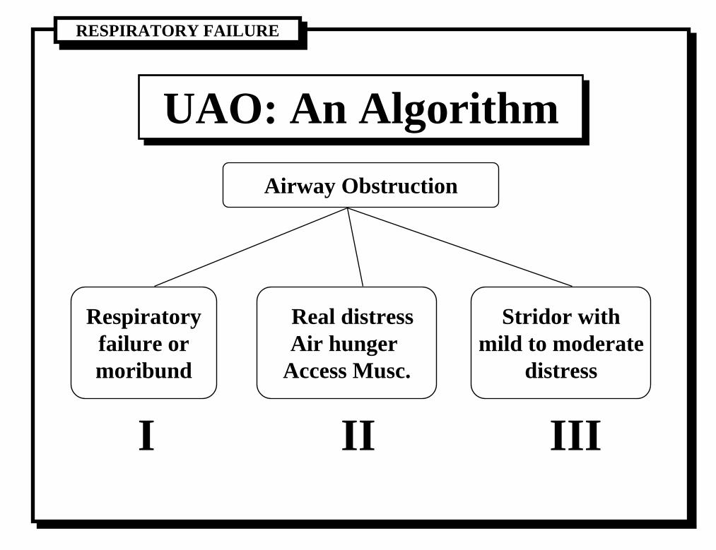
UAO: An AlgorithmUAO: An AlgorithmAirway Obstruction
Respiratoryfailure ormoribund
Real distressAir hunger
Access Musc.
Stridor withmild to moderate
distress
I II III

RESPIRATORY FAILURERESPIRATORY FAILURE
Algorithm IAlgorithm I
UAO & Respiratory Failure# Oxygen
# Artificial Airway if immediatly available
# Bag and Mask Ventilation, +Pressure
# Cricothyroidotomy
# Cardiac Assessment, and Recussitation
# To ICU or OR

RESPIRATORY FAILURERESPIRATORY FAILURE
Algorithm IIAlgorithm II
UAO & Severe Respiratory Distress# Allow to remain sitting up
# Oxygen, preferably with humidity
# Pulse Oximeter
# Minimize Perturbation
# Arrange transfer to ICU or OR for controlled airway management i.e. Intubation
# No decrease in proximate expertise
RESPIRATORY FAILURERESPIRATORY FAILURE
Algorithm IIIAlgorithm IIIUAO & Mild to Moderate Distress
# Clinical Impression
# If Suspect epiglottitis -- Lateral Neck X-Ray
# Accompany by a physician
# If epiglottitis -- protocol
# If not -- further exam, other studies
# Hospital admission to appropriate unit.

RESPIRATORY FAILURERESPIRATORY FAILURE
X-Ray FeaturesX-Ray Features# Find the epiglottis
valecula, arytenoids, hyoid
# Enlarged epiglottis, lack of central lucency
# Balooning of hypopharynx
# Supraglottitis
ary-epiglottic folds
# "Steeple" sign in croup
# Foreign bodies