respiratory distress in the newborn, not rds dr. alona bin-nun nicu shaare zedek
TRANSCRIPT
Respiratory Distress in the Newborn, not RDS
Dr. Alona Bin-Nun
NICU
Shaare Zedek
Respiratory Distress in the Newborn – Clinical Presentation
• Cyanosis• Grunting• Retractions• Tachypnea• Nasal flaring
• Extreme: Apnea, Shock
More Common Causes of Respiratory Distress
• RDS
• Pneumonia
• Meconium Aspiration
• Transient Tachypnea
• Hypothermia
• Hypoglycemia
Acute Life Threatening Emergencies Presenting in Respiratory Distress
• Choanal Stenosis
• Meconium Aspiration
• Tension Pneumothorax
• Diaphragmatic Hernia
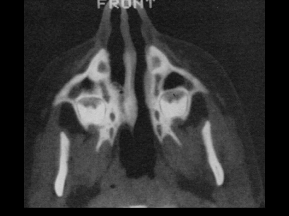
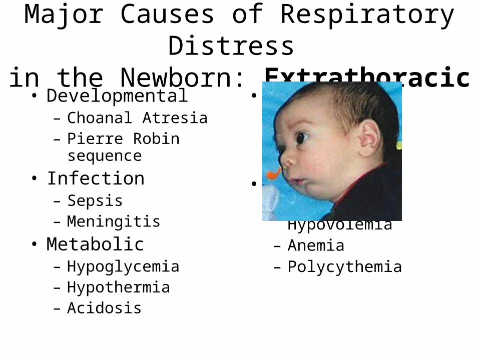
Major Causes of Respiratory Distress in the Newborn: Extrathoracic
• Developmental– Choanal Atresia– Pierre Robin sequence
• Infection– Sepsis– Meningitis
• Metabolic– Hypoglycemia– Hypothermia– Acidosis
• CNS– Infection– Hemorrhage– Edema
• Blood– Blood loss,
Hypovolemia– Anemia– Polycythemia
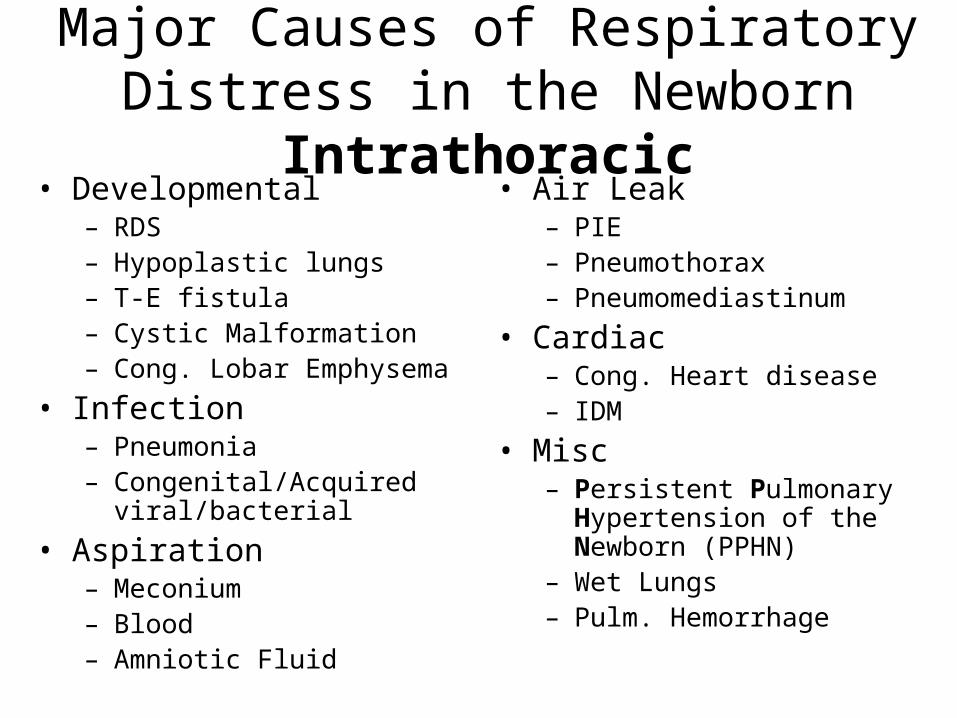
Major Causes of Respiratory Distress in the Newborn Intrathoracic
• Developmental– RDS– Hypoplastic lungs– T-E fistula– Cystic Malformation– Cong. Lobar Emphysema
• Infection– Pneumonia– Congenital/Acquired
viral/bacterial
• Aspiration– Meconium– Blood– Amniotic Fluid
• Air Leak– PIE– Pneumothorax– Pneumomediastinum
• Cardiac– Cong. Heart disease– IDM
• Misc– Persistent Pulmonary
Hypertension of the Newborn (PPHN)
– Wet Lungs– Pulm. Hemorrhage
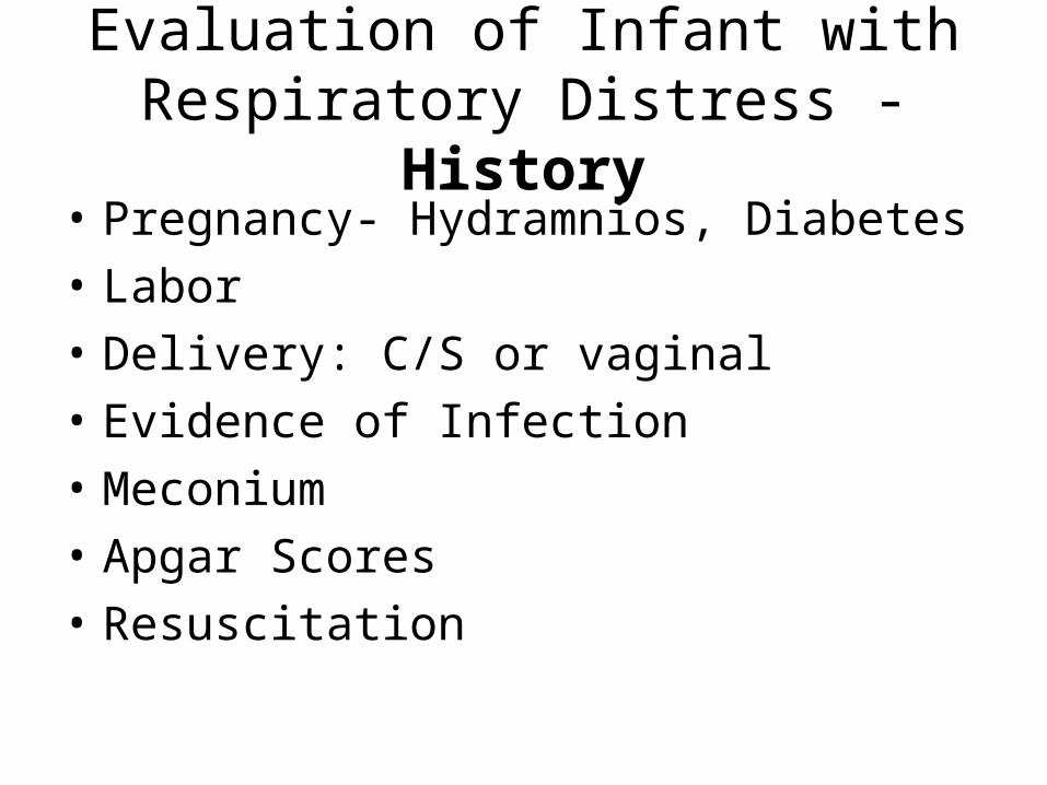
Evaluation of Infant with Respiratory Distress - History
• Pregnancy- Hydramnios, Diabetes
• Labor
• Delivery: C/S or vaginal
• Evidence of Infection
• Meconium
• Apgar Scores
• Resuscitation
Evaluation of Infant with Respiratory Distress – Physical Examination
• Degree of respiratory distress
• Cyanosis
• Air entry
• Heart murmur
• Temperature
• Scaphoid abdomen
• Position of PMI
Laboratory Tests
• O2 saturation• X-ray: AP+lateral. Assess both lungs and heart• Blood gas• Hct• Dextrostix• BP• Transillumination• Hyperoxia test• Nasogastric catheter (radio opaque)• Evaluate for sepsis

Management of Newborn with Respiratory Distress (1)
• Clear airway, esp. meconium
• Oxygen
• Ventilation– mask bagging → intubation– Cyanosis– CO2 retention– apnea
• Correct Acidosis

Management of Newborn with Respiratory Distress (2)
• Arterial Catheter, follow blood gases• Correct
– Hypoglycemia– Hypothermia– Shock– Anemia or polycythemia
• Drain Pneumothorax• Antibiotics (for unexplained persistent
respiratory distress)
Transient Tachypnea
• Clinical Presentation– Frequently term infant– C/S– Mild respiratory distress
– Moderate O2 requirement
– Duration: 2-5 days
• X-ray– Ill defined hazy central
markings– Fade towards periphery– Slight cardiomegaly
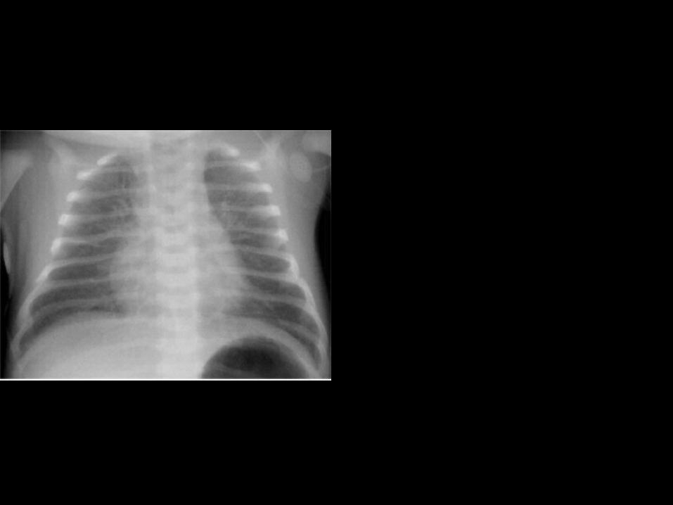
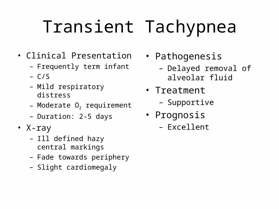
Transient Tachypnea
• Clinical Presentation– Frequently term infant– C/S– Mild respiratory distress
– Moderate O2 requirement
– Duration: 2-5 days
• X-ray– Ill defined hazy central
markings– Fade towards periphery– Slight cardiomegaly
• Pathogenesis– Delayed removal of
alveolar fluid
• Treatment– Supportive
• Prognosis– Excellent
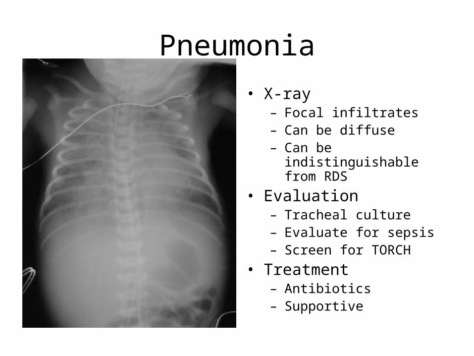
Pneumonia
• Bacterial– GBS, E.coli, other Gram
negative
• Viral– CMV, rubella, herpes, RSV
• Routes of Infection– Ascending (PROM)– Hemtogenous– Aspiration of infected
material
• Time of Infection– Before, during or after
delivery
• X-ray– Focal infiltrates– Can be diffuse– Can be indistinguishable
from RDS
• Evaluation– Tracheal culture– Evaluate for sepsis– Screen for TORCH
• Treatment– Antibiotics– Supportive

Meconium Aspiration Syndrome (MAS)

Effects of Meconium AspirationMeconium Aspiration
Chemical pneumonitis
Bacterial pneumonitis
Proximal Airway
Occlusion
Peripheral Airway
Occlusion
Extra-alveolar air
Partial
Atelectasis
Intrapulmonary Shunt
Ball valve
Complete
Hypoxemia and Acidodis
PPHN
Asphyxia

Treatment of MAS
• Prevention• Oxygen, CPAP • Assisted ventilation• NO• Drain pneumothorax• Antibiotics
• General measures, correct:– hypovolemia– metabolic acidosis– hypoglycemia– hypocalcemia– anemia
• Further Sequelae– CP– ATN– Anoxic liver + coagulopathy– NEC– Anoxic Myocardial damage

T-E Fistula classification
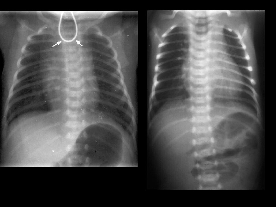
Esophagial Atresia and T-E Fistula
• Embryology– Interruption of division of foregut into trachea and esophagus
• Clinical Picture– Associated with prematurity and hydramnios– Increased salivation– Choking and dyspnea on feeding– Aspiration pneumonia– Other abnormalities (VACTER association)
• Diagnosis– X-ray: dilated proximal esophageal pouch, curling of NG catheter– Dye studies– Air in abdomen: presence or absence of fistula– Endoscopy
• Preoperative Care– Treat Pneumonia– Prevent gastric reflux – upright position– Suctioning of proximal pouch
• Definitive treatment– Surgery
• Prognosis– Survival– Depends on birth weight, prematurity, other
congenital abnormalities

Diaphragmatic Hernia
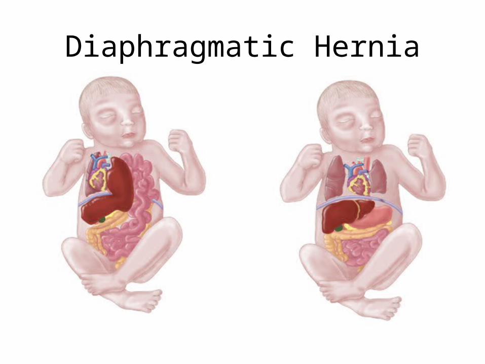
Diaphragmatic Hernia
• Treatment– Intubate and ventilate– Do not mask bag– Gastric tube– Beware of
pneumothorax– Surgery
• Post op:– Ventilation and
oxygenation: problematic
• Outcome– Poor due to lung
hypoplasia
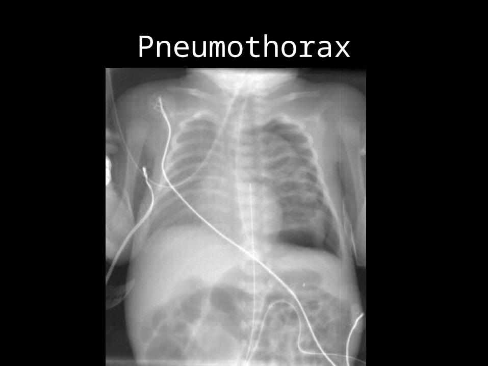
Pneumothorax
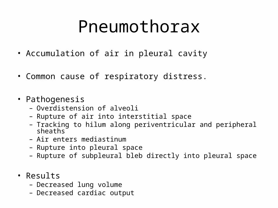
Pneumothorax
• Accumulation of air in pleural cavity
• Common cause of respiratory distress.
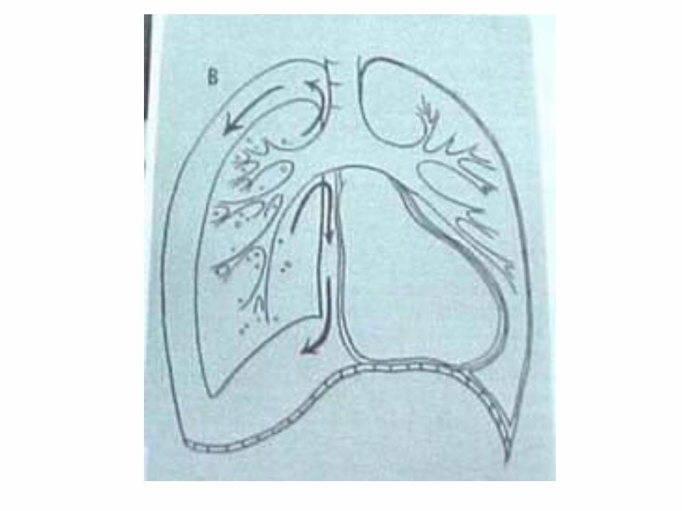
• Pathogenesis– Overdistension of alveoli– Rupture of air into interstitial space– Tracking to hilum along periventricular and peripheral sheaths– Air enters mediastinum– Rupture into pleural space– Rupture of subpleural bleb directly into pleural space
• Results– Decreased lung volume– Decreased cardiac output
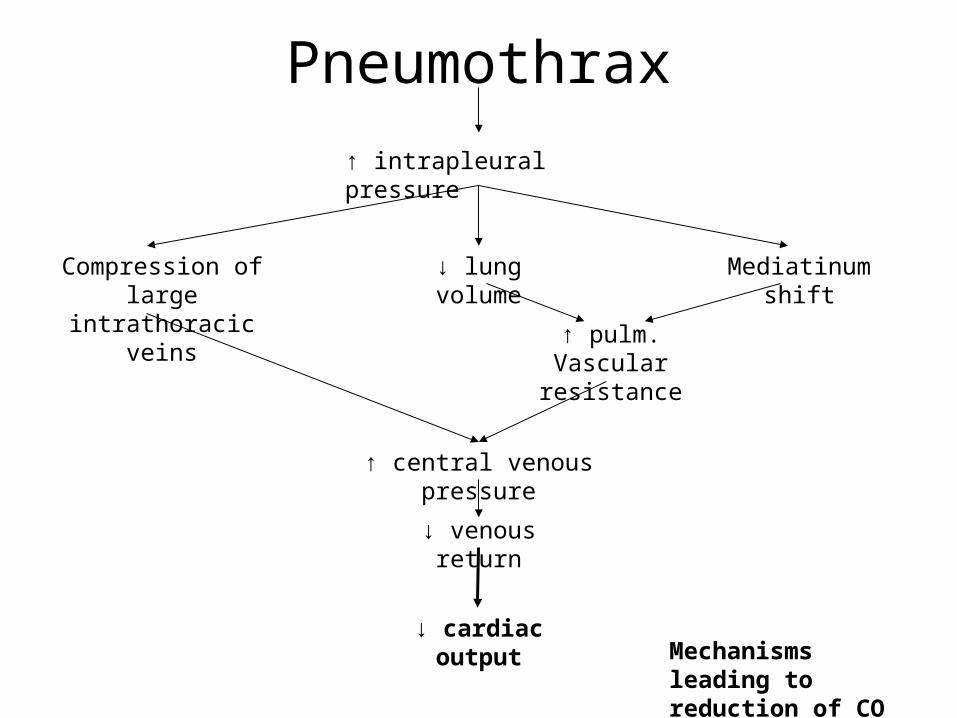
Pneumothrax
↑ intrapleural pressure
Compression of large intrathoracic veins
↓ lung volume Mediatinum shift
↑ pulm. Vascular resistance
↑ central venous pressure
↓ venous return
↓ cardiac outputMechanisms leading to reduction of CO
Clinical Presentation of Pneumothorax
• Grunting• Tachypnea• Apnea• Cyanosis• Bradycardia• Shock• Sudden deterioration in ventilated infant• Shifting of heart sounds• Chest asymmetry• Decreased air entry
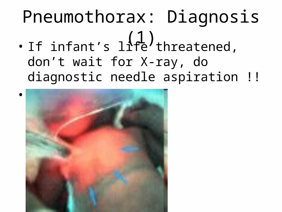
Pneumothorax: Diagnosis (1)• If infant’s life threatened, don’t wait for X-
ray, do diagnostic needle aspiration !!
• Transillumination

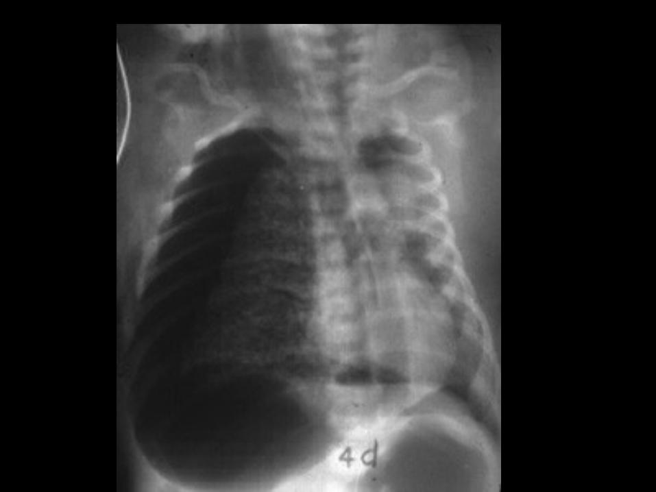
Pneumothorax: Diagnosis (2)
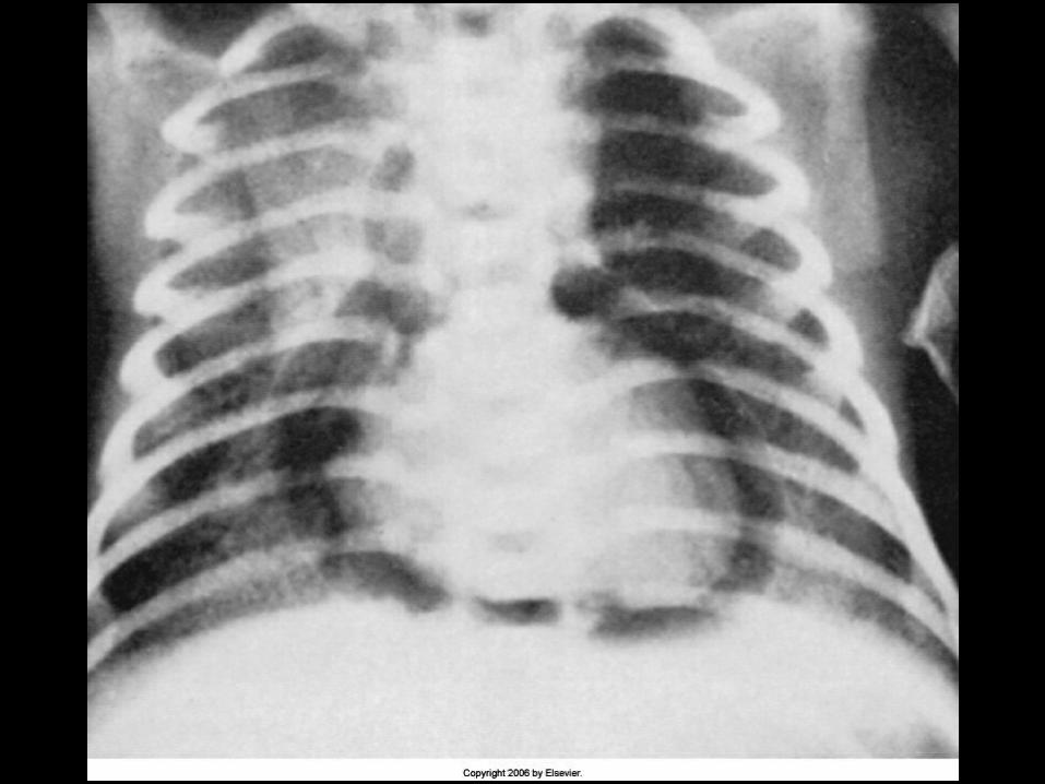
• X-ray– Seperation of lung from chest wall– Absent lung marking peripherally– Shift of mediastinum in tension pneumothorax– Bilateral tension: no shift, small heart– Lateral: air collection beneath sternum
Pneumothorax: Diagnosis (3)
• Associated with PIE– Pneumomediastinum– Pneumopericardium
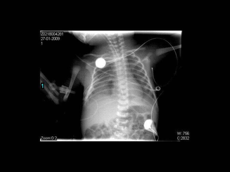
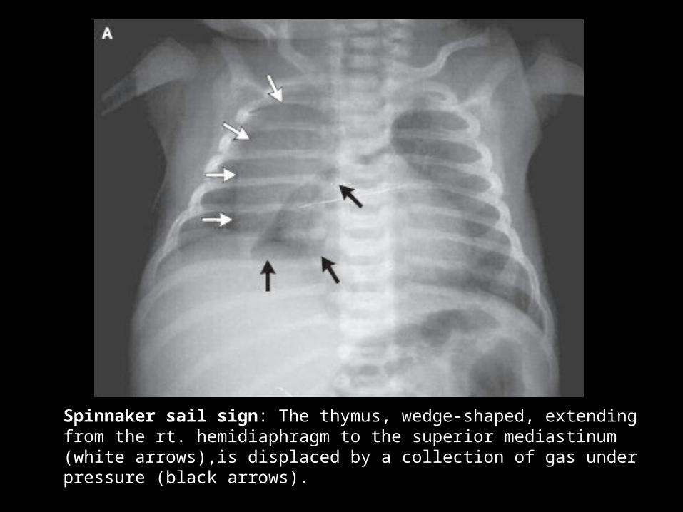
Spinnaker sail sign: The thymus, wedge-shaped, extending from the rt. hemidiaphragm to the superior mediastinum (white arrows),is displaced by a collection of gas under pressure (black arrows).
Causes of Pneumothorax
• Spontaneous
• RDS
• CPAP and mechanical ventilation
• Resuscitation
• Pulmonary hypoplasis
• Post thoracotomy
Treatment of Pneumothorax
• Observe only if:– Minimal respiratory distress– Minimal oxygen requirement– Breathing spontaneously– Maintaining good BP
• Indications for drainage– Tension pneumothorax– Cyanosis– Apnea– Deteriorating blood gases– Assisted ventilation– Shock
