resident handbookinitial stabilization of the critically ill child. (pc) 2. demonstrate mastery of...
TRANSCRIPT

UNC Pediatric Intensive Care Unit
Resident Handbook
Disclaimer: This handbook contains general guidelines for
the management of critically ill pediatric patients. Always
discuss specific patient care issues with the ICU attending or
fellow. Always communicate changes in patient status or plan
with the ICU attending and all services involved.
Contents: Page
Section I : UNC specific
Goals and Objectives 2
Clinical/Educational Responsibilities 4
Important Phone Numbers 8
Section II : General pearls
The ICU Progress Note 9
PALS resuscitation algorithms 12
Common PICU medication dosages 15
Respiratory 17
Cardiovascular 23
Electrolytes 34
Endocrine 39
Heme/Onc 42
Infectious Disease 44
Neurology 46

Page | 2
Goals and Objectives
OVERALL EDUCATIONAL GOAL:
To teach residents to care for children with serious medical
and surgical problems and to learn the principles to identify
and manage children with a critical illness. Residents will
acquire an understanding of the pathophysiologic basis of
common disease processes in the PICU, learn the technical
skills to resuscitate and care for critically ill children, and
understand end of life care for children and their families.
OBJECTIVES:
This rotation should allow the
resident to accomplish the
following objectives (in no
particular order):
1. Recognize and appropriately
respond to acute life
threatening events. Acquire
the necessary skills to
resuscitate and provide
initial stabilization of the critically ill child. (PC)
2. Demonstrate mastery of basic airway skills (use of
oxygen delivery devices and demonstration of bag-mask
ventilation) and placement of peripheral intraosseous and
intravenous catheters. Advanced airway skills
(intubation) and advanced intravenous access skills
(central venous catheter and arterial line placement) may
be performed if mastery of basic skills. (PC)
3. Understand the pathophysiology and treatment of
common medical disorders in the PICU: respiratory
failure (bronchiolitis, asthma, ARDS), shock (septic,
cardiogenic, hypovolemic), neurologic critical care (status
epilepticus, altered mental status, traumatic brain injury,
brain death), renal and liver failure. (PC, MK)
Core Competency Codes
PC=Patient Care
MK=Medical Knowledge
I&CS=Interpersonal & Communication Skills
P=Professionalism
PBL=Practice-Based Learning
SBP=Systems-Based Practice

Page | 3
4. Understand the indications, perioperative management
and complications of common surgical admissions to the
PICU: congenital heart disease, trauma, ENT, orthopedic,
neurosurgical and organ transplant. (PC, PBL)
5. Understand the different monitoring techniques in critical
care: vascular hemodynamics, intracranial devices, blood
pressure, arterial saturations, end-tidal CO2, and a variety
of common laboratory tests. (PC, PBL)
6. Understand pediatric critical care pharmacology:
inotropes and vasoactive agents, basic antibiotic therapy,
common sedatives and analgesics, drug pharmacokinetics
and monitoring of side effects. (MK, PBL)
7. Understand techniques for enteral and parenteral
nutritional supplementation in the PICU patient. (MK)
8. Understand the ethical and legal issues which emerge
during the care of critically ill and/or dying children (do
not resuscitate orders, withholding and withdrawing life
support, right of patients). (MK, SBP)
9. Understand the importance of psychosocial issues related
to the care of critically ill or dying children. Learn to
provide support and deliver difficult information to the
family of a critically ill child. (MK, PBL)
10. Succinctly present an ICU patient on rounds, formulate a
coherent assessment of a patient’s problems and present
an appropriate therapeutic/diagnostic plan. Further,
effectively communicate this plan to nurses, respiratory
therapists, and sub-specialists/ consultants. (PC)

Page | 4
Clinical Responsibilities
Rounds are standardized in the following manner:
Rounds start at 0730 with the PICU fellow/attending
running through the list of patients at the board to assess
which patients can be transferred/discharged and to go
over potential admissions.
X-rays are often reviewed next on PACS. Each resident
is responsible for interpreting their patient’s x-rays
Rounds then begin at Room 1
The format for rounds is:
o Brief synopsis of major events in the past 24 hrs by
the post-call resident
o RT report of ventilator settings and airway
maintenance issues
o RN report of drips, access, and any concerns
o Pharmacy report of current meds for reconciliation
with CPOE
o Resident report of assessment and plan
o Family questions and concerns
o PICU fellow summary of plan
Responsibilities include
Reading about your patient’s condition, disease process,
medications, etc.
Pre-rounding on your patients (includes knowing
overnight events, reviewing vital signs, conducting full
physical exam, reviewing and reconciling patient
medications from CPOE and MAR)
Writing daily progress notes, transfer notes, accept notes,
significant event notes and discharge notes
Entering orders in CPOE (please use the same dosing
weight (usually the admission weight) on every patient
every day despite any new documented weights. You

Page | 5
should only change the dosing weight after discussion
with PICU fellow/attending)
Reviewing and correcting/updating orders on all patients
every day (for example, post surgical patients may return
to the PICU with orders written by surgery that you need
to evaluate)
Don’t forget your prophylaxis if applicable
o Famotidine for gastritis prophylaxis in patients who
are NPO, on steroids, or on post-pyloric feeds
o SCDs for DVT prophylaxis in post-pubertal or high
risk patients
o Peri-op antibiotics (check with the surgeons)
When performing a “rule out” for fever, panculture your
patient (blood, urine, sputum, CSF) wherever applicable.
Don’t forget to get a peripheral blood culture as well in a
patient with a central line
When leaving the unit (for a break, to nap, to eat, etc),
provide a quick sign out to resident/s who remain in the
unit. Please also notify the fellow/attending that you are
leaving and summarize any patient care tasks that still
need to be done prior to your return
Specific responsibilities
On call resident
Listen to everyone’s presentations as you are responsible
for knowing every patient in the unit
Take notes on the post-call resident’s “to-do” list as their
presentation is considered sign out
Divide the patients for pre-rounding among the 3
residents who will be present on your post-call day
(including yourself)

Page | 6
Post call resident
Complete daily progress notes prior to rounds
Present the overnight events for each person
Depart immediately after rounds
Short call resident
Enter orders in CPOE
Sign out to on call resident prior to noon lecture
Attend noon lecture prior to departure
Afternoon resident
Arrive at 1200 for lecture
Obtain sign out from the on call resident regarding things
that need to be done at 1300
Helpful tips
PICU nurses are very experienced and invested in the
care of these patients. Learn from them. Take their advice
and concerns seriously.
If a nurse asks you to call the fellow/attending, do it.
If in doubt, call the fellow/attending.
The only stupid question is the one you didn’t ask.
Follow up on anything that was supposed to happen
(including labs and x-rays and CT scans. Even if you
aren’t a neurologist, you will likely notice something
really bad that we should know about).
Keep the surgical residents apprised of any changes in
their patients.
If in doubt about orders on surgical patients, ask the
fellow/attending the best course of action.
The PICU nurses perform arterial punctures and place
peripheral IV access. You should make an effort to ask
them to take over these procedures.

Page | 7
When a PICU patient requires a central line, chest tube,
or endotracheal tube, they are often too ill for a resident
(and sometimes a fellow) to complete. These procedures
are fellow level procedures but may be offered to you if
you are readily available at the bedside and the patient is
deemed appropriate by the fellow/attending.
Educational Responsibilities
Educational conferences
Monday Tuesday Wednesday Thursday Friday
* Mock
code/Chalk
talk 12:00
# Cath
conf
(3:30-5:30)
# Fellow
lecture
(11-12)
# Fellow
lecture
(11-12)
* Required
# Optional
Take charge of your education by reading about your patients,
asking questions, using the resources discussed during
orientation to find and read pertinent articles, and making sure
the attendings and fellows give your twice weekly formal
lectures. Remember that there can be a lot of teaching on
rounds so listen up and ask questions.

Page | 8
PICU PHONE NUMBERS
Phone #s
Fellow: 45488
Unit:
Charge Nurse:
RT: 5-7250 and 5-7251
Radiology reading room: 67554

Page | 9
ICU Progress Notes
Past 24 Hour Summary
Include only pertinent events such as
intubation/extubation, CPR, new neurologic deficit, etc
Do not include non-acute events such as titration of
infusions, dietary changes, etc.
Care Checklist
DVT prophylaxis required in every post-pubertal and/or
high risk patient
GI prophylaxis indicated in any patient who is NPO for
longer than 24 hours, on steroids, or receiving post-
pyloric feeds
Spont breathing trials and daily awakenings are indicated
in most intubated patients on sedation/paralytic
HOB should be at 30◦ for every patient except those in the
first 48 hours of a suspected ischemic stroke
Beta blockade is only recommended in patients > 18
years of age
Central line assessment is required in every patient with a
central line including PICC
Medications
Unclick non-pertinent medications such as IVF, most
PRN meds such as electrolyte replacement
Only include pertinent meds such as infusions,
antibiotics, cardiac meds, etc.
Physical Exam
Document at least General, HEENT, CV, Pulm, Abd, Ext,
and Neuro exam on every patient
Physician Assessment
First line should include patient age, pertinent PMH,
reason for admission, and current status.
Examples:

Page | 10
o 3 yo with Trisomy 21 and AVSD admitted s/p AVSD
repair on 11/22. Remains intubated and on inotropic
support.
o 12 yo with panhypopituitarism involving DI admitted
for hypernatremia in the setting of dehydration.
Following rehydration with IVF, sodium has
stabilized between 145-155 on vasopressin infusion.
o 6 yo with no sig PMH admitted s/p MVC with the
following injuries: 1. Grade II splenic lac, 2. Right
femur fracture, 3. Multiple abrasions. s/p ORIF of
right femur. Remains in c-collar.
A plan by systems should follow. The systems should
include those that are pertinent to your patient. There is
no need for repetition.
o For example, if you decide to include the diuresis plan
in the CV system, you do not need to mention it in the
FEN/GI system.
Remember, your note is geared toward the patient’s
primary physician and subspecialists. It is not as
necessary to mention the details of the plan. It is more
important to mention the overall goals and how you
anticipate achieving them.
Examples:
o the patient will need to remain NPO until return of
bowel function. The patient may need supplemental
nutrition with TPN if he remains NPO beyond 4-5
days.
o patient has been on narcotics and benzos for a
prolonged period of time and is at risk for withdrawal
as we titrate down his infusions. We will plan to
titrate the midazolam infusion first, supplement with
enteral lorazepam and monitor for signs/symptoms of
withdrawal
Page | 11
Avoid use of the word “stable” when possible. A patient’s
vitals can be stable without being appropriate and
therefore does not convey any information to your reader.
Instead, use “hemodynamically appropriate” or “Hgb
unchanged at range of 10-11”.
Avoid use of the phrase “as tolerated”. Instead define
specific parameters.
o For example, “titrate vent rate for pH > 7.25 and <
7.4” or “titrate supplemental oxygen to maintain
SpO2 > 93%”.
Avoid the use of the word “wean” when possible. Instead
use “titrate” or “titrate down”.
Use generic drug names when possible.
Use medical terminology such as “hypertension” instead
of “elevated BP” or “thrombocytopenia” instead of “low
platelets”.
DO NOT “cut and paste” the note each day. This sets you
up for contradictions and unnecessary repetition.

Page | 12
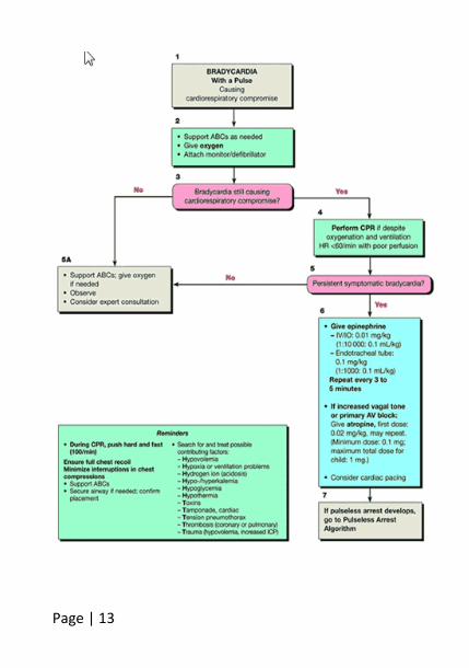
PALS Algorithms
Page | 13

Page | 14
Page | 15
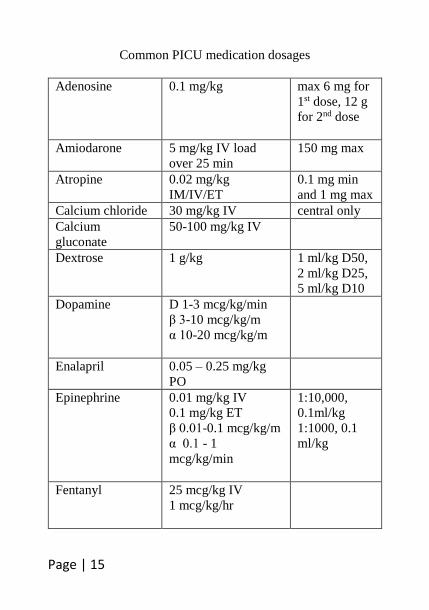
Common PICU medication dosages
Adenosine 0.1 mg/kg max 6 mg for
1st dose, 12 g
for 2nd dose
Amiodarone 5 mg/kg IV load
over 25 min
150 mg max
Atropine 0.02 mg/kg
IM/IV/ET
0.1 mg min
and 1 mg max
Calcium chloride 30 mg/kg IV central only
Calcium
gluconate
50-100 mg/kg IV
Dextrose 1 g/kg 1 ml/kg D50,
2 ml/kg D25,
5 ml/kg D10
Dopamine D 1-3 mcg/kg/min
β 3-10 mcg/kg/m
α 10-20 mcg/kg/m
Enalapril 0.05 – 0.25 mg/kg
PO
Epinephrine 0.01 mg/kg IV
0.1 mg/kg ET
β 0.01-0.1 mcg/kg/m
α 0.1 - 1
mcg/kg/min
1:10,000,
0.1ml/kg
1:1000, 0.1
ml/kg
Fentanyl 25 mcg/kg IV
1 mcg/kg/hr

Page | 16
Hydralazine 0.1 – 0.2 mg/kg IV max 20
mg/day
Magnesium 25-50 mg/kg IV 2 g max
Midazolam 0.05 mg/kg
IM/IN/IV
0.05 mg/kg/hr
Milrinone 0.25 – 1
mcg/kg/min
Morphine 0.05 mg/kg IV
Norepinephrine β 0.01-0.1 mcg/kg/m
α 0.1 - 1
mcg/kg/min
Sodium
bicarbonate
1 – 2 meq/kg IV
Vasopressin 0.5 – 40 mU/kg/hr
Vecuronium 0.1 mg/kg IV

Page | 17
Respiratory
1. Airway / intubation
a. Decision to intubate is clinical and based on poor
respiratory effort, difficulty oxygenating or
ventilating, need for decreased oxygen consumption,
unable to protect airway, altered mental status, need
for anesthesia, etc.
b. Equipment to prepare: SOAP ME
i. Suction: Yankauer and flexible catheter hooked to
suction canister and ready to use
ii. Oxygen source: bag and appropriate size mask,
pre-oxygenate patient
iii. Airway equipment: ETT size = (16 + Age) / 4 for
uncuffed tube, subtract 0.5 size for cuffed tube,
have other sizes available; check bulb on
laryngoscope
iv. Pharmacy: choose a sedative and a paralytic, make
sure you have secure IV access, premedicate with
atropine in patients < 5 yo
v. Monitoring Equipment: have SpO2, HR, cycle BP
q3 min, CO2 detector
c. Confirmation: auscultate bilateral breath sounds,
check end tidal CO2, obtain CXR
d. Troubleshooting: DOPE
i. Displaced ETT (Are you still intubated? Do you
have end tidal?)
ii. Obstructed ETT (Have you tried suctioning?)
iii. Pneumothorax (Have you listened for bilateral
breath sounds?)
iv. Equipment failure (Do you have disconnected vent
tubing?)

Page | 18
e. Rapid sequence intubation: do not apply PPV, do not
place NG prior to intubation, hold cricoid pressure
until ETT placement is confirmed
f. Assess for air leak around the ETT q12 hrs. A leak
should be audible between 20 and 30 cmH2O
g. Obtain CXR qAM to check ETT placement
2. Infant Airway
a. larynx more cephalad (C2-3 vs. C4-5) and anterior
b. larger tongue (and head), larger tonsils and adenoids.
c. hyoid bone attached to thyroid cartilage epiglottis
protrudes into airway (omega shaped)
d. cricoid cartilage—narrowest pointcylindrical
shaped larynx (glottis is narrowest in adults)
3. Predicting Difficult Airway
a. mallampati class > 2
b. thyromental distance < 3 fingerbreaths (< 6 cm)
c. submandibular space, neck movement, mouth opening
d. cricoid pressure and BURP: move thyroid cartilage
back, up, right, and with pressure
4. Mechanical ventilation
a. Conventional ventilation
i. Pressure control (set PIP) vs Volume control (set
tidal volume): choose PEEP, Rate, FiO2 in both
modes. Monitor variable parameter (PIP in volume
control and tidal volume in pressure control). Aim
for tidal volume of 5-7 ml/kg.
ii. Pressure Control: improved patient comfort,
improved delivery of breath to lung units with
different compliance and resistance
iii. Volume Control: guaranteed minute ventilation

Page | 19
b. PRVC is a pressure mode. The desired tidal volume is
set but the pressure used per breath varies and is based
on patient involvement and preset limit. So, the tidal
volume per breath may actually vary significantly.
c. APRV and HFOV are modes that are used to improve
oxygenation. HFOV is also used for air leaks and for
pulmonary hemorrhage
i. HFOV settings include FiO2 and MAP that affect
oxygenation and Amplitude and Frequency that
affect ventilation
1. Set the MAP about 2 cmH2O above previous
MAP and adjust based on oxygenation
parameters, lung expansion on CXR
2. Set the Amp based on patient “jiggle” and
ventilation parameters
3. Frequency: 1 Hz = 60 cycles/sec. lower limit is
3 Hz. the lower the frequency, the lower the
CO2
4. exhalation is active
5. cytokine production is ↓
6. good for air leak: prevents overdistension of
more compliant alveoli
ii. APRV settings include FiO2, Phigh set near MAP
on conventional vent, Plow set at zero, Thigh and
Tlow set to maximize recruitment.
d. Monitor serum pH. Aim for pH 7.3-7.4. Correlate at
least one serum pH with continuous End tidal
monitoring
5. Minute ventilation = tidal volume x RR. In volume
control, tidal volume is guaranteed. So if you want to
decrease CO2 by 25%, increase RR by 25%
6. Titrating vent settings toward extubation: patient must
demonstrate ability to initiate spontaneous breaths,

Page | 20
achieve adequate tidal volumes per each spontaneous
breath, have intact bulbar reflexes, have adequate muscle
strength to initiate breaths, and be sufficiently alert.
7. Hypoxia differential diagnosis
a. Low partial pressure of oxygen (high altitude)
b. Hypoventilation
c. Shunt (intracardiac or intrapulmonary)
i. Calculating PAO2: [FiO2 (Patm – PH2O vapor)] –
[PaCO2/RQ] where RQ is 0.8
ii. Normal A-a gradient is 10 accounting for
physiologic shunting from bronchial and thebesian
circulations
d. Alveolar dead space
i. Physiologic dead space = anatomic dead space +
alveolar dead space
ii. Anatomic dead space = ventilated but not perfused
areas (vent tubing, large airways)
iii. Alveolar dead space = lung disease
iv. Physiologic dead space = (PaCO2 – PETCO2) /
PaCO2
e. Diffusion abnormality
f. Shunt, alveolar dead space, and diffusion abnormality
give you an A-a gradient > 10
8. Monitoring
a. SpO2: Can be falsely low with nail polish, methylene
blue, indocyanine green, Methemoglobin levels <
15%, increased venous pulsations, and other light
sources. Can be falsely high with CO poisoning,
vasoocclusive disease in HgbSS, and
methemoglobinemia > 15%. Hyperbilirubinemia does
not affect the SpO2.
b. SaO2: estimated on blood gas, measured on co-
oximetry
Page | 21
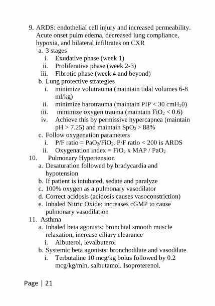
9. ARDS: endothelial cell injury and increased permeability.
Acute onset pulm edema, decreased lung compliance,
hypoxia, and bilateral infiltrates on CXR
a. 3 stages
i. Exudative phase (week 1)
ii. Proliferative phase (week 2-3)
iii. Fibrotic phase (week 4 and beyond)
b. Lung protective strategies
i. minimize volutrauma (maintain tidal volumes 6-8
ml/kg)
ii. minimize barotrauma (maintain PIP < 30 cmH20)
iii. minimize oxygen trauma (maintain FiO2 < 0.6)
iv. Achieve this by permissive hypercapnea (maintain
pH > 7.25) and maintain SpO2 > 88%
c. Follow oxygenation parameters
i. P/F ratio = PaO2/FiO2. P/F ratio < 200 is ARDS
ii. Oxygenation index = FiO2 x MAP / PaO2
10. Pulmonary Hypertension
a. Desaturation followed by bradycardia and
hypotension
b. If patient is intubated, sedate and paralyze
c. 100% oxygen as a pulmonary vasodilator
d. Correct acidosis (acidosis causes vasoconstriction)
e. Inhaled Nitric Oxide: increases cGMP to cause
pulmonary vasodilation
11. Asthma
a. Inhaled beta agonists: bronchial smooth muscle
relaxation, increase ciliary clearance
i. Albuterol, levalbuterol
b. Systemic beta agonists: bronchodilate and vasodilate
i. Terbutaline 10 mcg/kg bolus followed by 0.2
mcg/kg/min. salbutamol. Isoproterenol.

Page | 22
c. Subcutaneous beta agonists: epinephrine 0.01 ml/kg
of 1:1000
d. Corticosteroids: anti-inflammatory, act on leukocytes,
eicosanoids, and vascular endothelium
i. Methylprednisone 2 mg/kg IV followed by 1
mg/kg q12
ii. Inhaled steroids: beclomethasone, fluticasone,
budesonide
e. Anticholinergics: inhibit parasympathetic
bronchoconstriction
i. Ipratropium bromide
f. Methylxanthines: unclear mechanisms. Possibly via
adenosine receptor antagonism or decreasing cGMP
g. Magnesium sulfate: inhibits Ca channels to cause
smooth muscle relaxation. can cause hypotension,
respiratory depression, heart block
h. Heliox: Helium/Oxygen mixture. Helium is colorless,
odorless. lowest density of any gas (except hydrogen)
and higher viscosity than nitrogen so produces more
laminar flow. requires tight fitting mask for delivery
and an oxygen requirement <30% for effectiveness

Page | 23
Cardiovascular
1. Cardiopulmonary interactions
a. During spontaneous inspiration, negative intrathoracic
pressure decreases venous return to the heart. This is
the basis of pulsus paradoxus.
b. During PPV, positive intrathoracic pressure decreases
venous return to the heart. Thus, it is important to
assure adequate preload prior to initiating PPV.
Patients with obstructive lung disease such as asthma
also have decreased venous return which is
dramatically worsened upon initiation of PPV and can
lead to cardiac arrest.
c. PPV decreases afterload on left ventricle. This
decrease is most clinically relevant in heart failure.
However, PPV also increases afterload to the right
ventricle which can be clinically relevant even in a
heart with normal function.
2. Oxygen Delivery: The main goal in critical care medicine
is to provide adequate oxygen delivery to meet tissue
requirements and balance that with oxygen consumption
in order to maintain aerobic metabolism and organ
function.
a. Oxygen delivery = cardiac output x arterial oxygen
content
b. Cardiac output depends on HR and SV. SV depends
on preload, contractility, and afterload
c. Arterial oxygen content = bound content + dissolved
content = (1.36 x Hgb x SaO2) + (0.003 x PaO2)
d. Note that the dissolved content does not contribute
significantly to overall arterial oxygen content
especially when the hemoglobin is normal.
e. The bound content is affected by the oxyhemoglobin
dissociation curve. Factors that shift the curve to the

Page | 24
right make it easier for oxygen to unload to the
tissues.
f. Note that if you want to increase BP, you can either
increase the cardiac output, increase the SVR, or both.
If just SVR is increased, then the heart is working
against a resistance and the cardiac output can
actually decrease.
g. Normally, 1 L O2/min is delivered to tissues and 250
ml O2/min is consumed by the tissues giving an
oxygen extraction ratio of 25%. You can see this
number as the SvO2 on a VBG obtained from a
central venous line positioned near the RA. Assuming
an SaO2 of 100% in a patient with no lung disease,
the SvO2 should be approximately 75%. In sepsis,
this number will often be lower implying an increase
in oxygen consumption.
h. Assessment of oxygen delivery and consumption: end
organ perfusion (skin perfusion, mental status, UOP),
vital signs including NIRS, ABG with pH, base deficit
and lactate, Hgb, SvO2
3. Shock: inadequate oxygen delivery +/- increased oxygen
consumption or maldistribution of blood flow. Evidenced

Page | 25
by tachycardia and normotension if compensated,
hypotension if uncompensated.
a. Compensated shock maintains BP. Uncompensated
shock does not and thus CO decreases.
b. Types of shock pulse SV
R
CO CVP PCWP
Hypo-
volemic
thready ↑ ↓ ↓ ↓
Septic thready ↓ ↓ unless
compen
sated
↓ (can
↑ w/
cardiac dysfxn
)
↓ (can
↑ w/
cardiac dysfxn
)
Distributive (anaphy-
laxis or
spinal shock)
bounding w/
widened
pulse pressure
↓ ↑ ↓ ↓
Cardio-
genic
thready ↑ ↓ (can
be nml
with diastolic
dysfxn)
↑ ↑
i. Cardiogenic shock can be an issue with
1. Preload (dilated cardiomyopathy, regurgitant
valves)
2. Contractility (systolic or diastolic dysfunction)
a. Diastolic dysfunction can maintain BP and
CO for a longer period of time
3. Afterload (aortic coarctation, pulmonary or
aortic stenosis, pHTN, etc)
4. Obstruction (tamponade, tension
pneumothorax)
ii. Treatment
Page | 26
1. Volume resuscitate hypovolemic, septic, and
distributive shock
a. Use isotonic fluids (NS or LR)
b. Reassess after each bolus
c. After 60 ml/kg, consider supplementing
with inotrope/pressor support but do not
stop providing isotonic fluids if the patient
is still in shock
d. Epinephrine is often used for cold shock
and NE for warm shock
e. For shock unresponsive to fluids and
pressors, consider vasopressin and steroid
replacement
2. Broad spectrum antibiotics for suspected septic
shock
3. Cardiogenic shock: optimize preload with
diuretics. Inotropic support with low dose
epinephrine, milrinone, calcium without
worsening relaxation. Optimize pH. Lusitropic
support with milrinone.
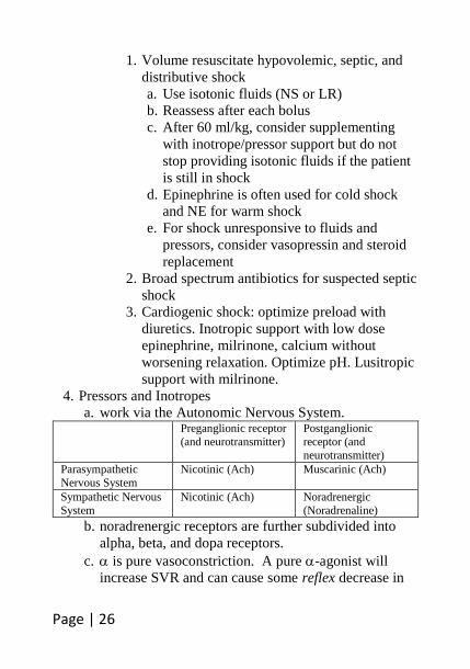
4. Pressors and Inotropes
a. work via the Autonomic Nervous System. Preganglionic receptor
(and neurotransmitter)
Postganglionic
receptor (and
neurotransmitter)
Parasympathetic
Nervous System
Nicotinic (Ach) Muscarinic (Ach)
Sympathetic Nervous
System
Nicotinic (Ach) Noradrenergic
(Noradrenaline)
b. noradrenergic receptors are further subdivided into
alpha, beta, and dopa receptors.
c. is pure vasoconstriction. A pure -agonist will
increase SVR and can cause some reflex decrease in

Page | 27
cardiac output, although it has no direct effect on the
heart itself.
d. is much more complicated in that it has several
effects:
i. Inotropy (desired)
ii. Chronotropy (generally not desired as it increases
MvO2)
iii. Mild vasodilation through 2 effects (explains
how this can work against SVR)
e. An and agonist can often work together in blood
pressure control. The -agonist will increase BP by
increasing SVR. The -agonist will increase cardiac
output. Pure > = > Pure
Phenyl-
ephrine
NE Epi dobutamine Isopro-
terenol
In an effort at simplification, anything to the left of
middle in the chart above is a pressor and anything to
the right of the middle is an inotrope.
Epinephrine at doses > 0.1 mcg/kg/min becomes
almost all
Norepinephrine may have enough to counteract the
reflex decrease in CO but remember that any
vasoconstricts more than any vasodilates
Dopamine is difficult to put on the chart as it is the
most dose dependent. At < 10 mcg/kg/min, it is
mainly an inotrope. At > 10 mcg/kg/min, it is mainly
a pressor. It also has a tendency to increase PVR.
Remember that calcium is a short acting inotrope and
vasoconstrictor. There are many other inotropes
including milrinone and methylene blue
5. CHD

Page | 28
a. Acyanotic lesions
i. Increased pulmonary blood flow (L R shunts,
present with CHF, FTT)
ii. Decreased pulmonary blood flow
b. Cyanotic lesions
i. Increased pulmonary blood flow
ii. Decreased pulmonary blood flow
c. Pre-op management
i. Secure diagnosis (Echo, cardiac cath) and
management plan
ii. Evaluate for syndrome associated with cardiac
defects such as Trisomy 21 (AV canal defects,
pulmonary HTN), Turners (coarctation), DiGeorge
(conotruncal and aortic arch defects), etc.
iii. Ensure adequate oxygen delivery
iv. Prevent acidosis
v. PGE for ductal dependent lesions
1. Ductal dependent pulmonary blood flow:
monitor PaO2
2. Ductal dependent systemic blood flow:
monitor end organ perfusion, limit
supplemental oxygen
d. Post-op management
i. Know the surgical procedure and the maneuvers
required to achieve them (for example, was a
ventriculotomy required? Was the ASD closed
primarily or with a patch?)
ii. Know the results of the post-op TEE for
assessment of heart function and residual defects
iii. Obtain CXR immediately post-op to assess
placement of new lines and tubes
Page | 29
iv. Dysrhythmias: obtain EKG immediately post-op
as new baseline, assess tachycardias with atrial
EKG
v. Bleeding: monitor platelet count, Hgb, coags (PT,
PTT, Fibrinogen). Goal Hgb > 10 (> 13 for single
ventricle physiology)
vi. Adequate oxygen delivery: monitor pH, base
deficit, lactate, NIRS. Assess volume status and
maintain adequate preload
vii. Know of any alterations in loading conditions:
1. Residual shunts or stenotic valves may have an
adverse effect on preload
2. Shunt closures or PA banding may have an
adverse effect on afterload
viii. Monitor electrolytes closely post-op. Patients
should be on ¾ maintenance IVF without
potassium as the urine output will diminish over
time and there is potential for renal injury. Avoid
TPN immediately post-op.
ix. For single ventricle physiology (post-op Norwood
and Glenn): maintain SpO2 75-85% as a non-
invasive way to maintain PaO2 40-45 mmHg as a
way to optimize pulmonary and system blood
flows (Qp:Qs). Qp:Qs is determined using
saturations: [(SaO2 – SvO2) / (SpvO2 – SpaO2)]
where SpvO2 is assumed to be 100% if there is no
lung disease and SpaO2 is assumed to be SaO2 in
complete mixing lesions.
x. For the first 12-18 hours post-op, any decrease in
urine output may indicate low cardiac output, poor
cardiac function, or hypovolemia. After the first
18-24 hours, a low urine output may indicate the
need for diuresis.

Page | 30
xi. Patients with a BT shunt or CVL may be at risk
for clots and may need a non-titrated, prophylactic
heparin infusion at 10 U/kg/hr (discuss with
fellow/attending)
xii. Tamponade: triad of muffled heart sounds, JVD,
and hypotension. On EKG, look for low voltages.
Clinically look for electrical alternans, narrow
pulse pressure and tachycardia. Management
involves supporting preload and relieving the
tamponade with pericardiocentesis
xiii. Chylothorax: normally, thoracic duct takes lymph,
fatty acids, and lymphocytes from the small
intestine to the subclavian vein.
1. Causes: surgical trauma, high CVP, congenital
malformations, thrombosis
2. Contents of chylothorax: triglycerides, fat
soluble vitamins, T-lymphocytes, proteins
(albumin, immunoglobulins), all blood
components except RBCs and platelets
3. Management: MCTs still meet the fatty acid
needs of the body but are absorbed directly
into the portal blood. Octreotide is a long
acting somatostatin analog that decreases
splanchnic blood flow. Surgical duct ligation
for drainage > 100 ml / year of age for 5 days
or persistent drainage for 2 weeks.
e. Cardiopulmonary bypass
i. On bypass, patient is receiving fully oxygenated
blood to all organs. However, the blood flow is
non-pulsatile and the blood is being continuously
exposed to tubing. Therefore, there is potential for
end organ injury with prolonged bypass. There is
also an inflammatory response that peaks 10-12

Page | 31
hours post bypass that manifests as low cardiac
output, capillary leak, decreased lung complains,
etc. Patients also have thrombocytopenia and/or
platelet dysfunction and hyperglycemia post-op.
ii. Cross clamp prevents blood flow inside the heart
and maintains non-pulsatile flow to the rest of the
organs. Watch for signs of cardiac ischemia post-
op.
iii. Deep hypothermic circulatory arrest (DHCA):
Patient is cooled to 18◦C and there is no flow
through the bypass circuit. This allows a bloodless
field where the surgeon can also remove all
clamps and tubing that sits in the way for intricate
repairs external to the heart. Data suggests that up
to 30 min of DHCA can be safe. Monitor for end
organ injury including renal and neurologic injury.
f. Repairs for management of the single ventricle
i. Stage I Norwood with BT shunt or Sano: The
pulmonary artery trunk is used to create a neo-
aorta. A shunt is placed to direct part of the
systemic blood flow to the PAs. Remember that
shunt flow is determined by the balance of PVR
and SVR. Thus, blood from the IVC/SVC enters
the RA, then the RV, then the neo-aorta where part
enters the pulmonary system and part continues
down the descending aorta. From the pulmonary
veins, blood enters the LA and then the RA
through an ASD.
ii. Stage II bidirectional Glenn: The BT shunt or
Sano is taken down. The SVC is disconnected
from the RA and attached directly to the PA.
Blood from the SVC must passively enter the
pulmonary circulation.

Page | 32
iii. Stage III Fontan: The IVC is now also
disconnected from the RA and attached to the PA
often via an extracardiac conduit. This conduit
may or may not be fenestrated to provide a right to
left shunt to maintain cardiac output in the setting
of pulmonary disease or pHTN. All blood flow to
the lungs is now passive and the RV functions as
the systemic ventricle. SpO2 should now be in the
90s.
6. Pacemaker Notation
Chamber
Paced
Chamber
Sensed
Response to
sensing
Rate
Modulation
O = none O = none O = none R for rate
modulation
A = atrium A = atrium T = triggered
V = ventricle V = ventricle I = inhibited
D = dual D = dual D = dual
Ex: VVIR pacer implies ventricle is paced,
ventricular impulses are sensed, inhibited from
pacing if pulse is greater than the programmed
threshold, is rate responsive (adjusts rate according to
patient’s activity)
7. Extracorporeal Membrane Oxygenation (ECMO) or
Extracorporeal Life Support (ECLS)
a. Requires the presence of a reversible disease process
b. Requires the use of systemic heparin (screen with
head ultrasound for any intraventricular hemorrhage)
c. Types
i. VV ECMO (veno-venous): provides pulmonary
support only. Often employs one cannula with two
lumens placed via the right IJ into the RA.
Page | 33
ii. VA ECMO (veno-arterial): provides pulmonary
and cardiac support. Employs two catheters. The
venous catheter is directed into the RA via the
right IJ and the arterial cannula is directed into the
ascending aorta via the right common carotid. The
right carotid artery is then ligated. The cannulas
can also be positioned directly in the RA and aorta
via an open chest.
d. Cardiac output is determined by pump flow and LV
output. Pump flow is determined by the amount of
venous blood withdrawn from the patient which is
dependent on cannula size and systemic venous
return. Avoid hypovolemia, pneumothorax, and
tamponade. LV output is determined by patient’s
heart function.
e. Monitoring
i. Ask perfusionist about the pump (presence of
clots, membrane oxygenator failure)
ii. Patient ABG evaluates the blood passing through
the circuit and through the pulmonary circulation.
The circuit ABG and VBG assess the adequacy of
the membrane function and of the oxygen delivery
to the tissues.
iii. Examine patient’s perfusion, lines, tubes, sites of
entry
iv. Monitor for excessive bleeding, heparin dose
required
v. Daily CXR to assess lung fields, position of lines
and tubes
f. Emergency management: If there is a problem with
the circuit, disconnect the patient from the circuit
(clamp the venous line first, then unclamp the bridge,
then clamp the arterial line), increase the ventilator
Page | 34
settings to the predetermined emergency settings,
proceed to routing PALS protocols.

Page | 35
FEN/GI
1. IVFs
a. Isotonic fluids include NS and LR
b. normal saline can cause a non-anion gap metabolic
acidosis when given in significant amounts
c. lactated ringers has lower chloride and sodium
concentrations compared to NS
2. Potassium
a. Hyperkalemia: manifests as EKG changes with
peaked T waves initially (K 6-8 meq/L) that
progresses to widened QRS, then disappearance of the
P wave, and finally a sine wave. The etiology can
range from acidosis to tumor lysis to renal failure.
Treatment:
i. Make sure to d/c exogenous sources (IVF, TPN, K
supps)
ii. Calcium will not decrease the potassium level but
will act quickly to increase threshold potential
iii. Medications that shift K intracellularly (temporary
solution, takes approx 30 min): sodium
bicarbonate, beta agonists, insulin (0.2 U/kg),
glucose (1 g/kg)
iv. Increase elimination (permanent solution but can
take longer): Polystyrene sulfonate with sorbitol
(Kayexalate – 1-2 g/kg PO or PR), dialysis
b. Hypokalemia: manifests as weakness and ileus with
EKG changes with flat T waves, U waves. Remember
that potassium supplements can inadvertently cause
hyperkalemia. PO supplementation is safer but should
be given with feeds. KCl dose is 0.5 – 1 meq/kg.
3. Sodium
a. Hypernatremia

Page | 36
i. Effects: cellular dehydration from osmotic fluid
shifts, cerebral dehydration (brain cell volume ↓
10-15%) venous sinus thrombosis, bridging
vein rupture (SDH), ICH), high pitched irritable
cry, rhabdomyolysis, hyperreflexia,
hyperglycemia from peripheral insulin resistance
ii. Causes
1. Too much intake: Na polystyrene
2. Not enough fluid (↑ serum osm): hypothalamic
d/o or abnormal thirst function
3. Water loss
a. In kidney (urine osm < 800, plasma osm >
275)
i. Renal water loss: DI, diuretics
ii. Osmotic water loss: ↑ glucose,
mannitol, ↑ solute feeds
b. Outside of kidney (urine osm > 800)
i. Insensible losses
ii. GI losses
iii. Management: rapid drop in Na leads to cerebral
edema. Aim to decrease the Na by 12 meq/day. If
the hypernatremia is chronic, body compensated
with idiogenic osmoles.
b. Hyponatremia
i. Causes
1. Serum osm > 280
a. No osmolar gap pseudohyponatremia
(lipids and proteins)
b. Osmolar gap translational hyponatremia
(glucose, mannitol, maltose, etc)
i. Corrected Na = measured Na + [((gluc
– 100)/100) x 2]
2. Serum osm < 280

Page | 37
a. Urine osm < 100: polydipsia, iatrogenic
water intake
b. Urine osm > 100: impaired water excretion
i. Hypovolemia: V/D, burns, CSW
ii. Euvolemia: SIADH, hypothyroidism
iii. Hypervolemia: CHF, nephritic
syndrome, cirrhosis
ii. Management: rapid increase in Na leads to central
and extrapontine demyelination (quadriplegia,
seizures, pseudobulbar palsy). Aim to increase Na
by 12 meq/day unless symptomatic. If
symptomatic, correct rapidly to Na 125 meq/L.
Assuming ongoing isotonic fluid losses, 1
ml/kg/hr of 3% will increase Na by 1 meq/L/hr.
4. Calcium
a. Hypercalcemia
i. Causes: hyperparathyroid, adrenal insuff, MEN,
thyrotoxicosis, Vit D and A excess,
immobilization, thiazides, lithium, theophylline,
glucocorticoid withdrawal
Phos 1,25(OH)
-Vit D
PTH Urin
e Ca
Immobilization Nml/
↑
↓ ↓ ↑
Hyperparathyroidis
m
↓ ↑ ↑ ↑
Malignancy ↓ Nml/↑ ↓ ↑
Abrupt
glucocorticoid w/d
Nml/
↑
Nml ↓ ↑
Sarcoidosis Nml/
↑
↑ ↓ ↑

Page | 38
Familial
Hypercalciuric
Hypercalcemia
Nml/
↓
Nml/↑ Nml/
↑
↓
ii. Signs/symptoms: Bone pain, Kidney stones,
Polyuria/polydipisia, Constipation, ↓ reflexes,
EKG: heart block, wide QRS, short QT
iii. Management: IVFs, furosemide, calcitonin,
bisphosphonates, edentate disodium chelation
b. Hypocalcemia
i. Causes: hypoparathyroid, Vit D deficiency,
alkalosis (iCa ↓ 0.42 mmol/L for every 0.1
mmol/L change in pH), hyperalbuminemia
(corrected Ca = measured Ca + (0.8 x (4-
albumin)), drugs (AEDs, barbs), DKA, TLS,
rhabdomyolysis
ii. Signs/symptoms: perioral paresthesias, Trousseau,
Chvostek, bronchospasm, laryngospasm, stridor,
seizures, cramps, EKG: prolonged QT, inverted T
waves
iii. Management: CaCl comes already ionized but
must be given centrally. CaGluconate can be given
peripherally but must be conjugated in the liver
5. Magnesium
a. Hypermagnesemia
i. Causes: renal failure (initially low Mg from
tubular injury then high Mg from decreased
elimination)
ii. Signs/symptoms: anticholinergic signs (inhibits
prejxnal release of ACh due to displacement of
memb bound Ca), ↓ reflexes, hypotension, resp
arrest, EKG: short QT (breaks torsades)
iii. Management: CaGluc, lasix, dialysis

Page | 39
b. Hypomagnesemia
i. Causes: ↓ intake, refeeding, malnutrition,
hyperthyroidism, transfusion with citrated blood,
acidosis shift, ↑ loss (lasix, ampho B, cisplatin,
cyclosporin, V/D, laxatives, malabsorption, gent
(toxic to proximal tubule where 30% Mg
reabsorbed), volume expansion (SIADH, burns),
sweating, hyperaldosteronism, ↓ albumin
ii. Signs/symptoms: muscle weakness, seizures, VT,
nystagmus, Trousseau/Chvostek, low K/Ca,
suppressed parathyroid hormone, EKG: ↑ PR, ↑
QRS, ↑QT, flat T
6. Phosphorus
a. Hypophosphatemia
i. Causes: refeeding syndrome,malabsorption,
respiratory alkalosis, β agonists, hypomagnesemia,
antacids, sepsis, burns, renal losses
ii. Effects
1. ↓ 2,3 DPG decreased oxygen delivery
2. ↓ ATP hemolytic anemia
3. ↓ cardiac caontractility
4. Muscle weakness
5. Paresthesias and neuropathy
6. Seizures
7. Platelet dysfunction
8. Liver failure
7. NPO guidelines
a. Nothing by mouth at least 2 hours before
b. Clears/breast milk at least 4 hours before (8 oz limit)
c. Simple solid foods, formula at least 6 hours before
d. Heavy and fatty foods at least 8 hours before
8. Gastritis prophylaxis with famotidine for patients who are
NPO, on post-pyloric feeds, or on corticosteroids

Page | 40

Page | 41
Endocrine
1. DKA
a. State of relative or absolute insulin deficiency with
increases in glucagon, cortisol, and growth hormone
leading to hepatic gluconeogenesis, hepatic and
skeletal muscle glycogenolysis, and hepatic lipolysis
b. Patients will be hypovolemic but remember that rapid
shifts in osmolality with aggressive fluid
administration may result in cerebral edema
c. The increased glucose and osmolality causes the brain
to grab idiogenic osmoles to avoid shrinkage. Thus,
lack of adequate compensation and/or aggressive
treatment of DKA results in cerebral edema
d. Risk factors for development of cerebral edema
include age < 5, male patients, new onset diabetes,
and protracted illness.
e. Note their initial mental status and order q1 neuro
checks for any altered mental status
f. Monitor acidosis, hypokalemia, and
hypophosphatemia with Chemistries q2
g. Monitor ketonuria with U/As each void and serum
ketones (Acetest for acetoacetate and β
hydroxybutyrate)
h. Monitor glucose by finger stick q1
i. Remember that you will have a pseudohyponatremia
while hyperglycemic
i. Corrected Na = measured Na + [(serum glucose –
100) / 100] x 1.6
j. Start insulin gtt at 0.1 units/kg/hr and do not titrate
while patient is acidotic and ketotic
k. 2 bag correction method
i. NS + 20 meq/L KPhos + 20 meq/L KCl (initial
fluids for any BG > 250)

Page | 42
ii. D10NS + 20 meq/L KPhos + 20 meq/L KCl (start
for any BG < 250)
l. Run total fluids above maintenance fluid requirements
(discuss with PICU fellow)
m. Do not treat acidosis with sodium bicarbonate as this
may cause a paradoxical CNS acidosis, impair oxygen
extraction, further hypokalemia, and hypocalcemia.
2. DI
a. Causes: CNS infection, TBI, intraventricular
hemorrhage, neurosurgery, brain death
b. Signs/Symptoms: polyurea, ↓urine osm, ↑ serum osm
and Na, hypovolemia
c. Management: fluid resuscitation, replace UOP > 3
ml/kg/hr 1:1 with NS, electrolyte correction,
vasopressin infusion 1-3 mU/kg/hr and titrate to UOP
3. SIADH
a. Causes: CNS infection, TBI, drugs, PPV
b. Signs/Symptoms: ↑ urine osm, ↓ serum osm and Na,
low UOP
c. Management: fluid restriction, furosemide, 3% saline
for symptomatic patients
4. Adrenal Insufficiency
a. Causes
i. Primary: CAH, Addisons
ii. Secondary: insufficient ACTH production or
destruction of pituitary (tumors, autoimmune)
iii. Tertiary: suppression from exogenous steroids
b. Signs/Symptoms: weight loss, dehydration, N/V,
hypotension, shock unresponsive to fluids/inotropes,
↓Na, ↑K, ↑Ca, acidosis
c. Cortisol stim test:
i. Obtain blood sample for cortisol and ACTH
ii. Inject synthetic ACTH

Page | 43
1. low-dose short test: inject 1 µg
2. conventional-dose short test: inject 250 µg
iii. obtain blood sample 60 minutes after injection
d. Management: fluid resuscitation, hydrocortisone 50
mg/m2/d divided q8, electrolyte correction

Page | 44
Heme/Onc
1. DVT
a. Risk factors: central venous lines, cancer, L-
asparaginase, trauma, immobilization, pregnancy,
obesity, smoking, HgbSS, vasculitis, sepsis,
dehydration, cardiomyopathy
2. Blood replacement
a. Older pRBCs from the blood bank have undergone
metabolic, chemical, and molecular changes including
decreased NO containing groups, loss of
deformability, decreased 2,3 DPG, decreased ATP
3. Transfusion associated disease
a. Acute lung injury
i. Immune response directed against HLA antibodies
that occurs within 30 minutes to 6 hours after
transfusion manifested as respiratory distress and
hypoxia with bilateral fluffy infiltrates on CXR.
May also see fever and hypotension.
ii. Management: for future transfusions, consider
obtaining washed pRBCs, newer pRBCs and
minimizing the number of donors
b. Transfusion associated GVHD
i. Associated with pRBCs, platelets, granulocytes
but not FFP
ii. Occurs in immunocompromised patients or those
with HLA haplotype issues (1st degree relatives)
iii. Fever, rash, cough, cholestatic jaundice,
lymphopenia
iv. Diagnosis: HLA analysis of lymphocytes
v. Prevention: gamma irradiation, HLA matched
blood products
4. Leukocytosis
a. WBC count > 100,000/mL

Page | 45
b. Hyperviscous state leading to pulmonary and CNS
findings
c. Management: avoid diuresis, avoid pRBCs.
Leukapherese with tumor lysis precautions
5. Tumor Lysis Syndrome
a. Physiology: patients with a high tumor burden or
those undergoing chemotherapy are at risk for
massive lysis of cells.
b. At risk patients include those with T cell lymphoma or
leukemia that present with leukocytosis.
c. Monitor q6 labs for hyperkalemia, hyperuricemia, and
hyperphosphatemia
d. Management: aggressive IVF hydration, alkalinize
urine, mannitol. Allopurinol competitively inhibits
xanthine oxidase and will prevent further production
of uric acid. Rasburicase is a recombinant urate
oxidase. Avoid giving calcium to prevent calcium-
phosphorus binding.
6. Anticoagulation
a. Heparin induced thrombocytopenia: type I is not
immune mediated and is transient, resolving without
intervention. Type II is immune mediated antibodies
formed to platelet factor 4. All sources of heparin
must be removed from the patient. Platelet counts will
normalize within one week of discontinuation but the
risk for thrombotic events can persist for weeks.
Management involves use of heparinoids or direct
thrombin inhibitors.

Page | 46
Infectious Disease
1. Hospital acquired infections
a. Ventilator associated pneumonia: more likely to occur
in the first week with early onset VAP usually from
gram positive organisms and late onset VAP from
gram negative organisms
b. Central line associated blood stream infection
2. Necrotizing Fasciitis
a. Necrosis of subcutaneous tissue with mix of aerobic
and anaerobic organisms. Single organisms are
usually GAS. Oral anaerobes such as fusobacteria can
especially cause necrotizing fasciitis of the head and
neck. Management is with beta lactam or beta
lactamase inhibitors (Pip/Tazo). If GAS is suspected,
treat with penicillin and clindamycin.
3. Staph Toxic Shock
a. Can occur with any Staph infection with 2-3 days of
prodrome preceding fever and erythroderma, followed
by desquamation in 1-2 weeks resulting from TSST-1
and enterotoxins
b. 25% of S. aureus strains are toxigenic but Blood
culture is only positive in 5-15% for S. aureus
c. Treatment: Nafcillin, cephalosporins, vancomycin.
Clindamycin as an adjunt inhibits toxin production.
Consider IVIg.
4. Strep Toxic Shock
a. Excruciating pain, confusion, hypoalbuminemia
resulting from strep pyogenic exotoxins A and B
b. Blood culture positive in 50%
c. Mortality rate 5 x that of staph toxic shock
d. Treatment: vancomycin + cefuroxime. Clindamycin
can be used as an adjunt. Consider IVIg.
5. Meningitis

Page | 47
a. Neonates are at risk for GBS, E.coli. Children are at
risk for S. pneumo, N. meningitides, Hib
WBC
count
%PMNs Glucose Protein
Normal 0-5 0-15 45-65 20-45
TB/fungal 25-
100
Lymph/
monos
30-45 100-
500
Viral 25-
500
< 50 45-65 50-100
Bacterial > 100 90 < 40 > 150
Aseptic/Partially
treated
Monos Normal Normal
b. Treatment: ceftriaxone covers H. flu, N. mening, and
S. pneumo. Vancomycin covers resistant S. pneumo
c. N. meningitidis: encapsulated gram negative
diplococcus with humans as the only natural host
i. Serogroup B causes the most cases but is not
included in the quadrivalent conjugate vaccine
ii. Complications result from malfunction of the
coagulation system from endotoxin release
d. HSV meningitis: EEG may show periodic lateralized
epileptiform discharges (PLEDs) early in the course.
Head CT may show temporal lobe edema. Diagnosis:
HSV PCR.
e. High CSF protein can also result from prolonged
seizures and disrupted blood brain barrier with
albumin-cytologic dissociation (malignancy, Guillain
Barre, multiple sclerosis)

Page | 48
Neurology
1. Sedation and paralytic medications
a. Opiates
i. Morphine, Fentanyl, Hydromorphone, Nalbuphine
ii. Side effects include respiratory depression,
itching, hypotension especially if combined with a
benzodiazepine, histamine release with morphine
iii. Reversal agent = naloxone. Remember that
naloxone has a shorter ½ life than your opiates so
the dose may need to be repeated
iv. Watch for withdrawal signs and symptoms
(diarrhea, sweating, agitation) if your patient is
coming off an opiate infusion after a greater than
5-7 day exposure
v. Uses
1. Post-op pain control – for extubated patients,
use morphine 0.05-0.1 mg/kg/dose q3-4 hours
PRN
2. For older post-op patients – consider morphine
PCA using demand dosing only (no continuous
without discussing with PICU fellow)
3. For intubated patients, typically start fentanyl
infusion at 1 mcg/kg/hr with an equivalent
PRN (i.e. 1mcg/kg/dose q1 PRN) combined
with a benzodiazepine
vi. For itching, consider a mu antagonist/kappa
agonist such as nalbuphine
b. Benzos
i. Midazolam, Diazepam, Lorazepam
ii. Watch for propylene glycol toxicity with
lorazepam (solvent). Watch for benzyl alcohol
toxicity with continuous infusion of midazolam
(antibacterial).

Page | 49
iii. Side effects include respiratory depression,
hypotension
iv. Watch for withdrawal signs and symptoms
(diarrhea, sweating, agitation) if your patient is
coming off a benzodiazepine infusion after a
greater than 5-7 day exposure
v. Uses
1. For intubated patients, typically start
midazolam 0.05 mg/kg/hr or lorazepam 0.1
mg/kg IV q4 with an equivalent PRN (i.e.
midazolam 0.05 mg/kg/dose q1 PRN or
lorazepam 0.1 mg/kg/dose q4 PRN)
2. For posterior spinal fusion and similar
surgeries, use diazepam 0.05-0.1 mg/kg/dose
q6 PRN muscle spasm
vi. Reversal agent = flumazenil
c. Barbiturates
i. Thiopental: rapid onset. Decreases cerebral
oxygen consumption and ICP. Side effects:
cardiac depression, bronchospasm.
d. Propofol
i. GABA agonist and NMDA-R inhibitor
ii. Very lipid soluble so quickly redistributes. No
active metabolite. Hepatic metabolism
iii. Contraindications: egg yolk allergy, metabolic
disorder
iv. Side effects: decreases inotropy. Hypotension.
impairs free fatty acid use and mitochondrial
activity leading to propofol infusion syndrome
(refractory metabolic acidosis, rhabdomyolysis,
hepatomegaly)
e. Chloral hydrate

Page | 50
i. Metabolized by liver to its active metabolite
trichloroethanol.
ii. ½ life 4-6 hours, excreted in urine
iii. Side effects: arrhythmias, withdrawal, agitation
f. Ketamine
i. NMDA-R antagonist, increases sympathetic
stimulation, bronchodilates, increases cerebral
blood flow
ii. Side effects include tachycardia, hypertension.
Large doses can cause laryngospasm and apnea.
Muscarinic effect of drooling.
iii. Contraindications: catecholamine depleted state
such as septic shock, increased ICP
g. Dexmedetomidine
i. α 2 agonist
ii. biphasic response with transient hypertension
initially with bolus followed by hypotension and
bradycardia
iii. potential for hypothermia in neonates with
decrease in brown fat use
h. Etomidate
i. Carboxylated imidazole derivative, GABA agonist
ii. Effect lasts 5-10 minutes with ½ life of 75 minutes
iii. No cardiac effects. Decreased cerebral oxygen
consumption
iv. Side effects: adrenal insufficiency (reversibly
inhibits 11 β hydroxylase and 17 α hydroxylase),
decreased seizure threshold
i. Depolarizing neuromuscular blocker (succinylcholine)
i. Side effects
1. Fasciculations, constipation, myoglobinemia
2. Increased intraocular, gastric, and intracranial
pressures

Page | 51
3. Hyperkalemia from increased acetylcholine
receptors from 48 hours up to 16 months s/p
any disease process resulting in large mass of
denervated muscle (burns, crush injuries,
neuromuscular dx)
4. Bradycardia, malignant hyperthermia, Guillain
Barre
ii. Succs effects can be prolonged with plasma
pseudocholinesterase deficiency, liver disease,
pregnancy, or with repeated dosing
j. Non-depolarizing neuromuscular blockers:
competitive inhibitors of acetylcholine. Muscles of the
eyelids and fingers are the first to be affected and the
last to recover. Muscles of the intercostals,
diaphragm, and larynx are the last to be affected and
the first to recover.
i. Aminosteroids: vecuronium, pancuronium,
rocuronium. Roc is safe to give in renal failure and
can be given IM. All aminsteroids have some
vagolytic activity with panc having the most.
ii. Benzylisoquinolines: Cisatracurium, mivacurium.
not vagolytic but can cause histamine release and
bronchospasm. Cisatracurium uses Hofmann
degradation. Mivacurium is degraded via plasma
cholinesterase.
2. Status epilepticus
a. Goal is to control seizures ASAP as the longer the
seizure continues, the harder it is to break
b. Remember that it’s still ABCs first
i. Might need to establish an airway if the patient’s
mental status is altered, the patient has poor
cough/gag, the patient is apneic

Page | 52
ii. Might need to support blood pressure – consider
phenylephrine infusion
c. Video EEG is necessary to assess for subclinical
seizures
d. Look for treatable causes (electrolyte imbalance,
hypoglycemia, etc)
e. Start with lorazepam 0.1 mg/kg
f. If the patient is still seizing, continue to provide
lorazepam but quickly add a second and then a third
agent:
i. Fosphenytoin 20 mg/kg
ii. Phenobarbital 20 mg/kg
iii. Levetiracetam 20 mg/kg
g. Consider pentobarbital infusion with goal to achieve
burst suppression on EEG
3. Glascow Coma Scale
a. Used in ER setting to assess neurologic status
b. Scored from 3-15 with a score of 9-12 suggesting
moderate head injury and a score below 9 suggesting
severe head injury
Motor Verbal Eye 1 – none 1 – none 1 - none
2 – extension,
decerebrate
2 – incomprehensible
sounds, inconsolable
2 – opens eyes to pain
3 – abnormal flexion,
decorticate
3 – inappropriate
words, irritable, inconsistently
consolable
3 – opens eyes to
command/speech
4 – flexion withdrawal to pain
4 – confused, cries but consoles
4 – opens eyes spontaneously
5 – localize,
withdraw to touch
5 – oriented, coos,
smiles
6 – obeys commands
4. TBI and stroke / neuroprotective guidelines
a. Goal is to prevent secondary brain injury

Page | 53
i. Avoid hyperthermia (place continuous rectal
probe, may need around the clock acetaminophen)
ii. Avoid hyponatremia (must place all patients on
isotonic fluid (normal saline))
iii. Avoid hyper and hypoglycemia (older children
should have NS only, younger children can have
D5NS, everyone should have frequent glucose
checks)
iv. Treat seizures aggressively
b. Order q1 hour neuro checks
c. If patient has an EVD, monitor ICPs
i. Call Fellow and Neurosurgery for any sustained
ICP > 20 mmHg
d. Signs of increased ICP: Cushing’s triad (bradycardia,
hypertension, agonal respirations), dilated or unequal
pupils.
e. Cerebral perfusion pressure (CPP) = Mean arterial
pressure – ICP (or CVP if it’s higher like in SVC
syndrome or Glenn physiology). Avoid hypotension.
f. Exam:
i. evenly distributed high pressure: CN VI palsy,
HA, vomiting;
ii. in early herniation: CN III palsy, poor pupillary
response, posturing
g. Types of herniation
i. Transtentorial results from any supratentorial mass
or cerebral edema
ii. Uncal results when the mass is lateralized.
anteromedial portion of the hippocampus herniates
over the edge of the tentorium
h. Cerebral edema
i. Types

Page | 54
1. Vasogenic: increased capillary membrane
permeability secondary to endothelial injury.
Examples include meningoencephalitis,
vasculitis, tumors, DKA, and hypertensive
encephalopathy
2. Interstitial: transudation of CSF into the brain
from increased ICP. Examples include
communicating and non-communicating
hydrocephalus
3. Cytotoxic: intracellular edema secondary to
brain cell (oligodendrocytes, astrocytes,
neurons) injury seen in the gray matter.
Examples include diffuse axonal injury, HIE,
and chronic hepatic encephalopathy
ii. Imaging: head CT reveals hypodense cortical gray
matter and subcortical white matter with
obliterated cisterns around the midbrain
iii. Steroids: helpful in vasogenic edema only to
inhibit lipid peroxidation
i. Mannitol vs. 3% saline
i. Mannitol: Start with 0.25 g/kg. ↓ edema, alters
blood rheologyimproved blood flow to ischemic
areas. Its diuretic effect can lead to hypotension
which will adversely decrease CPP
ii. 3% saline: Start with 3-5 ml/kg bolus. Can
continue as an infusion. Monitor serum osm and
maintain < 360 mOsm/L
j. Corticosteroids do not benefit head trauma, HIE, or
stroke. only ↓ vasogenic edema around tumors and
abscesses
k. Cerebral autoregulation

Page | 55
l. C-spine precautions: maintain c-spine precautions
until the spine is cleared by trauma surgery.
Remember that c-spine cannot be clinically cleared in
a patient with altered mental status, distracting
injuries, or a patient that is non-verbal. These patients
will likely need a c-spine MRI prior to clearance.
Remember also that patients under age 8 are at risk
for SCIWORA.
m. Stroke
i. Types
1. Ischemic/thrombotic: worse long-term
outcome
2. hemorrhagic: higher immediate mortality ii. Specific treatment
1. Maintain NPO status
2. Elevate HOB to 30 degrees for hemorrhagic
stroke and lay patient flat for ischemic stroke
3. Anticoagulation should only be considered in
conjunction with a stroke team
5. Pediatric brain death criteria
a. Varies from hospital to hospital.
a. The 1987 Ad Hoc Task Force guidelines for
determination of brain death in children involves:

Page | 56
a. Rule out any reversible causes of coma such
as high levels of sedating/paralyzing
medications, electrolyte disturbances, etc
b. Patient must not be hypothermic (defined by
individual hospital)
c. Physical exam includes absence of brain and
brainstem function
i. Midposition or fully dilated/non-
reactive pupils
ii. Absence of oculocephalic and
oculovestibular responses
iii. Absence of movement of bulbar
musculature, corneal, gag, cough,
sucking, and rooting reflexes
iv. Flaccid tone and absence of
posturing or other brainstem
mediated movement
v. Absence of respiratory effort on
apnea test
d. Spinal reflexes such as myoclonus, triple
flexor withdrawal of lower extremities to
pain, and hiccups do not preclude brain
death
b. Observation period according to age
a. Less than 7 days: cannot be done
b. 7 days to 2 months: 2 examinations and
EEGs 48 hours apart
c. 2 months to 12 months: 2 examinations and
EEGs 24 hours apart
d. > 12 months: 2 examinations 12 hours apart
c. Confirmatory tests include
a. Radionuclide imaging
b. Cerebral angiography