reshaping therapeutic cd4 antibody
TRANSCRIPT

Proc. Natl. Acad. Sci. USAVol. 88, pp. 4181-4185, May 1991Immunology
Reshaping a therapeutic CD4 antibody(humanized antibody/chimeric antibody/tolerance/autoimmune disease/transplantation)
SCOTT D. GORMAN, MICHAEL R. CLARK, EDWARD G. ROUTLEDGE, STEPHEN P. COBBOLD,AND HERMAN WALDMANNDepartment of Pathology, University of Cambridge, Tennis Court Road, Cambridge CB2 1QP, United Kingdom
Communicated by Cesar Milstein, February 11, 1991
ABSTRACT An immunosuppressive rat antibody (Cam-path-9) against human CD4 has been reshaped for use in themanagement of autoimmunity and the prevention of graftrejection. Two different forms of the reshaped antibody wereproduced that derive their heavy chain variable region frame-work sequences from the human myeloma proteins KOL orNEW. When compared to a chimeric form of the CD4 anti-body, the avidity of the KOL-based reshaped antibody was onlyslightly reduced, whereas that of the NEW-based reshapedantibody was very poor. The successful reshaping to theKOL-based framework was by a procedure involving thegrafting ofhuman framework sequences onto the cloned rodentvariable region by in vitro mutagenesis.
At present, unwanted immune responses in autoimmunityand graft rejection are managed by the long-term adminis-tration of immunosuppressants such as steroids, azathio-prine, and cyclosporine. Patients receiving such long-termtherapy are at continuous risk of infection and unwanted sideeffects of the drugs. An ideal alternative to sustained immu-nosuppression would be to establish a state of immunologicaltolerance to the inciting antigens. Here the intent is to provideshort-term treatment to modulate the host immune systemsuch that antigen-responsive T cells are either deleted orrendered anergic. In rodents, a short course of therapy withCD4 and CD8 monoclonal antibodies can tolerize to antigensas diverse as bone marrow, skin, and heart grafts (1-4) as wellas preventing induction of a wide range of experimentalautoimmune diseases (4-6). Remarkably, large doses of ratCD4 antibodies administered to mice can induce tolerance tothemselves, thus avoiding an antiimmunoglobulin responsethat might neutralize their biological activity. However,lower doses fail to self-tolerize although they can still betolerogenic for other antigens given under the CD4 umbrella(5, 6). In humans, rodent CD4 antibodies have thus far provenquite immunogenic despite their immunosuppressive prop-erties (7, 8). This problem can be minimized by reshaping therodent antibody to a human form. In this way a humanantibody is created that contains only the six complementa-rity-determining regions (CDRs) from the heavy and lightchain variable (VH and VL) regions of the rodent antibody ofinterest (9). To maximize the opportunities to use CD4antibodies for tolerance therapy, we have converted a knownimmunosuppressive CD4 monoclonal antibody of rat origininto a human form. The rat form of this antibody, Campath-9(Wellcome Foundation), was demonstrated to be therapeu-tically useful in combination with Campath-1H (CDw52)antibody to achieve a long-lasting remission in a patient withautoimmune systemic vasculitis (10).Reshaping antibodies is a relatively new procedure where
success cannot necessarily be guaranteed for any individualantibody. Here, we describe a further approach of reshaping
that grafts onto a rodent variable (V) region the frameworksequences from a human V region that is most homologousto that of the rodent. We compare the effectiveness of tworeshaped versions of the rat Campath-9 antibody, where onehas derived its human VH region framework sequence fromthe myeloma protein KOL and the other from NEW.*
MATERIALS AND METHODScDNA Cloning. cDNA encoding the VL and VH regions
were generated by a polymerase chain reaction (PCR)-basedmethod (11) except that the primer 5'-TGA GGA GAC GGTGAC CGT GGT CCC TTG GCC-3' was substituted forVH1FOR and 370C and 50'C PCR annealing temperatureswere used for VL and VH region cDNA amplifications,respectively. VL and VH cDNA regions were cloned into theM13-based vectors M13-VKPCR1 and M13-VHPCR1, re-spectively, as described (11). VL region clones were screenedby hybridization with a 32P-labeled probe (5'-GTT TCA TAATAT TGG AGA CA-3') specific for CDR 3 of the Y3-Agl.2.3VL region cDNA (12); clones not hybridizing to this probe,and VH region clones, were sequenced by the dideoxymethod (13).
Construction of Genes for Chimeric Antibodies. The plas-mid pVHrat/CG1 encoding the chimeric heavy chain consistsofthe following adjacently ligated fragments: the 6.6-kilobase(kb) vector pHBAPr-1 (14) linearized at its cloning site withHindIll and BamHI containing the f3-actin promoter, xan-thine-guanine phosphoribosyltransferase, and ampicillin-resistance genes; a 39-base-pair (bp) HindIII-Nco I syntheticlinker fragment (5'-AAG CTT TAC AGT TAC TGA GCACAC AGG ACC TCA CCA TGG-3'); a 698-bp Nco I-BamHIfragment from an M13-VHPCR1 clone containing the Cam-path-9 antibody VH region cDNA; a 2.3-kb BamHI-Sph Ifragment containing the human G1 constant (CG1) region gene(15); and a 20-bp Sph I-Bgl II synthetic linker fragment(5'-GCA TGC GCG GCC GCA GAT CT-3').The plasmid pVLrat/CK encoding the chimeric light chain
consists of the following adjacently ligated DNA fragments:a 7.5-kb BamHI-HindIII fragment from the plasmid pLD9(16) containing the /8-actin promoter, dihydrofolate reduc-tase, and ampicillin-resistance genes; the 39-bp HindIII-NcoI synthetic linker; a 587-bp Nco I-BamHI fragment from anM13-VKPCR1 clone containing the Campath-9 antibody VLregion cDNA; and a 4.9-kb BamHI fragment containing thehuman K constant (CK) region gene (17).
Construction of Genes for Reshaped Antibodies. The plas-mid PVHNEW/CGI encoding the NEW-based reshapedheavy chain is identical to the plasmid pNH316 (16) exceptthat the three CDRs of the Campath-9 antibody VH region
Abbreviations: V, variable; VH, heavy chain variable; VL, light chainvariable; CG1, G1 constant; CK, K constant; CDR, complementarity-determining region; PCR, polymerase chain reaction; ADCC, anti-body-dependent cell-mediated cytotoxicity.*The sequences reported in this paper have been deposited in theGenBank data base (accession nos. M61884 and M61885).
4181
The publication costs of this article were defrayed in part by page chargepayment. This article must therefore be hereby marked "advertisement"in accordance with 18 U.S.C. §1734 solely to indicate this fact.
Dow
nloa
ded
by g
uest
on
Janu
ary
7, 2
022

4182 Immunology: Gorman et al.
were CDR-grafted (9) into the 1.5-kb HindIII fragment frompNH316 encoding the Campath-1H antibody heavy chain byin vitro mutagenesis with three oligonucleotides.The KOL-based reshaped VH region was created by in
vitro mutagenesis of a Campath-9 antibody VH region cDNAclone in M13-VHPCR1 with five oligonucleotides that weredesigned to mutate the Campath-9 VH region frameworkresidues into the corresponding residues of the KOL anti-body (18). These five mutagenic oligonucleotides were si-multaneously introduced in a single mutagenesis reaction.Twelve clones were sequenced and each clone had incorpo-rated the five mutagenic oligonucleotides. The plasmidPVHKOL/CGL encoding the KOL-based reshaped heavychain consists of the following adjacently ligated fragments:the 9.8-kb vector pHPAPr-1-gpt (14) linearized at its cloningsite with HindIII and BamHI containing the (3-actin pro-moter, xanthine-guanine phosphoribosyltransferase, and am-picillin-resistance genes; the 39-bp HindIII-Nco I linker; a698-bp Nco I-BamHI fragment encoding the KOL-basedreshaped VH region; a 2.3-kb BamHI-Sph I fragment con-taining a human CG1 region gene (15); and the 20-bp Sph I-BglII synthetic linker.The plasmid PVLREI/CK encoding the reshaped light chain
is identical to the plasmid pLD9 (16) except that the threeCDRs of the Campath-9 antibody VL region were CDR-grafted (9) into the 748-bp HindIII fragment from pLD9encoding the Campath-1H antibody light chain by in vitromutagenesis with three oligonucleotides.
Transfections and Antibody Purification. The CD4-expressing cell line HCD4-NB2 is a clone ofthe rat T-cell lineNB2-6TG stably transfected by electroporation with theexpression vector pSFSVneo (19) containing cDNA encod-ing the human CD4 antigen (20).
Plasmids encoding antibody chains were cotransfected asdescribed (21) into dihydrofolate reductase-deficient Chinesehamster ovary cells (106 cells per 75-cm2 flask) using 9 pg and1 ug of the appropriate heavy and light chain constructs,respectively. Transfectants were selected in medium con-taining 5% dialyzed fetal bovine serum for 2-3 weeks, andantibody-secreting clones were identified by ELISA of cul-ture supernatants. Chimeric and reshaped antibodies werepurified from culture supernatants using protein A-SepharoseCL-4B (Pharmacia) column chromatography as described(22). Antibody concentrations were determined by absor-bance at 280 nm.
Immunofluorescence and Flow Cytometry. HCD4-NB2cells were washed with staining medium (phosphate-bufferedsaline containing 0.1% bovine serum albumin, 1% heat-inactivated normal rabbit serum, and 0.1% sodium azide) andthen incubated with either the chimeric or reshaped antibod-ies (105 cells per 0.1 ml) diluted in staining medium for 1 hrat 4°C. The cells were washed and then incubated withfluorescein isothiocyanate-conjugated anti-human IgGl (chain-specific) antibodies (The Binding Site, Birmingham,U.K.) diluted 1:30 in staining medium for 1 hr at 4°C.Propidium iodide (100 pg/ml final concentration) was addedduring the last 10 min of incubation. Cells were thoroughlywashed and resuspended in 0.5 ml of staining medium. Meancellular fluorescence (3000 live cells per sample) was deter-mined with a Cytofluorograph (model 50-H Ortho Instru-ments). Propidium iodide-stained dead cells were gated-out.Fifty percent antigen binding titers were determined by fittingthe data to a sigmoid curve by a least squares iterativeprocedure (23).
Antibody-Dependent Cell-Mediated Cytotoxicity (ADCC).Antibodies were assayed by ADCC with activated humanperipheral blood mononuclear cells (24). Briefly, 5 x 10⁴HCD4-NB2 cells were labeled with 51Cr and incubated for 1hr at room temperature with different concentrations ofantibodies. A 75-fold excess of activated cells was added as
effectors. After 4 hr at 37°C, cell death was determined bymeasuring 51Cr release.
RESULTSCloning of VL and VH Region cDNA. cDNAs encoding the
VL and VH regions from the Campath-9 antibody-secretingclone YNB46.1.8SG2B1.19 (10) were isolated by PCR usingprimers that amplify the segment of cDNA encoding theamino-terminal region through the joining region (11). VLregion clones were first screened by hybridization with a32P-labeled oligonucleotide probe complementary to CDR 2of the light chain expressed by the rat Y3-Agl.2.3 myelomacell line (12) that was used as the fusion partner to generatethe Campath-9 antibody-secreting hybridoma. Subsequentnucleotide sequence analysis was restricted to clones that didnot contain sequence complementary to this probe (about 5%of clones). In this manner, two cDNA clones from indepen-dent PCR amplifications were identified that encoded iden-tical VL regions. Nucleotide sequence analysis ofrandom VHregion clones from two independent PCR amplificationsrevealed a single species of VH region cDNA. These cDNAsequences have been submitted to the GenBank data base,and their predicted amino acid sequences are shown (Fig. 1).As no additional VL or VH region-encoding clones wereidentified, it was assumed that these sequences were derivedfrom the Campath-9 antibody genes.
Chimeric Antibody Constructs. Plasmids were constructedthat encoded a rat/human chimeric version ofthe Campath-9antibody. The plasmid pVHrat/CG, encodes a chimeric heavychain consisting of the Campath-9 VH region (Fig. L4) and ahuman CG1 region. The plasmid pVLrat/C, encodes a chi-meric light chain consisting of the Campath-9 VL region (Fig.1B) and a human CK. These chimeric heavy and light chainswere coexpressed in Chinese hamster ovary cells to producea chimeric antibody.Reshaped Antibody Heavy Chain Constructs. Possibly the
largest unknown variable when reshaping an antibody is theselection of the human immunoglobulin V region from whichthe framework sequences are derived. Because the frame-work regions hold the CDRs in their correct spatial orienta-tion and can sometimes even participate in antigen binding(29), this selection could be important. At present, there areinsufficient published reshaping results to generalize a "bestframework" selection strategy. Reshaping experiments todate (9, 30-32) have not compared the effectiveness ofdifferent human frameworks incorporating the same rodentCDRs.To investigate the importance of framework selection and
to maximize our chances of producing a functional reshapedCD4 antibody, we have designed two different versions ofreshaped VH regions. In the first case, we designed a re-shaped VH region that derives its CDRs from the Campath-9VH region and its framework sequences from the NEW-basedframework that had been used previously for the reshapedantibody Campath-1H (9) and others (30, 31). Given thedemonstrable antigen binding of these antibodies, it wasreasonable to try the same framework sequences as well. Aplasmid was thus constructed, pVHNEW/CGl, that encodesa reshaped heavy chain consisting of an NEW-based VHregion with Campath-9 VH region CDRs (Fig. U4) and ahuman CG1 region.
In the second case, we designed a reshaped VH region thatderives its CDRs from the Campath-9 VH region and itsframework sequences from the VH region of the humanmyeloma protein KOL (18). The VH region of KOL waschosen because of all known human heavy chain V regions itsoverall amino acid sequence is very homologous to theCampath-9 VH region (Fig. UA) containing 72% identicalresidues (excluding gaps introduced for alignment purposes).
Proc. Natl. Acad. Sci. USA 88 (1991)
Dow
nloa
ded
by g
uest
on
Janu
ary
7, 2
022

Proc. Natl. Acad. Sci. USA 88 (1991) 4183
A
CAMPATH-9NEW
CAMPATH-1HNEW-based resh.
KOLKOL-based resh.
CAMPATH-9NEW
CAMPATH-1HNEW-based resh.
KOLKOL-based resh.
10 20 30 40QVQLQESGGG LVQPGRSLKL SCAASGLTFS NYGMAWVRQA... EQ ..P. ..R.SQT.S. T.TV ..S... D.Y .... P.. P. . .R.SQT.S . T.TV ..F . .T DFY.N .. . . P.. P. . .R.SQT.S. T.TV ..F. .T ..... . . .P
. V V. R. ..SS .... S.A.Y.... .. V. R. ..SS. .FI ..........
80 90TISRDNGKST LYLQMDSLRS.MLV.TS.NQ FS.RLS.VTA.MLV.TS.NQ FS.RLS.VTA.MLV.TS.NQ FS.RLS.VTA.......S.N........ P.......S.N........ P
50 60 70PTKGLEWVAT ISHDGSD--T YFRDSVKGRF....... IGY VFYH.TSDD. ---TPLRS.V.GR.... IGF .RDKAKGYT. EYNP.....V.......IG. --. V
.G I .WD... .Q-- HYA.
.G........ .........
100 110 120 129EDTATYYCAR QG-------- TIAG-IRHWG QGTTVTVSSA.. V. N--------- L ...C.DV.. ..SL.A..V.. E.H------- -T.APFDY.. .. SL.A... . .....V. . SL........GV.F... D.GHGFCSSA SCF.P-DY.. ...P......GV F. --------
B
CAMPATH-9REI
CAMPATH-1HREI-based resh.
CAMPATH-9REI
CAMPATH-1HREI-based resh.
10DIQLTQSPVS...M....S....M....S....M.... S.
20LSASLGETVN....V.DR.T....V.DR.T....V.DR.T
30 40IECLASEDIY SDLAWYQQKP.T.Q..Q..I KY.N... .T..T.K..QN.D KY.N......
50 60GKSPQLLIYN TDTLQNGVPS..A.K.... E ASN .A.....A.K. .N. ...
.T.....................A.K...........D.............D
80 90 100 107YSLKINSLQS EDVATYFCQQ YNNYPWTFGG GTKLEIK.TFT.S.. .P. .....Y... .QSL.Y...Q ....Q.TFTFT.S ..P. .Y.L. HISR.R Q VFTFT.S ... P ..I............... Q ..V
FIG. 1. Comparison of the amino acid sequences of the heavy (A) and light (B) chain V regions described in the text. Dots indicate residuesthat are identical to the corresponding residue in Campath-9. Hyphens represent spaces introduced in the sequences by GAP (25) to aid thealignment. CDRs of Campath-9 are underlined and residues encoded within the amplification primers and cloning vectors are overlined. resh.,Reshaped. Sequences of NEW, KOL, and REI are from the Swiss-Prot protein sequence data base, release 14. It should be noted that thereare some minor sequence differences of NEW and KOL as recorded in the various data bases-for example, Swiss-Prot and Brookhaven (26).The actual framework sequences of the NEW- and REI-based reshaped V regions described here are identical to those of the Campath-1Hantibody (9), which differ only slightly from the reported framework sequences of NEW (27) and REI (28). For consistency, and given thedemonstrable antigen binding of this reshaped antibody, identical framework sequences were used here.
This was determined by a computer search of several databases. By contrast, the NEW VH region sequence has only47% identical residues. We reasoned that since the primaryfunction of the framework sequence is to hold the CDRs intheir correct spatial orientation, we could maximize thechances of retaining correct CDR structure (and hence anti-gen affinity) by deriving framework sequences from a humanVH region that is most homologous to that of the rodent. Ofthe several homologous human VH regions available, thechoice of KOL was made because its three-dimensionalstructure is well characterized. A plasmid was thus con-structed, pVHKOL/CG1, that encodes a reshaped heavychain consisting of a KOL-based VH region with Campath-9VH region CDRs (Fig. 1A) and a human CG1 region.Reshaped Antibody Light Chain Construct. We have de-
signed a reshaped VL region that derives its CDRs from theCampath-9 VL region and its framework sequences from theREI-based framework that has been used previously for thereshaped antibody Campath-1H (9). Again, given the demon-strable antigen binding of this antibody, it was reasonable totry the same framework as well. A plasmid was constructed,pVLREI/CK, that encodes a reshaped light chain consisting ofan REI-based VL region with Campath-9 VL region CDRs(Fig. 1B) and a human CK region. A second reshaped VLversion was not created as with the reshaped VH regionbecause REI is already highly homologous (67% identicalresidues) to the rat VL region of Campath-9. Thus thisreshaped light chain was coexpressed with the reshapedheavy chains in Chinese hamster ovary cells to produce tworeshaped antibodies (KOL- and NEW-based) differing onlyin their human-derived VH region framework sequences.
Properties of Chimeric and Reshaped Antibodies. The abil-ities of the chimeric and reshaped antibodies to bind theCD4+ cell line HCD4-NB2 were compared by immunofluo-rescence staining (Fig. 2). The chimeric and KOL-basedreshaped antibodies stained CD4+ cells well. The titrationcurves of these two antibodies were fitted to a sigmoid curve,and the concentrations (mean ± SEM) of chimeric andKOL-based reshaped antibodies needed to achieve 50%antigen saturation were determined to be 2.21 ± 0.16 and 7.16± 0.45 yg/ml, respectively. Thus the avidity of the KOL-based reshaped antibody is only slightly reduced as it only
<)0
c
'
a0
D
8001Bool ;/ 1*700- 07I- -
600- o/° *
500 /400 -0
300 1 10 10
200-A
100~~~~~110
.1 1 10 100
Antibody concentration, ,tg/ml
FIG. 2. Fluorescence of CD4+ cells stained with chimeric andreshaped antibodies. o, Campath-9 chimeric antibody; *, KOL-based reshaped antibody; A, NEW-based reshaped antibody; A,
Campath-1H antibody (isotype-matched negative control). TheKOL- and NEW-based reshaped antibodies have the same REI-based reshaped light chain.
70RFSGSGSGTQ.........D.........D
Immunology: Gorman et al.
Dow
nloa
ded
by g
uest
on
Janu
ary
7, 2
022
Proc. Natl. Acad. Sci. USA 88 (1991)
20-
0
.0
a.
15-
10-
5-
0
.01 .1 1 10
Antibody concentration, gg/mI
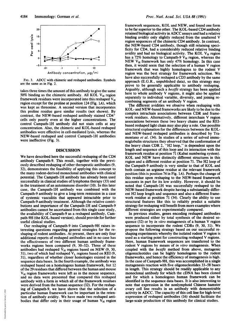
FIG. 3. ADCC with chimeric and reshaped antibodies. Symbolsare the same as in Fig. 2.
takes three times the amount of this antibody to give the same50% binding as the chimeric antibody. All KOL VH regionframework residues were incorporated into this reshaped VHregion except for the proline at position 124 (Fig. LA), whichwas kept as threonine. A second version that incorporatesthis proline residue gave similar results (not shown). Bycontrast, the NEW-based reshaped antibody stained CD4Icells only poorly even at the higher concentrations. Thecontrol Campath-1H antibody did not stain cells at any
concentration. Also, the chimeric and KOL-based reshapedantibodies were effective in cell-mediated lysis, whereas theNEW-based reshaped and control Campath-1H antibodieswere ineffective (Fig. 3).
DISCUSSIONWe have described here the successful reshaping of the CD4antibody Campath-9. This result, together with the previ-ously described reshaping of other therapeutic antibodies (9,32), demonstrates the feasibility of applying this concept tothe many rodent-derived monoclonal antibodies with clinicalpotential. The Campath-1H antibody has already been usedsuccessfully in clinical studies of lymphoma therapy (33) andin the treatment of an autoimmune disorder (10). In this latercase, the Campath-1H antibody was combined with theCampath-9 antibody to give a long-term remission in a caseof systemic vasculitis that appeared intractable prior toCampath-9 antibody treatment. Although the relative contri-butions and importance of the Campath-1H and Campath-9antibodies cannot be ascertained from this single case study,the availability of Campath-9 as a reshaped antibody, Cam-path-9H (the KOL-based version), should provide for furtheruseful clinical studies.Our reshaping of Campath-9 into Campath-9H raises in-
teresting questions regarding general strategies for the re-shaping of rodent antibodies. At present, there are only fouradditional reports of reshaped antibodies and in no case hasthe effectiveness of two different human antibody frame-works regions been compared (9, 30-32). Three of theseantibodies had reshaped VH regions based on NEW (9, 30,31), two of which had reshaped VL regions based on REI (9,31), regardless of whether closer homologies existed in thesequence data bases. In the fourth example, the antibody wasreshaped based on a homologous human framework, but 12of the 29 residues that differed between the human and mouseVH region frameworks were left as in the mouse sequence,and no data were presented that compared this reshapedantibody with a form in which all of the framework residueswere derived from the human sequence (32). For the reshap-ing of Campath-9, we have shown that the selection of aparticular human framework can be important in the reten-tion of antibody avidity. We have made two reshaped anti-bodies that differ only in their usage of human VH region
framework sequences, KOL and NEW, and found one formto be far superior to the other. The KOL-based CD4 antibodyretained biological activity inADCC assays and had a relativebinding avidity only slightly reduced from the unaltered Vregion sequences of the chimeric CD4 antibody. In contrast,the NEW-based CD4 antibody, though still retaining speci-ficity for CD4, had a considerably reduced relative bindingavidity and had no biological activity. The KOL VH regionhas a 72% homology to Campath-9 VH region, whereas theNEW VH framework has only 47% homology. In this casethen, it would seem that the selection of a human V regionframework that was highly homologous to the rodent Vregion was the best strategy for framework selection. Wehave also successfully reshaped a CD3 antibody by the sameapproach (E.G.R., unpublished data), so this strategy mayprove to be generally applicable to antibody reshaping.Arguably, although such a bestfit strategy has been appliedhere to whole antibody V regions, it might also be appliedseparately to individual variable, diversity, and joining re-combining segments of an antibody V region.The different avidities we observe when reshaping with
KOL- and NEW-based frameworks are likely to be due to thecomplex intrachain associations between CDR and frame-work residues. Alternatively, different interchain V regionassociations between these two heavy chains and the REI-based reshaped light chain may also play a role. One possiblestructural explanation for the differences between the KOL-and NEW-based reshaped antibodies is described by Tra-montano et al. (34). In studies of a series of solved immu-noglobulin structures they observed that the conformation ofthe heavy chain CDR 2, "H2 loop," is dependent upon thelength and sequence of this loop and its interaction with theframework residue at position 71 (Kabat numbering system).KOL and NEW have distinctly different structures in thisregion and a different residue at position 71. The H2 loop ofthe Campath-9 antibody is very similar to that of KOL andboth contain an arginine residue at the relevant frameworkposition (this is position 74 in Fig. 1A). Perhaps the change ofthis residue upon reshaping to the NEW-based frameworkaccounts in part for its low avidity. However, it should benoted that Campath-1H was successfully reshaped to theNEW-based framework despite having a substantially differ-ent H2 loop length and sequence and also a different frame-work residue at position 71 (34). Clearly the ability to usestructural features like this to reliably predict a suitablestrategy for reshaping will benefit from more examples wheredifferent strategies are experimentally compared.
In previous studies, genes encoding reshaped antibodieswere produced either by total synthesis of the desired se-quence (32) or by in vitro mutagenesis of a human V regionsequence to incorporate the rodent CDRs (9, 30, 31). Wepropose the following strategy based on our successful re-shaping experiments whereby the isolated rodent V region isused as a starting point for constructing reshaped V regions.Here, human framework sequences are transferred to therodent V regions by means of in vitro mutagenesis. Whencoupled with the bestfit method from above, mutagenicoligonucleotides can be highly homologous to the rodentframeworks, and hence the efficiency of mutagenesis is high.In the case of Campath-9H, this was accomplished in a singlemutagenesis reaction with five oligonucleotides 33-58 basesin length. This strategy should be readily applicable to anymonoclonal antibody for which the cDNA has been clonedand for which a homologous human framework can beidentified in the sequence data bases. It is also interesting tonote that expression in the nonlymphoid Chinese hamsterovary cell line results in an antibody with demonstrableactivity in ADCC. The capacity of this cell line for high-levelexpression of reshaped antibodies (16) should facilitate thelarge-scale production of this antibody for clinical studies.
0
o
220I---0z
4184 Immunology: Gorman et al.
Dow
nloa
ded
by g
uest
on
Janu
ary
7, 2
022

Proc. Natl. Acad. Sci. USA 88 (1991) 4185
We thank the following for their helpful discussions and assistance:G. Winter and P. Jones for the vectors M13-VKPCR1 and M13-VHPCR1, J. S. Crowe for Campath-1H cDNA, M. Page for theplasmids pLD9 and pNH316, J. Ivanyi and J. Howard for the cell lineNB2-6TG, A. Lesk, and C. Chothia. We thank H. Spence forsynthetic oligonucleotide synthesis, H. Kruger-Gray for flow cytom-etry assistance, and M. Frewin for technical assistance. This workwas supported by the Medical Research Council, United Kingdom,Wellcome Biotech PLC, and the Gilman Foundation. S.D.G. is arecipient of a Special Fellowship from the Leukemia Society ofAmerica.
1. Qin, S. X., Cobbold, S., Benjamin, R. & Waldmann, H. (1989)J. Exp. Med. 169, 779-794.
2. Qin, S. X., Wise, M., Cobbold, S. P., Leong, L., Kong, Y. M.,Parnes, J. & Waldmann, H. (1990) Eur. J. Immunol. 20,2737-2745.
3. Cobbold, S. P., Martin, G. & Waldmann, H. (1990) Eur. J.Immunol. 20, 2747-2755.
4. Madsen, J. C., Superina, R. A., Wood, K. J. & Morris, P. J.(1988) Nature (London) 332, 161-164.
5. Benjamin, R. J. & Waldmann, H. (1986) Nature (London) 320,449-451.
6. Gutstein, N. L. & Wofsy, D. (1986) J. Immunol. 137, 3414-3419.
7. Herzog, C., Walker, C., Muller, W., Rieber, P., Reiter, C.,Riethmuller, G., Wassmer, P., Stockinger, H., Madic, 0. &Pichler, W. J. (1989) J. Autoimmun. 2, 627-642.
8. Hafler, D. A., Ritz, J., Schlossman, S. F. & Weiner, H. L.(1988) J. Immunol. 141, 131-138.
9. Riechmann, L., Clark, M., Waldmann, H. & Winter, G. (1988)Nature (London) 332, 323-327.
10. Mathieson, P. W., Cobbold, S. P., Hale, G., Clark, M. R.,Oliveira, D. B. G., Lockwood, C. M. & Waldmann, H. (1990)N. Engl. J. Med. 323, 250-254.
11. Orlandi, R., Gussow, D. H., Jones, P. T. & Winter, G. (1989)Proc. Natl. Acad. Sci. USA 86, 3833-3837.
12. Crowe, J. S., Smith, M. A. & Cooper, H. J. (1989) NucleicAcids Res. 17, 7992.
13. Sanger, F., Nicklen, S. & Coulson, A. R. (1977) Proc. Natl.Acad. Sci. USA 74, 5463-5467.
14. Gunning, P., Leavitt, J., Muscat, G., Ng, S. Y. & Kedes, L.(1987) Proc. Natl. Acad. Sci. USA 84, 4831-4835.
15. Takahashi, N., Ueda, S., Obata, M., Nikaido, T., Nakai, S. &Honjo, T. (1982) Cell 29, 671-679.
16. Page, M. J. & Sydenham, M. A. (1991) Biotechnology 9, 64-68.
17. Hieter, P. A., Max, E. E., Seidmann, J. G., Maizel, J. V., Jr.,& Leder, P. (1980) Cell 22, 197-207.
18. Schmidt, W. E., Jung, H.-D., Palm, W. & Hilschmann, N.(1983) Hoppe-Seyler's Z. Physiol. Chem. 364, 713-747.
19. Ballhausen, W. G., Reske-Kunz, A. B., Tourvieille, B.,Ohashi, P. S., Parnes, J. R. & Mak, T. W. (1988) J. Exp. Med.167, 1493-1498.
20. Maddon, P. J., Littman, D. R., Godfrey, M., Maddon, D. E.,Chess, L. & Axel, R. (1985) Cell 42, 93-104.
21. Wigler, M., Pellicer, A., Silverstein, S., Axel, R., Urlaub, G.& Chasin, L. (1979) Proc. Natl. Acad. Sci. USA 76, 1373-1376.
22. Oi, V. T. & Herzenberg, L. A. (1980) in Selected Methods inCellular Immunology, eds. Mishell, B. B. & Shiigi, S. M.(Freeman, San Francisco), pp. 351-372.
23. Hale, G., Hoang, T., Prospero, T., Watt, S. M. & Waldmann,H. (1983) Mol. Biol. Med. 1, 305-319.
24. Clark, M. R. & Waldmann, H. (1987) J. Natl. Cancer Inst. 79,1393-1401.
25. Devereux, J., Haeberli, P. & Smithies, 0. (1984) Nucleic AcidsRes. 12, 387-395.
26. Lesk, A. M., Boswell, D. R., Lesk, V. I., Lesk, V. E. &Bairoch, A. (1989) Protein Sequences Data Anal. 2, 295-308.
27. Poljak, R. J., Nakashima, Y., Chen, B. L. & Konigsberg, W.(1977) Biochemistry 16, 3412-3420.
28. Epp, O., Lattman, E. E., Schiffer, M., Huber, R. & Palm, W.(1975) Biochemistry 14, 4943-4952.
29. Kabat, E. A., Wu, T. T., Reid-Miller, M., Perry, H. M. &Gottesmann, K. S. (1987) Sequences of Proteins ofImmuno-logical Interest (U.S. Dept. of Health and Human Services)(GPO, Washington).
30. Jones, P. T., Dear, P. H., Foote, J., Neuberger, M. S. &Winter, G. (1986) Nature (London) 321, 522-525.
31. Verhoeyen, M., Milstein, C. & Winter, G. (1988) Science 239,1534-1536.
32. Queen, C., Schneider, W. P., Selick, H. E., Payne, P. W.,Landolfi, N. F., Duncan, J. F., Avdalovic, N. M., Levitt, M.,Junghans, R. P. & Waldmann, T. A. (1989) Proc. Natl. Acad.Sci. USA 86, 10029-10033.
33. Hale, G., Dyer, M. J. S., Clark, M. R., Phillips, J. M., Marcus,R., Riechmann, L., Winter, G. & Waldmann, H. (1988) Lancetii, 1394-1399.
34. Tramontano, A., Chothia, C. & Lesk, A. M. (1990) J. Mol.Biol. 215, 175-182.
Immunology: Gorman et al.
Dow
nloa
ded
by g
uest
on
Janu
ary
7, 2
022