researchsenescent mouse cells fail to overtly regulate the ... · this is an open access article...
TRANSCRIPT

Kennedy et al. Cell Division 2010, 5:16http://www.celldiv.com/content/5/1/16
Open AccessR E S E A R C H
ResearchSenescent mouse cells fail to overtly regulate the HIRA histone chaperone and do not form robust Senescence Associated Heterochromatin FociAlyssa L Kennedy1,2, Tony McBryan3, Greg H Enders2, F Brad Johnson4, Rugang Zhang2 and Peter D Adams*3
AbstractBackground: Cellular senescence is a permanent growth arrest that occurs in response to cellular stressors, such as telomere shortening or activation of oncogenes. Although the process of senescence growth arrest is somewhat conserved between mouse and human cells, there are some critical differences in the molecular pathways of senescence between these two species. Recent studies in human fibroblasts have defined a cell signaling pathway that is initiated by repression of a specific Wnt ligand, Wnt2. This, in turn, activates a histone chaperone HIRA, and culminates in formation of specialized punctate domains of facultative heterochromatin, called Senescence-Associated Heterochromatin Foci (SAHF), that are enriched in the histone variant, macroH2A. SAHF are thought to repress expression of proliferation-promoting genes, thereby contributing to senescence-associated proliferation arrest. We asked whether this Wnt2-HIRA-SAHF pathway is conserved in mouse fibroblasts.
Results: We show that mouse embryo fibroblasts (MEFs) and mouse skin fibroblasts, do not form robust punctate SAHF in response to an activated Ras oncogene or shortened telomeres. However, senescent MEFs do exhibit elevated levels of macroH2A staining throughout the nucleus as a whole. Consistent with their failure to fully activate the SAHF assembly pathway, the Wnt2-HIRA signaling axis is not overtly regulated between proliferating and senescent mouse cells.
Conclusions: In addition to the previously defined differences between mouse and human cells in the mechanisms and phenotypes associated with senescence, we conclude that senescent mouse and human fibroblasts also differ at the level of chromatin and the signaling pathways used to regulate chromatin. These differences between human and mouse senescence may contribute to the increased propensity of mouse fibroblasts (and perhaps other mouse cell types) to become immortalized and transformed, compared to human cells.
BackgroundCellular senescence is an irreversible proliferation arrestthat is an important tumor suppression mechanism andis also thought to contribute to organismal aging [1].Senescence occurs in response to various cell stresses,including activated oncogenes, critically short telomeresor DNA damage. Senescence as a response to shortenedtelomeres is termed replicative senescence, and as aresponse to oncogene activation is termed oncogene-induced senescence. By permanently exiting the cell cyclein the presence of an activated oncogene or exposedtelomere ends, the cell is thought to prevent acquisition
of additional genetic alterations and possible transforma-tion. In this way, senescence is thought to contribute totumor suppression. However, senescence may come at acost to the organism, as this process is also thought tolead to exhaustion of stem cell populations and subse-quent tissue and organismal aging.
Comparison of senescence signaling pathways inmouse and human cells has revealed some similarities,but also many differences, between the senescence pro-grams of these two most-studied species [2]. These differ-ences might bear on the different longevity and tumorsuppression capacity of these species. Cellular senescenceis induced by concerted activity of the p53 and pRBtumor suppressor pathways in most primary human cells,including fibroblasts [3,4]. The pRB protein contributes
* Correspondence: [email protected] CR-UK Beatson Labs, Glasgow University, Glasgow, UKFull list of author information is available at the end of the article
© 2010 Kennedy et al; licensee BioMed Central Ltd. This is an Open Access article distributed under the terms of the Creative CommonsAttribution License (http://creativecommons.org/licenses/by/2.0), which permits unrestricted use, distribution, and reproduction inany medium, provided the original work is properly cited.
Kennedy et al. Cell Division 2010, 5:16http://www.celldiv.com/content/5/1/16
Page 2 of 11
to cell cycle arrest via its inhibitory effects on E2F familymembers, transcriptional activators of S-phase genes [5].The p53 protein also drives many aspects of the senes-cence program, including cell cycle arrest via transcrip-tional activation of its downstream effector p21, a cyclindependent kinase inhibitor [6]. In mouse fibroblasts,inactivation of either the p53 pathway or pRB (togetherwith related proteins, p107 and p130) is generally suffi-cient to abrogate senescence [7-9]. In contrast, in humanfibroblasts, inactivation of both pathways is required toinactivate senescence [3,4,10]. Another important differ-ence between mouse and human cells with respect tosenescence induction is expression of telomerase.Because of the 'end-replication problem' and lack of arobust telomere maintenance mechanism, in most prolif-erating human cells telomeres have potential to becomecritically short, and thus be sensed as DNA damage andso induce senescence [1]. Mouse somatic cells, however,express telomerase and contain markedly longer telom-eres, and thus telomere shortening does not appear tocontribute to mouse cell senescence [11-14].
A hallmark of many cultured human cells that haveundergone senescence is the formation of regions ofcharacteristically punctate heterochromatin, termedSenescence Associated Heterochromatin Foci (SAHF)[15,16]. The formation of heterochromatin in senescentcells is thought to contribute to permanent growth arrestby silencing proliferating promoting genes, such as cyclinA2, within dense regions of transcriptionally inactive het-erochromatin. In human cells, SAHF can be visualized byconventional epifluorescence microscopy as punctatedomains of DAPI-stained chromatin. These areas of het-erochromatin also have been shown to harbor variouswell-characterized heterochromatin proteins, such asHeterochromatin Protein 1 (HP1) and histone variantmacroH2A.
We have shown previously that in human cells canoni-cal Wnt signaling is an important regulator of SAHF for-mation [17]. Wnt ligands are extracellular signalingmolecules that are important regulators of development,body axis formation and stem cell renewal [18]. Canoni-cal Wnts ligands, when they bind to their cognate trans-membrane frizzled receptors, cause disruption of acomplex of proteins, including Axin, APC and GlycogenSynthase Kinase 3 (GSK3 (α and β isoforms). In turn, thisresults in a block to phosphorylation and degradation of aGSK3 substrate, β-catenin, a key transcription effector ofcanonical Wnt ligands. In pre-senescent primary humanfibroblasts, expression of the canonical Wnt ligand,Wnt2, is down-regulated, leading to activation of GSK3βwhich phosphorylates another of its substrates, the his-tone chaperone protein, HIRA. As a histone chaperone,HIRA is able to assemble DNA and histones intonucleosomes [19]. When phosphorylated by GSK3β on
serine 697, HIRA translocates to specific subnuclearorganelles, termed nuclear PML bodies [17]. This translo-cation of HIRA to PML bodies is a pre-requisite for HIRAto promote formation of punctate SAHF in human cells[20], presumably dependent on its histone chaperoneactivity. In human cells, formation of SAHF also dependson a functional pRB pathway [15,21]. Hence, in humanfibroblasts, the Wnt2-HIRA-SAHF signaling axis and thepRB tumor suppressor pathway coordinately regulate for-mation of SAHF.
In light of differences between mouse and human fibro-blasts in their utilization of the p53 and pRB pathways,and the impact of telomeres on senescence, we hypothe-sized that there might be differences in the Wnt2-HIRA-SAHF signaling pathway between senescence in mouseand human fibroblasts. Therefore, we sought to deter-mine whether this pathway is also regulated as mousefibroblasts enter senescence.
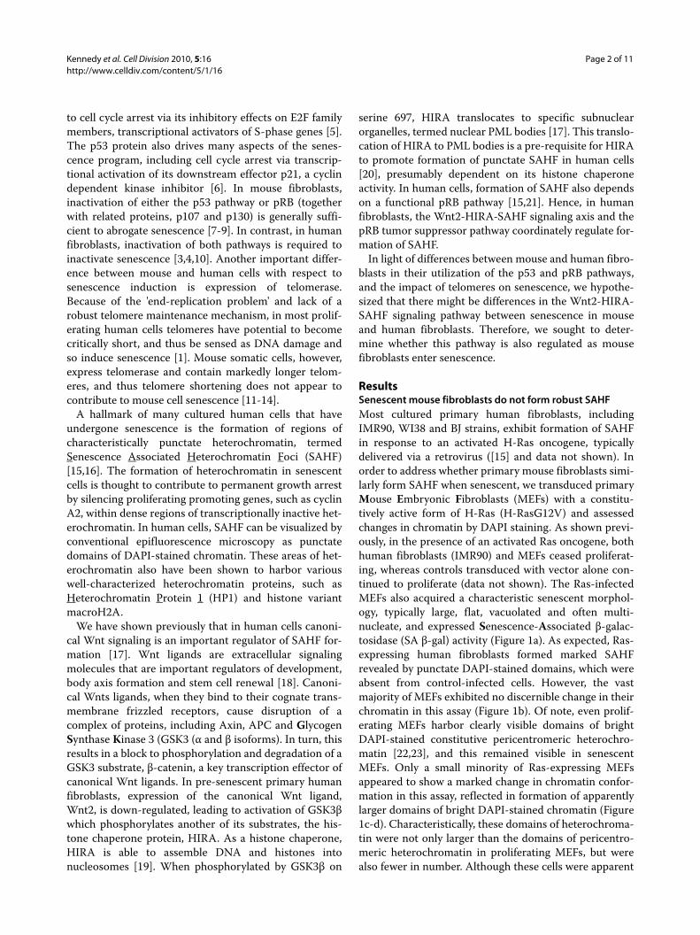
ResultsSenescent mouse fibroblasts do not form robust SAHFMost cultured primary human fibroblasts, includingIMR90, WI38 and BJ strains, exhibit formation of SAHFin response to an activated H-Ras oncogene, typicallydelivered via a retrovirus ([15] and data not shown). Inorder to address whether primary mouse fibroblasts simi-larly form SAHF when senescent, we transduced primaryMouse Embryonic Fibroblasts (MEFs) with a constitu-tively active form of H-Ras (H-RasG12V) and assessedchanges in chromatin by DAPI staining. As shown previ-ously, in the presence of an activated Ras oncogene, bothhuman fibroblasts (IMR90) and MEFs ceased proliferat-ing, whereas controls transduced with vector alone con-tinued to proliferate (data not shown). The Ras-infectedMEFs also acquired a characteristic senescent morphol-ogy, typically large, flat, vacuolated and often multi-nucleate, and expressed Senescence-Associated β-galac-tosidase (SA β-gal) activity (Figure 1a). As expected, Ras-expressing human fibroblasts formed marked SAHFrevealed by punctate DAPI-stained domains, which wereabsent from control-infected cells. However, the vastmajority of MEFs exhibited no discernible change in theirchromatin in this assay (Figure 1b). Of note, even prolif-erating MEFs harbor clearly visible domains of brightDAPI-stained constitutive pericentromeric heterochro-matin [22,23], and this remained visible in senescentMEFs. Only a small minority of Ras-expressing MEFsappeared to show a marked change in chromatin confor-mation in this assay, reflected in formation of apparentlylarger domains of bright DAPI-stained chromatin (Figure1c-d). Characteristically, these domains of heterochroma-tin were not only larger than the domains of pericentro-meric heterochromatin in proliferating MEFs, but werealso fewer in number. Although these cells were apparent
Kennedy et al. Cell Division 2010, 5:16http://www.celldiv.com/content/5/1/16
Page 3 of 11
Figure 1 Lack of marked chromatin changes in senescent mouse fibroblasts. (a) SA β-gal staining in MEFs transduced with either control or H-RasG12V, drug selected and stained 7 days after drug selection. (b) DAPI staining of nuclei of cells shown in (a) and human IMR90. (c) MEF nucleus with exceptionally condensed heterochromatin. (d) Percent cells with condensed chromatin in senescent MEFs or IMR90 cells. In IMR90, cells with characteristic SAHF as in (b) were scored as positive. In MEFs, only cells with exceptional compaction as in (c) were scored as positive. Pericentromeric heterochromatin of normal MEFs as in (b) was not scored as positive. (e) DAPI staining of wild type mouse skin fibroblasts and generation 5 TERC-/-, WRN-/- skin fibroblasts.
Kennedy et al. Cell Division 2010, 5:16http://www.celldiv.com/content/5/1/16
Page 4 of 11
by microscopy, the extent of their increase in senescencedid not reach significance (Figure 1d). In sum, this resultshows that onset of oncogene-induced senescence inmost MEFs is not associated with marked changes inchromatin structure, as measured by DAPI staining.
Another trigger of senescence and SAHF formation inhuman cells is shortened telomeres, associated with repli-cative senescence [21]. To ask whether shortened telom-eres can trigger SAHF formation in mouse cells, wecompared wild type mouse skin fibroblasts and skinfibroblasts from generation seven mice lacking both thetelomerase catalytic subunit and the Werner's helicase(TERC-/-, WRN-/-). The telomeres of these cells arefunctionally compromised, leading to marked impair-ment of cell proliferation and indicators of senescence[24]. However, even when these cells arrested prolifera-tion, we did not observe marked changes in chromatinstructure by this assay (Figure 1e). These results indicatethat neither activated oncogenes nor shortened telomeresinduce changes in chromatin structure in mouse cells thatare as marked as those observed in human cells.
In order to investigate further the status of chromatinin senescent MEFs, we examined the incorporation ofone component of SAHF, the histone variant macroH2A,by immunofluorescence. MacroH2A is important in het-erochromatin mediated silencing of the female X chro-mosome and as such, in control female-derived humanfibroblast IMR90 cells, the barr body stains positively forthis protein (Figure 2a) [25]. In addition, as shown previ-ously, macroH2A is incorporated into SAHF in these cells[26]. When we examined MEFs made senescent by trans-duction of activated Ras (Figure 2b-c), we found thatthere was an increase in macroH2A staining in manysenescent cells, compared to proliferating cells (p < 1 ×10-11). Interestingly, this increase in macroH2A stainingwas most apparent in the small proportion of cells thatdid show some chromatin compaction in senescent cells(Figure 1c and 2c).
Together, these microscopy-based assays reveal bothdifferences and similarities in chromatin regulationbetween senescent human fibroblasts and MEFs. A size-able proportion of the population does show an increasein macroH2A staining throughout the cell nucleus. How-ever, unlike human fibroblasts, the majority of MEFs donot exhibit pronounced changes in chromatin as theybecome senescent, as judged by formation of DAPI- ormacroH2A-stained puncta.
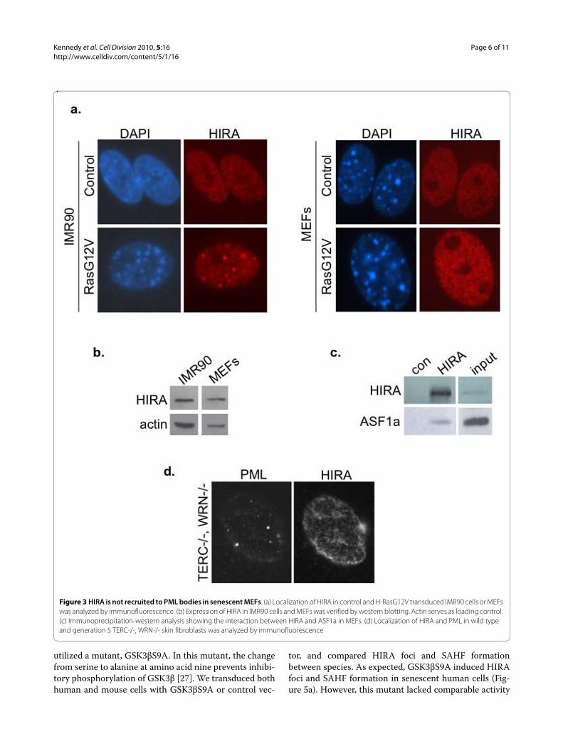
Senescent mouse fibroblasts do not recruit HIRA to PML bodiesGiven the established link between HIRA's localization toPML bodies and formation of SAHF in senescent humanfibroblasts [26], we questioned whether the failure ofsenescent mouse cells to form robust punctate SAHF is
linked to different regulation of HIRA in these cells. Asbefore, MEFs were made senescent by transduction withan activated Ras oncogene, and senescence confirmed bydecreased incoporation of BrdU and positive staining forSA β-gal (data not shown). However, when we comparedthe subcellular localization of HIRA in proliferating andsenescent MEFs and human fibroblasts, we found thatHIRA was recruited to PML bodies in senescent humanfibroblasts, but not senescent MEFs (Figure 3a and Addi-tional file 1: Figure S1). Of particular significance, HIRAdid not localize to PML bodies even in the small propor-tion of senescent MEFs that did contain compacted het-erochromatin, as revealed by DAPI staining (Figure 1cand data not shown). We confirmed that the HIRA pro-tein was expressed in MEFs, and reactive with the samemouse monoclonal antibody used for immunofluores-cence (Figure 3b). We also verified that in MEFs, HIRAbinds to its partner ASF1a, an interaction shown to becritical for formation of SAHF (Figure 3c) [26]. Similarconfirmatory results were also obtained with two rabbitpolyclonal antibodies to HIRA (data not shown). We alsofailed to observe HIRA localized to PML bodies in TERC-/-, WRN-/- skin fibroblasts (Figure 3d). These resultsindicate that the HIRA chaperone is regulated differentlyduring senescence of mouse and human fibroblasts. Spe-cifically, HIRA is not recruited to PML bodies as MEFsbecome senescent.
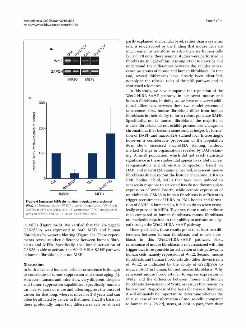
Senescent MEFs do not downregulate expression of Wnt2In human cells, an early trigger of senescence and recruit-ment of HIRA to PML bodies is repression of a specificWnt ligand, Wnt2 [17]. Therefore, we next examinedwhether MEFs downregulate expression of this specificWnt ligand. We performed RT-PCR analysis in proliferat-ing and senescent MEFs to determine whether theexpression of Wnt2 changed as the cells senesced. Senes-cence of MEFs was confirmed by appropriate morpholog-ical changes and expression of SA β-gal (data not shown).Unlike senescent IMR90 cells, which were compared inparallel as a control, we found that MEFs do not down-regulate expression of Wnt2 as they undergo oncogene-induced senescence (Figure 4a-b). This observationextends the differences in regulation of the Wnt2-HIRA-SAHF pathway in MEFs and human fibroblasts.
Forced activation of GSK3 does not regulate HIRA and SAHF in MEFsIn human fibroblasts, recruitment of HIRA to PML bod-ies depends on repression of expression of Wnt2 and con-sequent activation of GSK3 [17]. Forced activation ofGSK3, specifically the GSK3β isoform, has been shown totrigger HIRA's recruitment to PML bodies and formationof SAHF. To test whether GSK3β can similarly induceHIRA relocalization and formation of SAHF in MEFs, we

Kennedy et al. Cell Division 2010, 5:16http://www.celldiv.com/content/5/1/16
Page 5 of 11
Figure 2 Senescent MEFs stain more intensely with histone variant macroH2A. (a) Human IMR90 fibroblasts transduced with either control or H-RasG12V were stained for histone variant macroH2A and DNA (DAPI). Inactive X chromosome is enriched in macroH2A in female cells (arrow). (b) Control or H-RasG12V-transduced senescent MEFs were stained for histone variant macroH2A. Results were quantitated using metamorph and ex-pressed as histograms and box plots: The red line is the median (50th percentile); the box itself encompasses the 25th and 75th percentiles (Inter Quartile Range (IQR)); the whiskers are the most extreme data points within 1.5 × IQR; crosses outside the whiskers are outliers. (c) Images of cells stained as in (b). White arrows indicate a cell with increased macroH2A stain.
Kennedy et al. Cell Division 2010, 5:16http://www.celldiv.com/content/5/1/16
Page 6 of 11
utilized a mutant, GSK3βS9A. In this mutant, the changefrom serine to alanine at amino acid nine prevents inhibi-tory phosphorylation of GSK3β [27]. We transduced bothhuman and mouse cells with GSK3βS9A or control vec-
tor, and compared HIRA foci and SAHF formationbetween species. As expected, GSK3βS9A induced HIRAfoci and SAHF formation in senescent human cells (Fig-ure 5a). However, this mutant lacked comparable activity
Figure 3 HIRA is not recruited to PML bodies in senescent MEFs. (a) Localization of HIRA in control and H-RasG12V transduced IMR90 cells or MEFs was analyzed by immunofluorescence. (b) Expression of HIRA in IMR90 cells and MEFs was verified by western blotting. Actin serves as loading control. (c) Immunoprecipitation-western analysis showing the interaction between HIRA and ASF1a in MEFs. (d) Localization of HIRA and PML in wild type and generation 5 TERC-/-, WRN-/- skin fibroblasts was analyzed by immunofluorescence
Kennedy et al. Cell Division 2010, 5:16http://www.celldiv.com/content/5/1/16
Page 7 of 11
in MEFs (Figure 5a-b). We verified that the V5-tagged-GSK3βS9A was expressed in both MEFs and humanfibroblasts by western blotting (Figure 5c). These experi-ments reveal another difference between human fibro-blasts and MEFs. Specifically, that forced activation ofGSK3β is able to activate the Wnt2-HIRA-SAHF pathwayin human fibroblasts, but not MEFs.
DiscussionIn both mice and humans, cellular senescence is thoughtto contribute to tumor suppression and tissue aging [1].However, humans and mice show very different lifespansand tumor suppression capabilities. Specifically, humanscan live 80 years or more and often suppress the onset ofcancer for that long, whereas mice live 2-3 years and canoften be afflicted by cancer in that time. That the basis forthese profoundly important differences can be at least
partly explained at a cellular level, rather than a systemicone, is underscored by the finding that mouse cells aremuch easier to transform in vitro than are human cells[28,29]. Of note, these seminal studies were performed infibroblasts. In light of this, it is important to describe andunderstand the differences between the cellular senes-cence programs of mouse and human fibroblasts. To thatend, several differences have already been identified,notably in the relative roles of the pRB pathway and inshortened telomeres.
In this study, we have compared the regulation of theWnt2-HIRA-SAHF pathway in senescent mouse andhuman fibroblasts. In doing so, we have uncovered addi-tional differences between these two model systems ofsenescence. First, mouse fibroblasts differ from humanfibroblasts in their ability to form robust punctate SAHF.Specifically, unlike human fibroblasts, the majority ofmouse fibroblasts do not exhibit pronounced changes inchromatin as they become senescent, as judged by forma-tion of DAPI- and macroH2A-stained foci. Interestingly,however, a considerable proportion of the populationdoes show increased macroH2A staining, withoutmarked change in organization revealed by DAPI-stain-ing. A small population, which did not reach statisticalsignificance in these studies, did appear to exhibit nuclearreorganization and chromatin compaction, based onDAPI and macroH2A staining. Second, senescent mousefibroblasts do not recruit the histone chaperone HIRA toPML bodies. Third, MEFs that have been induced tosenesce in response to activated Ras do not downregulateexpression of Wnt2. Fourth, while ectopic expression ofuninhibitable GSK3β in human fibroblasts is sufficient totrigger recruitment of HIRA to PML bodies and forma-tion of SAHF in human cells, it fails to do so when ectop-ically expressed in MEFs. Together, these results indicatethat, compared to human fibroblasts, mouse fibroblastsare markedly impaired in their ability to activate and sig-nal through the Wnt2-HIRA-SAHF pathway.
More specifically, these results point to at least two dif-ferences between human fibroblasts and mouse fibro-blasts in this Wnt2-HIRA-SAHF pathway. First,senescence of mouse fibroblasts is not associated with thetrigger that is responsible for activation of this pathway inhuman cells, namely repression of Wnt2. Second, mousefibroblasts and human fibroblasts also differ downstreamof Wnt2, as indicated by the ability of GSK3βS9A toinduce SAHF in human, but not mouse, fibroblasts. Whysenescent mouse fibroblasts fail to repress expression ofWnt2, and the difference between mouse and humanfibroblasts downstream of Wnt2, are issues that remain tobe resolved. Regardless of the basis for these differences,it will ultimately be important to determine whether therelative ease of transformation of mouse cells, comparedto human cells [28,29], stems, at least in part, from their
Figure 4 Senescent MEFs do not downregulate expression of Wnt2. (a) Semiquantitative RT-PCR analysis of expression of Wnt2 and GAPDH in MEFs and IMR90 cells. (b) Quantitative RT-PCR analysis of ex-pression of Wnt2 and GAPDH in MEFs and IMR90 cells.

Kennedy et al. Cell Division 2010, 5:16http://www.celldiv.com/content/5/1/16
Page 8 of 11
failure to regulate this Wnt2-HIRA-SAHF axis, a candi-date tumor suppressor pathway.
The mechanism by which HIRA's recruitment to PMLbodies contributes to formation of SAHF is not well
understood. Previous studies have indicated that HIRA'srelocation to this subnuclear organelle is required for for-mation of SAHF, because blocking its relocalization,either with a dominant negative HIRA mutant or with the
Figure 5 The HIRA-SAHF pathway is not induced by activated GSK3βS9A in MEFs. (a) Localization of HIRA and PML in control virus and H-RasG12V-transduced senescent IMR90 cells was examined by immunofluorescence. (b) MEFs examined as in (a). (c) DAPI-staining of representative cells from (a) and (b). (d) Expression of transduced constructs was verified by western blotting V5-tagged GSK3βS9A or Ras.
Kennedy et al. Cell Division 2010, 5:16http://www.celldiv.com/content/5/1/16
Page 9 of 11
PML-RARα fusion protein which disrupts PML bodies,abolishes formation of SAHF [20,21]. This study furtheremphasizes the link between HIRA's localization to PMLbodies and formation of SAHF, because the failure ofmouse fibroblasts to obviously form punctate SAHF cor-relates with a failure of HIRA to enter PML bodies inthese cells. Conversely, these results imply that recruit-ment of HIRA to PML bodies is not directly linked to reg-ulation of macroH2A. This is consistent with HIRA beingprimarily involved in deposition of histone (H3/H4)2 het-erotetramers, rather than H2A/H2B heterodimers [19].
In this study, we note a level of similarity between theappearance of DAPI-stained nuclei of proliferating non-senescent MEFs and senescent human cells. This similar-ity at the microscopic level is not founded at the molecu-lar level. Specifically, the bright-stained DAPI puncta inmouse fibroblasts reflect the DNA sequence and struc-ture of mouse pericentromeric heterochromatin [22,23].However, the SAHF of senescent human cells largelyexclude pericentromeres and telomeres [20,21]. The simi-larity at the microscopic level is potentially confusing,and we caution that a punctate DAPI stain should not beused to score senescence of MEFs, as it sometimes is inhuman cells.
ConclusionsPrevious works of others have found important func-tional differences between the senescence programs ofmouse and human fibroblasts. As detailed in the Back-ground, there are important differences in the role of thepRB and p53 tumor suppressor pathways and telomeres.The p53 pathway is dominant in mouse fibroblasts,whereas in human cells either activation of the p53 orpRB pathway leads to senescence. To these key differ-ences between senescence of mouse and human fibro-blasts, we add differences in the regulation and functionof the Wnt2-HIRA-SAHF pathway. Based on severalmeasures, this pathway is not overtly regulated at theonset of senescence in MEFs, compared to its regulationin human fibroblasts. However, our results obviously donot exclude some role for HIRA in regulation of senes-cence in MEFs, and future more detailed studies will berequired to fully define the similarities and differences inchromatin regulation between mouse and human cells.
MethodsCell CultureIMR90 (ATCC) cell lines were cultured according toATCC guidelines in ambient oxygen. Experiments wereperformed on IMR90 cells between population doubling(PD) 20 and PD 35. Mouse Embryo Fibroblasts (MEFs)were prepared from pooled wild type C57BL/6J embryosthat were gestational age E12-E14. The head and internalorgans were removed, and the torso was minced and dis-
persed in 0.25% trypsin + EDTA for 15 minutes in shak-ing water bath at 37°C. Cells were spun, debris removedand then plated. These cells were considered passage 0.MEFs used in experiments were cultured in ambient oxy-gen and experiments were performed between PD 1 andPD 8. MEFs were cultured in Dulbecco's modified Eagle'smedium (DMEM) supplemented with 10% fetal bovineserum. Cells were harvested 7 days post drug selectionfor all assays. Mouse adult ear skin fibroblasts wereobtained as described [24].
DNA constructs and retroviral transductionThe following plasmids were used to produce retrovi-ruses: pBabe-puro-H-RasG12V (a gift of Bill Hahn andBob Weinberg); pBabe-puro-GSK3βS9A from Addgene(plasmid #14128). Retroviral-mediated gene transfer wasperformed using the Phoenix packaging cells (Dr. GaryNolan, Stanford University). Phoenix cells were trans-fected using Polyethylenimine (PEI), Linear MW 25,000(Polysciences, Inc. Warrington, PA) sterile solution 1 mg/ml. To one milliliter of serum free DMEM, 16 micro-grams of retroviral plasmid DNA and 2 micrograms of aplasmid encoding vesicular stomatitis virus glycoproteinG (VSV-G) plus 38 micrograms of PEI was added, thissolution was briefly vortexed, left to sit for 10 minutesand then added to Phoenix Cells. Twenty-four hourslater, media was changed on Phoenix cells. At 48 hourspost transfection, virus-containing medium was col-lected, supplemented with 8 μg/ml of polybrene (Sigma),and incubated with the target IMR90 or MEF cells at 37°Cfor 24 hours. A second round of infection was performedon the same target cells. MEFs and IMR90 cells weretransduced in parallel and drug selected with 3 μg/ml or 1μg/ml puromycin, respectively.
Immunofluorescence, antibodies, SAHF, and SA β-gal stainingTwo color indirect immunofluorescence assays were per-formed as described previously [26,30]. Anti-PML(AB1370, Chemicon) Anti-PML for Additional file 1: Fig-ure S1 (Upstate 05-718) Anti-V5 (Invitrogen) Anti-Ras(BD Transduction Labatories Cat. No. 610001), Anti-betaactin (Abcam clone AC-15) were from the indicated sup-pliers. Anti-macroH2A and anti-HIRA antibodies weredescribed previously [20,31]. SAHF (DAPI foci) weredetected by staining with 0.13 μg/ml DAPI for 2 min atroom temperature (as opposed to standard conditions of1 μg/ml for 5 min). SA beta-gal staining was performed asdescribed previously [32].
CoimmunoprecipitationsTo prepare cell lysates, cells were washed once with PBSand then collected by incubating in EBC-500 (50 mMTris-HCl pH8, 500 mM NaCl, 0.5% NP-40 + proteaseinhibitors) for 20 minutes while rocking at 4°C, scraped
Kennedy et al. Cell Division 2010, 5:16http://www.celldiv.com/content/5/1/16
Page 10 of 11
into microcentrifuge tubes, passed through a 22 gaugeneedle five times and then spun for 10 minutes at 10,000× g. Supernatant was used for immunoprecipitations.Immunoprecipitation was performed using 2 mg ofextracted protein for 3 hours at 4°C. Antibodies used forIP were WC15 [31] and M7023 secondary (Sigma, St.Louis, MO). Immunoprecipitates were recovered after 1hour incubation at 4°C with protein A Sepharose Beads(Amersham/GE healthcare, Baie d'Urfé, QC, Canada).Precipitates were washed five times with NETN (20 mMTris pH8, 1 mM EDTA, 0.5%NP-40, 100 mM NaCl). Pre-cipitates were eluted in 50 ul of 1 × Laemmli sample buf-fer, boiled 5 minutes, and separated by SDS-PAGE andtransferred to Immun-Blot PVDF membrane (Biorad,Hercules, CA).
RT-PCRTotal RNA was prepared using Trizol (Invitrogen),according to the manufacturer's instructions. Reversetranscription-PCR (RT-PCR) was performed using theQiagen one-step RT-PCR kit, according to the manufac-turer's instructions. Primers used for human GapDHGAGAGACCCTCACTGCTG and GATGGTACAT-GACAAGGTGC and for human Wnt2 ATGAACGCCCCTCTCGGTGGAA and TGTCCTTGGCGCTTCCCATC. For Mouse Wnt2: 5'-GTACATGAGAGCTACAG-GTGG-3' and 5'-ACGGGCAAACTTGATCCCGTAGTC-3' and GAPDH: 5'-GATGATGACCCTTTTGGCTC-CACC-3' and 5'-CCACTCACGGCAAATTCAACGGCA-3'. Quantitative RT-PCR was performed using theone-step RT-PCR Master mix (Applied Biosystems). Realtime PCR and cDNA synthesis was performed using thefollowing mixture: 25 μl 2 × reaction buffer, 1.25 μl 40 ×multiscribe, 2.5 μl 20 × primer/probe and 100 ng RNA ina 50 μl total volume. Reactions were amplified and ana-lyzed in triplicate using Chromo-4 real-time PCR detec-tion system (Bio-rad). The following primer/probe sets(Applied Biosystems) were used: human wnt2Hs00608222, human beta-actin (endogenous control-4352935E), mouse wnt2 Mm00470018, mouse beta-actinMm01205647.
Additional material
Competing interestsThe authors declare that they have no competing interests.
Authors' contributionsALK performed all the experiments and wrote the manuscript. TM performedstatistical analysis of data. GHE participated in design of the study and pro-vided funding for ALK. FBJ participated in design of the study. RZ participatedin design of the study. PDA conceived the study, participated in the design,provided funding and wrote the manuscript.
All authors read and approved the final manuscript.
AcknowledgementsThe lab of PDA is funded by NIA (P01 AG031862) and CR-UK (C10652/A10250). Work in the lab of GHE was funded by NIH R01GM062281. Work in the lab of FBJ is funded by NIA (P01 AG031862). We would like to acknowledge Anna Pecherskaya and Margret Einarson for help with quantitative image analysis; Michael Bouchard, Jane Clifford and Maureen Murphy for their helpful discus-sions and advice.
Author Details1Graduate Program in Molecular and Cellular Biology and Genetics, Drexel University College of Medicine, Philadelphia, USA, 2Fox Chase Cancer Center, Philadelphia, USA, 3CR-UK Beatson Labs, Glasgow University, Glasgow, UK and 4University of Pennsylvania, Philadelphia, USA
References1. Adams PD: Healing and hurting: molecular mechanisms, functions, and
pathologies of cellular senescence. Mol Cell 2009, 36(1):2-14.2. Campisi J: Senescent cells, tumor suppression, and organismal aging:
good citizens, bad neighbors. Cell 2005, 120(4):513-522.3. Shay JW, Wright WE, Werbin H: Defining the molecular mechanisms of
human cell immortalization. Biochim Biophys Acta 1991, 1072(1):1-7.4. Hahn WC, Dessain SK, Brooks MW, King JE, Elenbaas B, Sabatini DM,
DeCaprio JA, Weinberg RA: Enumeration of the simian virus 40 early region elements necessary for human cell transformation. Mol Cell Biol 2002, 22(7):2111-2123.
5. Dimova DK, Dyson NJ: The E2F transcriptional network: old acquaintances with new faces. Oncogene 2005, 24(17):2810-2826.
6. Sherr CJ, Roberts JM: CDK inhibitors: positive and negative regulators of G1-phase progression. Genes Dev 1999, 13(12):1501-1512.
7. Sage J, Mulligan GJ, Attardi LD, Miller A, Chen S, Williams B, Theodorou E, Jacks T: Targeted disruption of the three Rb-related genes leads to loss of G(1) control and immortalization. Genes Dev 2000, 14(23):3037-3050.
8. Dannenberg JH, van Rossum A, Schuijff L, te Riele H: Ablation of the retinoblastoma gene family deregulates G(1) control causing immortalization and increased cell turnover under growth-restricting conditions. Genes Dev 2000, 14(23):3051-3064.
9. Harvey M, Sands AT, Weiss RS, Hegi ME, Wiseman RW, Pantazis P, Giovanella BC, Tainsky MA, Bradley A, Donehower LA: In vitro growth characteristics of embryo fibroblasts isolated from p53-deficient mice. Oncogene 1993, 8(9):2457-2467.
10. Bond JA, Haughton MF, Rowson JM, Smith PJ, Gire V, Wynford-Thomas D, Wyllie FS: Control of replicative life span in human cells: barriers to clonal expansion intermediate between M1 senescence and M2 crisis. Mol Cell Biol 1999, 19(4):3103-3114.
11. Parrinello S, Samper E, Krtolica A, Goldstein J, Melov S, Campisi J: Oxygen sensitivity severely limits the replicative lifespan of murine fibroblasts. Nat Cell Biol 2003, 5(8):741-747.
12. Kipling D, Cooke HJ: Hypervariable ultra-long telomeres in mice. Nature 1990, 347(6291):400-402.
13. Prowse KR, Greider CW: Developmental and tissue-specific regulation of mouse telomerase and telomere length. Proc Natl Acad Sci USA 1995, 92(11):4818-4822.
14. Blasco MA, Lee HW, Hande MP, Samper E, Lansdorp PM, DePinho RA, Greider CW: Telomere shortening and tumor formation by mouse cells lacking telomerase RNA. Cell 1997, 91(1):25-34.
15. Narita M, Nunez S, Heard E, Lin AW, Hearn SA, Spector DL, Hannon GJ, Lowe SW: Rb-mediated heterochromatin formation and silencing of E2F target genes during cellular senescence. Cell 2003, 113(6):703-716.
16. Adams PD: Remodeling of chromatin structure in senescent cells and its potential impact on tumor suppression and aging. Gene 2007, 397(1-2):84-93.
17. Ye X, Zerlanko B, Kennedy A, Banumathy G, Zhang R, Adams PD: Downregulation of Wnt signaling is a trigger for formation of facultative heterochromatin and onset of cell senescence in primary human cells. Mol Cell 2007, 27(2):183-196.
Additional file 1 Figure S1: HIRA is not recruited to PML bodies in senescent MEFs. Localization of HIRA and PML in H-RasG12V transduced MEFs was analyzed by immunofluorescence.
Received: 1 February 2010 Accepted: 22 June 2010 Published: 22 June 2010This article is available from: http://www.celldiv.com/content/5/1/16© 2010 Kennedy et al; licensee BioMed Central Ltd. This is an Open Access article distributed under the terms of the Creative Commons Attribution License (http://creativecommons.org/licenses/by/2.0), which permits unrestricted use, distribution, and reproduction in any medium, provided the original work is properly cited.Cell Division 2010, 5:16
Kennedy et al. Cell Division 2010, 5:16http://www.celldiv.com/content/5/1/16
Page 11 of 11
18. Logan CY, Nusse R: The Wnt signaling pathway in development and disease. Annu Rev Cell Dev Biol 2004, 20:781-810.
19. Ray-Gallet D, Quivy J-P, Scamps C, Martini EM-D, Lipinski M, G A: HIRA Is Critical for a Nucleosome Assembly Pathway Independent of DNA Synthesis. Mol Cell 2002, 9:1091-1100.
20. Zhang R, Chen W, Adams PD: Molecular Dissection of Formation of Senescent Associated Heterochromatin Foci. Mol Cell Biol 2007, 27:2343-2358.
21. Ye X, Zerlanko B, Zhang R, Somaiah N, Lipinski M, Salomoni P, Adams PD: Definition of pRB- and p53-dependent and -independent steps in HIRA/ASF1a-mediated formation of senescence-associated heterochromatin foci. Mol Cell Biol 2007, 27(7):2452-2465.
22. Guenatri M, Bailly D, Maison C, Almouzni G: Mouse centric and pericentric satellite repeats form distinct functional heterochromatin. J Cell Biol 2004, 166(4):493-505.
23. Maison C, Bailly D, Peters AH, Quivy JP, Roche D, Taddei A, Lachner M, Jenuwein T, Almouzni G: Higher-order structure in pericentric heterochromatin involves a distinct pattern of histone modification and an RNA component. Nat Genet 2002, 30(3):329-334.
24. Du X, Shen J, Kugan N, Furth EE, Lombard DB, Cheung C, Pak S, Luo G, Pignolo RJ, DePinho RA, et al.: Telomere shortening exposes functions for the mouse Werner and Bloom syndrome genes. Mol Cell Biol 2004, 24(19):8437-8446.
25. Costanzi C, Pehrson JR: Histone macroH2A1 is concentrated in the inactive X chromosome of female mammals. Nature 1998, 393(6685):599-601.
26. Zhang R, Poustovoitov MV, Ye X, Santos HA, Chen W, Daganzo SM, Erzberger JP, Serebriiskii IG, Canutescu AA, Dunbrack RL, et al.: Formation of MacroH2A-containing senescence-associated heterochromatin foci and senescence driven by ASF1a and HIRA. Dev Cell 2005, 8(1):19-30.
27. Cross DA, Alessi DR, Cohen P, Andjelkovich M, Hemmings BA: Inhibition of glycogen synthase kinase-3 by insulin mediated by protein kinase B. Nature 1995, 378(6559):785-789.
28. Hahn WC, Counter CM, Lundberg AS, Beijersbergen RL, Brooks MW, Weinberg RA: Creation of human tumour cells with defined genetic elements. Nature 1999, 400(6743):464-468.
29. Land H, Parada LF, Weinberg RA: Tumorigenic conversion of primary embryo fibroblasts requires at least two cooperating oncogenes. Nature 1983, 304(5927):596-602.
30. Ye X, Franco AA, Santos H, Nelson DM, Kaufman PD, Adams PD: Defective S-phase chromatin assembly causes DNA damage, activation of the S-phase checkpoint and S-phase arrest. Molecular Cell 2003, 11:341-351.
31. Hall C, Nelson DM, Ye X, Baker K, DeCaprio JA, Seeholzer S, Lipinski M, Adams PD: HIRA, the human homologue of yeast Hir1p and Hir2p, is a novel cyclin-cdk2 substrate whose expression blocks S-phase progression. Mol Cell Biol 2001, 21(5):1854-1865.
32. Dimri GP, Lee X, Basile G, Acosta M, Scott G, Roskelley C, Medrano EE, Linskens M, Rubelj I, Pereira-Smith O, et al.: A biomarker that identifies senescent human cells in culture and in aging skin in vivo. Proc Natl Acad Sci USA 1995, 92(20):9363-9367.
doi: 10.1186/1747-1028-5-16Cite this article as: Kennedy et al., Senescent mouse cells fail to overtly regu-late the HIRA histone chaperone and do not form robust Senescence Associ-ated Heterochromatin Foci Cell Division 2010, 5:16