researcharticle post-exerciseheartraterecovery ... · pathway’,immunityis...
TRANSCRIPT

RESEARCH ARTICLE
Post-Exercise Heart Rate RecoveryIndependently Predicts Clinical Outcome inPatients with Acute Decompensated HeartFailureJong-Chan Youn1,2, Hye Sun Lee3, Suk-Won Choi1, Seong-Woo Han1, Kyu-Hyung Ryu1,Eui-Cheol Shin4, Seok-Min Kang2*
1 Division of Cardiology, Dongtan Sacred Heart Hospital, Hallym University College of Medicine, Hwaseong,Republic of Korea, 2 Division of Cardiology, Severance Cardiovascular Hospital, Yonsei University Collegeof Medicine, Seoul, Republic of Korea, 3 Department of Biostatistics, Yonsei University College of Medicine,Seoul, Republic of Korea, 4 Laboratory of Immunology and Infectious Diseases, Graduate School of MedicalScience and Engineering, KAIST, Daejeon, Republic of Korea
Abstract
Background
Post-exercise heart rate recovery (HRR) is an index of parasympathetic function associated
with clinical outcome in patients with chronic heart failure. However, its relationship with the
pro-inflammatory response and prognostic value in consecutive patients with acute decom-
pensated heart failure (ADHF) has not been investigated.
Methods
Wemeasured HRR and pro-inflammatory markers in 107 prospectively and consecutively
enrolled, recovered ADHF patients (71 male, 59 ± 15 years, mean ejection fraction 28.9 ±
14.2%) during the pre-discharge period. The primary endpoint included cardiovascular (CV)
events defined as CV mortality, cardiac transplantation, or rehospitalization due to HF
aggravation.
Results
The CV events occurred in 30 (28.0%) patients (5 cardiovascular deaths and 7 cardiac
transplantations) during the follow-up period (median 214 days, 11–812 days). When the
patients with ADHF were grouped by HRR according to the Contal and O’Quigley’s method,
low HRR was shown to be associated with significantly higher levels of serummonokine-
induced by gamma interferon (MIG) and poor clinical outcome. Multivariate Cox regression
analysis revealed that low HRR was an independent predictor of CV events in both enter
method and stepwise method. The addition of HRR to a model significantly increased
predictability for CV events across the entire follow-up period.
PLOS ONE | DOI:10.1371/journal.pone.0154534 May 2, 2016 1 / 12
a11111
OPEN ACCESS
Citation: Youn J-C, Lee HS, Choi S-W, Han S-W,Ryu K-H, Shin E-C, et al. (2016) Post-Exercise HeartRate Recovery Independently Predicts ClinicalOutcome in Patients with Acute DecompensatedHeart Failure. PLoS ONE 11(5): e0154534.doi:10.1371/journal.pone.0154534
Editor: Elena Cavarretta, Sapienza University ofRome, ITALY
Received: January 18, 2016
Accepted: April 14, 2016
Published: May 2, 2016
Copyright: © 2016 Youn et al. This is an openaccess article distributed under the terms of theCreative Commons Attribution License, which permitsunrestricted use, distribution, and reproduction in anymedium, provided the original author and source arecredited.
Data Availability Statement: All relevant data arewithin the paper.
Funding: This research was supported by BasicScience Research Program through the NationalResearch Foundation of Korea (NRF) funded by theMinistry of Science, ICT & Future Planning (NRF-2015R1C1A1A02036645). The funders had no role instudy design, data collection and analysis, decision topublish, or preparation of the manuscript.
Competing Interests: The authors have declaredthat no competing interests exist.

Conclusion
Impaired post-exercise HRR is associated with a pro-inflammatory response and indepen-
dently predicts clinical outcome in patients with ADHF. These findings may explain the rela-
tionship between autonomic dysfunction and clinical outcome in terms of the inflammatory
response in these patients.
IntroductionAutonomic imbalance in heart failure is characterized by increased sympathetic activity andwithdrawal of parasympathetic activity. This autonomic imbalance is associated with progres-sion of heart failure and a worse clinical outcome [1]. While beta-blockers have found a placeas a leading disease-modifying therapy that impacts the sympathetic nervous system, far less isknown about the withdrawal of parasympathetic activity. Moreover, it is difficult to measureparasympathetic function directly. Parasympathetic activity can be measured crudely by exam-ining responses to vagal nerve stimulation or pharmacologic blockade, heart rate variability,and post-exercise heart rate recovery (HRR) [2].
Among these, post-exercise HRR provides a non-invasive and clinically feasible method toquantitatively assess parasympathetic function. Impaired heart rate deceleration after exercisecessation is associated with poor clinical outcome in subjects referred for exercise testingregardless of cardiovascular disease history [3–5] or in patients with chronic heart failure [6–11]. However, its prognostic value has not been confirmed in prospectively and consecutivelyenrolled acute decompensated heart failure (ADHF) patients. Moreover, the underlying mech-anism of how impaired HRR is associated with poor clinical outcome is not well understood.One contributing factor could be that impaired HRR appears to correlate with an excessivepro-inflammatory status. According to the recent concept of ‘cholinergic anti-inflammatorypathway’, immunity is coordinated by neural circuits that operate reflexively, and this well-established neural circuit terminates excessive pro-inflammatory cytokine responses, prevent-ing immune-mediated damage [12,13]. Therefore, decreased parasympathetic activity mayresult in pro-inflammatory responses and increased morbidity and mortality [14–16]. Thus,we evaluated the prognostic value of post-exercise HRR, which is an index of parasympatheticfunction, in relation with the pro-inflammatory response in prospectively and consecutivelyenrolled, recovered ADHF patients.
Methods
Study populationPatients who were diagnosed with ADHF at Severance Cardiovascular Hospital were prospec-tively and consecutively enrolled between May 2012 and April 2014. Patients who displayedrapid onset of signs or symptoms of heart failure and one of the following criteria were eligiblefor the study: (i) lung congestion or (ii) objective findings of left ventricular systolic dysfunc-tion or structural heart disease. Lung congestion was defined as ‘congestion’ on a chest X-rayor as rales on physical examination. Recovered ambulatory ADHF patients during hospitaliza-tion were eligible for the study. Patients who could not perform the cardiopulmonary exercisetest (CPET) were excluded from this study. The primary endpoint included cardiovascular(CV) events defined as a composite of death, rehospitalization due to worsening heart failure,or urgent cardiac transplantation.
Impaired Heart Rate Recovery in ADHF
PLOSONE | DOI:10.1371/journal.pone.0154534 May 2, 2016 2 / 12

Ethics statementThe study protocol was approved by the institutional review board (IRB) of Yonsei UniversityCollege of Medicine, Severance Hospital, Seoul, Republic of Korea and IRB number was 4-2012-0027. All subjects provided written informed consent to participate in this study.
Biochemical and echocardiographic analysisBlood samples were obtained after an 8-h overnight fast by venipuncture into plain and EDTAtubes. Complete blood count, serum sodium, total cholesterol, albumin and serum creatinine(Cr) were measured using standard automated laboratory techniques. The blood samples weretested with NT-proBNP, which was kept at 4°C, using an electrochemiluminescence immuno-assay (Elecsys proBNP; Roche Diagnostics, Basel, Switzerland); intra- and inter-assay coeffi-cients of variations (CVs) were<1.3% and<1.7%, respectively. Plasma high sensitivity C-reactive protein (hsCRP) levels were measured using an immunonephelometric method on aBNII analyzer (Dade-Behring, Germany) with the manufacturer’s reagents. The detection limitfor hsCRP was 0.2 mg/L; intra- and inter-assay CVs were<5%, respectively.
Almost all echocardiographic measurements were performed at a relatively consistent timepoint for the entire cohort, that is, on the day of admission or the day after admission. Left ven-tricular ejection fraction (LVEF) was measured using the modified Quinones method. Inpatients with regional wall motion abnormalities, the LVEF was calculated using Simpson’sbiplane method with apical four- and two-chamber views.
Cytometric bead arrayWe tried to assess the relationship between levels of pro-inflammatory markers and clinicaloutcome in relation with HRR in this study. Blood for measuring various pro-inflammatorychemokines was drawn on the day of or before discharge, at about the same time as the CPETmeasurements. The concentrations of various serum chemokine markers (monokine-inducedby gamma interferon [MIG] (intra- and inter-assay CVs; 8.7% and 5.2%, respectively), inter-feron gamma-induced protein 10 [IP-10] (intra- and inter-assay CVs; 4.0% and 4.0%, respec-tively), chemokine (C-X3-C motif) ligand 1 [CX3CL1] (intra- and inter-assay CVs; 2.0% and3.0%, respectively), monocyte chemotactic protein 1 [MCP1] (intra- and inter-assay CVs; 4.0%and 6.9%, respectively), macrophage inflammatory protein 1α [MIP1α] (intra- and inter-assayCVs; 4.0% and 4.0%, respectively), macrophage inflammatory protein 1β [MIP1β] (intra- andinter-assay CVs; 3.0% and 3.0%, respectively), regulated on activation normal T cell expressedand secreted [RANTES] (intra- and inter-assay CVs; 6.0% and 4.2%, respectively)), granzymeB (intra- and inter-assay CVs; 2.0% and 5.0%, respectively), and tumour necrosis factor-α(TNF-α) (intra- and inter-assay CVs; 6.0% and 8.0%, respectively) were measured by flowcytometry using the BD cytometric bead array technique. Sample processing was performedaccording to the manufacturer-supplied instructions (BD Biosciences, San Jose, CA). Briefly,50 μl of mixed capture beads and 50 μl of each serum sample were incubated for 1 h at roomtemperature (RT). Next, 50 μl of mixed phycoerythrin detection reagents were added to thebead-sample mixture and incubated for 2 hour at RT. The samples were washed and assessedwith an LSR II Flow Cytometer (BD Biosciences, San Jose, CA). The data were analysed withFlowJo software version 9.2 for Mac (TreeStar, Ashland, OR).
Assessment of post-exercise heart rate recovery (HRR)A symptom-limited CPET was performed on a treadmill according to the modified Bruce rampprotocol. The CPET was performed after “clinical stabilization” according to current medical
Impaired Heart Rate Recovery in ADHF
PLOSONE | DOI:10.1371/journal.pone.0154534 May 2, 2016 3 / 12

guidelines. Clinical stabilization was defined as readiness for discharge from the hospital. Con-gestion was absent, and a stable, oral diuretic regimen was established for at least 48 hours. TheCPET was performed at a relatively consistent time point, that is, on the day of or before dis-charge. Patients were strongly encouraged to achieve a peak respiratory exchange ratio (RER)>1.10. Expired gases were collected continuously throughout exercise and analysed for ventilatorvolume, oxygen (O2) content, and carbon dioxide (CO2) content using a calibrated metaboliccart (Quark CPET, COSMED, Chicago, IL, USA). Expired gases were reported every 15 sec-onds. During the exercise test, monitoring consisted of continuous 12-lead electrocardiography,manual blood pressure measurements and heart rate recordings at every stage. CPET was ter-minated according to the following criteria: patient request, ventricular tachycardia,�2 mm ofhorizontal or down-sloping ST segment depression, or a drop in systolic blood pressure�20mm/Hg during exercise. A qualified exercise physiologist conducted each test with the supervi-sion of a physician. The following variables were derived from the CPET results: peak oxygenconsumption (peak VO2); peak RER, defined by the ratio of CO2 production to O2 consump-tion at peak effort; VE/VCO2 slope, defined as the slope of the increase in peak ventilation/increase in CO2 production throughout exercise. After completion of exercise, the treadmill wasstopped within 20 seconds and all subjects recovered in a seated position. HRR was calculatedas the difference between heart rate at peak exercise and after 1 or 2 minutes of recovery afterCPET. Because there have been no reports regarding HRR in ADHF patients, we analysed bothHRR(1 min) and HRR(2 min) in this study.
Statistical analysisContinuous variables are summarized as the mean ± standard deviation. Categorical variablesare summarized as a percentage of the total group. Discrete variables were compared using thechi-squared method. Regarding the cut-off value of the post-exercise HRR, we chose a cut-offpoint according to the Contal and O’Quigley's method [17] to maximize the hazard ratio (HRR[1 min]< 13, HRR[2 min]< 27). The cumulative incidence of CV events was assessed with theKaplan-Meier method. The statistical significance of the curves was calculated using the log-rank test. Univariate and multivariate Cox regression analysis were performed to identify inde-pendent predictors of CV events in patients with ADHF. Because HRR(1min) and HRR(2min)do not show normal distribution even after logarithmic transformation, we analysed HRR(1min) and HRR(2min) as a categorical variables in Cox regression analysis. In enter methodmultivariate Cox regression analysis, all significant variables in the univariate Cox analysis wereincluded to adjust relevant variables as many as possible. In addition, stepwise method multi-variate Cox regression analyses were also performed to evaluate the independent associations ofHRR(1min) and HRR(2min) with the most relevant minimum variables to avoid the over-fit-ting. We compared a model with HRR (full model) to a model without HRR (reduced model)using the likelihood ratio test. To further evaluate the predictability of HRR across the entirefollow-up period, we applied a time-dependent ROC curve method for censored CV eventsdata. We then compared the global concordance probability (integrated area under the curve,iAUC) of the model with HRR(1 min) and HRR(2 min) to that of the model without HRR.iAUC is a weighted average of the area under the curve (AUC) across a period of follow-up thatmeasures the predictive accuracy of a model during that period. The differences and 95% confi-dence interval were calculated by bootstrapping method. Two-sided p values of less than 0.05and excluding 0 in the 95% confidence interval were considered to indicate statistical signifi-cance. All statistical analyses were performed with SPSS version 20.0 (IBM Corporation,Armonk, NY, USA), SAS (version 9.2, SAS Inc., Cary, NC, USA), and the R Statistical Package(Institute for Statistics and Mathematics, Vienna, Austria, ver. 3.1.2, www.R-project.org).
Impaired Heart Rate Recovery in ADHF
PLOSONE | DOI:10.1371/journal.pone.0154534 May 2, 2016 4 / 12

Results
Baseline characteristics of study subjects according to CV eventsThe CV events occurred in 30 (28.0%) patients (5 cardiovascular deaths and 7 cardiac trans-plantations) during the follow-up period (median 214 days, 11–812 days). Clinical characteris-tics, echocardiographic, laboratory, and cardiopulmonary exercise test parameters of the 107ADHF patients studied (71 male, 59 ± 15 years, mean ejection fraction 28.9 ± 14.2%) are sum-marized in Table 1 according to CV events. Patients with CV events were more likely to exhibitexacerbated chronic HF rather than de novo HF, showed significantly lower BMI, hemoglobinlevel, and higher NT-proBNP. The beta-blocker prescription rate of patients with CV eventswas lower than that of patients without CV events. CPET parameters, including HRR(1 min)and HRR(2 min), differed significantly between the two groups. Various markers for the pro-inflammatory response, including MIG, IP-10, CXCL1, MCP-1, MIP1α, MIP1β, RANTES,granzyme B, and TNF-α, showed no significant differences between patients with and withoutCV events.
Prognostic value of HRR in relation to markers for the pro-inflammatoryresponseWe evaluated whether HRR is associated with the pro-inflammatory response and clinical out-comes in ADHF patients. When ADHF patients were divided by HRR based on Contal andO’Quigley's method, we found that Ln MIG, which is a T cell chemokine was significantlyhigher in both the low HRR(1 min) (HRR[1 min]< 13) and low HRR(2 min) groups (HRR[2min]<27) (HRR[1 min], 7.40 ± 1.10 pg/mL vs. 6.62 ± 1.02 pg/mL, p = 0.006; HRR[2 min],7.42 ± 1.14 pg/mL vs. 6.78 ± 1.03 pg/mL, p = 0.024). Other pro-inflammatory markers showedno significant differences relative to HRR group. Ln hsCRP, which is another marker of sys-temic inflammation, displayed no significant differences in the HRR(1 min) and HRR(2 min)groups (HRR[1 min], 1.54 ± 1.56 mg/L vs. 1.55 ± 1.51 mg/L, p = 0.978; HRR[2 min], 1.80 ±1.52 mg/L vs. 1.38 ± 1.48 mg/L, p = 0.232). Serum levels of MIG and hsCRP according to HRRin ADHF patients are shown in Fig 1.
Regarding the prognostic value of HRR, both low HRR(1 min) (< 13) and HRR(2 min)(< 27), were shown to be associated with poor clinical outcome (p = 0.001 for HRR[1 min],p = 0.006 for HRR[2 min]) in a Kaplan-Meier analysis (Fig 2).
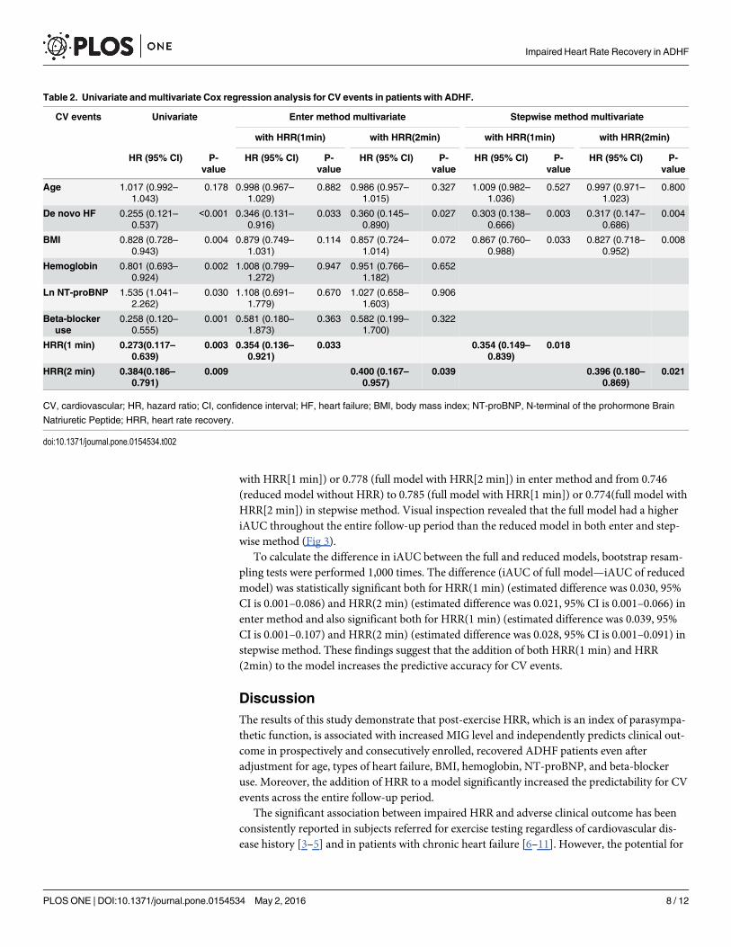
To determine which factors are relevant to the clinical outcome of ADHF patients, we stud-ied univariate and multivariate analysis for CV events. Due to the small number of CV events,we performed both enter method and stepwise method multivariate Cox regression analysis inthis study (Table 2).
In enter method multivariate Cox regression analysis, both HRR(1min) (p = 0.033) andHRR(2min) (p = 0.039) showed independent prognostic value when controlled for age, typesof heart failure, BMI, hemoglobin, NT-proBNP, and beta-blocker use. Stepwise method multi-variate Cox regression analysis also revealed independent association of both HRR(1min)(p = 0.018) and HRR(2min) (p = 0.021) with the clinical outcome even after adjusting the mostrelevant minimum variables (age, types of heart failure and BMI) to avoid the over-fitting.
In the likelihood ratio test, the full model with HRR(1 min) or HRR(2 min) exhibited signif-icantly lower –2 log likelihood than the reduced model without HRR in both enter method(HRR[1 min], 180.827 vs. 195.486, p<0.001; HRR[2 min], 191.154 vs. 195.486, p = 0.037) andstepwise method (HRR[1 min], 209.009 vs. 225.768, p<0.001; HRR[2 min], 220.303 vs.225.768, p = 0.019). Time-dependent ROC curves also demonstrated that the concordanceprobability (iAUC) increased from 0.756 (reduced model without HRR) to 0.787 (full model
Impaired Heart Rate Recovery in ADHF
PLOSONE | DOI:10.1371/journal.pone.0154534 May 2, 2016 5 / 12
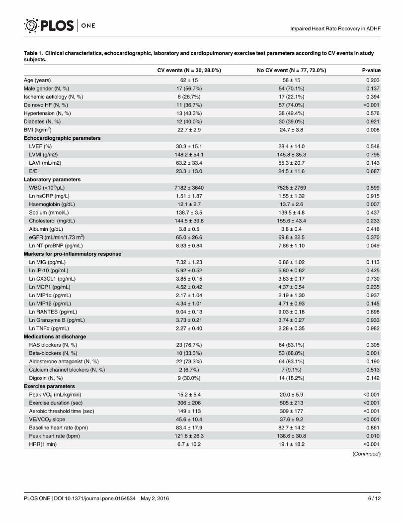
Table 1. Clinical characteristics, echocardiographic, laboratory and cardiopulmonary exercise test parameters according to CV events in studysubjects.
CV events (N = 30, 28.0%) No CV event (N = 77, 72.0%) P-value
Age (years) 62 ± 15 58 ± 15 0.203
Male gender (N, %) 17 (56.7%) 54 (70.1%) 0.137
Ischemic aetiology (N, %) 8 (26.7%) 17 (22.1%) 0.394
De novo HF (N, %) 11 (36.7%) 57 (74.0%) <0.001
Hypertension (N, %) 13 (43.3%) 38 (49.4%) 0.576
Diabetes (N, %) 12 (40.0%) 30 (39.0%) 0.921
BMI (kg/m2) 22.7 ± 2.9 24.7 ± 3.8 0.008
Echocardiographic parameters
LVEF (%) 30.3 ± 15.1 28.4 ± 14.0 0.548
LVMI (g/m2) 148.2 ± 54.1 145.8 ± 35.3 0.796
LAVI (mL/m2) 63.2 ± 33.4 55.3 ± 20.7 0.143
E/E’ 23.3 ± 13.0 24.5 ± 11.6 0.687
Laboratory parameters
WBC (×103/μL) 7182 ± 3640 7526 ± 2769 0.599
Ln hsCRP (mg/L) 1.51 ± 1.87 1.55 ± 1.32 0.915
Haemoglobin (g/dL) 12.1 ± 2.7 13.7 ± 2.6 0.007
Sodium (mmol/L) 138.7 ± 3.5 139.5 ± 4.8 0.437
Cholesterol (mg/dL) 144.5 ± 39.8 155.6 ± 43.4 0.233
Albumin (g/dL) 3.8 ± 0.5 3.8 ± 0.4 0.416
eGFR (mL/min/1.73 m2) 65.0 ± 26.6 69.8 ± 22.5 0.370
Ln NT-proBNP (pg/mL) 8.33 ± 0.84 7.86 ± 1.10 0.049
Markers for pro-inflammatory response
Ln MIG (pg/mL) 7.32 ± 1.23 6.86 ± 1.02 0.113
Ln IP-10 (pg/mL) 5.92 ± 0.52 5.80 ± 0.62 0.425
Ln CX3CL1 (pg/mL) 3.85 ± 0.15 3.83 ± 0.17 0.730
Ln MCP1 (pg/mL) 4.52 ± 0.42 4.37 ± 0.54 0.235
Ln MIP1α (pg/mL) 2.17 ± 1.04 2.19 ± 1.30 0.937
Ln MIP1β (pg/mL) 4.34 ± 1.01 4.71 ± 0.93 0.145
Ln RANTES (pg/mL) 9.04 ± 0.13 9.03 ± 0.18 0.898
Ln Granzyme B (pg/mL) 3.73 ± 0.21 3.74 ± 0.27 0.933
Ln TNFα (pg/mL) 2.27 ± 0.40 2.28 ± 0.35 0.982
Medications at discharge
RAS blockers (N, %) 23 (76.7%) 64 (83.1%) 0.305
Beta-blockers (N, %) 10 (33.3%) 53 (68.8%) 0.001
Aldosterone antagonist (N, %) 22 (73.3%) 64 (83.1%) 0.190
Calcium channel blockers (N, %) 2 (6.7%) 7 (9.1%) 0.513
Digoxin (N, %) 9 (30.0%) 14 (18.2%) 0.142
Exercise parameters
Peak VO2 (mL/kg/min) 15.2 ± 5.4 20.0 ± 5.9 <0.001
Exercise duration (sec) 306 ± 206 505 ± 213 <0.001
Aerobic threshold time (sec) 149 ± 113 309 ± 177 <0.001
VE/VCO2 slope 45.6 ± 10.4 37.6 ± 9.2 <0.001
Baseline heart rate (bpm) 83.4 ± 17.9 82.7 ± 14.2 0.861
Peak heart rate (bpm) 121.8 ± 26.3 138.6 ± 30.8 0.010
HRR(1 min) 6.7 ± 10.2 19.1 ± 18.2 <0.001
(Continued)
Impaired Heart Rate Recovery in ADHF
PLOSONE | DOI:10.1371/journal.pone.0154534 May 2, 2016 6 / 12
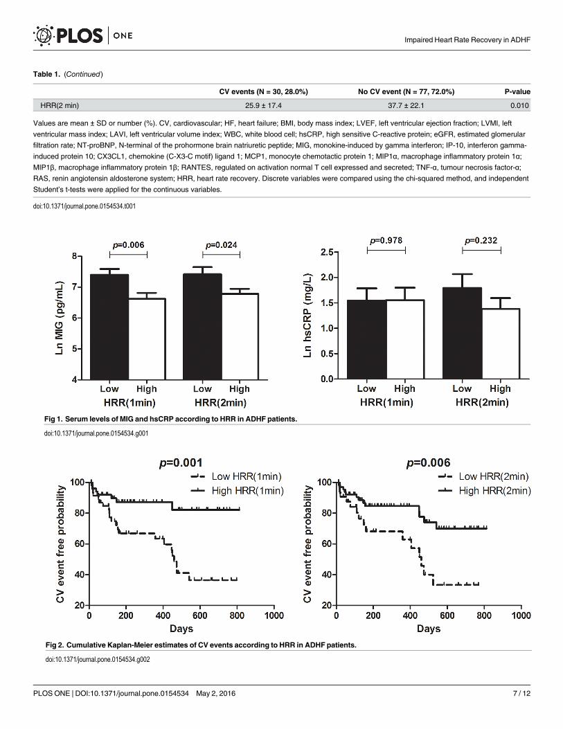
Table 1. (Continued)
CV events (N = 30, 28.0%) No CV event (N = 77, 72.0%) P-value
HRR(2 min) 25.9 ± 17.4 37.7 ± 22.1 0.010
Values are mean ± SD or number (%). CV, cardiovascular; HF, heart failure; BMI, body mass index; LVEF, left ventricular ejection fraction; LVMI, left
ventricular mass index; LAVI, left ventricular volume index; WBC, white blood cell; hsCRP, high sensitive C-reactive protein; eGFR, estimated glomerular
filtration rate; NT-proBNP, N-terminal of the prohormone brain natriuretic peptide; MIG, monokine-induced by gamma interferon; IP-10, interferon gamma-
induced protein 10; CX3CL1, chemokine (C-X3-C motif) ligand 1; MCP1, monocyte chemotactic protein 1; MIP1α, macrophage inflammatory protein 1α;
MIP1β, macrophage inflammatory protein 1β; RANTES, regulated on activation normal T cell expressed and secreted; TNF-α, tumour necrosis factor-α;
RAS, renin angiotensin aldosterone system; HRR, heart rate recovery. Discrete variables were compared using the chi-squared method, and independent
Student’s t-tests were applied for the continuous variables.
doi:10.1371/journal.pone.0154534.t001
Fig 1. Serum levels of MIG and hsCRP according to HRR in ADHF patients.
doi:10.1371/journal.pone.0154534.g001
Fig 2. Cumulative Kaplan-Meier estimates of CV events according to HRR in ADHF patients.
doi:10.1371/journal.pone.0154534.g002
Impaired Heart Rate Recovery in ADHF
PLOSONE | DOI:10.1371/journal.pone.0154534 May 2, 2016 7 / 12
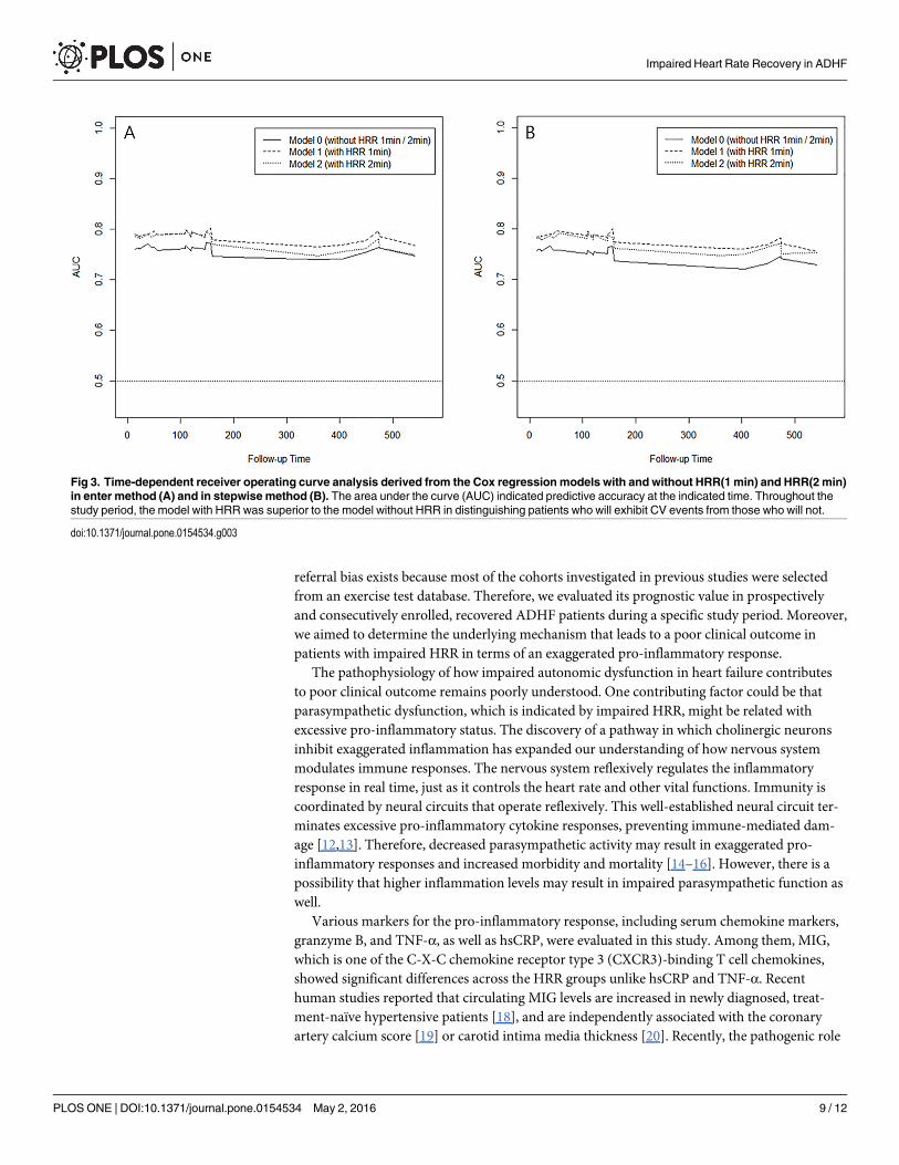
with HRR[1 min]) or 0.778 (full model with HRR[2 min]) in enter method and from 0.746(reduced model without HRR) to 0.785 (full model with HRR[1 min]) or 0.774(full model withHRR[2 min]) in stepwise method. Visual inspection revealed that the full model had a higheriAUC throughout the entire follow-up period than the reduced model in both enter and step-wise method (Fig 3).
To calculate the difference in iAUC between the full and reduced models, bootstrap resam-pling tests were performed 1,000 times. The difference (iAUC of full model—iAUC of reducedmodel) was statistically significant both for HRR(1 min) (estimated difference was 0.030, 95%CI is 0.001–0.086) and HRR(2 min) (estimated difference was 0.021, 95% CI is 0.001–0.066) inenter method and also significant both for HRR(1 min) (estimated difference was 0.039, 95%CI is 0.001–0.107) and HRR(2 min) (estimated difference was 0.028, 95% CI is 0.001–0.091) instepwise method. These findings suggest that the addition of both HRR(1 min) and HRR(2min) to the model increases the predictive accuracy for CV events.
DiscussionThe results of this study demonstrate that post-exercise HRR, which is an index of parasympa-thetic function, is associated with increased MIG level and independently predicts clinical out-come in prospectively and consecutively enrolled, recovered ADHF patients even afteradjustment for age, types of heart failure, BMI, hemoglobin, NT-proBNP, and beta-blockeruse. Moreover, the addition of HRR to a model significantly increased the predictability for CVevents across the entire follow-up period.
The significant association between impaired HRR and adverse clinical outcome has beenconsistently reported in subjects referred for exercise testing regardless of cardiovascular dis-ease history [3–5] and in patients with chronic heart failure [6–11]. However, the potential for
Table 2. Univariate andmultivariate Cox regression analysis for CV events in patients with ADHF.
CV events Univariate Enter method multivariate Stepwise method multivariate
with HRR(1min) with HRR(2min) with HRR(1min) with HRR(2min)
HR (95% CI) P-value
HR (95% CI) P-value
HR (95% CI) P-value
HR (95% CI) P-value
HR (95% CI) P-value
Age 1.017 (0.992–1.043)
0.178 0.998 (0.967–1.029)
0.882 0.986 (0.957–1.015)
0.327 1.009 (0.982–1.036)
0.527 0.997 (0.971–1.023)
0.800
De novo HF 0.255 (0.121–0.537)
<0.001 0.346 (0.131–0.916)
0.033 0.360 (0.145–0.890)
0.027 0.303 (0.138–0.666)
0.003 0.317 (0.147–0.686)
0.004
BMI 0.828 (0.728–0.943)
0.004 0.879 (0.749–1.031)
0.114 0.857 (0.724–1.014)
0.072 0.867 (0.760–0.988)
0.033 0.827 (0.718–0.952)
0.008
Hemoglobin 0.801 (0.693–0.924)
0.002 1.008 (0.799–1.272)
0.947 0.951 (0.766–1.182)
0.652
Ln NT-proBNP 1.535 (1.041–2.262)
0.030 1.108 (0.691–1.779)
0.670 1.027 (0.658–1.603)
0.906
Beta-blockeruse
0.258 (0.120–0.555)
0.001 0.581 (0.180–1.873)
0.363 0.582 (0.199–1.700)
0.322
HRR(1 min) 0.273(0.117–0.639)
0.003 0.354 (0.136–0.921)
0.033 0.354 (0.149–0.839)
0.018
HRR(2 min) 0.384(0.186–0.791)
0.009 0.400 (0.167–0.957)
0.039 0.396 (0.180–0.869)
0.021
CV, cardiovascular; HR, hazard ratio; CI, confidence interval; HF, heart failure; BMI, body mass index; NT-proBNP, N-terminal of the prohormone Brain
Natriuretic Peptide; HRR, heart rate recovery.
doi:10.1371/journal.pone.0154534.t002
Impaired Heart Rate Recovery in ADHF
PLOSONE | DOI:10.1371/journal.pone.0154534 May 2, 2016 8 / 12
referral bias exists because most of the cohorts investigated in previous studies were selectedfrom an exercise test database. Therefore, we evaluated its prognostic value in prospectivelyand consecutively enrolled, recovered ADHF patients during a specific study period. Moreover,we aimed to determine the underlying mechanism that leads to a poor clinical outcome inpatients with impaired HRR in terms of an exaggerated pro-inflammatory response.
The pathophysiology of how impaired autonomic dysfunction in heart failure contributesto poor clinical outcome remains poorly understood. One contributing factor could be thatparasympathetic dysfunction, which is indicated by impaired HRR, might be related withexcessive pro-inflammatory status. The discovery of a pathway in which cholinergic neuronsinhibit exaggerated inflammation has expanded our understanding of how nervous systemmodulates immune responses. The nervous system reflexively regulates the inflammatoryresponse in real time, just as it controls the heart rate and other vital functions. Immunity iscoordinated by neural circuits that operate reflexively. This well-established neural circuit ter-minates excessive pro-inflammatory cytokine responses, preventing immune-mediated dam-age [12,13]. Therefore, decreased parasympathetic activity may result in exaggerated pro-inflammatory responses and increased morbidity and mortality [14–16]. However, there is apossibility that higher inflammation levels may result in impaired parasympathetic function aswell.
Various markers for the pro-inflammatory response, including serum chemokine markers,granzyme B, and TNF-α, as well as hsCRP, were evaluated in this study. Among them, MIG,which is one of the C-X-C chemokine receptor type 3 (CXCR3)-binding T cell chemokines,showed significant differences across the HRR groups unlike hsCRP and TNF-α. Recenthuman studies reported that circulating MIG levels are increased in newly diagnosed, treat-ment-naïve hypertensive patients [18], and are independently associated with the coronaryartery calcium score [19] or carotid intima media thickness [20]. Recently, the pathogenic role
Fig 3. Time-dependent receiver operating curve analysis derived from the Cox regression models with and without HRR(1min) and HRR(2min)in enter method (A) and in stepwise method (B). The area under the curve (AUC) indicated predictive accuracy at the indicated time. Throughout thestudy period, the model with HRR was superior to the model without HRR in distinguishing patients who will exhibit CV events from those who will not.
doi:10.1371/journal.pone.0154534.g003
Impaired Heart Rate Recovery in ADHF
PLOSONE | DOI:10.1371/journal.pone.0154534 May 2, 2016 9 / 12
of CD4+ T cells in pressure overload-induced cardiac remodelling and in the transition to heartfailure was demonstrated in animal models [21]. Moreover, the enhanced CD4+ T cell activa-tion observed in patients with heart failure in proportion to severity suggests that T cell-driveninflammation is one important pathophysiology of heart failure [22,23]. Consequently, ourfindings that patients with autonomic dysfunction showed increased serumMIG levels furthersupport the involvement of T cell-driven inflammation in patients with heart failure.
It has been suggested that specific medication, such as beta-blockers, may influence theHRR. Lipinski, et al. [7] and Kubrychtova, et al. [10] showed improved HRR in patients takingbeta-blockers while Racine, et al. [24] reported that beta-blocker therapy does not significantlyimprove HRR, suggesting that controlling for beta-blocker use in the evaluation of HRR maynot be necessary. In our study, both HRR(1 min) and HRR(2 min) showed no significant dif-ferences between beta-blocker users and non-users. However, impaired post-exercise HRRindependently predicted clinical outcome irrespective of beta-blocker use in this study.
Several device-based therapies to target specific aspects of autonomic imbalance are activelyunder investigation in patients with heart failure. Among them, vagal nerve stimulation hasbeen shown to improve clinical heart failure symptoms, New York Heart Association (NYHA)heart failure class, and evidence of left ventricular reverse remodelling in an open label pilotstudy [25]. More recently, the results of two other human vagal nerve stimulation studies,NECTAR-HF [26] and ANTHEM-HF [27], were reported. While NECTAR-HF did not showimprovements in the primary endpoint of reduction in left ventricular end-systolic diameter(LVESD), ANTHEM-HF demonstrated significant improvement in left ventricular ejectionfraction and LVESD in left and right vagal stimulation of chronic symptomatic heart failurepatients. Currently, the INOVATE HF clinical trial [28], using the CardioFit System, is beingconducted in 90 centres worldwide to evaluate its long-term safety and efficacy in patients.This trial will provide more definitive data on the safety and efficacy of vagal nerve stimulationin patients with heart failure. Another recent first-in-human trial of high thoracic spinal cordstimulation revealed that this approach is safe and can potentially improve symptoms, func-tional capacity, and cardiac function in patients with advanced HF [29]. Regarding the use ofdevice-based therapies to modulate autonomic dysfunction, it is important to investigate theproper patient population, one that will benefit from these device therapies. Post-exerciseHRR, which is an index of parasympathetic function, can be used in patient selection for auto-nomic modulation similar to left bundle branch block pattern QRS morphology and QRS dura-tion for cardiac resynchronization therapy.
The present study has several potential limitations. First, due to the maximal exercise proto-col, the intensity of exercise by each patient is not controlled. Second, our study populationcannot represent all heart failure patients. The analysed subjects were hospitalized and ambula-tory, recovered ADHF patients. Moreover, these data were collected from a Korean population.The generalizability of the current findings to other populations (e.g. Caucasian populations) isunknown. Third, the lack of adjustment for diet pattern and socioeconomic status might benoted as well. Fourth, due to the small number of CV events, our multivariate Cox regressionmodel included a limited number of variables. Peak VO2, which is a well-known prognosticfactor in patients with HF is not adjusted in the multivariate analysis due to the significant co-linearity with both HRR(1 min) and HRR(2 min). Finally, lack of other parasympathetic activ-ity measurements, such as heart rate variability or baroreflex sensitivity, could be another limi-tation of this study. However, we evaluated the prognostic value of post-exercise HRR inprospectively and consecutively enrolled, recovered ADHF patients, thereby minimizing thepotential for referral bias. Moreover, we sought to examine the underlying mechanism thatleads to poor clinical outcome in patients with impaired HRR in terms of ‘cholinergic anti-inflammatory pathway’.
Impaired Heart Rate Recovery in ADHF
PLOSONE | DOI:10.1371/journal.pone.0154534 May 2, 2016 10 / 12

ConclusionsPost-exercise HRR is an index of parasympathetic function associated with clinical outcome inpatients with chronic heart failure. However, its prognostic value has not been confirmed inprospectively and consecutively enrolled ADHF patients. Here, we confirm that impaired post-exercise HRR is associated with an exaggerated pro-inflammatory response and independentlypredicts clinical outcome in patients with ADHF. Measurement of post-exercise HRR is a sim-ple and feasible assessment, which might have important implications on risk stratification ofADHF patients.
Author ContributionsConceived and designed the experiments: JCY SMK. Performed the experiments: JCY SMK.Analyzed the data: JCY HSL SWC SWH KHR ECS SMK. Contributed reagents/materials/anal-ysis tools: JCY HSL SMK. Wrote the paper: JCY HSL SMK.
References1. Olshansky B, Sabbah HN, Hauptman PJ, Colucci WS. Parasympathetic nervous system and heart fail-
ure: pathophysiology and potential implications for therapy. Circulation. 2008; 118: 863–871. doi: 10.1161/CIRCULATIONAHA.107.760405 PMID: 18711023
2. Lahiri MK, Kannankeril PJ, Goldberger JJ. Assessment of autonomic function in cardiovascular dis-ease: physiological basis and prognostic implications. J Am Coll Cardiol. 2008; 51: 1725–1733. doi: 10.1016/j.jacc.2008.01.038 PMID: 18452777
3. Vivekananthan DP, Blackstone EH, Pothier CE, Lauer MS. Heart rate recovery after exercise is a pre-dictor of mortality, independent of the angiographic severity of coronary disease. J Am Coll Cardiol.2003; 42: 831–838. PMID: 12957428
4. Nishime EO, Cole CR, Blackstone EH, Pashkow FJ, Lauer MS. Heart rate recovery and treadmill exer-cise score as predictors of mortality in patients referred for exercise ECG. JAMA. 2000; 284: 1392–1398. PMID: 10989401
5. Cole CR, Blackstone EH, Pashkow FJ, Snader CE, Lauer MS. Heart-rate recovery immediately afterexercise as a predictor of mortality. N Engl J Med. 1999; 341: 1351–1357. PMID: 10536127
6. Imai K, Sato H, Hori M, Kusuoka H, Ozaki H, Yokoyama H, et al. Vagally mediated heart rate recoveryafter exercise is accelerated in athletes but blunted in patients with chronic heart failure. J Am Coll Car-diol. 1994; 24: 1529–1535. PMID: 7930286
7. Lipinski MJ, Vetrovec GW, Gorelik D, Froelicher VF. The importance of heart rate recovery in patientswith heart failure or left ventricular systolic dysfunction. J Card Fail. 2005; 11: 624–630. PMID: 16230267
8. Arena R, Guazzi M, Myers J, Peberdy MA. Prognostic value of heart rate recovery in patients with heartfailure. Am Heart J. 2006; 151: 851 e857–813.
9. Sheppard RJ, Racine N, Roof A, Ducharme A, Blanchet M, White M. Heart rate recovery—a potentialmarker of clinical outcomes in heart failure patients receiving beta-blocker therapy. Can J Cardiol.2007; 23: 1135–1138. PMID: 18060099
10. Kubrychtova V, Olson TP, Bailey KR, Thapa P, Allison TG, Johnson BD. Heart rate recovery and prog-nosis in heart failure patients. Eur J Appl Physiol. 2009; 105: 37–45. doi: 10.1007/s00421-008-0870-zPMID: 18797918
11. Tang YD, Dewland TA, Wencker D, Katz SD. Post-exercise heart rate recovery independently predictsmortality risk in patients with chronic heart failure. J Card Fail. 2009; 15: 850–855. doi: 10.1016/j.cardfail.2009.06.437 PMID: 19944361
12. Borovikova LV, Ivanova S, Zhang M, Yang H, Botchkina GI, Watkins LR, et al. Vagus nerve stimulationattenuates the systemic inflammatory response to endotoxin. Nature. 2000; 405: 458–462. PMID:10839541
13. Tracey KJ. Reflex control of immunity. Nat Rev Immunol. 2009; 9: 418–428. doi: 10.1038/nri2566PMID: 19461672
14. Thayer JF, Lane RD. The role of vagal function in the risk for cardiovascular disease and mortality. BiolPsychol. 2007; 74: 224–242. PMID: 17182165
15. Haensel A, Mills PJ, Nelesen RA, Ziegler MG, Dimsdale JE. The relationship between heart rate vari-ability and inflammatory markers in cardiovascular diseases. Psychoneuroendocrinology. 2008; 33:1305–1312. doi: 10.1016/j.psyneuen.2008.08.007 PMID: 18819754
Impaired Heart Rate Recovery in ADHF
PLOSONE | DOI:10.1371/journal.pone.0154534 May 2, 2016 11 / 12
16. Thayer JF, Fischer JE. Heart rate variability, overnight urinary norepinephrine and C-reactive protein:evidence for the cholinergic anti-inflammatory pathway in healthy human adults. J Intern Med. 2009;265: 439–447. doi: 10.1111/j.1365-2796.2008.02023.x PMID: 19019182
17. Contal C, O'Quigley J. An application of changepoint methods in studying the effect of age on survivalin breast cancer. Computational Statistics & Data Analysis. 1999; 30: 253–270.
18. Youn JC, Yu HT, Lim BJ, Koh MJ, Lee J, Chang DY, et al. Immunosenescent CD8+ T cells and C-X-Cchemokine receptor type 3 chemokines are increased in human hypertension. Hypertension. 2013; 62:126–133. doi: 10.1161/HYPERTENSIONAHA.113.00689 PMID: 23716586
19. Yu HT, Oh J, Chang HJ, Lee SH, Shin EC, Park S. Serummonokine induced by gamma interferon as anovel biomarker for coronary artery calcification in humans. Coron Artery Dis. 2015; 26: 317–321. doi:10.1097/MCA.0000000000000236 PMID: 25756330
20. Yu HT, Lee J, Shin EC, Park S. Significant Association between SerumMonokine Induced by GammaInterferon and Carotid Intima Media Thickness. J Atheroscler Thromb. 2015; 22: 816–822. doi: 10.5551/jat.28886 PMID: 25739534
21. Laroumanie F, Douin-Echinard V, Pozzo J, Lairez O, Tortosa F, Vinel C, et al. CD4+ T cells promotethe transition from hypertrophy to heart failure during chronic pressure overload. Circulation. 2014; 129:2111–2124. doi: 10.1161/CIRCULATIONAHA.113.007101 PMID: 24657994
22. Fukunaga T, Soejima H, Irie A, Sugamura K, Oe Y, Tanaka T, et al. Relation between CD4+ T-cell acti-vation and severity of chronic heart failure secondary to ischemic or idiopathic dilated cardiomyopathy.Am J Cardiol. 2007; 100: 483–488. PMID: 17659933
23. Moro-Garcia MA, Echeverria A, Galan-Artimez MC, Suarez-Garcia FM, Solano-Jaurrieta JJ, Avanzas-Fernandez P, et al. Immunosenescence and inflammation characterize chronic heart failure patientswith more advanced disease. Int J Cardiol. 2014; 174: 590–599. doi: 10.1016/j.ijcard.2014.04.128PMID: 24801091
24. Racine N, Blanchet M, Ducharme A, Marquis J, Boucher JM, JuneauM, et al. Decreased heart raterecovery after exercise in patients with congestive heart failure: effect of beta-blocker therapy. J CardFail. 2003; 9: 296–302. PMID: 13680550
25. De Ferrari GM, Crijns HJ, Borggrefe M, Milasinovic G, Smid J, Zabel M, et al. Chronic vagus nerve stim-ulation: a new and promising therapeutic approach for chronic heart failure. Eur Heart J. 2011; 32: 847–855. doi: 10.1093/eurheartj/ehq391 PMID: 21030409
26. Zannad F, De Ferrari GM, Tuinenburg AE, Wright D, Brugada J, Butter C, et al. Chronic vagal stimula-tion for the treatment of low ejection fraction heart failure: results of the NEural Cardiac TherApy foRHeart Failure (NECTAR-HF) randomized controlled trial. Eur Heart J. 2015; 36: 425–433. doi: 10.1093/eurheartj/ehu345 PMID: 25176942
27. Premchand RK, Sharma K, Mittal S, Monteiro R, Dixit S, Libbus I, et al. autonomic regulation therapyvia left or right cervical vagus nerve stimulation in patients with chronic heart failure: results of theANTHEM-HF trial. J Card Fail. 2014; 20: 808–816. doi: 10.1016/j.cardfail.2014.08.009 PMID:25187002
28. Hauptman PJ, Schwartz PJ, Gold MR, Borggrefe M, Van Veldhuisen DJ, Starling RC, et al. Rationaleand study design of the increase of vagal tone in heart failure study: INOVATE-HF. Am Heart J. 2012;163: 954–962 e951. doi: 10.1016/j.ahj.2012.03.021 PMID: 22709747
29. Tse HF, Turner S, Sanders P, Okuyama Y, Fujiu K, Cheung CW, et al. Thoracic Spinal Cord Stimulationfor Heart Failure as a Restorative Treatment (SCS HEART study): First-in-man experience. HeartRhythm. 2015; 12: 588–595. doi: 10.1016/j.hrthm.2014.12.014 PMID: 25500165
Impaired Heart Rate Recovery in ADHF
PLOSONE | DOI:10.1371/journal.pone.0154534 May 2, 2016 12 / 12