researcharticle divergenceoftheresponseinducedby...
TRANSCRIPT

RESEARCH ARTICLE
Divergence of the Response Induced byXenogenic Immunization in the SepsisSurvival of RatsMagdiel Perez-Cruz1, Cristina Costa1, Rafael Manez1,2*
1 Infectious Diseases and Transplantation Division, Bellvitge Biomedical Research Institute (IDIBELL),L’Hospitalet de Llobregat, Barcelona, Spain, 2 Intensive Care Department, Bellvitge University Hospital,L’Hospitalet de Llobregat, Barcelona, Spain
AbstractWe have previously described that boosted natural xenoantibodies in rats cross-react to
bacteria by targeting carbohydrate antigens. This type of immunization is associated with
reduced survival after cecal ligation and puncture (CLP). In the present study, we investigat-
ed further this phenomenon by immunizing Lewis rats with three intraperitoneal injections,
every other day, of hamster blood compared to saline-injected control animals. One day
after the last injection, CLP was performed to produce a low-grade sepsis. Induction of
xenoantibodies was associated with a reduction in animal survival after CLP relative to con-
trols (45% vs. 90%, p<0.01). No bacterial blood load was observed after CLP in this model
either with or without xenoantibody enhancement, indicating that the augmented mortality
was not mediated by a direct effect of boosted xenoantibodies over blood bacteria. Never-
theless, the xenoimmunization produced a systemic inflammatory response in all rats. Addi-
tionally, a lack of weight gain at the time of CLP was present in animals that died after the
procedure, which was not observed in surviving rats and controls. The cytokine profile at
the time of CLP in animals that died after the procedure was characterized by an increase in
the serum level of several cytokines, particularly adipokines. In contrast, the cytokine profile
at CLP of xenoimmunized rats that survived the procedure was characterized by a reduction
in the level of cytokines. In conclusion, this study failed to show a direct effect of boosted
xenoantibodies over blood bacterial isolates as cause for the decreased survival after CLP.
However, it evidenced that non-infectious systemic inflammation may lead to a pattern of
augmented cytokines, particularly adipokines, which impairs survival after subsequent
CLP. Therefore, the profile of cytokines existing before the infectious insult appears more
crucial than that resulting from the condition for the outcome of sepsis.
PLOS ONE | DOI:10.1371/journal.pone.0125472 May 18, 2015 1 / 14
OPEN ACCESS
Citation: Perez-Cruz M, Costa C, Manez R (2015)Divergence of the Response Induced by XenogenicImmunization in the Sepsis Survival of Rats. PLoSONE 10(5): e0125472. doi:10.1371/journal.pone.0125472
Academic Editor: Vladimir Trajkovic, School ofMedicine, University of Belgrade, SERBIA
Received: November 18, 2014
Accepted: March 24, 2015
Published: May 18, 2015
Copyright: © 2015 Perez-Cruz et al. This is an openaccess article distributed under the terms of theCreative Commons Attribution License, which permitsunrestricted use, distribution, and reproduction in anymedium, provided the original author and source arecredited.
Data Availability Statement: All relevant data arewithin the paper and its Supporting Information files.
Funding: This work was supported by “Fondo deInvestigaciones Sanitarias” (FIS) grants PI05/0861and PI10/01727 from Carlos III Health Institute,Spanish Ministry of Health (www.isciii.es), andEuropean Commission’s Seventh FrameworkProgramme grant HEALTH-F4-2013-603049TRANSLINK Collaborative Project (www.ec.europa.eu/research/fp7). MPC was funded by an IDIBELLfellowship (www.idibell.cat) and CC by the Ramón yCajal program from the Spanish Ministry of Educationand Science (www.idi.mineco.gob.es) and IDIBELL.

IntroductionNatural antibodies are characterized by their recognition of antigens in the absence of any evi-dence of exogenous exposure to them. These antibodies are mainly IgM, are polyreactive, ex-hibit modest antigen binding affinity, are directed against carbohydrate antigens, and are dueto the direct stimulation of antibody production by T-cell-independent (TI) pathways involv-ing B-1 lymphocytes [1]. Natural antibodies recognize self, altered-self and foreign antigens,comprising an important first line of defense against invading pathogens. Thus, natural IgMantibodies play a critical role in bacterial clearance [2], making mice lacking this isotype of an-tibodies more susceptible to cecal ligation and puncture (CLP) [3]. In addition, natural anti-bodies are also important for tissue homeostasis and for inhibiting or preventing inflammatoryreactions [4]. Natural IgM antibodies contribute to the removal of apoptotic and transformedcells through a complement-dependent pathway, suppression of inflammation, elimination ofaltered proteins, and control of autoreactive IgG and antibody-producing B cells capable ofcausing diseases [5].
Natural antibodies comprise a subgroup that bind antigens depicted on cells and tissues ofdissimilar species defined as xenoantibodies [6]. These antibodies appear to be produced in re-sponse to bacteria contained in the gut that cross-react with structurally alike xenoantigens. Inhumans, xenoantibodies include IgM and IgG directed against the galactose α1,3 galactose(αGal) carbohydrate epitope [7], which is expressed in most mammalian species. Anti- αGalantibodies react with various bacteria, including strains of Escherichia coli, Klebsiella and Sal-monella [8,9] and drop after antibiotic treatment that removes Gram-negative enteric flora[10]. Interestingly, the binding of anti- αGal antibodies to blood Gram-negative bacteria iso-lates was higher than their binding to normal stool isolates [9]. In blood isolates, anti- αGalIgG antibodies may bind the capsular polysaccharide, increasing the alternative complementpathway lysis of the microorganism, or the lipopolysaccharide that makes the bacteria resistantto lysis [9].
We have previously described higher titers of natural anti- αGal IgM antibodies in some pa-tients at the time of initiation of renal replacement therapy, which correlated with increasedserum levels of TNF-α [11]. This condition predicted later risk for enteric peritonitis in perito-neal dialysis patients and mortality in both peritoneal dialysis and hemodialysis patients [11].In addition, we have recently shown that boosted TI xenoantibodies in rats by exposure tohamster or pig antigens cross-react to Enterococcus faecalis by targeting melibiose and L-rham-nose carbohydrates [12]. This circumstance was associated with reduced survival of these ani-mals after CLP, which was not observed with the generation of IgG antibodies. To elucidate themechanisms underlying these findings, we analyzed the xenoantibody and inflammatory re-sponses caused by exposure to xenogeneic antigens and their impact on CLP-induced sepsis ina rat model. The production of xenoantibodies in rats is characterized by the predominant ini-tial expansion of TI natural IgM antibodies (first week), followed by the generation of T-cell-dependent adaptive IgG antibodies (days 21 to 28) [6]. This allows the assessment of the impactof the TI xenoantibodies in the outcome of sepsis.
Material and Methods
AnimalsLewis rats (200–250 g weight) and Golden Syrian hamsters (100–150 g weight) were purchasedfrom Interfauna Harlan Iberica SL (Barcelona, Spain). Animals were maintained at Universityof Barcelona (Bellvitge Campus) animal facility under controlled conditions of temperature(21 ± 1°C) and humidity (55 ± 5%), cycles of light/dark of 12/12 h, and with food and water
Xenoantibodies and Sepsis
PLOS ONE | DOI:10.1371/journal.pone.0125472 May 18, 2015 2 / 14
Competing Interests: The authors have declaredthat no competing interests exist.
given ad libitum. Mice were acclimatized for 1 week before entering the study. Animals wereanesthetized by isoflurane inhalation for blood draw and injections: deep anesthesia (at 4%) forhamsters (cardiac puncture), middle (at 2%) for rat blood draw and light (at 1%) for rat bloodinjection. All animal procedures were supervised and approved by Bellvitge Biomedical Re-search Institute (IDIBELL) ethics committee for animal experimentation and the CataloniaGovernment (Permit Number: DMA 3225). The care and handling of the animals conforms tothe Guide for the Care and Use of Laboratory Animals published by the US National Institutesof Health (NIH Publication n° 85–23 revised 1996) and the European Agreement of VertebrateAnimal Protection for Experimental Use (86/609).
Rat immunizationTo produce a pattern of predominantly TI anti-hamster xenoantibodies [6], three intraperito-neal (ip) injections of 1 ml of hamster blood were given on alternating days (days -5, -3 and -1relative to CLP). Control animals were subjected to three ip injections of phosphate-bufferedsaline (PBS) on the same days. Hamster blood was collected heparinized from cardiac punc-ture, pooled and immediately injected ip into rats.
Determination of xenoantibodiesIgM and IgG anti-hamster xenoantibodies were determined by flow cytometry, measuring onlythe surface-bound immunoglobulins. Lymphocytes obtained from the hamster spleen (1x10⁶
cells per sample) were incubated with test sera diluted 1/50 in PBS/1% bovine serum albumin(BSA) at 4°C for 30 minutes in a final volume of 100 μl in V-bottom 96-well microtiter plates(NUNC Denmark). After the first incubation, a wash with PBS/1% BSA was performed fol-lowed by a second incubation (4°C 30 minutes in the dark) with 100 μl of a mixture of bothpolyclonal secondary antibodies (goat F(ab')2 fragment anti-rat IgG (H+L) conjugated withdichloro triazinyl aminofluorescein (DTAF) [dilution 1/100] and goat F(ab')2 fragment anti-rat IgM (μ) conjugated with phycoerythrin (PE) [dilution 1/200] (Beckman Coulter Immuno-tech) in PBS/1% BSA. Finally, cells were washed, resuspended in PBS and transferred to FACStubes. For the determination of fluorescence intensity, a FACSCalibur cytometer (BD Biosci-ences) was used with three detectors/photomultipliers, which detect the light emitted at 530nm (FL1), 585 nm (FL2) and> 670 nm (FL3), along with the help of programs for acquisitionand analysis (Cell Quest) and for verification (FACS Comp).
CLPCLP was performed as described elsewhere [13]. Briefly, Lewis rats were anesthetized with3–4% isoflurane and under sterile conditions a 1–2 cm midline incision was made and thececum was exteriorized and ligated (4–0 Safil Violet, B/Braum, Germany) distal to the ileocecalvalve. To generate a low-grade sepsis, defined as ~ 0–15% mortality during the acute phase ofsepsis, 25% of the cecum (~ 10 mm) was ligated and punctured twice with a 19-gauge needle.The abdomen was closed in two layers and animal recovery was facilitated by keeping the ani-mal on a thermal blanket. Access to water and food was facilitated 2 h after surgery when ani-mals were placed in their corresponding cages. Body weight, mobility, food intake, cutaneousfeatures and respiratory frequency of animals were monitored twice a day over a period of 15days, providing a score (S1 Table) that led to different corrective measures depending on ani-mal status (S2 Table).
Xenoantibodies and Sepsis
PLOS ONE | DOI:10.1371/journal.pone.0125472 May 18, 2015 3 / 14

Blood bacterial loadBlood collected after cardiac puncture was serially diluted and immediately plated on Trypti-case Soy Agar II plates supplemented with 5% Sheep Blood (Becton Dickinson). Colony-form-ing units (CFU) were counted after 24 h incubation at 37°C.
Blood hematology and serum biochemistryOne ml of blood was collected from the tail vein at different times, centrifuged (2 x 10 min,5000 rpm) and the sera were frozen at -20°C. ALT, AST, urea and triglycerides were measuredaccording to protocols of the International Federation of Clinical Chemistry by spectrophoto-metric analysis at the Veterinarian School of Barcelona Autonomous University. EDTA-antic-oagulated blood (0.5 ml) was also obtained for hematological analysis by cytometry utilizingperoxidase staining (ADVIA 120 Hematology System, Siemens Healthcare), according to pro-tocols of Veterinarian School of Barcelona Autonomous University.
Cytokine/Chemokine AnalysisRat serum was assessed for the presence of 33 cytokines including proinflammatory and anti-inflammatory cytokines, chemokines, growth factors and soluble cell receptors (S3 Table). An-tibody array membranes (rat cytokine antibody array I; RayBiotech, Norcross, GA) were firstblocked for 30 min with the provided blocking buffer, to which rat sera were subsequentlyadded for a final 10-fold dilution of the sera. Membranes were then incubated for 2 h at roomtemperature with shaking. After addition of a biotin-conjugated anti-cytokine antibody mix-ture, the membrane was incubated with horseradish peroxidase-conjugated streptavidin anddeveloped. Signal intensity was quantified with the Quantity One software (Bio-Rad Laborato-ries). The advantages of using this multiple cytokine analysis have been previously described[14]: 1) is a panel commercially available permitting easily replication studies; 2) allows the si-multaneous analysis of cytokines, chemokines, soluble cell receptors and growth factors; 3)many of them have been engaged in the pathogenesis of sepsis; 4) includes cytokines with bothpro- and anti-inflammatory activities allowing a large evaluation of immune responses.
Power of the study and statistical analysisWe calculated that a sample size of 20 animals per group would provide the appropriate power(1-β = 0.8) to identify a significant (α = 0.05) reduction in rat survival after CLP from 90% to50%. The results are expressed as the mean ± standard error of the mean (SEM). The differ-ences for each group at different times were compared by one-way ANOVA for repeated mea-sures with the Bonferroni correction. Survival data were compared by the log-rank test. Toevaluate the interactive and independent effects on cytokine expression in three groups (con-trol-TI, TI-alive, TI-death) and at three times (days -5, 0, 2), a repeated-measures two-wayANOVA was used. As the interaction term was significant (p<0.001), individual One-wayANOVAs for repeated measures with the Bonferroni correction were performed for eachcytokine to assess differences at the three times evaluated. Statistical results were consideredsignificant if p-value<0.05. Statistical analyses were performed using R 2.15: A language andenvironment for statistical computing (R Foundation for Statistical Computing, Vienna,Austria).
Xenoantibodies and Sepsis
PLOS ONE | DOI:10.1371/journal.pone.0125472 May 18, 2015 4 / 14
Results
Impact of anti-hamster xenoantibodies on Lewis rat survival after CLPExposure of Lewis rats to three injections of hamster blood, with one injection administeredevery other day, caused an increase of IgM and IgG anti-hamster xenoantibodies at day 5 com-pared to control animals. On this day, the average level of IgM was three-fold higher than IgGxenoantibodies (Fig 1A), following contrary patterns thereafter to day 20 when the averagelevel of IgG was 5-fold higher than IgM (Fig 1A). No change was observed in the level of IgMand IgG xenoantibodies with PBS injections in control animals (data not shown). CLP sepsiswas performed in Lewis rats on day 5 after xenoimmunization or the corresponding control in-jections. Augmentation of anti-hamster xenoantibodies was directly associated with lower sur-vival after CLP compared to controls (45% vs. 90%, p<0.01) (Fig 1B). No differences wereobserved in the levels of anti-hamster IgM and IgG xenoantibodies before CLP between ratsthat died after the procedure and those that did not (Fig 1C). Bacterial loads were also assessed24 h after CLP. However, no bacteria were detected in blood samples after CLP either afterboosting anti-hamster xenoantibodies or not, despite the presence of signs of inflammationand abscesses in the abdomen.
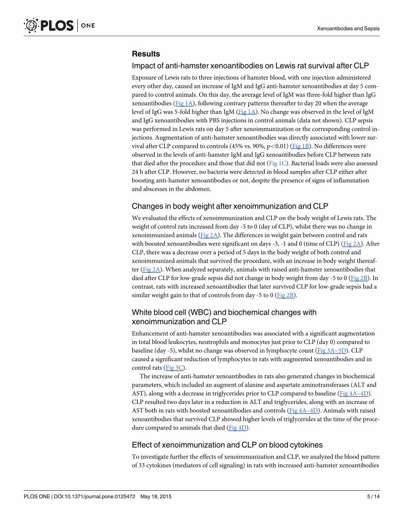
Changes in body weight after xenoimmunization and CLPWe evaluated the effects of xenoimmunization and CLP on the body weight of Lewis rats. Theweight of control rats increased from day -5 to 0 (day of CLP), whilst there was no change inxenoimmunized animals (Fig 2A). The differences in weight gain between control and ratswith boosted xenoantibodies were significant on days -3, -1 and 0 (time of CLP) (Fig 2A). AfterCLP, there was a decrease over a period of 5 days in the body weight of both control andxenoimmunized animals that survived the procedure, with an increase in body weight thereaf-ter (Fig 2A). When analyzed separately, animals with raised anti-hamster xenoantibodies thatdied after CLP for low-grade sepsis did not change in body weight from day -5 to 0 (Fig 2B). Incontrast, rats with increased xenoantibodies that later survived CLP for low-grade sepsis had asimilar weight gain to that of controls from day -5 to 0 (Fig 2B).
White blood cell (WBC) and biochemical changes withxenoimmunization and CLPEnhancement of anti-hamster xenoantibodies was associated with a significant augmentationin total blood leukocytes, neutrophils and monocytes just prior to CLP (day 0) compared tobaseline (day -5), whilst no change was observed in lymphocyte count (Fig 3A–3D). CLPcaused a significant reduction of lymphocytes in rats with augmented xenoantibodies and incontrol rats (Fig 3C).
The increase of anti-hamster xenoantibodies in rats also generated changes in biochemicalparameters, which included an augment of alanine and aspartate aminotransferases (ALT andAST), along with a decrease in triglycerides prior to CLP compared to baseline (Fig 4A–4D).CLP resulted two days later in a reduction in ALT and triglycerides, along with an increase ofAST both in rats with boosted xenoantibodies and controls (Fig 4A–4D). Animals with raisedxenoantibodies that survived CLP showed higher levels of triglycerides at the time of the proce-dure compared to animals that died (Fig 4D).
Effect of xenoimmunization and CLP on blood cytokinesTo investigate further the effects of xenoimmunization and CLP, we analyzed the blood patternof 33 cytokines (mediators of cell signaling) in rats with increased anti-hamster xenoantibodies
Xenoantibodies and Sepsis
PLOS ONE | DOI:10.1371/journal.pone.0125472 May 18, 2015 5 / 14

Fig 1. Generation of rat anti-hamster xenoantibodies and their impact on sepsis survival after CLP. (A)Levels of anti-hamster IgM and IgG xenoantibodies in Lewis after three injections of hamster blood, with oneinjection administered every other day. Mean ± SEM of 5 animals per group. MFI: Mean fluorescenceintensity. (B) Percent survival of Lewis rats with and without enhanced anti-hamster xenoantibodiessubmitted to CLP (n = 20 per group; *p<0.01). (C) Level of anti-hamster IgM and IgG xenoantibodies atbaseline (day—5) and before CLP (day 0) in Lewis rats immunized with hamster blood and subsequentlysubmitted to CLP that survived (Xeno-A; n = 9) or died (Xeno-D; n = 11) after the procedure. Values are themean ± SEM.
doi:10.1371/journal.pone.0125472.g001
Xenoantibodies and Sepsis
PLOS ONE | DOI:10.1371/journal.pone.0125472 May 18, 2015 6 / 14
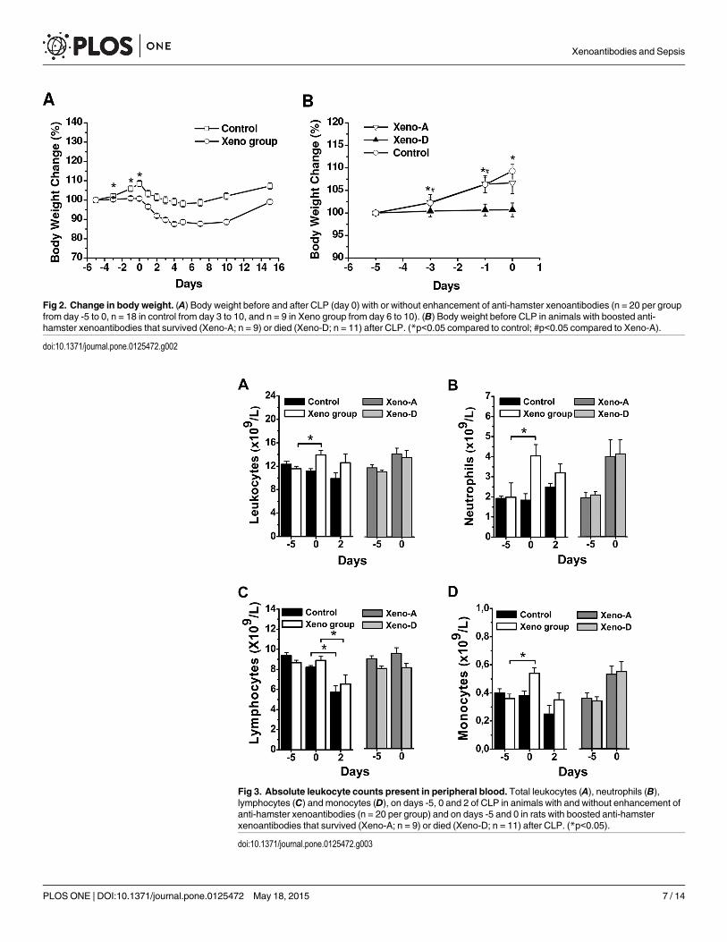
Fig 2. Change in body weight. (A) Body weight before and after CLP (day 0) with or without enhancement of anti-hamster xenoantibodies (n = 20 per groupfrom day -5 to 0, n = 18 in control from day 3 to 10, and n = 9 in Xeno group from day 6 to 10). (B) Body weight before CLP in animals with boosted anti-hamster xenoantibodies that survived (Xeno-A; n = 9) or died (Xeno-D; n = 11) after CLP. (*p<0.05 compared to control; #p<0.05 compared to Xeno-A).
doi:10.1371/journal.pone.0125472.g002
Fig 3. Absolute leukocyte counts present in peripheral blood. Total leukocytes (A), neutrophils (B),lymphocytes (C) and monocytes (D), on days -5, 0 and 2 of CLP in animals with and without enhancement ofanti-hamster xenoantibodies (n = 20 per group) and on days -5 and 0 in rats with boosted anti-hamsterxenoantibodies that survived (Xeno-A; n = 9) or died (Xeno-D; n = 11) after CLP. (*p<0.05).
doi:10.1371/journal.pone.0125472.g003
Xenoantibodies and Sepsis
PLOS ONE | DOI:10.1371/journal.pone.0125472 May 18, 2015 7 / 14

that survived or died after CLP, and in control animals. No change was observed for any cyto-kine in the control group at the time of CLP (day 0) compared to baseline (day -5) (Fig 5A).Xenoimmunization of animals that subsequently survived CLP produced at pre-CLP time sig-nificant decreases compared to baseline in ICAM-1, IL-1 R6, IL-4, IL-6, IL-10, LIX, MMP-8,and VEGF along with a significant augment of TIMP-1 (Fig 5B). In contrast, the boosting ofxenoantibodies in animals that died after CLP did not result in any reduction of cytokines atthe time of the procedure compared to day -5 and was associated with significant increases ofB7-2/CD86, β-NGF, leptin, L-selectin and TIMP-1 (Fig 5C).
Two days after CLP, control rats showed a significant decrease in IL-4, IL-6, IL-10, LIX, andVEGF, along with increases in agrin, ICAM-1 and TIMP-1, compared to day 0 (pre-CLP) (Fig5A). In animals with boosted xenoantibodies that survived CLP, there was a significant increaseat 48 h post-CLP in agrin, CINC-2α, ICAM-1 and TIMP-1 compared to baseline and pre-CLPsamples, but no major changes for the other cytokines (Fig 5B).
DiscussionThe present study confirmed that exposuring Lewis rats to hamster cells resulted in mainly ex-panded TI natural xenoantibodies, systemic alterations and impaired survival after CLP-
Fig 4. Clinical chemistry determinations in peripheral blood. ALT (A), AST (B), urea (C) and triglycerides(D), on days -5, 0 and 2 of CLP in animals with and without enhancement of anti-hamster xenoantibodies(n = 20 per group) and on days -5 and 0 in rats with boosted anti-hamster xenoantibodies that survived(Xeno-A; n = 9) or died (Xeno-D; n = 11) after CLP. (*p<0.05).
doi:10.1371/journal.pone.0125472.g004
Xenoantibodies and Sepsis
PLOS ONE | DOI:10.1371/journal.pone.0125472 May 18, 2015 8 / 14

Fig 5. Cytokine concentrations. Fold changes of 33 cytokines on days -5 (baseline), 0 (before CLP; blackcolumn) and 2 (two days after CLP; white column). (A) Control; (B) animals with increased anti-hamster
Xenoantibodies and Sepsis
PLOS ONE | DOI:10.1371/journal.pone.0125472 May 18, 2015 9 / 14

induced sepsis. The exacerbated mortality was not directly influenced by the amount ofxenoantibodies. Similar levels of IgM and IgG anti-hamster xenoantibodies at the pre-CLPtime point were observed both in xenoimmunized rats that survived and those dying after theprocedure. Likewise, the data did not show an association between increased mortality and aparticular effect of xenoantibodies on blood bacterial isolates. No blood bacterial loads wereobserved after this CLP model regardless of whether xenoantibodies were boosted or not. Thelack of blood bacteria isolates after CLP in our model indicates that the enhancement of anti-melibiose and L-rhamnose antibodies reactive to E. faecalis caused by the hamster blood expo-sure is not responsible for the observed increased mortality [11]. However, it does not rule outan impairment of sepsis if bacteremia is present and caused by microorganisms expressing car-bohydrate antigens targeted by natural antibodies, as has been suggested for αGal and poly-N-acetyl glucosamine antigens [9,15].
Xenoimmunization to boost mainly TI anti-hamster antibodies in rats produced a systemicinflammatory response at the pre-CLP time point, as evidenced by leukocytosis resulting froman increase in blood neutrophils and monocytes, along with increases in ALT and AST. Therewas also a lack of weight gain and a drop in triglyceride levels, though no animal died beforeCLP. Interestingly, the impairment of the nutritional status coinciding with the non-infectioussystemic inflammation affected mostly those rats that subsequently died after CLP. A processof peritonitis can be suspected in these animals. The malnutrition signs were even more pro-nounced after CLP in these animals, affecting also xenoimmunized animals that survived CLPand controls. CLP did not change blood leukocyte counts compared to pre-CLP besides thelymphopenia 48 h after the procedure, as was previously observed [16], both in xenoimmu-nized rats and controls. However, there was an increase in serum AST and a decrease in ALT.To our knowledge, this is the first time that a decrease in serum ALT levels has been reportedafter CLP, which in our study involved xenoimmunized animals and controls. Previous studiesdescribe a significant increase in ALT resulting from liver damage after CLP, particularly inmoderate-severe sepsis [17], which returns to baseline levels by day 4 in low-severity models.The decrease in ALT after CLP may reflect the impairment of the nutritional status of the ani-mals, as has been suggested in humans [18], though it was not observed initially in xenoimmu-nized rats with lack of weight gain.
Our investigations did not include the phenotype assessment of the diverse leukocytes in-volved in the inflammatory response of xenoimmunization and CLP, which certainly is a short-coming of the study. However, immune cells implement their tasks primarily through thesecretion of cytokines that mediate functions such as direct killing, self-renewal, recruitment ofother cells, and promotion or inhibition of inflammation. These cytokines may be produceddifferently by a particular cell and not always correlates with a specific cell phenotype [19].Therefore, we consider that a high-throughput multiplex cytokine assay could also characterizethe magnitude and quality of the immune response. In this study, CLP in rats without previousxenoimmunization led to minimal mortality and (on day two) the augmentation in blood ofagrin (soluble cell receptor), ICAM-1 (soluble cell receptor) and TIMP-1 (metalloproteinasewith pleiotropic activities). However, there was a predominant pattern of reduction of cyto-kines that included IL-4, IL-6, IL-10, LIX and VEGF (pro-inflammatory and anti-inflammatorycytokines, chemokines and growth factors). The impaired production of cytokines has beenpreviously reported with non-lethal CLP [20], which contrasts to the elevated cytokine levels
xenoantibodies that survived CLP (Xeno-A); (C) animals with boosted anti-hamster xenoantibodies that diedafter CLP (Xeno-D). Mean ± SEM of 3 animals per group. (*p<0.05 compared to day -5;#p<0.05 compared todays -5 and 0).
doi:10.1371/journal.pone.0125472.g005
Xenoantibodies and Sepsis
PLOS ONE | DOI:10.1371/journal.pone.0125472 May 18, 2015 10 / 14

observed in sepsis of increased mortality [16,21,22]. In these studies that assessed only particu-lar cytokines, mortality after CLP related to the augmentation of only proinflammatory cyto-kines such as IL-6 [16,23,24], or both pro- and anti-inflammatory cytokines [22,25]. However,when a cytokine profile using a multiplex cytokine measurement is employed as in our study,patterns including the simultaneous augment and reduction of different cytokines are detected[14,26,27], similar to what we observed in our analysis.
Xenoimmunized rats that later survived CLP showed a more clear profile of blood cytokinereduction at the pre-CLP time point, which included cytokines which also decreased after CLPin control animals along with others such as ICAM-1, IL-1R6 and MMP-8, being TIMP-1 theonly cytokine that was augmented. In these rats all the cytokine levels increased in response toCLP, displaying a pattern with no cytokine decreases. A similar cytokine profile and outcomewith secondary bacterial infections has been described after peritoneal lypopolysaccharide(LPS) challenge in animals previously exposed to peritoneal bacterial products or cytokinessuch as tumor necrosis factor (TNF) [20]. Unfortunately, no information was provided aboutthe impact of the latter procedures in the cytokine profile before LPS challenge. Suppression ofTLR and inflammatory responses has been described with IgM natural antibodies and in endo-toxin tolerance [28–30]. This condition is a phenomenon observed after exposure to low con-centrations of LPS that produce a transient unresponsive state to further challenges withendotoxin [31,32]. Thus, xenoantibodies and/or immune complexes generated by xenoimmu-nization may provide a predominant profile of negative regulation of TLR-triggered inflamma-tory responses, leading to a response similar to that of non-lethal CLP.
In contrast, no cytokine was reduced at the pre-CLP time point in xenoimmunized animalsthat died after CLP. Instead, there were increases in B7-2/CD86 (soluble cell receptor), β-NGF(growth factor, adipokine), leptin (adipokine), L-selectin (soluble cell receptor) and TIMP-1.Interestingly, the increase of cytokines in these rats included leptin and β-NGF that are identi-fied as adipokines since they are secreted by adipose tissue [33]. Leptin has been shown to be el-evated in early stages of inflammation and in the exacerbation of sepsis mortality [34,35].There is also evidence that leptin is produced by intraperitoneal adipocytes [36,37], and that in-creased concentrations of this and other adipokines have a very high sensitivity and specificityfor an early diagnosis of peritonitis in patients undergoing peritoneal dialysis [38]. In ourstudy, they may have contributed to the lack of weight gain and metabolic disorders observedin animals during xenoimmunization [39], and later mortality after CLP.
This study has failed to show a particular effect of boosted xenoantibodies in the increasedmortality of CLP. However, it indicates that the inflammatory profile before infectious injuriesis crucial to the response that will occur afterwards. This concept was already recognized in se-vere non-infectious situations, such as trauma or hemorrhage, along with the impact of theseconditions on the development of sepsis [40,41]. Likewise, it may be extended to non-criticalconditions or even, apparently, to normal circumstances. Using single cytokine analysis, higherpre-infection levels have been associated with an elevated risk of community-acquired pneu-monia requiring hospitalization and mortality in dialysis patients [11,42]. In the latter case, itwas also associated with an increase of natural IgM xenoantibodies. Also, the metabolic syn-drome is associated with increased levels of adipokines [43], which in our study showed a par-ticular involvement in increasing the severity of sepsis. Overall, our results suggest thatpreventive strategies acting on the inflammatory status of susceptible patients prior to infectionmay help averting the harmful consequences of sepsis
Xenoantibodies and Sepsis
PLOS ONE | DOI:10.1371/journal.pone.0125472 May 18, 2015 11 / 14

ConclusionsEnhancement of predominantly natural anti-hamster xenoantibodies in Lewis rats with axenoimmunization protocol was associated with increased mortality from low-grade sepsisafter CLP. The impairment of sepsis survival in rats could not be correlated with the level ofxenoantibodies or bacteremia. However, it was associated with a lack of weight gain duringxenoimmunization and a cytokine profile at the time of CLP characterized by increased levelsof cytokines, particularly adipokines such as leptin and β-NGF. This profile contrasted with thereduction in cytokine levels observed in xenoimmunized rats that gained weight and survivedCLP. Thus, the outcome of sepsis appears to depend more on the cytokine profile existing be-fore sepsis than that resulting from the condition, with a special harmful effect of adipokines.
Supporting InformationS1 Table. Scoring to evaluate rat body weight, general aspect, self-mutilation or signs ofpain and response to stimulus.(DOC)
S2 Table. Corrective measures applied to rats depending on final score.(DOC)
S3 Table. Cytokine class and effect included in array analysis (from http://copewithcytokines.de).(DOC)
Author ContributionsConceived and designed the experiments: CC RM. Performed the experiments: MPC CC. Ana-lyzed the data: MPC RM. Wrote the paper: MPC CC RM.
References1. Mond JJ, Lees A, Snapper CM (1995) T cell-independent antigens type 2. Annu Rev Immunol, 13:
655–692. PMID: 7612238
2. Ochsenbein AF, Fehr T, Lutz C, Suter M, Brombacher F, Hengartner H, et al. (1999) Control of earlyviral and bacterial distribution and disease by natural antibodies. Science, 286:2156–2159. PMID:10591647
3. Boes M, Prodeus AP, Schmidt T, Carroll MC, Chen J (1998) A critical role of natural immunoglobulin Min immediate defense against systemic bacterial infection. J Exp Med, 188: 2381–2386. PMID:9858525
4. Schwartz-Albiez R, Monteiro RC, Rodriguez M, Binder CJ, Shoenfeld Y (2009) Natural antibodies, in-travenous immunoglobulin and their role in autoimmunity, cancer and inflammation. Clin Exp Immunol,158 (Suppl 1): 43–50. doi: 10.1111/j.1365-2249.2009.04026.x PMID: 19883423
5. Kaveri SV, Silverman GJ, Bayry J (2012) Natural IgM in immune equilibrium and harnessing their thera-peutic potential. J Immunol, 188: 939–945. doi: 10.4049/jimmunol.1102107 PMID: 22262757
6. Cramer DV (2000) Natural antibodies and the host immune responses to xenografts. Xenotransplanta-tion, 7: 83–92. PMID: 10961291
7. Galili U, Anaraki F, Thall A, Hill-Black C, Radic M (1993) One percent of human circulating B lympho-cytes are capable of producing the natural anti-Gal antibody. Blood, 82: 2485–2493. PMID: 7691263
8. Galili U, Mandrell RE, Hamadeh RM, Shohet SB, Griffiss JM (1988) Interaction between human naturalanti-alpha-galactosyl immunoglobulin G and bacteria of the human flora. Infect Immun 56: 1730–1737.PMID: 3290105
9. Hamadeh RM, Jarvis GA, Galili U, Mandrell RE, Zhou P, Griffiss JM (1992) Human natural anti-Gal IgGregulates alternative complement pathway activation on bacterial surfaces. J Clin Invest, 89: 1223–1235. PMID: 1556184
Xenoantibodies and Sepsis
PLOS ONE | DOI:10.1371/journal.pone.0125472 May 18, 2015 12 / 14

10. Mañez R, Blanco FJ, Diaz I, Centeno A, Lopez-Pelaez E, Hermida M, et al. (2001) Removal of bowelaerobic gram-negative bacteria is more effective than immunosuppression with cyclophosphamide andsteroids to decrease natural α-galactosyl IgG antibodies. Xenotrasplantation, 8: 15–23. PMID:11208187
11. Pérez-Fontan M, Máñez R, Rodríguez-Carmona A, Peteiro J, Martínez V, García-Falcón T, et al.(2006) Serum levels of anti-αgalactosyl antibodies predict survival and peritoneal dialysis-related enter-ic peritonitis rates in patients undergoing renal replacement therapy. Am J Kidney Dis, 48: 972–982.PMID: 17162152
12. Perez-Cruz M, Costa C, Mañez R (2014) Boosted Rat Natural Xenoantibodies Cross-React with En-terococcus faecalis by Targeting Melibiose and L-Rhamnose. J Innate Immun, 6: 140–151. doi: 10.1159/000355305 PMID: 24246417
13. Rittirsch D, Huber-Lang MS, Flierl MA, Ward PA (2009) Immunodesign of experimental sepsis by cecalligation and puncture. Nat Protoc, 4: 31–36. doi: 10.1038/nprot.2008.214 PMID: 19131954
14. Lvovschi V, Arnaud L, Parizot C, Freund Y, Juillien G, Ghillani-Dalbin P, et al. (2011) Cytokine profilesin sepsis have limited relevance for stratifying patients in the emergency department: a prospective ob-servational study. PLoS One, 6: e28870. doi: 10.1371/journal.pone.0028870 PMID: 22220196
15. Skurnik D, Kropec A, Roux D, Theilacker C, Huebner J, Pier GB (2012) Natural antibodies in normalhuman serum inhibit Staphylococcus aureus capsular polysaccharide vaccine efficacy. Clin Infect Dis,55: 1188–1197. doi: 10.1093/cid/cis624 PMID: 22806596
16. Xiao H, Siddiqui J, Remick DG (2006) Mechanisms of mortality in early and late sepsis. Infect Immun,74: 5227–5235. PMID: 16926416
17. Deng M, Scott MJ, Loughran P, Gibson G, Sodhi C, Watkins S, et al. (2013) Lipopolysaccharide clear-ance, bacterial clearance, and systemic inflammatory responses are regulated by cell type-specificfunctions of TLR4 during sepsis. J Immunol, 190: 5152–5160. doi: 10.4049/jimmunol.1300496 PMID:23562812
18. Elinav E, Ackerman Z, Maaravi Y, Ben-Dov IZ, Ein-Mor E, Stessman J (2006) Low alanine aminotrans-ferase activity in older people is associated with greater long-termmortality. J Am Geriatr Soc, 54:1719–1724. PMID: 17087699
19. Yamanaka YJ, Gierahn TM, Love JC (2013) The dynamic lives of T cells: new approaches and themes.Trends Immunol 34: 59–66. doi: 10.1016/j.it.2012.10.006 PMID: 23200626
20. Sterns T, Pollack N, Echtenacher B, Männel DN (2005) Divergence protection induced by bacterialproducts and sepsis-induced immune suppression. Infect Immun, 73: 4905–4912. PMID: 16041004
21. Ebong SJ, Call DR, Bolgos G, Newcomb DE, Granger JI, O'Reilly M, et al. (1999) Immunopathologic re-sponses to non-lethal sepsis. Shock, 12: 118–126. PMID: 10446892
22. Ebong S, Call D, Nemzek J, Bolgos G, Newcomb D, Remick D (1999) Immunopathologic alterations inmurine models of sepsis of increasing severity. Infect Immun, 67: 6603–6610. PMID: 10569781
23. Remick DG, Bolgos G, Copeland S, Siddiqui J (2005) Role of interleukin-6 in mortality from and physio-logic response to sepsis. Infect Immun, 73: 2751–2757. PMID: 15845478
24. Osuchowski MF, Connett J, Welch K, Granger J, Remick DG (2009) Stratification is the key: inflamma-tory biomarkers accurately direct immunomodulatory therapy in experimental sepsis. Crit Care Med,37: 1567–1573. doi: 10.1097/CCM.0b013e31819df06b PMID: 19325479
25. Osuchowski MF, Welch K, Siddiqui J, Remick DG (2006) Circulating cytokine/inhibitor profiles reshapethe understanding of the SIRS/CARS continuum in sepsis and predict mortality. J Immunol, 177:1967–1974. PMID: 16849510
26. Cui X, Li Y, Xuemei L, Haley M, Moayeri M, Fitz Y, et al.(2006) Sublethal doses of Bacillus anthracisletal toxin inhibit inflammation with lypopolysaccharide and Escherichia coli challenge but have oppo-site effects on survival. J Infect Dis, 193: 829–840. PMID: 16479518
27. Fjell CD, Thair S, Hsu J, Walley KR, Rusell JA, Boyd J (2013) Cytokines and signalling molecules pre-dict clinical outcomes in sepsis. PLoS One, 8: e79207. doi: 10.1371/journal.pone.0079207 PMID:24244449
28. Chen Y, Khanna S, Goodyear CS, Park YB, Raz E, Thiel S, et al. (2009) Regulation of dendritic cellsand macrophages by anti-apoptotic cell natural antibody that suppresses TLR responses and inhibitsinflammatory arthritis. J Immunol, 183: 1346–1359. doi: 10.4049/jimmunol.0900948 PMID: 19564341
29. Draisma A, Pickkers P, BouwMP, van der Hoeven JG (2009) Development of endotoxin tolerance inhumans in vivo. Crit Care Med, 37: 1261–1267. doi: 10.1097/CCM.0b013e31819c3c67 PMID:19242351
30. Biswas SK, Lopez-Collado E (2009) Endotoxin tolerance: new mechanisms, molecules and clinical sig-nificance. Trends Immunol, 30: 475–487. doi: 10.1016/j.it.2009.07.009 PMID: 19781994
Xenoantibodies and Sepsis
PLOS ONE | DOI:10.1371/journal.pone.0125472 May 18, 2015 13 / 14

31. Takeda K, Kaisho T, Akira S (2003) Toll-like receptors. Annu Rev Immunol, 21: 335–376. PMID:12524386
32. Zhang Y, Liu S, Liu J, Zhang T, Shen Q, Yu Y, et al. (2009) Immune complex/Ig negatively regulateTLR4-triggered inflammatory response in macrophages through Fc gamma RIIb-dependent PGE2 pro-duction. J Immunol, 182: 554–562. PMID: 19109188
33. Hrischev P, Nikolov P, Atanassova P, Nikolova J, Orbetzova M, Penkova N (2012) Leptin, leptin recep-tors, nerve growth factor and TrkA in women with metabolic syndrome. J BioSci Biotech, SE/ONLINE:63–66.
34. Shapiro NI, Khankin EV, Van Meurs M, Shih SC, Lu S, Yano M, et al. (2010) Leptin exacerbates sep-sis-mediated morbidity and mortality. J Immunol, 185: 517–524. doi: 10.4049/jimmunol.0903975PMID: 20519646
35. Barbier M, Cherbut C, Aubé AC, Blottière HM, Galmiche JP (1998) Elevated plasma leptin concentra-tions in early stages of experimental intestinal inflammation in rats. Gut, 43: 783–790. PMID: 9824605
36. Heimburger O, Wang T, Lonnqvist F, Stenvinkel P (1999) Peritoneal clearance of leptin in CAPD pa-tients: impact of local insulin administration. Nephrol Dial Transplant, 14: 723–727. PMID: 10193827
37. Krempler F, Breban D, Oberkofler H, Esterbauer H, Hell E, Paulweber B, et al. (2000) Leptin, peroxi-some proliferator-activated receptor-γ, and CCAAT/enhancer binding protein-αmRNA expression inadipose tissue of humans and their relation to cardiovascular risk factors. Arterioscler Thromb VascBiol, 20: 443–449. PMID: 10669642
38. Kolonko A, Chudek J, Wiecek A (2008) Concentration of adiopokines in peritoneal effluent: a newmark-er of acute peritonitis in peritoneal dialysis patients? Perit Dial Int, 28: 527–532. PMID: 18708547
39. Friedman JM, Halaas JL (1998) Leptin and the regulation of body weight in mammals. Nature, 395:763–770. PMID: 9796811
40. Saadia R, Schein M (1999) Multiple organ failure. How valid is the "two hit" model? J Accid Emerg Med,16: 163–166. PMID: 10353038
41. Cai B, Deitch EA, Ulloa L (2010) Novel insights for systemic inflammation in sepsis and hemorrhage.Mediators Inflamm, 2010: 642462. doi: 10.1155/2010/642462 PMID: 20628562
42. Yende S, Tuomanen EI, Wunderink R, Kanaya A, Newman AB, Harris T, et al. (2005) Preinfection sys-temic inflammatory markers and risk of hospitalization due to pneumonia. Am J Respir Crit Care Med,172: 1440–1446. PMID: 16166617
43. Yu D, Yu Z, Sun Q, Sun L, Li H, Song J, et al. (2011) Effects of body fat on the associations of high-mo-lecular-weight adiponectin, leptin and soluble leptin receptor with metabolic syndrome in Chinese.PLoS One, 6: e16818. doi: 10.1371/journal.pone.0016818 PMID: 21347230
Xenoantibodies and Sepsis
PLOS ONE | DOI:10.1371/journal.pone.0125472 May 18, 2015 14 / 14