researcharticle …amphibia,urodela)fromthelate jurassicofqinglong,hebeiprovince,china.plos...
TRANSCRIPT
RESEARCH ARTICLE
A New Basal Salamandroid (Amphibia,Urodela) from the Late Jurassic of Qinglong,Hebei Province, ChinaJia Jia, Ke-Qin Gao*
School of Earth and Space Sciences, Peking University, 5 Yiheyuan Road, Beijing, 100871, China
AbstractA new salamandroid salamander, Qinglongtriton gangouensis (gen. et sp. nov.), is named
and described based on 46 fossil specimens of juveniles and adults collected from the
Upper Jurassic (Oxfordian) Tiaojishan Formation cropping out in Hebei Province, China.
The new salamander displays several ontogenetically and taxonomically significant fea-
tures, most prominently the presence of a toothed palatine, toothed coronoid, and a unique
pattern of the hyobranchium in adults. Comparative study of the new salamander with previ-
ously known fossil and extant salamandroids sheds new light on the early evolution of the
Salamandroidea, the most species-diverse clade in the Urodela. Cladistic analysis places
the new salamander as the sister taxon to Beiyanerpeton, and the two taxa together form
the basalmost clade within the Salamandroidea. Along with recently reported Beiyanerpe-ton from the same geological formation in the neighboring Liaoning Province, the discovery
ofQinglongtriton indicates that morphological disparity had been underway for the sala-
mandroid clade by early Late Jurassic (Oxfordian) time.
IntroductionSalamandroidea [1] are the most species-diverse group of salamanders (Caudata, Urodela),including approximately 610 extant species in seven families [2, 3]. Whether Sirenidae (sirensand their closely related fossil taxa) should be classified in Salamandroidea is a matter ofdebate, as recent phylogenetic results based on molecular, morphological, or combined dataincompatibly place the family as the basalmost clade of Urodela [4, 5], sister clade with the Sal-amandroidea [6], or nested within Salamandroidea [7–13]; see [14] for a review). The membersof Salamandroidea have a worldwide distribution except for Antarctica, Ethiopian realms, andthe Oceanian regions of the world [15].
The suborder Salamandroidea differs from its sister clade Cryptobranchoidea [16] primarilyby fusion of the angular bone to the prearticular in the lower jaw, bicapitate trunk ribs, andinternal fertilization. The evolutionary history of the salamandroids can be traced back to theMesozoic, and recent fossil discoveries from China have provided critical evidence indicatingthat the split of Salamandroidea from Cryptobranchoidea had taken place no later than
PLOSONE | DOI:10.1371/journal.pone.0153834 May 4, 2016 1 / 35
a11111
OPEN ACCESS
Citation: Jia J, Gao K-Q (2016) A New BasalSalamandroid (Amphibia, Urodela) from the LateJurassic of Qinglong, Hebei Province, China. PLoSONE 11(5): e0153834. doi:10.1371/journal.pone.0153834
Editor:William Oki Wong, Institute of Botany, CHINA
Received: December 11, 2015
Accepted: April 2, 2016
Published: May 4, 2016
Copyright: © 2016 Jia, Gao. This is an open accessarticle distributed under the terms of the CreativeCommons Attribution License, which permitsunrestricted use, distribution, and reproduction in anymedium, provided the original author and source arecredited.
Data Availability Statement: All relevant data arewithin the paper and its Supporting Information files.
Funding: The research was funded by grants fromthe National Natural Science Foundation of China(NSFC 41072007, 41272016). Jia’s research wasalso supported by the China Scholarship Council.The funders had no role in study design, datacollection and analysis, decision to publish, orpreparation of the manuscript.
Competing Interests: The authors have declaredthat no competing interests exist.
Abbreviations: CIB, Chengdu Institute of Biology,Chinese Academy of Sciences, Chengdu, China;
Oxfordian time [8]. The earliest fossil record known for Salamandroidea is documented byspecimens of Beiyanerpeton jianpingensis from western Liaoning Province [8], from fossil bedsdated to the Late Jurassic at about 157 Ma [17]. The split of the two major salamander clades asa critical cladogenetic event apparently had a profound impact on shaping the evolutionarytree of salamanders as one of the three major groups of modern amphibians [18].
The purposes of this paper are to describe a new salamandroid based on fossils from UpperJurassic beds cropping out in the northeastern part of Hebei Province, northern China; to dis-cuss the evolution of several key characters within the salamandroid clade and to assess thephylogenetic relationships of the new salamander with other salamandroids. A total of 46 spec-imens used in this study were collected in the summer of 2009 from a locality near the smallvillage of Nanshimenzi, close to the provincial border with Liaoning (Fig 1). All 46 specimensare referable to the same genus and species, despite differences in body size and palatal mor-phology, which we interpret as ontogenetic variation. All of the specimens were collected fromthe same horizon of grayish-yellowish silty shales, and all have the cranial and postcranial skel-etons preserved in articulation without post-mortem decomposition of the bones or disruptionof them by fluvial transportation. In addition, most specimens retain evidence of soft-anatomi-cal structures, including mineralized denticles (gill rakers) of the internal gills, impressions ofthe eyes and external gill filaments, and body outline. The fossil beds cropping out at the Nan-shimenzi locality pertain to the Upper Jurassic (Oxfordian) Tiaojishan Formation, from whicha fairly diverse vertebrate fossil fauna and a species-rich fossil flora have been revealed byrecent discoveries (see Geological Setting below).
The Mesozoic fossil record of Salamandroidea is scanty, in contrast to the relatively abun-dant fossils of the group known from the Cenozoic. To date, the recently published Beiyanerpe-ton from China represents the only salamandroid known from the Jurassic Period [8]. TheLate Jurassic Iridotriton from the Morrison Formation of North America was originally
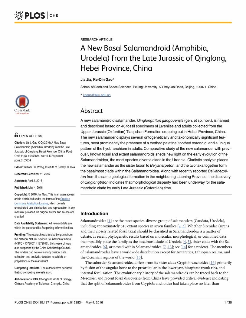
Fig 1. Map showing geographic location of the Nanshimenzi fossil site in relation to other Jurassic salamander fossil localities in Hebei, Liaoning,and Inner Mongolia. Stratigraphic position of the fossil beds at the Nanshimenzi site is denoted by the salamander figured on the left column.
doi:10.1371/journal.pone.0153834.g001
Late Jurassic Salamandroid from China
PLOS ONE | DOI:10.1371/journal.pone.0153834 May 4, 2016 2 / 35
PKUP, Peking University Paleontological Collections,Beijing, China; UALVP, University of Alberta,Laboratory for Vertebrate Paleontology, Edmonton,Canada; adf, anterodorsal fenestra; amf,anteromedial fenestra; anf, angular foramen; at,atlas; bb, basibranchial; co, coronoid; d, dentary;exo, exoccipital; fe, femur; fi, fibula; fr, frontal; gr, gillraker; hb, hypobranchial; hu, humerus; icaf, internalcarotid foramen; il, ilium; isc, ischium; lac, lacrimal;m, maxilla; na, nasal; opi, opisthotic; pa, parietal;pal, palatine; pm, premaxilla; pra, prearticular; praf,prearticular foramen; prf, prefrontal; pro, prootic; ps,parasphenoid; pt, pterygoid; qu, quadrate; ra, radius;sca, scapulocoracoid; sm, septomaxilla; sq,squamosal; st, stapes; ti, tibia; ul, ulna; vo, vomer.
described as a putative salamandroid [19], but subsequently shown in a cladistic analysis toinstead be a cryptobranchoid [8]. Hence, the new taxon described herein adds significantly tothe Jurassic record, documenting a second salamandroid besides Beiyanerpeton as recentlyreported from the same geologic formation, but farther to the north in China. Compared tolimited discoveries from the Late Jurassic, salamandroids have an improved fossil record inCretaceous. Besides the amphiumid Proamphiuma in North America [20] and Valdotriton inEurope [21], there are a number of taxa that are viewed as possible salamandroids in NorthAmerica and Europe. These are classified in Batrachosauroididae, Scapherpetontidae, andsome are classified in extant families of Proteidae and Salamandridae (see [22, 23] for review).However, most of these are based on isolated vertebrae or jaws, while articulated fossils arerare. The discovery of the exquisitely preserved specimens in this collection enables a study ofthe detailed morphology of the new salamandroid, and a discussion on the evolution of severalkey characters within the salamandroid clade as presented below in the text.
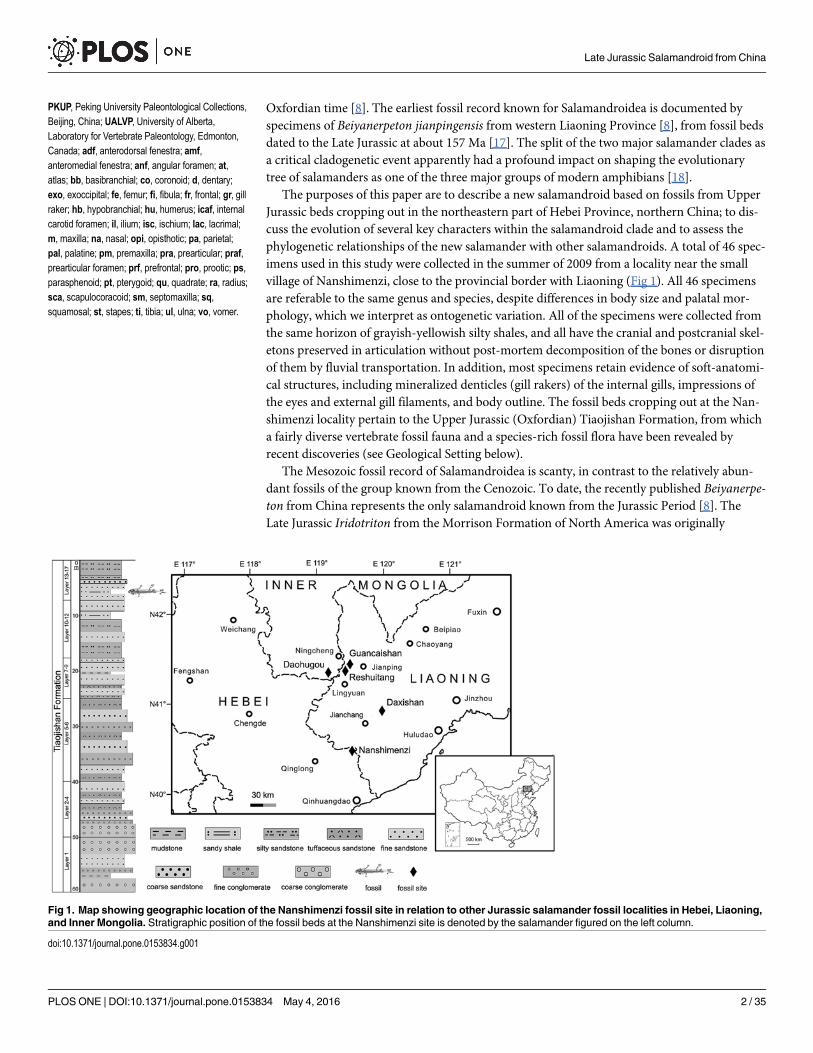
Geologic Setting and Associated Vertebrate AssemblageThe new salamander fossil locality (N40°31'52"/E119°29'11") lies on a hill (Fig 2) to the northof the small village of Nanshimenzi, Qinglong Manchu Autonomous County, QinhuangdaoCity, Hebei Province. The salamander fossil-bearing beds cropping out at the locality pertainto the Upper Jurassic Tiaojishan Formation; the stratigraphic equivalents of this unit in Hebeiand Liaoning provinces have been termed the Lanqi Formation in some literature. The Tiao-jishan (Lanqi) Formation is composed of a set of grayish purple andesite, andesite agglomeratelavas, and grayish black basalts, all intercalated with tuffaceous sandstones and ignimbrites[24]. With the thickness of the rock sequence ranging from 130 m to over 900 m, deposits ofthe Tiaojishan Formation crop out widely in the Western Hills of Beijing, Liaoning and Hebeiprovinces.
The Tiaojishan Formation has yielded high-precision 40Ar/39Ar dates of 161.8 ± 0.4 Manear the base and 159.5 ± 0.6 Ma near the top of the formation [25, 26]. Other published radio-metric dates of the formation cover a wide range, from 152 Ma to 167 Ma [17, 27–32].
Fig 2. Type locality outside the Nanshimenzi Village, Qinglong Manchu Autonomous County, Qinhuangdao City, Hebei Province. Arrow pointing tothe fossil beds as cropping out at the type locality.
doi:10.1371/journal.pone.0153834.g002
Late Jurassic Salamandroid from China
PLOS ONE | DOI:10.1371/journal.pone.0153834 May 4, 2016 3 / 35

Published dates compatible with those of high-precision 40Ar/39Ar dates obtained by Changet al. [25, 26] include a 40Ar/39Ar date of 161 ± 1 Ma for the lower part of the formation at Xia-bancheng, Hebei Province [27], and SIMS U-Pb Zircon dates of 158.7 ± 1.8 Ma–160.7 ± 1.7Ma for the Daxishan section, Jianchang Basin, western Liaoning Province [32]. These conflict-ing results were probably affected by specimen sampling (e.g., whole rock, biotite, feldspar andzircon), use of different dating methods (40Ar/39Ar, U-Pb SHRIMP, U-Pb LA-ICP-MS), andcalculation differences [33].
No radiometric dates are directly available from the fossil beds at Nanshimenzi village. Therelative age of the rock sequence has been determined by stratigraphic correlation with theDaxishan section in the Jianchang Basin [34], some 70 km northeast of Nanshimenzi village.As the Daxishan section has been recently dated at 158–160 Ma [32], the fossil beds at Nanshi-menzi village can be reasonably hypothesized as falling within that time interval. In addition tothe salamander fossils described in this paper, the Upper Jurassic rock sequence exposed atNanshimenzi village and its vicinity has recently yielded other important vertebrate fossils,including several pterosaurs [35, 36], a non-avian dinosaur [37], and two mammals [38, 39].
In the past few years, important vertebrate fossil discoveries have been made at several otherlocalities in the Tiaojishan Formation of western Liaoning Province. These include: salamanderfossils from the Reshuitang and Guancaishan localities, near Lingyuan and Jianping, respec-tively [8, 40, 41]; and paravian dinosaur, pterosaur, and mammal fossils from the Daxishanlocality near Jianchang [42–46]. Along with those from the geologically older Haifanggou (Jiu-longshan) Formation (see below), fossils from the Tiaojishan Formation are components of theYanliao Biota that immediately predates the world-famed Jehol Biota [47].
The Tiaojishan (Lanqi) Formation is disconformably underlain by the Middle Jurassic Hai-fanggou Formation; correlative beds of the latter in the Western Hills of Beijing are known asthe Jiulongshan Formation. The Haifanggou (Jiulongshan) Formation is mainly composed ofyellowish green sandstones and shales with tuffs, varying in thickness from 118 m to 533 m[24]. The Haifanggou Formation as exposed at Daohugou village, Inner Mongolia, has yieldedsignificant vertebrate, invertebrate, and plant fossils that are important components of the Yan-liao Biota (= Daohugou Biota in Sullivan et al. [48]). It must be stressed that essentially all thefossils described in the literature so far (e.g., the mammalMagaconus Zhou et al. [49]) from theDaohugou beds (sensu stricto) are from the Haifanggou Formation, where published radiomet-ric dates for the “Daohugou beds” at the Daohugou section [17, 25, 28, 30, 50] are derived fromthe overlying Tiaojishan Formation. Dating of the Haifanggou (Jiulongshan) Formation is onlyavailable by analysis of rock samples from the Chengde area in Hebei Province ([51]: U-Pb163.4 ± 1.1 Ma) and from the Beipiao Basin in Liaoning Province ([26]: 40Ar/39Ar 166.7 ± 1Ma).
Materials and MethodsAll specimens of this study are deposited in the Peking University Paleontological Collections(PKUP), Beijing, China, and are accessible by qualified professionals. No permits were requiredfor the described study. PKUP V0226 is designated as the holotype because it is a large speci-men with well-preserved cranial and postcranial skeleton, and it shows diagnostic features inthe palate. Specimens were manually prepared using fine needles under a Leica MZ 16 micro-scope. Selected specimens (PKUP V0226, V0228, V0254) were scanned at China University ofGeosciences (Beijing) using a High-Resolution X-ray CT scanner (Nikon XT H 320 LC). Scan-ning of specimens was performed using no filter, at voltage of 180 kV and a current of 105 uAfor PKUP V0226, 170 kV and 63 uA for PKUP V0228, 170 kV and 47 uA for PKUP V0254.Voxel size (mm) is 0.066624×0.066624 for PKUP V0226, 0.118609×0.118609 for PKUP
Late Jurassic Salamandroid from China
PLOS ONE | DOI:10.1371/journal.pone.0153834 May 4, 2016 4 / 35

V0228, 0.078569×0.078569 for PKUP V0254. These specimens were scanned along the coronalaxis for a total of 1998 slices at an image resolution of 2000×2000. Segmentation and 3D recon-struction of the specimens are made by using VG Studio Max 2.2 (Volume Graphics, Heidel-berg, Germany). Line drawings of anatomical structures were prepared using AdobePhotoshop (CS4).
All measurements were obtained from manually held calipers. Total length (TL) refers tothe distance between the tip of the snout and the posterior extremity of the last caudal vertebra;snout-pelvic length (SPL) is the measurement from the snout tip to the posterior end of the pel-vis; skull length (SKL) is the maximum dimension from snout tip to the posterior extremity ofthe occipital condyles; skull width (SKW) is the greatest distance between cranio-mandibularjoints. The mean of three independent measurements of each parameter was taken to minimizeerrors. As most of the specimens have part of the tail missing, TL is obtainable from few speci-mens (PKUP V0233, V0236, V0263, V0266). The SPL from 27 measurable specimens rangesfrom 45.53 mm to 155.71 mm, with PKUP V0226 (holotype) the largest, and PKUP V0261 thesmallest. The remaining 19 specimens have the snout or pelvis partly missing, making the SPLunobtainable. Differences in SPL or SKL in combination with extent of ossification of skull ele-ments were used to estimate the relative age of individual specimens. In addition, ossificationof the septomaxilla, presence of posterolateral processes of basibranchial II and extensive ossifi-cation of epiphysis in long bones were used as criteria to distinguish adults from juveniles.
We follow Milner [52] in using the term ‘Caudata’ for total-group, and the term ‘Urodela’for crown-group, salamanders. However, we accept the commonly used classification of Cryp-tobranchoidea and Salamandroidea as two suborders within Urodela, regardless of the contro-versial position of Sirenidae as a basal urodele clade or an ingroup clade of Salamandroidea.Anatomical descriptions were mainly based on observations from the holotype, but were alsosupplemented with information from referred specimens.
Nomenclatural ActsThe electronic edition of this article conforms to the requirements of the amended Interna-tional Code of Zoological Nomenclature, and hence the new names contained herein are avail-able under that Code from the electronic edition of this article. This published work and thenomenclatural acts it contains have been registered in ZooBank, the online registration systemfor the ICZN. The ZooBank LSIDs (Life Science Identifiers) can be resolved and the associatedinformation viewed through any standard web browser by appending the LSID to the prefix“http://zoobank.org/”. The LSID for this publication is: urn:lsid:zoobank.org:pub:582E2169-76E0-4592-8CEB-94AF6D5C3353. The electronic edition of this work was published in a jour-nal with an ISSN, and has been archived and is available from the following digital repositories:PubMed Central (http://www.ncbi.nlm.nih.gov/pmc), LOCKSS (http://www.lockss.org).
Systematic PaleontologyClass Amphibia Linnaeus, 1758Subclass Lissamphibia Haeckel, 1866Superorder Caudata Scopoli, 1777Order Urodela Duméril, 1806Suborder Salamandroidea Dunn, 1922Family Incertae Sedis
GenusQinglongtriton gen. nov.urn:lsid:zoobank.org:act:DE89F7C6-1870-4B3E-A4BD-8C2BB208D193
Late Jurassic Salamandroid from China
PLOS ONE | DOI:10.1371/journal.pone.0153834 May 4, 2016 5 / 35

Qinglongtriton gangouensis gen. et sp. nov.urn:lsid:zoobank.org:act:B8CFC9E4-051C-4DD9-8C9F-0EC6AF0C7C0A
(Figs 3–9)
Fig 3. Holotype ofQinglongtriton gangouensis (PKUP V0226). Photograph (left) and line drawing (right) of incomplete skeleton in ventral view.
doi:10.1371/journal.pone.0153834.g003
Late Jurassic Salamandroid from China
PLOS ONE | DOI:10.1371/journal.pone.0153834 May 4, 2016 6 / 35
Holotype. PKUP V0226, an incomplete skeleton including articulated cranium and post-cranium, with part of the tail missing (Figs 3, 5A, 6A, 7C and 7D; S1 Fig; S1 and S3 Movies).
Diagnosis. A basal salamandroid sharing with Beiyanerpeton derived features including:palatine present as discrete and dentate element; sensory groove present on external surface ofpremaxilla and maxilla; lateral surface of dentary deeply grooved; presacral vertebrae 15 innumber. Differing from closely related Beiyanerpeton in having: lacrimal dorsally grooved fornasolacrimal duct; anterior ramus of pterygoid bearing teeth and directing anteromedially;ossification of orbitosphenoid absent; free operculum lacking; coronoid present as a dentateelement; marginal teeth pedicellate; metacarpal II longest in manus; phalangeal formula 2-2-3-
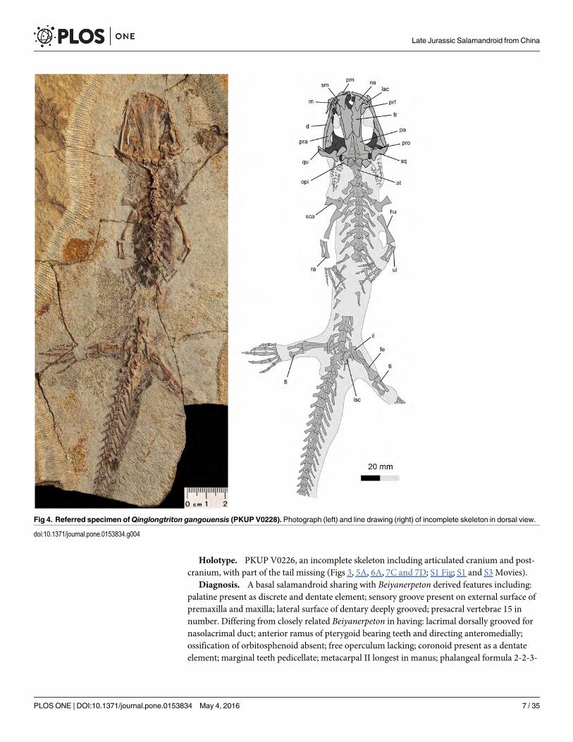
Fig 4. Referred specimen ofQinglongtriton gangouensis (PKUP V0228). Photograph (left) and line drawing (right) of incomplete skeleton in dorsal view.
doi:10.1371/journal.pone.0153834.g004
Late Jurassic Salamandroid from China
PLOS ONE | DOI:10.1371/journal.pone.0153834 May 4, 2016 7 / 35
3-3 in pes. The new taxon has the basibranchial II ossified with paired anterolateral and pos-terolateral processes fused with a median rod as a unique feature among salamanders.
Etymology. “Qinglong” refers to the Qinglong Manchu Autonomous County; “triton”,suffix commonly used for salamander names; “gangou” refers to Gangou Township, in whichthe fossil locality occurs.
Referred Specimens. PKUP V0227–V0271, all specimens from the same locality and the13th layer of the stratigraphic section as the holotype.
Type Locality and Horizon. Fossil locality approximately 300 m northwest of Nanshi-menzi village (N40°31'52"/E119°29'11"), Gangou Township, Qinglong Manchu AutonomousCounty, Qinhuangdao City, Hebei Province, China; the 13th layer of the stratigraphic sectionmeasured as outlined in Fig 1; Upper Jurassic (Oxfordian) Tiaojishan Formation.
Anatomical Description
General FeaturesIn all specimens, the snout is short and broadly rounded, although minor variations in blunt-ness can be seen in some specimens (Figs 3–6). Among the specimens without significant
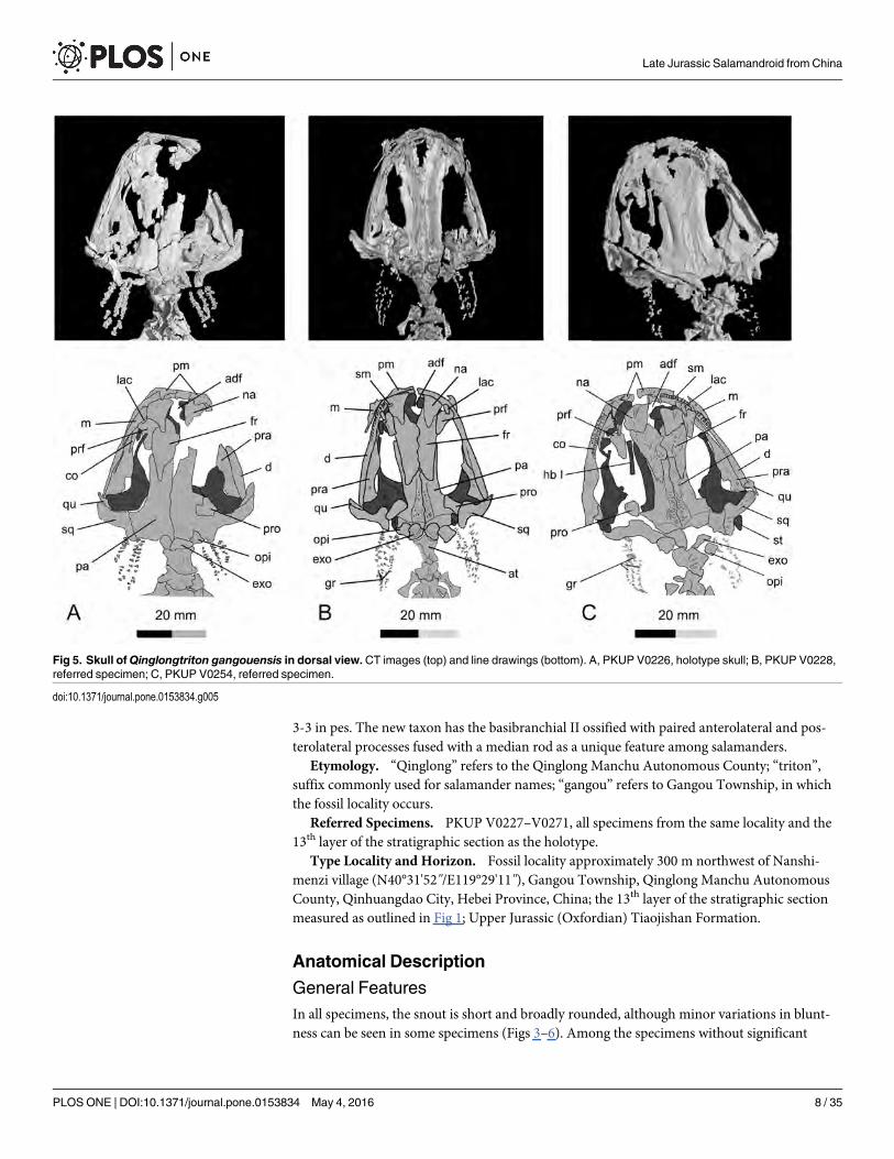
Fig 5. Skull ofQinglongtriton gangouensis in dorsal view.CT images (top) and line drawings (bottom). A, PKUP V0226, holotype skull; B, PKUP V0228,referred specimen; C, PKUP V0254, referred specimen.
doi:10.1371/journal.pone.0153834.g005
Late Jurassic Salamandroid from China
PLOS ONE | DOI:10.1371/journal.pone.0153834 May 4, 2016 8 / 35
distortion, the skull is slightly longer than wide (PKUP V0226–V0229, V0233, V0235, V0237,V0238, V0240, V0242, V0253, V0254, V0256–V0258). In several other specimens, the skull isslightly wider than long, but only on account of crushing (e.g., PKUP V0230, V0231, V0236,V0241, V0245, V0250, V0251).
The new salamander is a neotenic form as clearly indicated by the presence of gill structuresin the adult stage (Figs 3 and 4; S2 and S3 Figs; S1 and S2 Movies). Posterolaterally on eitherside of the skull the gill rakers of the internal gills are preserved as three rows of mineralizeddenticles, whereas the three external gill filaments are preserved as impressions. In addition,several other features in adult individuals are indicative of the neotenic nature of the new sala-mander (see Discussion below): a toothed palatine remains as a discrete element; the pterygoiddisplays a larval configuration, having a slender, toothed anterior process curving anterome-dially; the coronoid is retained as a discrete and toothed element in the lower jaw; the atlasbears a pair of transverse processes; and all of the mesopodial elements are unossified.
Compared to metamorphosed salamanders, neotenic forms tend to have a large body size[53]. This is also the case with Qinglongtriton gangouensis, the holotype of which (PKUPV0226) has a snout-pelvic length of 155.71 mm, and a total length of ~270 mm. All other largespecimens have a snout-pelvic length exceeding 100 mm. The maturity of these individuals is
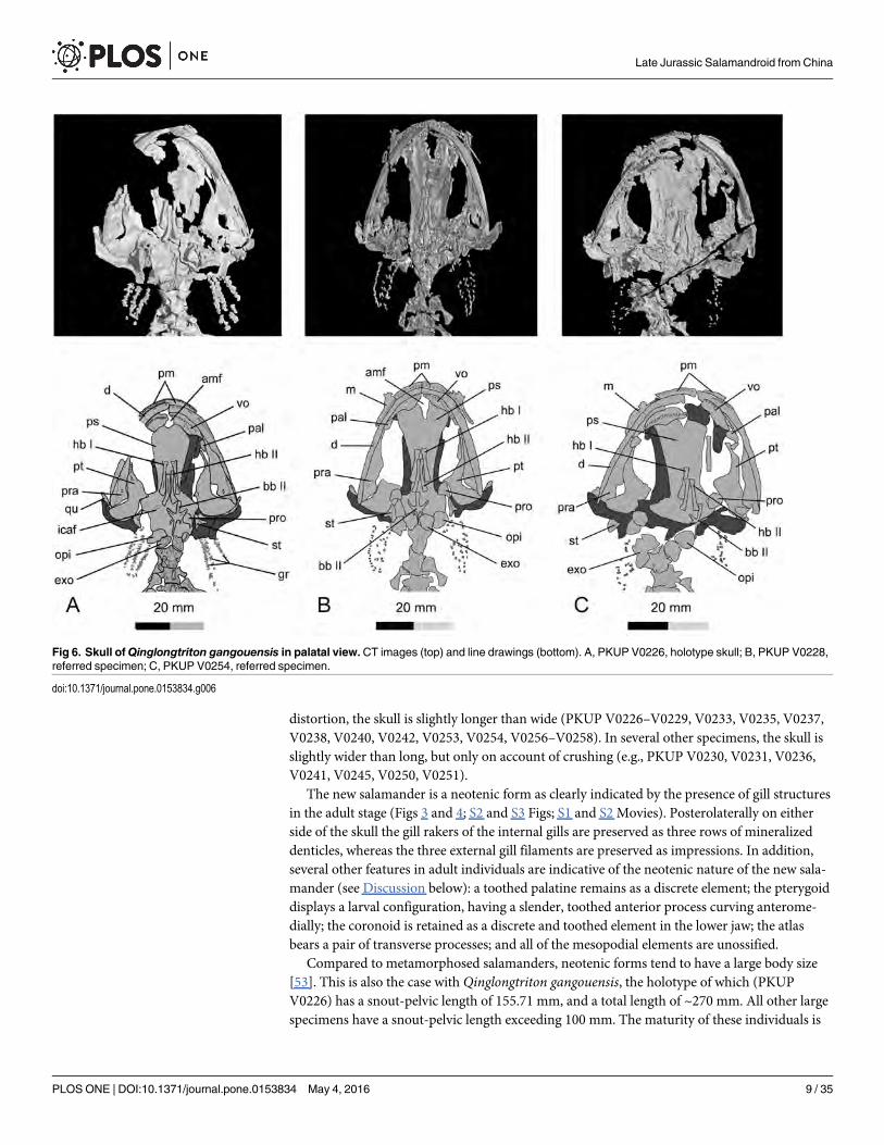
Fig 6. Skull ofQinglongtriton gangouensis in palatal view.CT images (top) and line drawings (bottom). A, PKUP V0226, holotype skull; B, PKUP V0228,referred specimen; C, PKUP V0254, referred specimen.
doi:10.1371/journal.pone.0153834.g006
Late Jurassic Salamandroid from China
PLOS ONE | DOI:10.1371/journal.pone.0153834 May 4, 2016 9 / 35
determined by the ossified septomaxilla, quadrate, and the hyobranchium, which are amongthe last cranial elements to be ossified ontogenetically [54, 55].
Dermal Skull RoofThe premaxillae are paired, and are slightly arched anteriorly to form the bluntly roundedsnout (Fig 5; S1 Fig; S3 Movie). The external surface of the premaxilla is penetrated by two orthree sensory foramina for the mesial branch of the profundus nerve (CN V) as seen in extantSalamandra [56]. The pars dorsalis (alary process) of the premaxilla is roughly triangular,
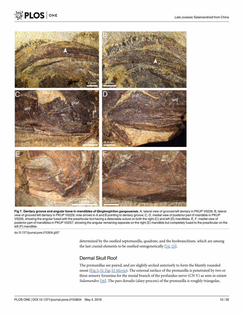
Fig 7. Dentary groove and angular bone in mandibles ofQinglongtriton gangouensis. A, lateral view of grooved left dentary in PKUP V0226; B, lateralview of grooved left dentary in PKUP V0229; note arrows in A and B pointing to dentary groove; C, D, medial view of posterior part of mandible in PKUPV0226, showing the angular fused with the prearticular but having a detectable suture on both the right (C) and left (D) mandibles; E, F, medial view ofposterior part of mandibles in PKUP V0257, showing the angular remaining separate on the right (E) mandible but completely fused to the prearticular on theleft (F) mandible.
doi:10.1371/journal.pone.0153834.g007
Late Jurassic Salamandroid from China
PLOS ONE | DOI:10.1371/journal.pone.0153834 May 4, 2016 10 / 35

extensively overlapping the nasal, but failing to reach the frontal. The partes dorsalis of the pre-maxillae are medially separated by an anterodorsal fenestra. At the base of the pars dorsalis, ahorizontal groove (S4 Fig) is present in association with the sensory foramina for the mesialbranch of the profundus nerve (CN V) and its associated blood vessels to supply the skin of thesnout [56]. The pars palatina of the premaxilla is a narrow, lingually projecting ledge connect-ing with the pars palatina of the maxilla to form the anterior part of the palate (e.g., PKUPV0228, V0229).
The paired nasals are broadly separated from one another by a large anterodorsal fenestra, asynapomorphy of the Salamandroidea [1, 8]. The nasal is broad anteriorly and narrowed poste-riorly, forming a triangular process that overlaps the frontal. The widened part of the nasal islaterally in articulation with the lacrimal, together forming the dorsal rim of the external naris,but is separated from the maxilla by the latter element (PKUP V0230, V0237, V0241). Severaltiny foramina penetrate the anterior part of the nasal (PKUP V0237, V0254), for passage of theultimate twigs of the mesial branch of the ophthalmicus profundus nerve (CN V) as seen inextant Salamandra [56]. The width across the nasals is slightly greater than that of the frontals,a plesiomorphic condition in urodeles [8].
The paired frontals diverge anteriorly to form the posterior border of the anterodorsal fenes-tra, but meet posteriorly along the skull midline to the point where the posterior processes ofthe frontals diverge again to overlap the parietals. The midline suture between the two frontalscurves irregularly, so that the suture is more or less sinuous rather than straight. The frontal-parietal suture is roughly “W”-shaped, resulting from the posterior process of the former ele-ment overlapping the parietal. The dorsal surface of the frontal is variably ornamented with
Fig 8. Marginal and palatal teeth ofQinglongtriton gangouensis. A, PKUP V0245, teeth in right premaxilla in lingual view; B, PKUP V0237, teeth in leftdentary in labial view; C, PKUP V0257, teeth in right palatine in medial view; D, PKUP V0237, teeth in right coronoid in lingual view. Arrow pointing to dividingzone between basal pedicel and crown.
doi:10.1371/journal.pone.0153834.g008
Late Jurassic Salamandroid from China
PLOS ONE | DOI:10.1371/journal.pone.0153834 May 4, 2016 11 / 35

Fig 9. Preservation of soft-tissue impressions ofQinglongtriton gangouensis. A, PKUP V0241,preserving mineralized denticles of internal gill, impressions of gill filaments, body outlines of trunk, limbs andtail with skeleton, in dorsal view; B, PKUP V0237, preserving mineralized denticles of internal gill, bodyoutlines of trunk and limbs with skeleton, in dorsal view; C, PKUP V0252, preserving soft-tissue outline of tailwith caudal series rotated post-mortem to lie in right lateral view.
doi:10.1371/journal.pone.0153834.g009
Late Jurassic Salamandroid from China
PLOS ONE | DOI:10.1371/journal.pone.0153834 May 4, 2016 12 / 35

dermal rugosities, especially in large, presumably old individuals (e.g., PKUP V0227, V0228,V0235, V0237, V0241, V0254, V0256).
The septomaxilla is small and irregular in shape, lying at the posterolateral border of thenaris (Fig 5B and 5C). As seen in PKUP V0237, the tiny element is medially concave for pas-sage of the nasolacrimal duct (see Discussion below), and it bears a pair of small arms pointingposteriorly. A septomaxilla is absent in Cryptobranchidae, Proteidae, Amphiumidae, and Sire-nidae [55]; its absence in these families has been interpreted as a “paedomorphic loss” [57].However, the absence of the septomaxilla in metamorphosed salamandrids and its presence inneotenic ambystomatids [55] are inconsistent with this interpretation.
The maxilla is reduced, being roughly the same length as the premaxilla (Fig 5). The shortanterior (premaxillary) process fits in a small groove of the premaxilla, and the posterior pro-cess has a pointed tip free from contacting any bony elements. The pars facialis (dorsal process)ascends from a wide base in the anterior half of the maxilla. The dorsal border of the process isslightly notched for articulation with the lacrimal and prefrontal, but is entirely separated fromthe nasal by the lacrimal (PKUP V0227, V0228, V0237, V0241, V0245, V0254, V0256). Theprocess is anteriorly notched for the external naris, and is posteriorly notched for the orbit. Sev-eral small projections are associated with these notches (PKUP V0234, V0245, V0253, V0254).Three to five foramina (S5 Fig) are scattered on the external surface of the pars facialis (PKUPV0234, V0237, V0254), and these correspond to the foramina laterale nasi for the lateralbranch of the ophthalmicus profundus nerve (CN V) as seen in extant Salamandra [56]. At thebase of the pars facialis is a horizontal groove (S5 Fig) in association with the foramina for thelateral branch of the ophthalmicus profundus nerve (CN V).
Internally, the pars palatine of the maxilla is a narrow ledge that together with the pars pala-tine of the premaxilla and the vomer contributes to the palate (PKUP V0228–V0230, V0237,V0241, V0245). A large foramen (occasionally two) is found at the base of the pars facialisslightly anterior to a deep fossa (PKUP V0230, V0237, V0241, V0245, V0248, V0256). This isinterpreted as the infraorbital foramen, i.e., the posterior opening of the superior alveolar canalcarrying a major branch of the trigeminal nerve (CN V) and its associated blood vessels.Another, smaller foramen slightly anterodorsal to the infraorbital foramen is for the lateral ter-minal branch of the ophthalmicus profundus of the trigeminal (CN V), as seen in extant Sala-mandra [56].
The prefrontal is wide anteriorly but is narrow posteriorly (PKUP V0227, V0228, V0230,V0233, V0237, V0241, V0245, V0254). It medially articulates with the nasal and frontal, andhas a blunt posterior end that fails to contact the anterolateral process of the parietal. Antero-laterally, the widened part of the prefrontal underlies the lacrimal, and has a small part in artic-ulation with the maxilla to close the orbital rim. Immediately above the orbital rim, theprefrontal bears a short groove (S6 Fig) that connects with the groove of the lacrimal for thepassage of the nasolacrimal duct (PKUP V0228, V0230, V0237, V0245, V0254; see Discussionbelow).
The lacrimal is a small plate with a shallow groove (S6 Fig) on its dorsal surface (PKUPV0227, V0228, V0230, V0237, V0241, V0253, V0254, V0256, V0260). Set between the nasaland maxilla, the lacrimal enters the external naris anteriorly and overlaps the prefrontal poste-riorly. The longitudinal groove on its dorsal surface carried the nasolacrimal duct, which in lifedrains secretions from eye glands into the nasal cavity [16, 58]. In one large specimen with askull length of 46.18 mm (PKUP V0254), the lacrimal becomes narrow, and is scrolled into adeep trough for the nasolacrimal duct. Despite differences in shape of the bone or the depth ofthe groove, the dorsally grooved lacrimal is a unique feature of the new salamander. In extantsalamanders, a lacrimal is found in hynobiids, dicamptodontids, and rhyacotritonids, but in
Late Jurassic Salamandroid from China
PLOS ONE | DOI:10.1371/journal.pone.0153834 May 4, 2016 13 / 35

these species the nasolacrimal duct passes through the lacrimal ventrally ([59]; see Discussionbelow).
The paired parietals contact one another along a straight suture at the midline. Anteriorly,the parietals are extensively overlapped by the frontals. A large part of the lateral border of theparietal curves downward as a well-developed descending flange, because an ossified orbito-sphenoid is absent (see below). A slender anterolateral process of the parietal extends along thelateral margin of the frontal, approaching but failing to contact the prefrontal; thus, the parietalcontributes to most of the medial border of the orbit (PKUP V0227–V0229, V0235, V0237,V0241, V0254). The parietal also bears a short, boot-like posterolateral process laterally in con-tact with the squamosal (PKUP V0227, V0229, V0230, V0233, V0235, V0237, V0241, V0245,V0246, V0250, V0253, V0254). In those large specimens (e.g., PKUP V0235, V0245, V0254),the parietal table is heavily ornamented with vermiculate rugosities along the midline suture,whereas in those smaller specimens (e.g., PKUP V0228, V0257, V0258) ornamentation ismuch less pronounced. The posterior border of the parietal table forms a transverse bonyridge, from which the parietal turns downward to form a flange that together with the opistho-tic and exoccipital closes the braincase posteriorly. The external concave surface of the flange isthe cervical epaxial fossa for attachment of the intervertebral epaxial muscles as seen in extantAmphiuma [60].
SuspensoriumThe squamosal is roughly “T”-shaped, with an expanded proximal end and a blunt distal end(Figs 4 and 5). The proximal end of the squamosal is in broad contact with the posterolateralprocess of the parietal. Presence of a parietal-squamosal contact is a plesiomorphic conditionat the level of Caudata, as known from the stem caudates Karaurus and Kokartus [61, 62]. Gen-erally, the squamosal is dorsally convex and ventrally concave. The dorsal surface bears aprominent ridge running from the proximal to the distal end along the anterior margin. Thisridge marks the attachment of the adductor mandibulae externus as known from extant sala-manders [63]. Ventrally, the squamosal overlaps the pterygoid and quadrate (Figs 4 and 5; S1Fig; S3 Movie).
The quadrate is small but well ossified as a principal element of the suspensorium. Severalspecimens (e.g., PKUP V0231, V0234, V0241, V0245, V0248, V0255, V0257, V0260) with thequadrate preserved in life position show that it is transversely oriented, whereas in other speci-mens (e.g., PKUP V0226, V0228, V0229, V0236, V0239, V0240, V0242, V0246, V0247, V0249,V0252, V0253, V0256, V0258, V0259) the bone is disarticulated and slightly rotated to an obli-que or even a perpendicular position in relation to the squamosal, clearly an artifact of preser-vation. The quadrate has a long ascending process extending about half the length of theanteroventral border of the squamosal. At the base of this process, the quadrate articulates withthe squamosal by clasping its anterodistal end, as evidenced by an articular facet on the squa-mosal in those specimens in which the quadrate has been disarticulated from the squamosal.The distal end of the quadrate is slightly expanded, and is ventrally convex forming a condylefor articulation with the mandible (PKUP V0227, V0230, V0235, V0237, V0241, V0245,V0254). The expanded distal end also meets the pterygoid at the craniomandibular joint. Asmall quadrate foramen is identified in several specimens (PKUP V0226, V0231, V0234,V0237, V0241, V0245, V0254) penetrating the expanded distal part of the element (Fig 5A and5C). This is the opening for the passage of the branch of the posterior condylar artery and veinas generally seen in other tetrapods [64].
The pterygoid is triradiate, but displays a larval configuration (Fig 6). That is, the anteriorpalatal process is markedly elongated and curved anteromedially. However, the process is free
Late Jurassic Salamandroid from China
PLOS ONE | DOI:10.1371/journal.pone.0153834 May 4, 2016 14 / 35
from contacting any bony element including the palatine (PKUP V0226–V0229, V0235–V0238, V0240, V0241, V0245, V0246, V0248, V0249). The process ventrally bears a singletooth row along its anterior portion. The medial process is extremely short, and it bears a smallfacet for articulation with the ala of the parasphenoid. The quadrate process is expanded pos-terolaterally, and distally articulates with the quadrate and the squamosal. In several specimens(PKUP V0226, V0237, V0240, V0254), the left pterygoid is penetrated by a small foramenbehind the tooth row. Such a foramen is probably the result of natural bone resorption, as com-monly seen in extant and extinct salamander species, such as Paradactylodon persicus (Hyno-biidae), Onychodactylus japonicus (Hynobiidae), Ambystoma gracile (Ambystomatidae),Triturus karelinii (Salamandridae) and Dicamptodon ensatus (Dicamptodontidae) [65, 66].
PalateThe palate consists mainly of the paired vomers and palatines, and the partes palatina of thepremaxillae and maxillae. In addition, the broad, anterior part of the parasphenoid contributesto the formation of the palate. The vomers are strip-like, obliquely arranged, and are separatedat the skull midline by the anteromedial fenestra (Figs 3 and 6). The vomers articulate with thepartes palatina of the premaxilla and maxilla anterolaterally, and in palatal view overlap theparasphenoid posteromedially (PKUP V0226, V0234, V0237). Each vomer bears a curvedtooth row, paralleling the maxillary arcade. Ventrally, the vomer carries two short groovesbehind the tooth row in all specimens having this part exposed (PKUP V0226, V0229, V0235,V0246). One groove is perpendicular to, and the other lying posteromedial to, the vomerinetooth row (Fig 6A). These were probably for nerves and blood vessels associated with the pal-ate. Close to the end of the vomerine tooth row, the posterolateral border of the vomer isindented an incipient notch for the choana (Figs 3 and 6).
The palatine is a small, almond-shaped plate, with its pointed end oriented posteriorly. Itlies close to the anterior process of the pterygoid, but clearly remains separate from the latterelement, although displaced in several specimens overlapping either the vomer or pterygoid.As observed in PKUP V0257, the palatine bears a straight tooth row along its lateral margin.Besides the basal salamandroid Beiyanerpeton, Qinglongtriton is the second Jurassic salaman-der known as having a discrete and toothed palatine in the adult stage. The accumulating evi-dence supports the argument by Gao and Shubin [8] that the presence of a separate palatine asa tooth-bearing element in the palate is probably a plesiomorphic condition forSalamandroidea.
The parasphenoid is a large, azygous bony plate that forms the braincase floor and part ofthe palate. The cultriform process is narrow at its base, but widens abruptly towards its anteriorend, where it underlies the vomers anterolaterally in palatal view. The anterior margin of theprocess is notched for the posterior border of the anteromedial fenestra (Figs 3 and 6). The pos-terolateral ala is short, triangular, and is laterally in contact with the medial process of the pter-ygoid. As observed in several specimens (PKUP V0226, V0246, V0257, V0267), the alae aregrooved for the passage of the internal carotid arteries, with the internal carotid foramen open-ing within the groove.
BraincaseThe large and azygous parasphenoid forms the braincase floor and part of the palate as hasbeen described above. No orbitosphenoid (sphenethmoid) has been observed in dorsal or pala-tal view of the available specimens. CT scans of several large specimens confirm that pairedorbitosphenoids are completely lacking (Fig 6; S1 Fig; S3 Movie). Among extant salamanders,proteids are the only group that lacks a free orbitosphenoid [55, 67]. However, the orbito-
Late Jurassic Salamandroid from China
PLOS ONE | DOI:10.1371/journal.pone.0153834 May 4, 2016 15 / 35
temporal region in proteids is laterally covered by a bony wall, which has no suture can bedelimited from the parietal but is penetrated by the foramen opticum and foramen oculomo-torium as commonly seen in other salamanders; this is obviously not the case in Qinglongtri-ton. Because there is no trace of an orbitosphenoid even in adult specimens, the element can beinterpreted as cartilaginous in the new salamander.
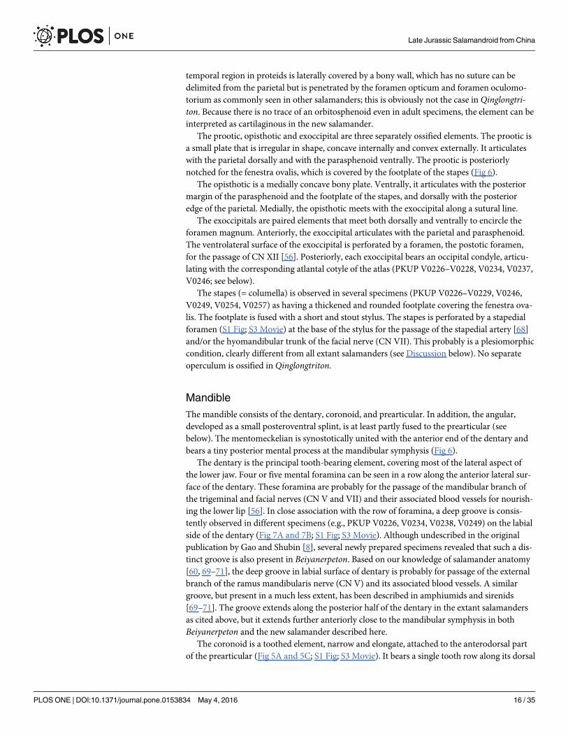
The prootic, opisthotic and exoccipital are three separately ossified elements. The prootic isa small plate that is irregular in shape, concave internally and convex externally. It articulateswith the parietal dorsally and with the parasphenoid ventrally. The prootic is posteriorlynotched for the fenestra ovalis, which is covered by the footplate of the stapes (Fig 6).
The opisthotic is a medially concave bony plate. Ventrally, it articulates with the posteriormargin of the parasphenoid and the footplate of the stapes, and dorsally with the posterioredge of the parietal. Medially, the opisthotic meets with the exoccipital along a sutural line.
The exoccipitals are paired elements that meet both dorsally and ventrally to encircle theforamen magnum. Anteriorly, the exoccipital articulates with the parietal and parasphenoid.The ventrolateral surface of the exoccipital is perforated by a foramen, the postotic foramen,for the passage of CN XII [56]. Posteriorly, each exoccipital bears an occipital condyle, articu-lating with the corresponding atlantal cotyle of the atlas (PKUP V0226–V0228, V0234, V0237,V0246; see below).
The stapes (= columella) is observed in several specimens (PKUP V0226–V0229, V0246,V0249, V0254, V0257) as having a thickened and rounded footplate covering the fenestra ova-lis. The footplate is fused with a short and stout stylus. The stapes is perforated by a stapedialforamen (S1 Fig; S3 Movie) at the base of the stylus for the passage of the stapedial artery [68]and/or the hyomandibular trunk of the facial nerve (CN VII). This probably is a plesiomorphiccondition, clearly different from all extant salamanders (see Discussion below). No separateoperculum is ossified in Qinglongtriton.
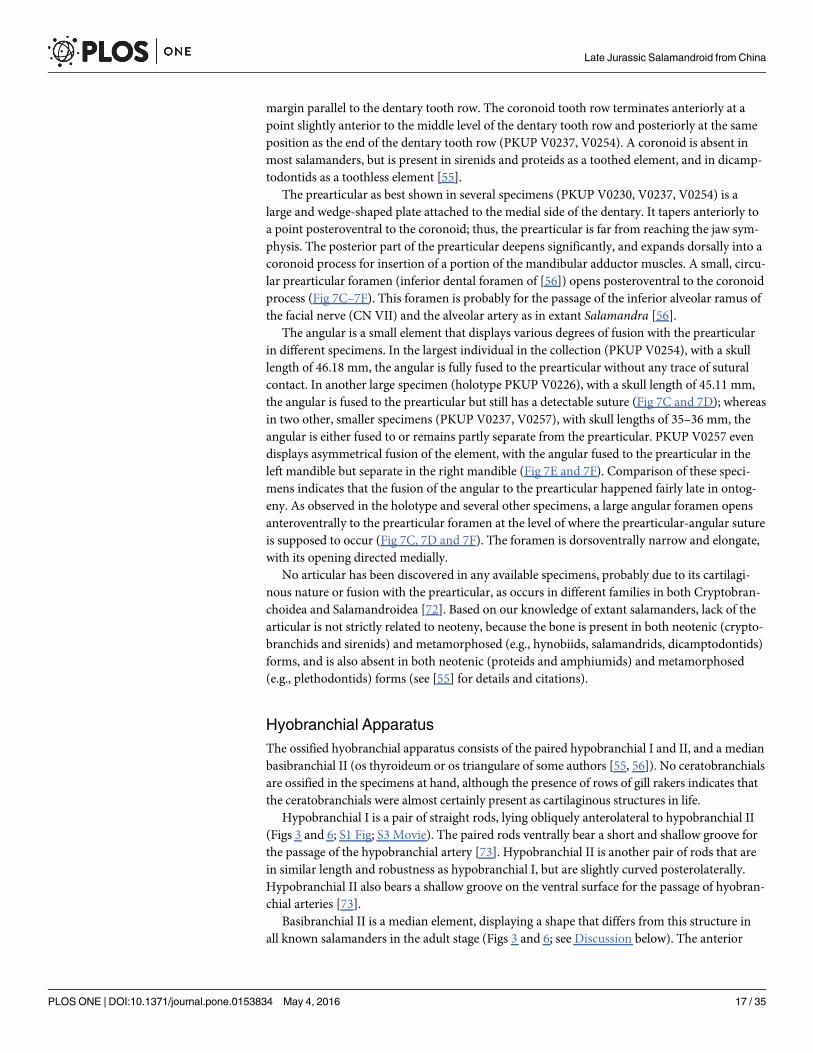
MandibleThe mandible consists of the dentary, coronoid, and prearticular. In addition, the angular,developed as a small posteroventral splint, is at least partly fused to the prearticular (seebelow). The mentomeckelian is synostotically united with the anterior end of the dentary andbears a tiny posterior mental process at the mandibular symphysis (Fig 6).
The dentary is the principal tooth-bearing element, covering most of the lateral aspect ofthe lower jaw. Four or five mental foramina can be seen in a row along the anterior lateral sur-face of the dentary. These foramina are probably for the passage of the mandibular branch ofthe trigeminal and facial nerves (CN V and VII) and their associated blood vessels for nourish-ing the lower lip [56]. In close association with the row of foramina, a deep groove is consis-tently observed in different specimens (e.g., PKUP V0226, V0234, V0238, V0249) on the labialside of the dentary (Fig 7A and 7B; S1 Fig; S3 Movie). Although undescribed in the originalpublication by Gao and Shubin [8], several newly prepared specimens revealed that such a dis-tinct groove is also present in Beiyanerpeton. Based on our knowledge of salamander anatomy[60, 69–71], the deep groove in labial surface of dentary is probably for passage of the externalbranch of the ramus mandibularis nerve (CN V) and its associated blood vessels. A similargroove, but present in a much less extent, has been described in amphiumids and sirenids[69–71]. The groove extends along the posterior half of the dentary in the extant salamandersas cited above, but it extends further anteriorly close to the mandibular symphysis in bothBeiyanerpeton and the new salamander described here.
The coronoid is a toothed element, narrow and elongate, attached to the anterodorsal partof the prearticular (Fig 5A and 5C; S1 Fig; S3 Movie). It bears a single tooth row along its dorsal
Late Jurassic Salamandroid from China
PLOS ONE | DOI:10.1371/journal.pone.0153834 May 4, 2016 16 / 35
margin parallel to the dentary tooth row. The coronoid tooth row terminates anteriorly at apoint slightly anterior to the middle level of the dentary tooth row and posteriorly at the sameposition as the end of the dentary tooth row (PKUP V0237, V0254). A coronoid is absent inmost salamanders, but is present in sirenids and proteids as a toothed element, and in dicamp-todontids as a toothless element [55].
The prearticular as best shown in several specimens (PKUP V0230, V0237, V0254) is alarge and wedge-shaped plate attached to the medial side of the dentary. It tapers anteriorly toa point posteroventral to the coronoid; thus, the prearticular is far from reaching the jaw sym-physis. The posterior part of the prearticular deepens significantly, and expands dorsally into acoronoid process for insertion of a portion of the mandibular adductor muscles. A small, circu-lar prearticular foramen (inferior dental foramen of [56]) opens posteroventral to the coronoidprocess (Fig 7C–7F). This foramen is probably for the passage of the inferior alveolar ramus ofthe facial nerve (CN VII) and the alveolar artery as in extant Salamandra [56].
The angular is a small element that displays various degrees of fusion with the prearticularin different specimens. In the largest individual in the collection (PKUP V0254), with a skulllength of 46.18 mm, the angular is fully fused to the prearticular without any trace of suturalcontact. In another large specimen (holotype PKUP V0226), with a skull length of 45.11 mm,the angular is fused to the prearticular but still has a detectable suture (Fig 7C and 7D); whereasin two other, smaller specimens (PKUP V0237, V0257), with skull lengths of 35–36 mm, theangular is either fused to or remains partly separate from the prearticular. PKUP V0257 evendisplays asymmetrical fusion of the element, with the angular fused to the prearticular in theleft mandible but separate in the right mandible (Fig 7E and 7F). Comparison of these speci-mens indicates that the fusion of the angular to the prearticular happened fairly late in ontog-eny. As observed in the holotype and several other specimens, a large angular foramen opensanteroventrally to the prearticular foramen at the level of where the prearticular-angular sutureis supposed to occur (Fig 7C, 7D and 7F). The foramen is dorsoventrally narrow and elongate,with its opening directed medially.
No articular has been discovered in any available specimens, probably due to its cartilagi-nous nature or fusion with the prearticular, as occurs in different families in both Cryptobran-choidea and Salamandroidea [72]. Based on our knowledge of extant salamanders, lack of thearticular is not strictly related to neoteny, because the bone is present in both neotenic (crypto-branchids and sirenids) and metamorphosed (e.g., hynobiids, salamandrids, dicamptodontids)forms, and is also absent in both neotenic (proteids and amphiumids) and metamorphosed(e.g., plethodontids) forms (see [55] for details and citations).
Hyobranchial ApparatusThe ossified hyobranchial apparatus consists of the paired hypobranchial I and II, and a medianbasibranchial II (os thyroideum or os triangulare of some authors [55, 56]). No ceratobranchialsare ossified in the specimens at hand, although the presence of rows of gill rakers indicates thatthe ceratobranchials were almost certainly present as cartilaginous structures in life.
Hypobranchial I is a pair of straight rods, lying obliquely anterolateral to hypobranchial II(Figs 3 and 6; S1 Fig; S3 Movie). The paired rods ventrally bear a short and shallow groove forthe passage of the hypobranchial artery [73]. Hypobranchial II is another pair of rods that arein similar length and robustness as hypobranchial I, but are slightly curved posterolaterally.Hypobranchial II also bears a shallow groove on the ventral surface for the passage of hyobran-chial arteries [73].
Basibranchial II is a median element, displaying a shape that differs from this structure inall known salamanders in the adult stage (Figs 3 and 6; see Discussion below). The anterior
Late Jurassic Salamandroid from China
PLOS ONE | DOI:10.1371/journal.pone.0153834 May 4, 2016 17 / 35

part of the basibranchial II is basically anchor-shaped, as the median rod is fused with a pair ofanterolateral processes. The process is flat, and medially has a thin crest presumably for attach-ment of the M. geniohyoideus and M. rectus cervicis superficialis as in extant salamanders [56,74]. The median rod extends posteriorly as a spike-like process, and is fused with a pair of pos-terolateral processes immediately posterior to the anchor-shaped portion (Figs 3 and 6). Injuvenile specimens (e.g., PKUP V0257, V0267), however, the posterolateral processes are lack-ing; therefore, ontogenetic change of the basibranchial II is documented by ossification of anextra pair of posterolateral processes in adults (see Discussion below).
DentitionTooth-bearing elements in Qinglongtriton include the premaxilla and maxillae in the maxillaryarch; vomers, palatines and pterygoids in the palate; and the dentary and coronoid in the lowerjaw. Each of these bones bears a single tooth row (monostichous), and all teeth are monocuspidwith a pointed tip (Fig 8).
As best observed from two specimens (PKUP V0237, V0254), marginal teeth are pedicellate,having the basal pedicel and crown separated by a poorly mineralized dividing zone (Fig 8).The teeth in the premaxilla and maxilla consistently number as 18–20 on each of these ele-ments in large specimens (PKUP V0226, V0229, V0234, V0237), the same number is also seenin a juvenile specimen (PKUP V0264: SKL = 18.28 mm, SPL = 64.56 mm), although the den-tary tooth row is not exposed in the latter specimen. The dentary tooth row has ~50 tooth posi-tions in two specimens (PKUP V0237, V0254). A complete coronoid tooth row is observed inPKUP V0254, which bears ~20 pedicellate teeth.
Palatal teeth are pedicellate, but sub-pedicellate teeth with a lingually globular/fibrous ring-like region can be occasionally observed as a developmental feature. Vomerine teeth are bestexposed in PKUP V0229, are about 23 in number, and are arranged in a single, curved row. Asimilar number is also revealed in a CT-scan of other two specimens (PKUP V0227, V0228).There are 10 or 11 teeth in the palatine (PKUP V0237, V0257), and ~22 in the pterygoid(PKUP V0226).
Axial SkeletonThe vertebral column of Qinglongtriton consists of 15 presacrals, including the atlas, 14 trunkvertebrae, one sacral, three caudosacrals, and as many as 33 caudals (Figs 3 and 4; S1 and S2Movies). The vertebral centrum is amphicoelous as in many other salamanders, except sala-mandrids and plethodontids and extinct batrachosauroidids that have opisthocoelous type ofcentrum [61, 75]. The number of the presacrals is invariably constant, whereas the number ofcaudals varies among different specimens. The tail is shorter than the snout-pelvic length inadult (PKUP V0233) and large juvenile individuals (PKUP V0236, V0266), but vice versa insmall juveniles (e.g., PKUP V0263).
The atlas is hourglass-shaped, and is slightly wider and longer than the trunk vertebrae as inmost other salamanders. The atlas bears a short, triangular odontoid process (tuberculuminterglenoideum) anteroventrally. The paired cotyles are cup-like facets for articulation withthe occipital condyles. The prominent crest extends along the entire length of the neural arch,and slightly over the first trunk vertebra. In ventral view, the atlas lacks a subcentral keel, buthas a large median depression immediately behind the odontoid process (Figs 3 and 6). A simi-lar median depression of the atlas is known from the stem caudate Urupia [76] and some butnot all extant salamanders (see Discussion below).
In lateral view of the atlas, a spinal nerve foramen opens on each side at the base of the neu-ral arch. Interestingly, just below the spinal nerve foramen the atlas bears a small projection,
Late Jurassic Salamandroid from China
PLOS ONE | DOI:10.1371/journal.pone.0153834 May 4, 2016 18 / 35

which corresponds to the “rudimentary transverse process” of Mivart [77] for a similar struc-ture in proteids and sirenids, followed by Gilbert [78] for Necturus, Skutschas and Martin [62]for the stem caudate Kokartus, and Skutschas and Krasnolutskii [76] for Urupia. The centrumalso displays a short ridge laterally, proceeding from the posterior edge to the transverse pro-cess (PKUP V0226, V0249, V0256).
In the trunk region, the vertebral centrum tends to increase in length from the second to the11th or the 12th vertebra, and then becomes shorter towards the sacral vertebra. As seen in theholotype (PKUP V0226), the shortest vertebra (first trunk) is only half the length of the longestone (11th trunk). In dorsal view, the neural arch dorsally carries a low crest that runs alongmuch of the length of the vertebra and continues onto the neural spine, which rises posterodor-sally as a short bar that is triangular in cross-section. The dorsal tip of the spine ends in a facet,probably for attachment of a tendon of the M. dorsalis trunci [56]. Spinal nerve foramina pene-trate the neural arch anterior to the transverse process for the first two trunk vertebrae, butposterior to the transverse process for the rest of the trunk series (PKUP V0228, V0229,V0231, V0251, V0256). The anterior two trunk vertebrae each ventrally lack a subcentral keel,but bear a depression with that of the second trunk often deeper than the first. The remainingtrunk vertebrae lack a ventral depression, but bear a subcentral keel with a foramen opening oneach side of the keel. The subcentral keel extends the length of the centrum in the anterior ver-tebrae, and then gradually shortens to become limited to the middle portion of the centrum inthe posterior portion of the trunk series. Laterally, all trunk vertebrae bear a pair of rod-liketransverse processes. The transverse process is weakly bifurcated, with two heads closelyappressed with each other. The process is half the length of the centrum, is directed posterolat-erally, and bears two facets distally for articulation with the bicapitate ribs.
The first three pairs of ribs are obviously more robust than the remaining ones, having anexpanded distal end for attachment of the pectoral muscles. In PKUP V0226 and V0258, ashort uncinate process is present on the first or the third trunk rib, for attachment of epaxialmuscles. Starting from the fourth pair, the trunk ribs tend to become shorter posteriorly, withthe last pair being merely triangular stubs. The anterior ribs are bicapitate, but have the tuber-culum and capitulum connected by an extremely thin bony web. Starting from the fourth pair,the remaining ribs have a small notch developed between the two heads; in some cases the dor-sal tuberculum is more prominent than the ventral capitulum (PKUP V0226, V0237, V0240,V0241). The possession of bicapitate trunk ribs gives a clear indication of membership of Qin-glongtriton in the suborder Salamandroidea.
The sacral vertebra is the same in length as the last trunk vertebra. Dorsally, the neuralspine is as prominent as in the trunk region. Ventrally, the subcentral keel is limited to the mid-dle portion of the centrum, with a subcentral foramen on each side of the keel. Laterally, thetransverse processes are stouter and longer than those of the trunk vertebrae. A spinal nerveforamen opens posterior to the transverse process. The sacral rib is long and curved, about thesame length as the ilium (Figs 3 and 4).
Several rib-bearing vertebrae immediately following the sacral are often referred to as thecaudosacrals [79]. In Qinglongtriton, three pertain to in this series: preserved in a dorsoventralposition, they clearly bear free ribs (Figs 3 and 4). The neural spine is limited to the posteriorpart of the neural arch (PKUP V0228, V0231, V0238). Ventrally, the subcentral keel andforamina on the three caudosacrals are unclear in the holotype, but a CT-scan of PKUP V0228reveals the same pattern as in the sacral and posterior trunk vertebrae. The transverse processin these caudosacrals is about half the length of the centrum, and is directed posterolaterally.The free ribs are slender and slightly curved, and have the same length as their correspondingtransverse process.
Late Jurassic Salamandroid from China
PLOS ONE | DOI:10.1371/journal.pone.0153834 May 4, 2016 19 / 35
Beginning from the first caudal vertebra, the tail commonly has been rotated taphonomi-cally at a right angle to the horizontal body plan. The neural and haemal arches are all well ossi-fied, and the haemal spine of the first caudal vertebra is bent at a greater angle than those of theother caudals. Spinal nerve foramina penetrate the neural arch posterior to the transverse pro-cess of the caudal vertebrae (PKUP V0231). The number of ossified caudal vertebrae increasesontogenetically, as small individuals possess fewer caudals (25 in PKUP V0263; 24 in PKUPV0266) than large individuals (>29 in PKUP V0226, 30 in PKUP V0233 and>27 in PKUPV0236).
Appendicular SkeletonThe ossified pectoral girdle consists of a pair of scapulocoracoids (Figs 3 and 4; S1 and S2 Mov-ies), as in other salamanders except for sirenids that have a separate coracoid [16, 57]. Thescapular portion is trapezoidal in shape, having the expanded distal end about twice the widthof the proximal constriction where it fuses with the coracoid plate. The coracoid portion isexpanded into a fan-shaped structure, which in lateral view is convex anteriorly and concaveposteriorly.
The glenoid fossa, an ovoid socket, is developed within a posterior concavity of the coracoidplate. The supracoracoid foramen (coracoid foramen) penetrates the thickened coracoid plateanterior to the glenoid fossa as can be seen in both the lateral and medial side of the plate. Theforamen in life is for the passage of the supracoracoideus nerve and the corresponding arteryand vein [56]. No suprascapula, procoracoid, or sternum is preserved; these elements are allcartilaginous in extant salamanders and presumably were in Qinglongtriton.
The humerus is straight, having a short shaft and slightly expanded proximal and distalends (S7A Fig). Both the proximal and distal ends are slightly concave, implying where werecapped with a cartilaginous humeral head and distal condyles, respectively. On the flexor side,the crista ventralis is merely a short smooth ridge running along the pre-axial border from theproximal end to the shaft, and lacks any recognizable projection. Opposite to the crista ventra-lis and on the extensor side, the proximal end bears a small tubercle connected with a smallridge as the crista dorsalis, presumably for insertion of M. subscapularis [56]. Distally, both theolecranon fossa on the extensor, and the fossa cubitalis ventralis on the flexor side are recogniz-able. The radial and ulnar condyles are not ossified.
The radius is roughly the same in length as the ulna or is slightly shorter than the latter. Theradius is slightly wider distally than proximally, whereas the ulna exhibits the converse, with itsproximal end wider than the distal end (S7A Fig). The ulna proximally bears a blunt olecranonprocess, anterodorsal to which is the articular facet for the humeral condyle.
No mesopodial elements are preserved in any of the specimens in the collection, and thislack of ossification of the mesopodium is obviously a neotenic feature of Qinglongtriton. Themetacarpals are all dumbbell-shaped, but are slightly different in length and robustness. Meta-carpal II is the longest and most robust in all available specimens; metacarpal I and IV are sub-equal in length and are the shortest.
The phalanges are also dumbbell-shaped, except the terminal phalanges, which have apointed tip. In all specimens, the third digit is the longest and the first the shortest. The phalan-geal formula is 2-2-3-2, a plesiomorphic pattern for salamanders [80].
The pelvic girdle consists of paired ilia and ischia, whereas the pubis and ypsiloid are notpreserved and are assumed to be cartilaginous as in extant taxa [57]. The ilium resembles acurved club, having its dorsal end slightly expanded for the attachment of the M. extensor ilio-tibialis [56]. The ventral end is expanded to form part of the acetabulum. In PKUP V0253, the
Late Jurassic Salamandroid from China
PLOS ONE | DOI:10.1371/journal.pone.0153834 May 4, 2016 20 / 35
acetabulum is a shallow fossa irregular in shape. The ischium is a plate, having a slightly con-cave lateral border and a straight medial edge for the symphysis (Figs 3 and 4).
The femur is straight, with a short shaft and expanded proximal and distal ends (S7B Fig).The femoral head and distal condyles were probably cartilaginous, as indicated by concave sur-faces on both ends of the shaft. The femur is slightly longer than the humerus, and is abouttwice the length of the tibia or fibula. The femur clearly lacks a hooked trochanter, with thisstructure merely a tiny process projecting anteroventrally, connected with the trochanter crestas a thin ridge along the proximal one-third of the femur. The trochanter groove is a deep fossadirectly posterior to the anterior trochanter crest. The distal end of the femur bears a shallowtrochlear groove, separating the relatively larger tibia condyle from the smaller fibular condyleas seen in several specimens (PKUP V0236, V0248, V0253).
The tibia and the fibula are both straight. The tibia is slightly wider proximally than distally,whereas the fibula is wider distally than proximally (S7B Fig). The tibial crest is a short triangu-lar ridge for insertion of the tendon of M. extensor iliotibialis [56]. The fibula is slightly shorterthan the tibia.
No tarsals are ossified. The metatarsals are all dumbbell-shaped, with metatarsal III consis-tently the longest, and metatarsal I the shortest. Metatarsal II and IV are subequal in length,and metatarsal V is the second shortest.
All phalanges are more or less dumbbell-shaped, except the terminal phalanges, which areslenderly pointed. The third digit is the longest and the first the shortest, corresponding to therelative length of the metatarsals. The phalangeal formula is 2-2-3-3-3 in all specimens butPKUP V0256, in which it is 2-2-3-4-3.
Soft TissuesMany specimens are preserved with soft-tissue impressions of the eye, body outline, and externalgill filaments (Figs 3, 4 and 9; S2 and S3 Figs). Mineralized denticles commonly termed gill rakersare not classified as soft tissues but are part of the internal gills and, hence, are described here.
All specimens are preserved with gill rakers, and many with gill filaments as evidence ofexternal gills. Gill rakers are tiny triangular denticles arranged in three rows, with the paireddenticles preserved in interlocked position like the teeth of a zipper. As in extant salamanders[74], the gill rakers were used for supporting the internal gills and as strainers in suction feeding.The external gills are preserved as impressions of gill filaments, three in number on each side.
Eyes are preserved as rounded dark stains within the orbit (PKUP V0227). Scleral cartilagesmight be expected to be present as typically seen in other neotenic salamanders, but left notrace in these specimens.
Body outlines of the trunk, tail and appendages are preserved in many specimens (e.g.,PKUP V0228, V0240, V0241, V0244, V0245, V0252, V0253). The trunk region is stout and thelimbs short and robust (Fig 9A and 9B; S2 and S3 Figs). A few relatively slim-bodied specimenswith a proportionally longer tail are probably male individuals as in extant salamanders [81].Those specimens (PKUP V0241, V0245, V0252) show that the tail is deep and laterally com-pressed, having a low dorsal fin and a deep ventral fin (Fig 9C). The laterally compressed tailand well-developed tail fin are consistent with the supposed aquatic life style of Qinglongtriton,as evidenced by its neotenic features.
Results and DiscussionCompared to other known fossil and extant salamanders, the new taxon has several significantfeatures that may shed light on character evolution in salamanders. We discuss these charactersas below.
Late Jurassic Salamandroid from China
PLOS ONE | DOI:10.1371/journal.pone.0153834 May 4, 2016 21 / 35

Significance ofQinglongtriton for Character Distribution and Evolution inSalamanders
Lacrimal and Nasolacrimal Duct. A lacrimal is present in extant hynobiids, dicampto-dontids, and rhyacotritonids, whereas other salamanders lack this element [55, 57]. Becausethe stem caudate Karaurus also has a lacrimal, the presence of this element in extant salaman-ders is probably a plesiomorphic feature. In those salamanders in which the lacrimal is presentat the adult stage, it is a small plate between the maxilla and the nasal/prefrontal (Fig 10). Theposition of the lacrimal relative to the narial opening or to the orbit is an informative feature inthe classification of hynobiids [82]. Within the Hynobiidae, the lacrimal variably enters thenaris, the orbit or both. The lacrimal enters only the naris in the Dicamptodontidae, whereas itenters both the naris and orbit in the Rhyacotritonidae. Taking the stem caudate Karaurus forout-group comparison, it seems that the lacrimal entering only the naris is a plesiomorphiccondition for salamanders.
Qinglongtriton displays the inferred plesiomorphic condition of the lacrimal, with a smalllacrimal entering only the naris (PKUP V0227, V0228, V0237, V0241, V0253, V0254, V0256,V0260). However, the new salamander uniquely has an open groove on the dorsal surface ofthe lacrimal (S6 Fig), different from all other lacrimal-bearing salamanders. The groove is forthe passage of the nasolacrimal duct, which is a soft-tissue tube to conduct secretions from theeye gland into the nasal cavity [16, 58]. In those salamanders having a lacrimal bone (hyno-biids, dicamptodontids, and rhyacotritonids), the nasolacrimal duct normally enters a smallforamen posterodorsally on the lacrimal bone, passes through the lacrimal canal, and thenexits from another foramen on the anteroventral edge of the bone. This pattern is seen inHynobius, Ranodon, and Onychodactylus [73, 84], and in Dicamptodon and Rhyacotriton[66, 83].
Fig 10. Comparison of position of lacrimal in relation to external naris and orbit in different groups of salamanders. A, Karaurus (Karauridae); B,Salamandrella (Hynobiidae); C, Batrachuperus (Hynobiidae); D, Hynobius (Hynobiidae); E, Ranodon (Hynobiidae); F, Rhyacotriton (Rhyacotritonidae); G,Dicamptodon (Dicamptodontidae); H,Qinglongtriton based on PKUP V0237. A, based on Estes [61]; B–E, based on Fei et al. [82]; F, based onAmphibiaTree [83]; and G, based onWake [66]. All are depicted in dorsal view, lacrimal is shaded in dark gray and opening of nasolacrimal duct is shaded indark. Not in scale.
doi:10.1371/journal.pone.0153834.g010
Late Jurassic Salamandroid from China
PLOS ONE | DOI:10.1371/journal.pone.0153834 May 4, 2016 22 / 35
Developmental evidence provides the clue for the dorsally open condition of the lacrimalcanal in Qinglongtriton. As known from extant salamanders (e.g., Ranodon and Onychodacty-lus), the lacrimal first occurs as a thin plate at or before metamorphosis, after the formation ofthe nasolacrimal duct; then the thin plate grows to enclose the canal at or soon after metamor-phosis [65, 85]. Based on this knowledge, we conclude that the ossification of the lacrimal bonein Qinglongtriton was terminated before complete ossification of the lacrimal canal; this docu-ments a previously unknown pattern of the canal in neotenic salamanders. All living salaman-ders that are obligate neotenes (cryptobranchids, proteids, amphiumids, sirenids) lack both thelacrimal bone and the nasolacrimal duct [16, 55, 72]. This seems to fit with the pattern in otheramphibians in which aquatic forms tend to lose the eye gland and nasolacrimal duct [86, 87].However, the discovery that neotenic Beiyanerpeton possesses a lacrimal [8] is in keeping withthe demonstration that Qinglongtriton is another neotenic form with clear evidence of a nasola-crimal duct; hence, the apparent loss of a lacrimal bone and nasolacrimal duct in extant obli-gate neotenes is a specialization that is not a necessary consequence of failure tometamorphose.
In those salamanders having a nasolacrimal duct but lacking a lacrimal bone (ambystoma-tids, salamandrids, plethodontids), the prefrontal and maxilla take over the roles of the lacrimalto carry the nasolacrimal duct. These include both neotenic (Ambystoma mexicanum) andmetamorphosed (salamandrids and plethodontids) forms. Medvedeva [88, 89] hypothesizedthat the prefrontal in these salamanders is actually a compound bone, the ‘prefronto-lacrimal’.Such a hypothesis seems to be supported by the Ambystoma mexicanum, in which the prefron-tal may be ossified from two anlages [65]. A more recent study by Vater [90] showed that theprefrontal in the salamandrid Triturus is indeed a bone of dual origin, specifically the lacrimaland prefrontal.
Palatine as a Discrete Element. The presence of a toothed palatine as a discrete elementin the adult stage is an unusual feature in salamanders. With the exception of sirenids, in mostother salamanders the palatine occurs only in early ontogeny, but is subsequently lost byresorption [65] or fusion with the pterygoid [55, 91] at metamorphosis. Exceptions are alsoknown from Cryptobranchus and Amphiuma, which show no trace of a palatine during theembryonic and larval stages [55]. Besides the recent report of Beiyanerpeton [8], Qinglongtritonis another Late Jurassic salamander that retained a palatine bone into the adult stage (Figs 3and 6). Because the small palatine in these two Jurassic salamanders is free, rather than part ofthe palatopterygoid, this element could be resorbed, but not be fused to the pterygoid if thesesalamanders had proceeded metamorphosis. It appears that retention of a discrete palatine inthese early salamanders likely resulted from delayed, incomplete resorption of the element, anontogenetic pattern previously unknown for fossil or extant salamanders.
Embryonically in salamanders, the common developmental pattern is ossification of thepalatine as a small plate connected to the anterior part of the palatopterygoid. At metamorpho-sis, the palatine along with anterior part of the pterygoid is resorbed [55, 65, 92]. This pattern isknown for hynobiids, salamandrids, ambystomatids, plethodontids, and proteids [55, 65].However, sirenids display a different pattern as has been documented in Siren intermedia byReilly and Altig [93]. According to these authors, early occurrence of the palatine in Siren atstage I was not much different from other salamanders, but at stage II the palatine was freedfrom the pterygoid as a consequence of resorption of the latter element (palatopterygoid disin-tegration). Then, the resorption of the pterygoid continued to stage III, in which the pterygoidwas completely lost, whereas the palatine persisted throughout ontogeny [93].
Because no metamorphosed salamanders are known with a palatine bone in adult stage,retention of the palatine in the two Jurassic salamandroids may be related to their neoteniccondition; however, this does not mean that the feature characterizes neoteny, as many other
Late Jurassic Salamandroid from China
PLOS ONE | DOI:10.1371/journal.pone.0153834 May 4, 2016 23 / 35

neotenic salamanders lack a palatine as adults. Furthermore, the palatine cannot be detectedeven in early embryonic stages in cryptobranchids as neotenic salamanders [55]. Retention ofthe palatine in Beiyanerpeton and Qinglongtriton obviously reflects a distinct developmentalfeature different from all other known salamanders.
Stapes and Stapedial Foramen. Among extant salamanders, a stapes is present in all fam-ily groups except salamandrids [55], neotenic species of plethodontids [59], and the sirenidPseudobranchus [55]. The lack of a stapes in all salamandrids is obviously a synapomorphy ofthe family, whereas that the similar condition in other two groups is probably independentlyacquired based on our knowledge of the phylogeny of Urodela [8].
The stapes in Qinglongtriton is penetrated by a stapedial foramen (Fig 6A; S1 Fig; S3Movie), different from all living salamanders with a stapes, in which the foramen is entirelylost [55, 64, 72]. Our re-examination of specimens of Beiyanerpeton jianpingensis (e.g., PKUPV0601–V0603) indicates that the foramen is also present in that taxon. Absence of a stapedialforamen was regarded as a synapomorphy of the Batrachia [94]. On the contrary, presence ofthis foramen in Jurassic crown-group salamanders refutes this notion. Because the stem cau-date Karaurus also has the stapes perforated [61, 95], the lack of a stapedial foramen in extantsalamanders is re-interpreted here as a derived feature acquired within the crown-group sala-manders. This is yet another example in which paleontological evidence reveals deep-time evo-lution of a particular feature that cannot be detected based on extant taxa only.
Fusion of Angular to Prearticular. Salamanders in Hynobiidae and Cryptobranchidaehave a separate angular in the lower jaw. Loss of the angular by fusion with the prearticular is aderived feature that is diagnostic of the salamandroid clade. Multiple specimens of Qinglongtri-ton show a varied extent of fusion of the angular to the prearticular as has been describedabove. Because some of the relatively large specimens including the holotype (PKUP V0226,V0237, V0254, V0257) have the angular partly or fully fused to the prearticular along a visiblesutural line (Fig 7C–7F), we conclude that the ontogenetic fusion of the angular to the prearti-cular in Qinglongtriton was delayed to a late developmental stage, even close to maturity.
In all known extant or extinct salamanders, a separate angular is only present in cryptobran-choids and their closely related fossil forms such as Chunerpeton [96]. Gardner [97] interpretedthe presence of an angular in a Paleocene amphiumid (genus and species indeterminate) basedon a notch on the posterolingual edge of the subdental shelf in an incomplete dentary (UALVP14316). However, this interpretation cannot be warranted, because the angular, if present, doesnot articulate with the subdental shelf of the dentary, but more ventrally between the prearticu-lar and the ventral edge of dentary. Instead, the notch that Gardner [97] described was likelyfor articulation with the prearticular.
Teeth and Crown Patterns. Pedicellate teeth with bicuspid crowns are two features thatare usually used to characterize the Lissamphibia following the work of Parsons and Williams[98]. Although many caecilians [99, 100], and some salamanders and frogs have monocuspidand even non-pedicellate teeth, many developmental studies have documented the ontogenetictransition of teeth from a monocuspid and non-pedicellate larval dentition to a bicuspid andpedicellate adult dentition as a general pattern [98, 101–104]. For example, a study of toothdevelopment in Amphiuma showed that the non-pedicellate and monocuspid larval teeth werereplaced by pedicellate and bicuspid teeth as adult dentition [105]. Another study implied thatthe monocuspid crown pattern in some caecilians, larval salamanders, and some adult frogs isprobably an ancestral condition, whereas the bicuspid condition is derived [106].
In extant salamanders, Siren has non-pedicellate teeth with monocuspid tooth-crowns,whereas the closely related Pseudobranchus has pedicellate teeth and monocuspid crowns [71,98, 102, 107–109]. The teeth of proteids have been described as monocuspid and pedicellate inNecturus [108, 109], and those in Proteus as having no crown/base constriction [110] or having
Late Jurassic Salamandroid from China
PLOS ONE | DOI:10.1371/journal.pone.0153834 May 4, 2016 24 / 35
slight indication of a crown/base constriction [111]. Carroll and Holmes [63] concluded that:“the generally poor development of pedicelly in sirenids and proteids may be attributed to theirneotenic nature.”
No bicuspid teeth have been found in specimens of Qinglongtriton. However, both marginaland palatal teeth are pedicellate, with a dividing zone separating the crown from the pedicel(Fig 8). This pattern demonstrates that bicuspid crown as a derived feature was not developedin association with pedicelly in this new salamander. The monocuspid crown is shared with itssister taxon Beiyanerpeton, but pedicelly is not. Of stem caudates, tooth morphology isunknown for Middle–Late Jurassic Karaurus and Urupia, but Skutschas and Martin [62] wereable to show that the Middle Jurassic Kokartus had non-pedicellate teeth with monocuspidcrowns. Gao and Shubin [8] determined that the dentition in the basal salamandroid Beiyaner-peton is non-pedicellate and monocuspid; they also argued that pedicelly and bicuspid crownsare derived features within Urodela, a notion supported by our study of the new salamander inthis paper.
Basibranchial II. Basibranchial II is the most posteromedian element in the hyobranchialapparatus of salamanders. The shape of the basibranchial II is highly variable among taxo-nomic groups [74]. In the stem caudate Karaurus and two Jurassic crown-group salamanders(Chunerpeton and Beiyanerpeton), basibranchial II is simply anchor-shaped, having a shortmedian rod fused to a pair of anterolateral processes ([61, 96]; Fig 11A and 11B). The EarlyCretaceous salamandroid Valdotriton from Spain has an inverted “Y”-shaped basibranchial II(Fig 11H). This configuration of the basibranchial II is basically similar to that in the Europeansalamandrid Chioglossa (Fig 11J), and is likely a derived, rather than a primitive conditionwithin the Salamandroidea (contra Evans and Milner [21]).
Among extant salamanders, a basibranchial II is absent in cryptobranchids [55], but is vari-ably present or absent in hynobiids. Most hynobiids (Ranodon, Pachyhynobius, Liua, Salaman-drella, and Batrachuperus) have a tri-radiate basibranchial II, with a horizontal bar fused to ashort median process, so that the element is basically an inverted “T-shape” [112]. Two otherhynobiids, Hynobius and Pseudohynobius, simply have a horizontal bar, lacking a median pro-cess, whereas basibranchial II is entirely absent in Onychodactylus [55, 82, 112].
Basibranchial II seems to be more variable in shape in salamandroids than in hynobiids.Most salamandrids (e.g., Salamandrina, Pleurodeles, Triturus, Neurergus, Notophthalmus, Tari-cha, Cynops,Hypselotriton, Tylototriton, Pachytriton, Paramesotriton) lack a basibranchial II atadult stage [82, 115], whereas those have it, it is either ossified as triangular plate as seen in Sal-amandra (Fig 11I) or an inverted Y-shaped structure as seen in Chioglossa (Fig 11J) andMer-tensiella [116]. Dicamptodontids and ambystomatids have the tri-radiate condition, more orless resembling that in most hynobiids, whereas rhyacotritonids have a simple horizontal barsimilar to other hynobiids. Amphiumids have basibranchial II entirely missing, whereas in pro-teids, it is a simple vertical bar (Fig 11N). In plethodontids, basibranchial II is basically anchor-shaped, but has all three rami oriented posteriorly as seen in Gyrinophilus (Fig 11M) and inEurycea [59], or is a horizontal bar strongly bowed anteriorly without a posteromedial ramusas in Hydromantes [117]. Sirenids have a relatively long vertical stem, but is fused with a vari-able number of processes at the posterior end (Fig 11O). Overall, the presence or absence, orthe shape of the basibranchial II are highly variable among salamanders, and no phylogeneticpattern can be unambiguously distinguished for this structure among salamander families.
Basibranchial II in mature individuals of Qinglongtriton (Fig 11G) differs from all knownstem caudates and crown-group salamanders in having a median rod fused with a pair of ante-rolateral and a pair of posterolateral rami. Comparison of adult with juvenile specimens revealsontogenetic change in hypobranchial II: small and presumably juvenile specimens (PKUPV0232, V0257, V0267; Fig 11F) display the primitive anchor-shaped basibranchial II, whereas
Late Jurassic Salamandroid from China
PLOS ONE | DOI:10.1371/journal.pone.0153834 May 4, 2016 25 / 35

large and presumably adult specimens (e.g., PKUP V0226, V0246, V0249, V0252) have addeda second pair of processes posteriorly (Fig 11G). Moreover, ossification of the second pair ofprocesses seems to be more extensive in keeping with increased body size.
Basibranchial II is the main force-transmitting element from the protractible musculatureto the tongue pad [113]. It has been documented that basibranchial II provides the attachmentfor three muscles: M. geniohyoideus, M. rectus cervicis, and M. subarcualis oblique [118–121].In those salamanders lacking basibranchial II (e.g., Onychodactylus japonicus and cryptobran-chids), M. geniohyoideus is continuous with M. rectus cervicis, whereas M. subarcualis obliqueinserts on the adjacent fascia of M. rectus cervicis [118, 119, 121]. In Qinglongtriton, the com-plex structure of basibranchial II may have strengthened the attachment of these muscles, thusenhancing the ability to control the hyobranchial apparatus during feeding and respiration.
Atlas and Anterior Trunk Vertebrae. Two notable features in the vertebral column ofQinglongtriton are worth discussion: a pair of transverse processes on the neural arch of theatlas and a median depression in the ventral surface of the centrum in the atlas and one or twoanterior trunk vertebrae. The transverse processes on the atlas of Qinglongtriton are rudimen-tary, but are comparable to those in extant salamanders that have the processes. In crown-group salamanders, an atlas with rudimentary transverse process can be found in proteids, sire-nids, and Ambystoma gracile, whereas the process is lacking in Cryptobranchus and Amphiuma
Fig 11. Comparison of basibranchial II of salamanders. A, Karaurus (Karauridae) based on Estes [61]; B,Chunerpeton (Cryptobranchidae) based on Gao and Shubin [96]; C, Ranodon (Hynobiidae); D, Hynobius(Hynobiidae); E, Salamandrella (Hynobiidae); C–E based on Xiong et al. [112]; F, G, juvenile and adult stageinQinglongtriton (based on PKUP V0267 and PKUP V0226, respectively); H, Valdotriton (Salamandroidea)based on Evans and Milner [21]; I, Salamandra (Salamandridae); J, Chioglossa (Salamandridae); K,Rhyacotriton (Rhyacotritonidae); L, Dicamptodon (Dicamptodontidae); M,Gyrinophilus (Plethodontidae); I–M based onWake and Deban [113]; N, Proteus (Proteidae) based on Rose [55]; O, Siren (Sirenidae) basedon AmphibiaTree [114]. Not in scale.
doi:10.1371/journal.pone.0153834.g011
Late Jurassic Salamandroid from China
PLOS ONE | DOI:10.1371/journal.pone.0153834 May 4, 2016 26 / 35

[77, 97, 114]. To our knowledge, no metamorphosed salamanders are known with such pro-cesses. In stem caudates, the transverse process on the atlas can be found in Kokartus and Uru-pia, both have been recognized as neotenic [62, 76]. None of the taxa mentioned above haveribs associated with the atlas.
Rogers [122] claimed that the transverse process of the atlas is present in larval individualsof Ambystoma tigrinum but the process is reabsorbed at metamorphosis. Later workers arguedthat possession of transverse processes on the atlas is probably a neotenic feature [62, 123].Our new fossil discovery, corroborated with evidence from other fossil and extant salamanders,appears to support the interpretation that presence of transverse processes on the atlas isindeed of a feature associated with neoteny with the exception of Cryptobranchus andAmphiuma.
The atlas of Qinglongtriton resembles to that of Kokartus and Urupia in having a mediandepression on the ventral surface (PKUP V0226, V0229, V0235, V0249, V0256, V0259). Inaddition, the first two trunk vertebrae in Qinglongtriton also bear a deep median depression onthe ventral surface of the centrum without known functions. A median depression on the ven-tral surface of the atlas or anterior trunk vertebrae is also known in several extant taxa (Siren,Andrias, Cryptobranchus, Necturus, Ambystoma, Amphiuma, and Eurycea), and in a hynobiidspecies Batrachuperus longdongensis (CIB 20020489, 20020499). Because all these are neotenicsalamanders, and no metamorphosed species have such a depression to our knowledge, itseems reasonable to interpret the presence of such a distinct depression in the atlas and thefirst two trunk vertebrae as another neotenic feature in addition to the rudimentary transverseprocess of the atlas as discussed above.
Phylogenetic Placement ofQinglongtritonThe data matrix used in this study is revised from Gao and Shubin ([8]: supplementary infor-mation appendix), with the new salamander Qinglongtriton added as the additional taxon.Revised character codings of some fossil taxa are made based on our examination of specimensat hand and are highlighted in the dataset (S1 Appendix). Of the 105 characters in the originaldataset, 74 were scored for the new taxon (see S1 Appendix). As in the original analyses, thestem-caudate Karaurus was designated as the outgroup, and all characters were unordered andequally weighted. As in the original analysis [8], several fossil taxa (e.g., Laccotriton, Jeholotri-ton) were excluded from the analysis due to their anatomical uncertainties are under revision.Several characters (11, 18, 47, 50, 72, 73, 92, 93, 103) were excluded from the current analysesbecause these are uninformative in relation to the taxon sampling in the current dataset. Theanalysis of the revised dataset was performed using the implicit enumeration search option inTNT (Version 1.1; Goloboff et al. [124, 125]). Three most parsimonious trees (MPTs) werefound (tree length = 241 steps, consistent index = 0.494, retention index = 0.725). As seen inthe strict consensus tree (Fig 12), all the three MPTs have the new taxon placed as the sistertaxon with Beiyanerpeton, and the two taxa together form the basalmost clade within the Sala-mandroidea. Thus, Qinglongtriton adds another salamandroid that documented the early evo-lution of the clade during the Late Jurassic in North China.
Qinglongtriton shares with Beiyanerpeton and other salamandroids derived character states asfollows: nasals separated by anterodorsal fenestra without a midline contact (8–1); anterolateralprocess of parietal extending beyond midlevel of orbit; angular fused to prearticular (26–1); artic-ular absent by fusion to prearticular (29–1); and spinal nerve foramina occur in trunk, sacral andcaudal vertebrae (51–3). The sister-group relationship ofQinglongtritonwith Beiyanerpetonwithin the Salamandroidea is supported by three characters: palatine in adult present as discreteelement (19–0); pterygoid teeth present (39–1); and monocuspid tooth crown (42–0).
Late Jurassic Salamandroid from China
PLOS ONE | DOI:10.1371/journal.pone.0153834 May 4, 2016 27 / 35
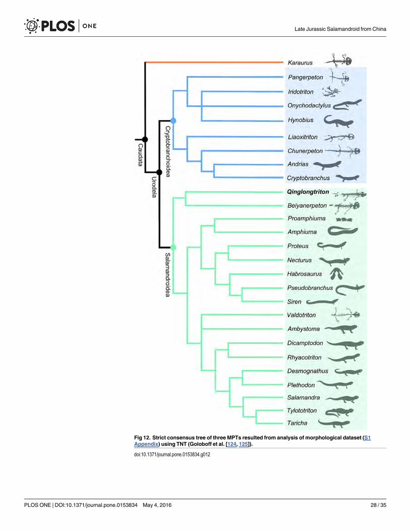
Fig 12. Strict consensus tree of three MPTs resulted from analysis of morphological dataset (S1Appendix) using TNT (Goloboff et al. [124, 125]).
doi:10.1371/journal.pone.0153834.g012
Late Jurassic Salamandroid from China
PLOS ONE | DOI:10.1371/journal.pone.0153834 May 4, 2016 28 / 35
In general, the new taxon Qinglongtriton adds another rare fossil salamander to the YanliaoBiota in addition to the previous report of Beiyanerpeton from the same geological formation.The new discovery provides further support for the hypothesis that the split of the Salaman-droidea from Cryptobranchoidea was deeply rooted in salamander phylogeny before the LateJurassic (Oxfordian) time.
ConclusionsThe following conclusions can be drawn from our study of Qinglongtriton gangouensis fromthe Nanshimenzi locality:
1. Qinglongtriton gangouensis is a neotenic salamandroid named and described based on mul-tiple specimens from the Upper Jurassic Tiaojishan Formation.
2. The new salamandroid displays several features of ontogenetic or taxonomic significance,including: lacrimal with a dorsally open groove for the nasolacrimal duct; presence of a sta-pedial foramen; lack of ossification of the orbitosphenoid; pedicellate teeth with monocus-pid crowns; presence of a toothed coronoid; ontogenetic fusion of the angular bone to theprearticular; and a unique shape of the basibranchial II.
3. Phylogenetic analysis places Qinglongtriton as the sister taxon to Beiyanerpeton, and the twotaxa together form the basalmost clade within the Salamandroidea. Along with the coexist-ing Beiyanerpeton the new discovery indicates that morphological disparity of the salaman-droid clade was already underway by early Late Jurassic (Oxfordian) time.
Supporting InformationS1 Appendix. Taxon-character data matrix used in phylogenetic analysis (A = 0/1; B = 0/2;C = 1/2). Character description see Gao and Shubin (2012; 10.1073/pnas.1009828109/-/DCSupplemental). Modified coding of characters (bold-faced and underlined) as explained intext below.(PDF)
S1 Fig. CT-rendered reconstruction of skull of PKUP V0226 (holotype of Qinglongtritongangouensis gen. et sp. nov.) in dorsal (left) and ventral (right) views. Abbreviations used asin Materials and Methods of main text.(TIF)
S2 Fig. Referred specimen of Qinglongtriton gangouensis gen. et sp. nov. (PKUP V0227).Photograph (left) and line drawing (right) of incomplete skeleton in dorsal view. Abbreviationsused as in Materials and Methods of main text.(TIF)
S3 Fig. Referred specimen of Qinglongtriton gangouensis gen. et sp. nov. (PKUP V0254).Photograph (left) and line drawing (right) of incomplete skeleton in dorsal view. Abbreviationsused as in Materials and Methods of main text.(TIF)
S4 Fig. Close-up photographs of referred specimens of Qinglongtriton gangouensis gen. etsp. nov., showing horizontal groove (arrows) and sensory foramina (sfm) in premaxilla. A,PKUP V0237; B, PKUP V0254.(TIF)
Late Jurassic Salamandroid from China
PLOS ONE | DOI:10.1371/journal.pone.0153834 May 4, 2016 29 / 35

S5 Fig. Close-up photographs of referred specimens of Qinglongtriton gangouensis showinghorizontal groove (arrows) and foramina laterale nasi (fln) in left maxilla. A, PKUP V0234;B, PKUP V0254.(TIF)
S6 Fig. Close-up photographs of referred specimens of Qinglongtriton gangouensis gen. etsp. nov., showing nasolacrimal groove (arrows) in lacrimal and prefrontal. A, PKUP V0237;B, PKUP V0254.(TIF)
S7 Fig. Close-up photographs of limbs in referred specimens of Qinglongtriton gangouensisgen. et sp. nov. A, PKUP V0245; B, PKUP V0251. Abbreviations used as in Materials andMethods in main text.(TIF)
S1 Movie. Reconstruction of the upper body of PKUP V0226 (holotype of Qinglongtritongangouensis gen. et sp. nov.).(MP4)
S2 Movie. Reconstruction of whole body of PKUP V0228 (referred specimen of Qinglong-triton gangouensis gen. et sp. nov.).(MP4)
S3 Movie. CT rendered reconstruction of skull of PKUP V0226 (holotype of Qinglongtritongangouensis gen. et sp. nov.).(MP4)
AcknowledgmentsThe research was funded by grants from the National Natural Science Foundation of China(NSFC 41072007, 41272016). Jia’s research was also supported by the China Scholarship Coun-cil. We thank Zhang Lijun and Zhou Chang-Fu (Shenyang Normal University) for their helpin collecting stratigraphic data and measuring the geologic section; and thank M. Ellison(American Museum of Natural History) for photography of specimens. J. Jia thanks N. Shubinand M. Coates (University of Chicago) for discussion. We thank R. C. Fox (University ofAlberta) for reading and improving the manuscript. We also thank three reviewers (J. Olori, J.Gardner and P. Skutschas) for their constructive comments and suggestions.
Author ContributionsConceived and designed the experiments: JJ KG. Performed the experiments: JJ KG. Analyzedthe data: JJ. Contributed reagents/materials/analysis tools: JJ KG. Wrote the paper: JJ KG.
References1. Dunn ER. The sound-transmitting apparatus of salamanders and the phylogeny of the Caudata.
American Naturalist. 1922; 56: 418–427.
2. AmphibiaWeb: Information on amphibian biology and conservation. Berkeley, California: Amphibia-Web. Available: http://amphibaweb.org/. Accessed 20 November 2015.
3. Frost DR. Amphibian species of the world: an online reference. Version 6.0. 2015. 2015. AmericanMuseum of Natural History, New York, USA. Available: http://research.amnh.org/herpetology/amphibia/index.html. Accessed 9 March 2015.
4. Larson A, DimmickW. Phylogenetic relationships of the salamander families: an analysis of congru-ence among morphological and molecular characters. Herpetological Monographs. 1993; 7: 77–93.
Late Jurassic Salamandroid from China
PLOS ONE | DOI:10.1371/journal.pone.0153834 May 4, 2016 30 / 35
5. Zhang P, Wake DB. Higher-level salamander relationships and divergence dates inferred from com-plete mitochondrial genomes. Molecular Phylogenetics and Evolution. 2009; 53: 492–508. doi: 10.1016/j.ympev.2009.07.010 PMID: 19595776
6. Pyron RA, Wiens JJ. A large-scale phylogeny of Amphibia including over 2800 species, and a revisedclassification of extant frogs, salamanders, and caecilians. Molecular Phylogenetics and Evolution.2011; 61: 543–583. doi: 10.1016/j.ympev.2011.06.012 PMID: 21723399
7. Gao KQ, Shubin NH. Late Jurassic salamanders from northern China. Nature. 2001; 410: 574–577.PMID: 11279493
8. Gao KQ, Shubin NH. Late Jurassic salamandroid from western Liaoning, China. Proceedings of theNational Academy of Sciences of the United States of America. 2012; 109: 5767–5772. doi: 10.1073/pnas.1009828109 PMID: 22411790
9. Larson A, Weisrock DW, Kozak KH. Phylogenetic systematics of salamanders (Amphibia: Urodela), areview. In: Sever DM, editor. Reproductive biology and phylogeny of Urodela (Amphibia). Enfield,North Hampshire: Science Publishers; 2003. pp. 31–108.
10. Larson A, Wake D, Devitt T. 2006. Caudata. Tree of life web project. Available: http://tolweb.org/Caudata/14939/2006.09.05. Accessed 20 March 2015.
11. Wiens JJ, Bonnet RM, Chippindale PT. Ontogeny discombobulates phylogeny: paedomorphosis andhigher-level salamander relationships. Systematic Biology. 2005; 54: 91–110. PMID: 15805013
12. Frost DR, Grant T, Faivovich J, Bain RH, Haas A, Haddad CFB, et al. The amphibian tree of life. Bulle-tin of the American Museum of Natural History. 2006; 297: 1–370.
13. Roelants K, Gower DJ, Wilkinson M, Loader SP, Biju SD, Guillaume K, et al. Global patterns of diver-sification in the history of modern amphibians. Proceedings of the National Academy of Sciences ofthe United States of America. 2007; 104: 887–892. PMID: 17213318
14. Struck TH. Data congruence, paedomorphosis and salamanders. Frontiers in Zoology. 2007; 4: 22.PMID: 17974010
15. DuellmanWE. Global distribution of amphibians: patterns, conservation, and future challenges. In:DuellmanWE, editor. Patterns of distribution of amphibians. Baltimore and London: The Johns Hop-kins University Press; 1999. pp. 1–30.
16. Noble GK. The biology of the Amphibia. 1st ed. New York and London: McGraw-Hill Book Company;1931.
17. Liu YQ, Liu YX, Ji SA, Yang ZQ. U-Pb zircon age for the Daohugou Biota at Ningcheng of Inner Mon-golia and comments on related issues. Chinese Science Bulletin. 2006; 51: 2634–2644.
18. Anderson JS. Fossils, molecules, divergence times, and the origin of Salamandroidea. Proceedingsof the National Academy of Sciences of the United States of America. 2012; 109: 5557–5558. doi: 10.1073/pnas.1202491109 PMID: 22460794
19. Evans SE, Lally C, Chure DC, Maisano JA. A Late Jurassic salamander (Amphibia: Caudata) from theMorrison Formation of North America. Zoological Journal of the Linnean Society. 2005; 143: 599–646.
20. Estes R. The fossil record of amphiumid salamanders. Breviora. 1969; 322: 1–11.
21. Evans SE, Milner AR. A metamorphosed salamander from the Early Cretaceous of Las Hoyas, Spain.Philosophical Transactions of the Royal Society B: Biological Sciences. 1996; 351: 627–646.
22. Gardner JD, DeMar DG Jr. Mesozoic and Palaeocene lissamphibian assemblages of North America:a comprehensive review. Palaeobiodiversity and Palaeoenvironments. 2013; 93: 459–515.
23. Skutschas PP. Mesozoic salamanders and albanerpetontids of Middle Asia, Kazakhstan, and Siberia.Palaeobiodiversity and Palaeoenvironments. 2013; 93: 441–457.
24. Chen PJ. Jurassic biostratigraphy of China. In: ZhangW, Chen PJ, Palmer AR, editors. Biostratigra-phy of China. Beijing, China: Science Press; 2003. pp. 423–463.
25. Chang SC, Zhang H, Renne PR, Fang Y. High-precision 40Ar/39Ar age constraints on the basal LanqiFormation and its implications for the origin of angiosperm plants. Earth and Planetary Science Let-ters. 2009; 279: 212–221.
26. Chang SC, Zhang H, Hemming SR, Mesko GT, Fang Y. 40Ar/39Ar age constraints on the Haifanggouand Lanqi formations: when did the first flowers bloom? In: Jourdan F, Mark DF, Verati C, editors.Advances in 40Ar/39Ar dating: from archaeology to planetary sciences. London: Geological Society;Special Publications 378. 2014. pp. 277–284.
27. Davis GA, Zhang Y, Wang C, Darby BJ, Zhang C, Gehrels G. Mesozoic tectonic evolution of the Yan-shan fold and thrust belt, with emphasis on Hebei and Liaoning provinces, northern China. GeologicalSociety of America Memoirs. 2001; 194: 171–197.
28. ChenW, Ji Q, Liu DY, Zhang Y, Song B, Liu XY. Isotope geochronology of the fossil-bearing beds inthe Daohugou area, Ningcheng, Inner Mongolia. Geological Bulletin of China. 2004; 23: 1165–1169.
Late Jurassic Salamandroid from China
PLOS ONE | DOI:10.1371/journal.pone.0153834 May 4, 2016 31 / 35

29. Zhao Y, Zhang H, Xu G, Yang Z, Hu J. The Jurassic major tectonic events of the Yanshanian intra-plate deformation belt. Geological Bulletin of China. 2004; 23: 854–863.
30. YangW, Li SG. Geochronology and geochemistry of the Mesozoic volcanic rocks in Western Liao-ning: Implications for lithospheric thinning of the North China Craton. Lithos. 2008; 102: 88–107.
31. Song B, Han H, Guo P. U-Pb Zircon dating of the Mesozoic welded pyroclastics in the QG2 well andits geological significance. Advances in Geosciences. 2012; 2012: 131–137.
32. Wang LL, Hu DY, Zhang LJ, Zheng SL, He HY, Deng CL, et al. SIMS U-Pb zircon age of Jurassic sed-iments in Linglongta, Jianchang, western Liaoning: constraint on the age of oldest feathered dino-saurs. Chinese Science Bulletin. 2013; 58: 1346–1353.
33. Faure Gunter. Principles of isotope geology ( 2nd edition). New York: Wiley; 1986.
34. Ma Y, Cui S, Zeng Q, WuM. Yanshanian compression and extension in the Yanshan area. GeologicalBulletin of China. 2002; 21: 218–223.
35. Lü J, Hone DWE. A new Chinese anurognathid pterosaur and the evolution of pterosaurian taillengths. Acta Geologica Sinica. 2012; 86: 1317–1325.
36. Jiang S, Wang X, Cheng X, Costa FR, Huang J, Kellner AWA. Short note on an anurognathid ptero-saur with a long tail from the Upper Jurassic of China. Historical Biology. 2015; 27: 718–722.
37. Xu X, Zheng X, Sullivan C, Wang X, Xing L, Wang Y, et al. A bizarre Jurassic mairaptoran theropodwith preserved evidence of membranous wings. Nature. 2015; 52: 70–73.
38. Zheng X, Bi S, Wang X, Meng J. A new arboreal haramiyid shows the diversity of crown mammals inthe Jurassic period. Nature. 2013; 500: 199–202. doi: 10.1038/nature12353 PMID: 23925244
39. Luo ZX, Meng QJ, Ji Q, Liu D, Zhang YG, Neander AI. Evolutionary development in basal mammlia-forms as revealed by a docodontan. Science. 2015; 347: 760–764. doi: 10.1126/science.1260880PMID: 25678660
40. Wang Y, Evans SE. A new short-bodied salamander from the Upper Jurassic/Lower Cretaceous ofChina. Acta Palaeontologica Polonica. 2006; 51: 127–130.
41. Gao KQ, Chen J, Jia J. Taxonomic diversity, stratigraphic range, and exceptional preservation ofJuro-Cretaceous salamanders from northern China. Canadian Journal of Earth Sciences. 2013; 50:255–267.
42. Xu X, Zhao Q, Norell MA, Sullivan C, Hone D, Erickson G, et al. A new feathered maniraptoran dino-saur fossil that fills a morphological gap in avian origin. Chinese Science Bulletin. 2009; 54: 430–435.
43. Li Q, Gao KQ, Vinther J, Shawkey MD, Clarke JA, d'Alba L, et al. Plumage color patterns of an extinctdinosaur. Science. 2010; 327: 1369–1372. doi: 10.1126/science.1186290 PMID: 20133521
44. Lü J, Unwin DM, Jin X, Liu Y, Ji Q. Evidence for modular evolution in a long-tailed pterosaur with apterodactyloid skull. Proceedings of the Royal Society of London B: Biological Sciences. 2010; 277:383–389.
45. Luo ZX, Yuan CX, Meng QJ, Ji Q. A Jurassic eutherian mammal and divergence of marsupials andplacentals. Nature. 2011; 476: 442–445. doi: 10.1038/nature10291 PMID: 21866158
46. Bi S, Wang Y, Guan J, Sheng X, Meng J. Three new Jurassic euharamiyidan species reinforce earlydivergence of mammals. Nature. 2014; 500: 579–584.
47. Zhou ZH,Wang Y. Vertebrate diversity of the Jehol Biota as compared with other Lagerstätte. ScienceChina Earth Sciences. 2010; 53: 1894–1907.
48. Sullivan C, Wang Y, Hone DWE,Wang Y, Xing X, Zhang F. The vertebrates of the Jurassic DaohugouBiota of northeastern China. Journal of Vertebrate Paleontology. 2014; 34: 243–280.
49. Zhou CF, Wu S, Martin T, Luo ZX. A Jurassic mammaliaform and the earliest mammalian evolutionaryadaptations. Nature. 2014; 500: 163–167.
50. He HY, Wang XL, Zhou ZH, Zhu RX, Jin F, Wang F, et al. 40Ar/39Ar dating of ignimbrite from InnerMongolia, northeastern China, indicates a post-Middle Jurassic age for the overlying Daohugou Bed.Geophysical Research Letters. 2004; 31: L20609.
51. Chen HY, Zhang YQ, Zhang JD, Fan YG, Peng QP, Lian Q, et al. LA-ICP-MS zircon U-Pb age andgeochemical characteristics of tuff of Jiulongshan Formation from Chengde basin, northern Hebei.Geological Bulletin of China. 2014; 33: 966–973.
52. Milner AR. Mesozoic and Tertiary Caudata and Albanerpetontidae. In: Heatwole H, Carroll R, editors.Amphibian Biology. Volume 4: Palaeontology: The Evolutionary History of Amphibians. Chipping Nor-ton, New South Wales: Surrey Beatty and Sons; 2000. pp. 1412–1444.
53. Wiens JJ, Hoverman HT. Digit reduction, body size, and paedomorphosis in salamanders. Evolutionand Development. 2008; 10: 449–463. doi: 10.1111/j.1525-142X.2008.00256.x PMID: 18638322
Late Jurassic Salamandroid from China
PLOS ONE | DOI:10.1371/journal.pone.0153834 May 4, 2016 32 / 35

54. Schoch RR. The early formation of the skull in extant and Paleozoic amphibians. Paleobiology. 2002;28: 278–296.
55. Rose CS. The developmental morphology of salamander skulls. In: Heatwole H, Davies M, editors.Amphibian Biology. Volume 5: Osteology. Chipping Norton, New South Wales: Surrey Beatty andSons; 2003. pp. 1684–1783.
56. Francis ETB. The anatomy of the salamander. Oxford: Clarendon Press; 1934.
57. DuellmanWE, Trueb L. Biology of amphibians. New York: McGraw-Hill Book Company; 1986.
58. Hillenius WJ, Rehorek SJ. From the eye to the nose: ancient orbital to vomeronasal communication intetrapods? In: Mason RT, LeMaster MP, Müller-Schwarze D, editors. Chemical signals in vertebrates10. New York: Springer; 2005. pp. 228–241.
59. Wake DB. Comparative osteology and evolution of the lungless salamanders, family Plethodontidae.Memoir of the Southern California Academy of Sciences. 1966; 4: 1–111.
60. Erdman S, Cundall D. The feeding apparatus of the salamander Amphiuma tridactylum: morphologyand behavior. Journal of Morphology. 1984; 181: 175–204.
61. Estes R. Encyclopedia of Paleoherpetology, Part 2: Gymnophiona, Caudata. Stuttgart, Germany:Gustav Fischer Verlag; 1981.
62. Skutschas PP. Martin T. Cranial anatomy of the stem salamander Kokartus honorarius (Amphibia:Caudata) from the Middle Jurassic of Kyrgyzstan. Zoological Journal of the Linnean Society. 2011;161: 816–838.
63. Carroll RL, Holmes R. The skull and jaw musculature as guides to the ancestry of salamanders. Zoo-logical Journal of the Linnean Society. 1980; 68: 1–40.
64. Olson EC. The middle ear—morphological types in amphibians and reptiles. American Zoologist.1966; 6: 399–419. PMID: 5949350
65. Lebedkina NS. Evolution of the amphibian skull. Sofia, Moscow, Russia: Pensoft Publishers; 2004.
66. Wake DB. "Dicamptodon ensatus", Digital Morphology. 2001. Available: http://digimorph.org/specimens/Dicamptodon_ensatus/. Accessed 29 April 2015.
67. Evans SE. "Necturus maculosus", Digital Morphology. 2003. Available: http://digimorph.org/specimens/Necturus_maculosus/head/. Accessed 9 May 2015.
68. Goodrich ES. Studies on the structure and development of vertebrates. Chicago, Illinois: The Univer-sity of Chicago Press; 1930.
69. Norris HW. The cranial nerves of Amphiuma means. The Journal of Comparative Neurology and Psy-chology. 1908; 18: 527–569.
70. Norris HW. The cranial nerves of Siren lacertina. Journal of Morphology. 1913; 24: 245–338.
71. Gardner JD. Revision of Habrosaurus Gilmore (Caudata; Sirenidae) and relationships among sirenidsalamanders. Palaeontology. 2003a; 46: 1089–1122.
72. Trueb L. Patterns of cranial diversity among the Lissamphibia. In: Hanken J, Hall BK, editors. Theskull, volume 2: patterns of structural and systematic diversity. Chicago and London: The Universityof Chicago Press; 1993. pp. 255–343.
73. Schmalhausen II. The origin of terrestrial vertebrates. London and New York: Academic Press;1968.
74. Deban SM, Wake DB. Aquatic feeding in salamanders. In: Schwenk K, editor. Feeding: form, functionand evolution in tetrapod vertebrates. San Diego, California: Academic Press; 2000. pp. 65–94.
75. Wake DB, Lawson R. Developmental and adult morphology of the vertebral column in the plethodon-tid salamander Eurycea bislineata, with comments on vertebral evolution in the Amphibia. Journal ofMorphology. 1973; 139: 251–300. PMID: 4684348
76. Skutschas PP, Krasnolutskii SA. A new genus and species of basal salamanders from the MiddleJurassic of western Siberia, Russia. Proceedings of the Zoological Institute RAS. 2011; 315: 167–175.
77. Mivart G. On the axial skeleton of the Urodela. Proceedings of the Zoological Society of London.1870; 1870: 260–278.
78. Gilbert SG. Pictorial anatomy of theNecturus. Seattle and London: University of Washington Press;1973.
79. Worthington RD, Wake DB. Patterns of regional variation in the vertebral column of terrestrial sala-manders. Journal of Morphology. 1972; 137: 257–277.
80. Shubin NH,Wake DB. Morphological variation, development, and evolution of the limb skeleton of sal-amanders. In: Heatwole H, Davies M, editors. Amphibian Biology. Volume 5: Osteology. ChippingNorton, New South Wales: Surrey Beatty and Sons; 2003. pp. 1782–1808.
Late Jurassic Salamandroid from China
PLOS ONE | DOI:10.1371/journal.pone.0153834 May 4, 2016 33 / 35

81. Fei L, Ye C, Jiang J. Colored atlas of Chinese amphibians. Chengdu, China: Sichuan PublishingHouse of Science and Technology; 2012.
82. Fei L, Hu S, Ye C, Huang Y. Fauna Sinica Amphibia Volume 1. Beijing, China: Science Press; 2006.
83. AmphibiaTree. "Rhyacotriton variegatus", Digital Morphology. 2007. Available: http://digimorph.org/specimens/Rhyacotriton_variegatus/head/. Accessed 29 April 2015.
84. Nishio K, Matsui M, Tasumi M. The lacrimal bone in salamanders of the generaHynobius and Pachy-palaminus: a reexamination of its taxonomic significance. Monitore zoologico italiano: Italian Journalof Zoology. 1987; 21: 307–315.
85. Lebedkina NS. The homologies of temporal bones in Amphibia and Reptilia. In: Roček Z, editor. Stud-ies in Herpetology. Prague, Czech Republic; Charles University Press; 1986. pp. 303–306.
86. Reiss JO, Eisthen HL. Comparative anatomy and physiology of chemical senses in amphibians. In:Thewissen JGM, Nummela S, editors. Sensory evolution on the threshold: adaptations in secondarilyaquatic vertebrates. Berkley and Los Angeles, California; University of California Press; 2008. pp.43–63
87. Rehorek SJ, Grand-Pierre AE, Cummings JR, Jewell B, Constantine J, Hillenius WJ. A re-examina-tion and re-evaluation of salamander orbital glands. The Anatomical Record. 2013; 296: 1789–1796.doi: 10.1002/ar.22782 PMID: 24106029
88. Medvedeva IM. The olfactory organ in amphibians and its phylogenetic significance. Leningrad, Rus-sia: Nauka Publisher; 1975.
89. Medvedeva IM. On the origin of nasolacrimal duct in Tetrapoda. In: Roček Z, editor. Studies in Herpe-tology. Prague, Czech Republic: Charles University Press; 1986. pp. 37–49.
90. Vater M. Is the prefrontal bone in Alpine newt (Triturus alpestris Laurenti, 1768) of dual origin? Journalof Anatomy. 2007; 211: 290–295. PMID: 17645458
91. Jömann N, Clemen G, Greven H. Notes on cranial ontogeny and delayed metamorphosis in the hyno-biid salamander Ranodon sibiricus Kessler, 1866 (Urodela). Annals of Anatomy. 2005; 187: 305–321.PMID: 16130831
92. Vasilieva AB. Zubnaya sistema amfibii: ontogenez I evolyutsiya [Ph. D Dissertation]. Moscow, Russia:Moscow State University; 1999.
93. Reilly SM, Altig R. Cranial ontogeny in Siren intermedia (Caudata: Sirenidae): paedomorphic, meta-morphic, and novel patterns of heterochrony. Copeia. 1996; 1996: 29–41.
94. Trueb L, Cloutier R. A phylogenetic investigation into the inter- and intrarelationships of the Lissamphi-bia (Amphibia: Temnospondyli). In: Schultze HP, Trueb L, editors. Origins of the higher groups of tetra-pods: controversy and consensus. Ithaca and London: Cornell University Press; 1991. pp. 223–313.
95. Ivachnenko M. Urodeles from the Triassic and Jurassic of Soviet Central Asia. Paleontological Jour-nal. 1978; 1978: 362–368.
96. Gao KQ, Shubin NH. Earliest known crown-group salamanders. Nature. 2003; 422: 424–428. PMID:12660782
97. Gardner JD. The fossil salamander Proamphiuma cretacea Estes (Caudata: Amphiumidae) and rela-tionships within the Amphiumidae. Journal of Vertebrate Paleontology. 2003b; 23: 769–782.
98. Parsons TS, Williams EE. The teeth of Amphibia and their relation to amphibian phylogeny. Journal ofMorphology. 1962; 110: 375–389.
99. Greven H. On the diversity of tooth crowns in Gymnophiona. Mémoires de La Société Zoologique deFrance. 1986; 43: 85–90.
100. Wake MH. The osteology of caecilians. In: Heatwole H, Davies M, editors. Amphibian Biology, volume5, Osteology. Chipping Norton, New SouthWales: Surrey Beatty and Sons; 2003. pp. 1811–1878.
101. Clemen G, Greven H. Morphologische Untersuchungen an der Mundhohle von Urodelen V. DieMunddachbezahnung von Triturus vulgaris (L.) (Salamandridae: Amphibia). Zoologische JahrbücherAbteilung für Anatomie und Ontogenie der Tiere. 1979; 102: 170–186.
102. Clemen G, Greven H. Morphological studies on the mouth cavity of Urodela IX. Teeth of the palateand the splenials in Siren and Pseudobranchus (Sirenidae: Amphibia). Sonderdruck aus Zeitschrift fürzoologische Systematik und Evolutionsforschung. 1988; 26: 135–143.
103. Greven H, Clemen G. Morphological studies on the mouth cavity of Urodela VIII. The teeth of theupper jaw and the palate in twoHynobius-species (Hynobiidae: Amphibia). Sonderdruck aus Zeit-schrift für zoologische Systematik und Evolutionsforschung. 1985; 23: 136–147.
104. Greven H, Jömann N, Clemen G. Dentigerous bones, dentition and dental laminae in the hynobiid sal-amander Ranodon sibiricus Kessler, 1866 (Amphibia, Urodela). Acta Biologica Benrodis. 2006; 13:195–217.
Late Jurassic Salamandroid from China
PLOS ONE | DOI:10.1371/journal.pone.0153834 May 4, 2016 34 / 35

105. Clemen G, Greven H. Morphologische untersuchungen an der Mundhöhle von urodelen VII. Diemunddachbezahnung von Amphiuma (Amphiumidae: Amphibia). Bonner Zoologische Beiträge.1980; 31: 357–362.
106. Greven H. The dentition of Gegeneophis ramaswamii Taylor, 1964 (Amphibia, Gymnophiona), withcomments on monocuspid teeth in the Amphibia. Journal of Zoological Systematics and EvolutionaryResearch. 1984; 22: 342–348.
107. Bishop SC. Handbook of salamanders. Ithaca, New York: Comstock Publishing Company; 1943.
108. Means DB. Comments on undivided teeth in urodeles. Copeia. 1972; 1972: 586–588.
109. Greven H, Clemen G. Morphological studies on the mouth cavity of urodeles IV. The teeth of theupper jaw and the palate in Necturus maculosus (Rafinesque) (Proteidae: Amphibia). Archivum Histo-logicum Japonicum. 1979; 42: 445–457. PMID: 543787
110. Oltmanns E. Zur morphologie der zahnerezenter amphibien. Anatomischer Anzeiger. 1952; 98:369–389. PMID: 14943982
111. Larsen JH Jr, Guthrie DJ. Parallelism in the Proteidae reconsidered. Copeia. 1974; 1974: 635–643.
112. Xiong JL, Sun P, Zhang JL, Liu XY. A comparative study of the hyobranchial apparatus in Hynobiidae(Amphibia: Urodela). Zoology. 2013; 116: 99–105. doi: 10.1016/j.zool.2012.10.004 PMID: 23474179
113. Wake DB, Deban SM. Terrestrial feeding in salamanders. In: Schwenk K, editor. Feeding: form, func-tion and evolution in tetrapod vertebrates. San Diego, California: Academic Press; 2000. pp. 95–116.
114. AmphibiaTree. "Siren lacertina", Digital Morphology. 2008. Available: http://digimorph.org/specimens/Siren_lacertina/head/. Accessed 24 March 2015.
115. Ötezi N, Wake DB. The morphology and evolution of the tongue and associated structures in sala-manders and newts (Family Salamandridae). Copeia. 1969; 1969: 91–123.
116. Vassilieva AB, Serbinova IA. Bony skeleton in the caucasian salamander,Mertensiella caucasica(Urodela: Salamandridae): ontogeny and embryonization effect. Russian Journal of Herpetology.2013; 20: 85–96.
117. Buckley D, Wake MH, Wake DB. Comparative skull osteology of Karsenia koreana (Amphibia, Cau-data, Plethodontidae). Journal of Morphology. 2010; 271: 533–558. doi: 10.1002/jmor.10816 PMID:20013790
118. Edgeworth FH. The cranial muscles of vertebrates. London, United Kingdom: Cambridge UniversityPress; 1935.
119. Eaton TH. The myology of salamanders with particular reference to Dicamptodon ensatus(Eschscholtz). Journal of Morphology. 1936; 37: 31–75.
120. Heiss E, Natchev N, Gumpenberger M, Weissenbacher A, Wassenbergh SV. Biomechanics andhydrodynamics of prey capture in the Chinese giant salamander reveal a high-performance jaw-powered suction feeding mechanism. Journal of the Royal Society Interface. 2013; 10: 20121028.
121. Kleinteich T, Herzen J, Beckmann F, Matsui M, Haas A. Anatomy, function, and evolution of jaw andhyobranchial muscles in cryptobranchoid salamander larvae. Journal of Morphology. 2014; 275:230–246. doi: 10.1002/jmor.20211 PMID: 24136411
122. Rogers KL. Facultative metamorphosis in a series of high altitude fossil populations of Ambystomatigrinum (Irvingtonian: Alamosa County, Colorado). Copeia. 1985; 4: 926–932.
123. Hellrung VH.Gerrothorax pustuloglomeratus, a temnospondyl (Amphibia) with a bony branchialchamber from the Lower Keuper of Kupferzell (South Germany). Staatliches Museum Für Natur-kunde. 2003; 330: D–70191.
124. Goloboff PA, Farris JS, Nixon KC. TNT: tree analysis using new technology, v.1.1 (Willi Hennig Soci-ety Edition). 2008a. Available: http://www.zmuc.dk/public/phylogeny/tnt. Accessed 5 May, 2015.
125. Goloboff PA, Farris JS, Nixon KC. TNT, a free program for phylogenetic analysis. Cladistics. 2008b;24: 774–786.
Late Jurassic Salamandroid from China
PLOS ONE | DOI:10.1371/journal.pone.0153834 May 4, 2016 35 / 35