research report p remotor cortex in observing … · s.manthey et al. / cognitive brain research 15...
TRANSCRIPT

Cognitive Brain Research 15 (2003) 296–307www.elsevier.com/ locate/bres
Research report
P remotor cortex in observing erroneous action: an fMRI study*Sophie Manthey , Ricarda I. Schubotz, D.Yves von Cramon
Max Planck Institute of Cognitive Neuroscience, Leipzig, Germany
Accepted 8 July 2002
Abstract
The lateral premotor cortex (PMC) is involved during action observation in monkeys and humans, reflecting a matching processbetween observed actions and their corresponding motor schemata. In the present study, functional magnetic resonance imaging (fMRI)was used to investigate if paying attention to the two observable action components, objects and movements, modulates premotoractivation during the observation of actions. Participants were asked to classify presented movies as showing correct actions, erroneousactions, or senseless movements. Erroneous actions were incorrect either with regard to employed objects, or to performed movements.The experiment yielded two major results: (1) The ventrolateral premotor cortex (vPMC) and the anterior part of the intraparietal sulcus(aIPS) are strongly activated during the observation of actions in humans. Premotor activation was dominantly located within BrodmannArea (BA) 6, and sometimes extended into BA 44. (2) The presentation of object errors and movements errors allowed to disentanglebrain activations corresponding to the analysis of movements and objects in observed actions. Left premotor areas were more involved inthe analysis of objects, whereas right premotor areas were dominant in the analysis of movements. It is suggested that the analysis ofcategorical information, like objects, and that of coordinate information, like movements, are pronounced in different hemispheres. 2002 Elsevier Science B.V. All rights reserved.
Theme: Motor systems and sensorimotor integration
Topic: Cortex
Keywords: Premotor cortex; Functional magnetic resonance imaging; Action observation; Action slip; Hemispheric lateralization; Objects
1 . Introduction observation, research in humans [9,26] and monkeys[21,38,45] indicates an outstanding role of the PMC.
How do we recognize others subjects’ actions? It is According to Rizzolatti and coworkers [44], in monkeyssuggested that when we observe actions, their corre- this cortex is a store of motor schemata. It respondssponding action schemata are triggered, including a ‘good- whenever an observed action triggers a stored motorness-to-fit’ evaluation between the observed action and the schema, and possibly also when both are subsequentlytriggered action schema [44,46]. An action schema can be subjected to a matching process. However, it remainsdescribed on two levels, the goal of an action and its unclear if observed objects and observed movements areimplementation [3], the latter of which can be defined by processed differently within the PMC. The aim of ourthe actors’ movements and involved objects. Since the goal study was to clarify this question in the human PMC, usingitself is not observable, the triggering of an action schema whole-brain fMRI.within the observing subject is necessarily based on at least We set out to dissociate both components of observedone of the two observable components of implementation, actions by manipulating objects and movements respec-i.e., objects and movements [7,13,14,25]. tively. Subjects were scanned while observing actions and
Regarding cortical areas that are involved in action action slips, i.e., actions in which the implementationimpedes the goal achievement [52]. Two types of actionslips were employed. By violating the choice of an object,*Corresponding author. Tel.:149-341-9940-258; fax:149-341-9940-we realized actions with action-inappropriate objects (ob-221.
E-mail address: [email protected](S. Manthey). ject errors). By violating a movement, we realized actions
0926-6410/02/$ – see front matter 2002 Elsevier Science B.V. All rights reserved.PI I : S0926-6410( 02 )00201-X

297S. Manthey et al. / Cognitive Brain Research 15 (2003) 296–307
with action-inappropriate movements (movement errors). Accordingly, in both conditionsobject error andmovementIn clinical research, these two error types are also classifiederror, the obviously intended goal was not achieved. In theas ‘substitutional action slips’ and ‘qualitative action slips’, baseline condition, an object was chosen and ‘thoughtless-respectively [51]. In case that object-related observations ly’ moved. Participants had to classify the observed scenesand movement-related observations were processed differ- as ‘correct actions’ (leftmost button), ‘erroneous actions’ently within PMC, we expected these two types of errors to (middle button), and ‘movements’ (rightmost button) usingyield significantly different premotor activations. a response box. Participants were asked to respond as soon
as possible while watching the movie. In case they had notresponded until the movies’ end, a question mark ap-
2 . Materials and methods peared, indicating that 2 s were still left to answer. Afeedback indicated whether the answer was correct (‘1’),
2 .1. Participants incorrect (‘2’) or missing (‘0’).
Twelve healthy right-handed students (five female and 2 .4. Scanning procedureseven male, aged 20–29 years, mean age 23.3) participatedin the study. All had normal or corrected-to-normal vision. Imaging was performed at 3 T on a Bruker MedspecParticipants gave written consent prior to testing. The 30/100 system equipped with the standard bird-cage headexperimental standards were approved by the local ethics coil. In order to reduce movements of the head, stabilizingcommittee of the University of Leipzig. cushions were used. Slices were positioned parallel to the
bicommissural plane (AC-PC), with 14 slices (thickness 62 .2. Stimuli and procedure mm, spacing 2 mm) covering the entire brain. Two sets of
two-dimensional anatomical images were acquired for eachMovies were presented that showed either correct participant immediately prior to the functional experiment,
actions, actions characterized by object errors, actions using an MDEFT and an EPI-T1 sequence (2563256 pixelcharacterized by movement errors, or aimless object matrix, respectively). Functional images in plane with themanipulations (baseline condition). The stationary camera, anatomical images were acquired using a single-shotfilming the actor, was positioned over the actor’s shoulder gradient EPI sequence (TR51000 ms, TE530 ms, 64364facing downwards onto the hands. Thus, the participants pixel matrix, flip angle 908, field of view 192 mm)observed the scenery from the actor’s perspective. The sensitive to BOLD contrast. One functional run with 2030number of presented hands (one or two) and objects (one to time points was measured, with each time point samplingthree) was balanced between conditions. Each movie lasted over all 14 slices.6 s, with 2 s showing a frozen image composed of themovie’s first picture and 4 s showing the movie itself. 2 .5. Data analysisFrozen pictures were presented in order to allow a firstperceptual orientation over the scenery before the proper The fMRI data were processed using the softwareaction observation started. Each trial was preceded by a package LIPSIA [30]. To correct for movements, theshort visual cue of 1 s that announced the next movie. The images of the fMRI time series were geometrically alignedinter-stimulus-interval was 7 s. Overall, 36 trials were using a matching metric based on linear correlation. Topresented per condition. Trials were presented randomly correct for the temporal offset between the slices acquiredthroughout the session such that successive trials always in one image, a sinc-interpolation algorithm based on thebelonged to different conditions. Nyquist Shannon-Theorem was employed. In the pre-
processing, low-frequency signals (frequencies due to2 .3. Tasks global signal changes like respiration) were suppressed by
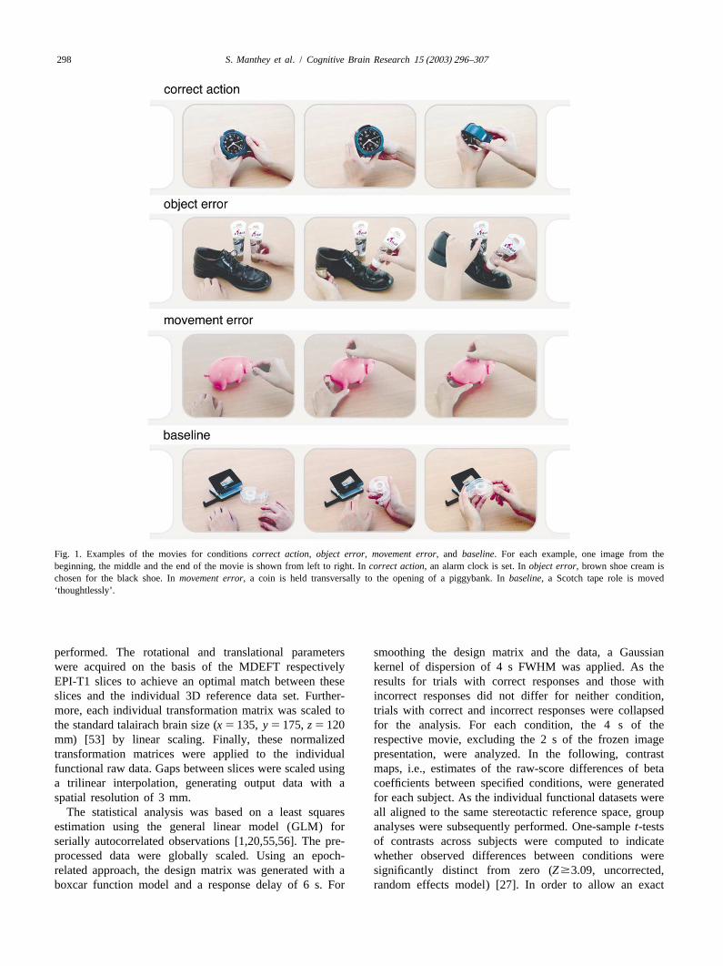
applying a 1/105 Hz highpass filter. The filter length wasIn the correct action condition, movies showed an calculated as 1.5 times the length of one complete oscilla-
action that was performed correctly, such that the intended tion, i.e., maximal interval between two trials of the samegoal was achieved (Fig. 1 and Table 1). In theobject error experimental condition (1.5370 s5105 s). Because lowcondition, the actors’ hands and several objects were frequencies were removed, temporal filtering also effectedpresented, like, e.g., a black shoe, black shoe cream, and the signal baseline correction. The increased autocorrela-brown shoe cream. Instead of using the black shoe cream, tion caused by the filtering was taken into account duringthe hands chose the context-inappropriate object, i.e., the statistical evaluation by an adjustment of the degrees ofbrown shoe cream, and put some of the brown shoe cream freedom. Spatial smoothing was performed using a Gaus-onto the black shoe. In themovement error condition, an sian kernel with a sigma of 0.8.action was performed, but a movement not appropriate to To align the functional images onto a 3D stereotacticmanipulate the concerned object was made, such as, e.g., coordinate reference system, a rigid linear registration withholding a coin transversal to the opening of a piggybank. six degrees of freedom (3 rotational, 3 translational) was
S. Manthey et al. / Cognitive Brain Research 15 (2003) 296–307298
Fig. 1. Examples of the movies for conditionscorrect action, object error, movement error, and baseline. For each example, one image from thebeginning, the middle and the end of the movie is shown from left to right. Incorrect action, an alarm clock is set. Inobject error, brown shoe cream ischosen for the black shoe. Inmovement error, a coin is held transversally to the opening of a piggybank. Inbaseline, a Scotch tape role is moved‘thoughtlessly’.
performed. The rotational and translational parameters smoothing the design matrix and the data, a Gaussianwere acquired on the basis of the MDEFT respectively kernel of dispersion of 4 s FWHM was applied. As theEPI-T1 slices to achieve an optimal match between these results for trials with correct responses and those withslices and the individual 3D reference data set. Further- incorrect responses did not differ for neither condition,more, each individual transformation matrix was scaled to trials with correct and incorrect responses were collapsedthe standard talairach brain size (x 5135, y 5 175,z 5 120 for the analysis. For each condition, the 4 s of themm) [53] by linear scaling. Finally, these normalized respective movie, excluding the 2 s of the frozen imagetransformation matrices were applied to the individual presentation, were analyzed. In the following, contrastfunctional raw data. Gaps between slices were scaled using maps, i.e., estimates of the raw-score differences of betaa trilinear interpolation, generating output data with a coefficients between specified conditions, were generatedspatial resolution of 3 mm. for each subject. As the individual functional datasets were
The statistical analysis was based on a least squares all aligned to the same stereotactic reference space, groupestimation using the general linear model (GLM) for analyses were subsequently performed. One-samplet-testsserially autocorrelated observations [1,20,55,56]. The pre- of contrasts across subjects were computed to indicateprocessed data were globally scaled. Using an epoch- whether observed differences between conditions wererelated approach, the design matrix was generated with a significantly distinct from zero (Z$3.09, uncorrected,boxcar function model and a response delay of 6 s. For random effects model) [27]. In order to allow an exact

299S. Manthey et al. / Cognitive Brain Research 15 (2003) 296–307
Table 1Description of stimulus material employed in the three experimental conditions and the baseline
Correct action Object error Movement error Baseline(appropriate object, (inappropriate movement)inappropriate object)
fold napkin set the table (plate, pan) correct text with white-out (slip off text rollline)
cut apple stir coffee (teaspoon, table open can (not pressing hard enough, can knifespoon) opener not stuck in can)
lace shoe stamp a letter (appropriate punch holes in A4 size paper (set hole cupsize stamp, too big stamp) puncher to punch holes in A5 size paper)
fold laundry prepay letter (stamp, pack suitcase (lay garment on edge of eraserairmail sticker) suitcase and close suitcase)
cut piece of put lid on plastic box stick band-aid on wound (stick partially markerpaper with (appropriate size lid, too beside wound)scissors big lid)put bookmark put lid on pot (appropriate tear out page with picture on it from rulerin book size lid, too small lid) magazine (tear out half the picture)paint fingernail insert whisk into left mixer clean glasses with cloth (rub fingers on hand lotion
opening (left whisk, right glasses)whisk)
screw top off cut bread (appropriate open letter with a letter opener (slip off so corkscrewbottle bread knife, too small that letter is not opened at its edge)
bread knife)put on gloves put refill into ball pen assemble jigsaw puzzle (place last piece pear
(appropriate size refill, too so it does not fit)big refill)
fasten with put cap on green pen write text with ballpoint pen (forget to spongesafety pin (green cap, red cap) make the ballpoint pen ready to write)set alarm clock put on second sock (brown put on ring (put on the wrong finger) calendar
sock, green sock)unzip backpack butter slice of bread (slice put on wristwatch (put on the wrong way stop watch
of bread, piece of cake) around)cut hair staple a batch of paper put money in piggybank (hold the coin vase
(appropriate size stapler, transverse to the piggybank opening)small stapler)
wipe floor sweep floor (broom, mop) mark place on city map (slip off the place passportto be marked)
take cookie out thread a needle (sewing light a candle (holding flame too far away egg cupof box needle, pin) from wick)mix cards cut fingernails (nail put on sandal (entangling toes in slings of bandage
scissors, too big scissors) shoe)peel orange add cocoa to milk (cocoa, stick text on noticeboard (text facing the sweetener
coffee) noticeboard — not readable)turn on heating screw a screw (slotted pan- pour water into glass (pour some of the purse
head screw, crossed pan- water slightly beside glass)head screw)
sew by hand add sugar to coffee (sugar, put flowers in vase (some flower stems money/salt) stick out of the vase) bottle of
shampooopen can (cola) add milk to tea (milk, open bag of sweets (bag not opened magazine/
buttermilk) enough so that no sweets can be taken) bookuse sand paper put frozen food in freezer switch on a fan (switch pressed not hard glass/bottle
(ice cream, milk) enough) of mineralwater
erase text open bottle of wine peel banana (pull upwards instead of fountain(corkscrew, bottle opener) downwards) pen/scissors
take butter put pot of water on stove sweep dirt onto dust pan (sweep beside tin /pepperwith knife (water, orange juice) sweeper)zip jacket unlock door (appropriate open bottle of beer (slip off) alarm clock/
size key, too big key) CDdry hair put lotion on hands (hand cut piece of meat (knife pressed not hard tomato/
lotion, hair wax) enough to cut meat) carrotclose window screw bulb in mounting pick flower (pick only part of the dish towel
(appropriate size bulb, too blossom) /platesmall bulb)

S. Manthey et al. / Cognitive Brain Research 15 (2003) 296–307300
Table 1. Continued
Correct action Object error Movement error Baseline(appropriate object, (inappropriate movement)inappropriate object)
dial telephone perfume oneself (perfume, unlock bicycle lock (hold the key pliers /number hair spray) transverse to the lock) hammerwash hands pour coffee in cup (cup, put on belt (put on belt inside out) cap/sun
glass) glassesclean window put shoe cream on black fax letter (insert sheet of paper ascew in thermos/
shoe (black shoe cream, the fax machine) teaspoonbrown shoe cream)
spray cream pack picknick basket copy text (lay sheet of paper wrong way spirit level /next to cake (bread, hair dryer) around in copy machine) spatulaplay dominos wipe up dirt with cloth close door (forgetting to press down stapler /
(cloth, washrag) doorhandle) staplerinse cup fizzy tablet in glass (fizzy string beads (letting the beads fall off elastic band/
tablet, cap of package) other end of the string) sharpenertyping on the peel potato (peeler, big shave leg (holding the shaver upside scarf /computer knife) down) bathing suitwipe tiles throw coin into cigarette play dice with shaker (not closing the Scotch tape/
automat (appropriate size opening of the shaker so dice fall out of paper blockcoin, too big coin) shaker)
pull up blinds cut flower stem with shears put toothpaste on toothbrush (not bookmark/(appropriate size shears, pressing hard enough to get toothpaste booktoo small shears) out of tube)
drop mix cake mixture (sugar, take piece of cake with fork (not pressing cigarette /newspaper in pepper) hard enough to separate an eatable piece ashtrayletter box of the cake)
anatomical localization of activation peaks in individual action, object error, movement error and baseline) indi-subjects,z-maps were generated for each subject usingt cated a main effect for error rates (F(3,33)514.6, P,
statistics. 0.0001) and reaction times (F(3,33)532.44, P,0.0001)(Fig. 2). Singlet-tests with a Bonferonia-level correctionrevealed that, compared to the classification of correct
3 . Results actions and erroneous actions, the movement classificationwas significantly easier (correct action, F(1,11)514.97,
3 .1. Behavioral performance P,0.003;object error, F(1,11)549.67,P,0.0001;move-ment error, F(1,11)526.15, P,0.0003) and faster (cor-
Behavioral performance was assessed by error rates andrect action, F(1,11)539.39, P,0.0001; object error,by reaction times of correct responses. A repeated mea-F(1,11)529.73, P,0.0002; movement error, F(1,11)5sures ANOVA with the factor CONDITION (correct 35.52, P,0.0001). Moreover, classifications of correct
Fig. 2. Behavioral performance. Error rates and reaction times of the correct responses for the four conditionscorrect action (CA), object error (OE),movement error (ME) and baseline (BA).

301S. Manthey et al. / Cognitive Brain Research 15 (2003) 296–307
actions took significantly longer than those of action slips (ROIs) were defined bilaterally for the vPMC and the aIPS(object error, F(1,11)526.27, P,0.0003; movement according to the respective conditions. ROIs were definederror, F(1,11)532.67,P,0.0001). as spheres with a radius of 6 mm. Their exact locations
were established as follows. A new groupz-map was3 .2. FMRI data generated which resulted from contrasting the conditions
correct action andmovement error against thebaseline, soSeveral brain areas showed significant activations during that the experimental conditions of interest were repre-
the observation of correct actions, actions with object sented in onez-map [8]. ROIs were then centered at theviolation, and actions with movement violation. All con- local maxima of thisz-map within right and left vPMC andditions caused activations within the left vPMC, the frontal aIPS, respectively. Thus, the locations of the ROIs did noteye fields (FEF), the posterior intraparietal sulcus (pIPS), differ across conditions or subjects. For all voxels of athe left pre-supplementary motor area (preSMA), and the ROI, mean contrast values were computed for eachmotion area (MT), relative to baseline (Table 2 and Fig. contrast and subject. These mean values subsequently3). Additionally, conditionscorrect action and movement entered a repeated measures ANOVA.error yielded activations within the right vPMC, the This ANOVA with the factor HEMISPHERE (left, right)supramarginal gyrus (SMG) extending into the aIPS, and indicated, firstly, that the vPMC activation was signifi-the caudal cingulate zone (CCZ). cantly stronger in the right hemisphere than in the left
In order to quantify the hemispheric distribution of hemisphere forcorrect action andmovement error (correctactivation incorrect action andmovement error within (a) action, F(1,11)524.02, P,0.0005; movement error,the vPMC and (b) the aIPS, spherical regions of interest F(1,11)58.04, P,0.016) and, secondly, that the aIPS
Table 2aBaseline contrasts:Correct action (CA), object error (OE), or movement error (ME) versusbaseline (BA)
CA-BA OE-BA ME-BA
Anatomical area x y z Z-score x y z Z-score x y z Z-score
vPMC L 249 0 32 4.1 246 6 23 4.3 243 0 29 4.8240 25 38 4.3
R 46 22 32 4.9 46 22 32 4.6
CCZ 1 0 29 4.3 24 0 32 3.8 4 3 29 4.5
FEF L 225 28 44 5.2 222 3 50 4.7 225 28 44 4.8R 28 22 53 5.1 25 22 47 4.2 22 22 47 4.7
preSMA L 27 21 53 3.6 24 16 55 3.9 24 15 55 3.5R 10 6 50 4.3
MFG L 243 27 26 3.2
SMG L 258 226 32 4.4 258 235 38 4.6258 238 38 3.9
R 55 232 44 4.6 58 232 41 3.9aIPS R 40 235 41 4.3 31 241 41 3.5
pIPS L 231 247 47 4.4 228 250 47 4.8 225 250 47 4.5216 268 50 4.5 219 268 47 5.1 225 277 32 5.1
R 22 274 38 5.0 28 280 41 4.4 25 280 41 5.222 256 55 5.0 22 256 53 4.2 10 253 58 5.1
MT L 243 268 0 5.4 240 268 0 5.0 243 265 0 5.2R 46 253 0 4.2 49 250 23 3.6 52 265 8 4.8
MTG R 40 265 20 3.8 40 265 20 4.2
INS L 237 25 17 4.1 –25 21 –3 4.1 228 18 0 4.1R 37 22 23 4.0
FG L 219 247 0 5.1 219 247 0 4.9 228 250 23 4.4R 28 256 0 4.0 25 244 25 3.6 34 238 0 4.1
CAU L 27 6 11 3.7 24 3 11 4.2R 4 9 11 3.9 4 6 11 4.5 7 9 8 4.2
THA L 24 214 5 4.4R 4 214 5 4.2
Anatomical areas are abbreviated as follows: aIPS anterior intraparietal sulcus, CAU caudate nucleus, CCZ caudal cingulate zone, FEF frontal eye field, FGfusiform gyrus, MFG middle frontal gyrus, INS insula, MT motion area, MTG middle temporal gyrus, pIPS posterior intraparietal sulcus, preSMApre-supplementary motor area, SMG supramarginal gyrus, THA thalamus, vPMC ventrolateral premotor cortex.a Mean location [56] andZ-score of peak activations from group statistics (N512).

S. Manthey et al. / Cognitive Brain Research 15 (2003) 296–307302
Fig. 3. Brain activations duringcorrect action andobject /movement errors. Group statistics (N512) indicating significant activations during observationof correct action, object error andmovement error, relative to the object manipulation baseline were superimposed onto a T1-weighted individual brain. Inorder to show activations without anatomical distortion, the outermost 3 to 5 mm of the brain were removed using a white matter segmentation. The firstcolumn shows the brain from the left side (x-coordinate5252 [53]), the second column shows the top-view (z-coordinate5136), the third column showsthe right side (x-coordinate5152), and the fourth column shows the medial perspective (x-coordinate50). Activation was noted incorrect action andmovement error bilaterally within the ventrolateral premotor cortex (vPMC), whereas inobject error activation was noted only within the left vPMC. Thebilateral activation of the anterior part of the intraparietal sulcus (aIPS) was noted only incorrect action and movement error.
activation was significantly stronger in the left hemisphere humans [9,13,24–26]. Particularly, the vPMC is reportedthan in the right hemisphere formovement error in studies that investigated the observation of manual(F(1,11)517.84,P,0.001). Accordingly, the ROI analy- actions, as e.g. grasping food [21,22,38], grasping a cupsis yielded a right hemisphere dominance within the vPMC and raising it to the mouth as well as the pantomime of thisfor correct action and movement error, and a left hemi- action [9], opening a bottle presented as a pantomimesphere dominance for aIPS activation formovement error. [13,25], grasping objects with precision grip [24] and
manipulating small objects [26].Premotor involvement in action observation is taken to
4 . Discussion reflect a system that matches observed actions onto corre-sponding motor representations [45]. The prerequisite for
The present study investigated whether the observable this matching process is a triggering of the action schemacomponents of action, i.e., employed objects and per- within the premotor cortex that corresponds to the ob-formed movements, are processed differently within the served action. It has been suggested, that this matchingPMC. To this end, each component was manipulated process underlies our ability to recognize observed actions.distinctly in an error detection paradigm. Our findings Moreover, the same matching processes might also under-revealed vPMC activation during the observation of correct lie the learning of actions by means of imitation.actions, and during the observation of both movement- In the present study, the vPMC was activated withinrelated and object-related violations, relative to baseline. both hemispheres during observation of correct actions andHowever, conditionscorrect action and movement error movement errors, and the activation was dominant in theinduced stronger activations within the right vPMC, right hemisphere, as evident from the ROI statistics.whereas conditionobject error induced stronger activa- During observation of object errors, in contrast, the lefttions within the left vPMC. vPMC was activated exclusively.
Premotor activations during action observation are in Note thatZ-scores of group-averaged data inmovementline with findings reported in monkeys [21,38] and in error (Table 2) did not reflect this effect. However, this

303S. Manthey et al. / Cognitive Brain Research 15 (2003) 296–307
lack of consistence can be explained by the fact that the has been indicated by a number of clinical and experimen-interindividual spatial variance of local activation maxima tal studies [15,28,40,54]. In line with these findings, event-was higher within the right PMC (Table 3). Moreover, related potential studies and imaging studies have revealedright hemisphere dominance in movement error was also lateralized activation patterns based on coordinate andreflected by individualZ-scores (Table 3). categorical processing of information, using, e.g., com-
Thinking about possible explanations for this finding, pound letter stimuli [17,42] or visuospatial polygon pat-we propose that the hemispheric lateralization might reflect terns [32].a bias towards one of two possible levels of information Along with these findings, we suggest that left hemi-processing, relating to coordinate (‘global’) and categorical sphere dominance in theobject error condition could(‘local’) representations. As proposed by Kosslyn and reflect the processing of categorical representations, andcolleagues [28], categorical representations capture general right hemisphere dominance in themovement error con-properties of a stimulus without making commitments to dition the processing of coordinate representations, respec-specific properties that are likely to change from instance tively.to instance. This also applies to the observation ofobject Like movement errors, however, thecorrect actionerrors in the present study, which were determined by the condition also revealed dominantly right hemisphere acti-combination of two or more objects inappropriate to vation. This could be due to the fact that both conditionsachieve an action goal. In contrast, coordinate representa- were rather similar in that both required to monitor thetions capture locations of a stimulus or parts of it in terms entire sequence of movements performed on an object inof metric units. This applies to the observation ofmove- order to decide whether the action was correctly performedment errors in the present study, which were determined until the end of the movie. In order to test this hypothesis,by the way that an object or tool is moved or manipulated a post-hoc behavioral study was conducted. Sixteen sub-in relation to another. The processing of coordinate and jects had to classify trials showingcorrect actions, objectcategorical representations have been suggested to beerrors andmovement errors in a speeded choice paradigm,lateralized, i.e., within right-handers, coordinate repre- i.e., the answer was required during the movie pre-sentations are processed dominantly within the right sentation. As a result, both the classification of correcthemisphere, whereas categorical representations are pro- actions (2630 ms) and that of movement errors (2677 ms)cessed dominantly within the left hemisphere [28]. This took significantly longer than the classification of object
Table 3aBaseline contrasts:Correct action (CA), object error (OE), or movement error (ME) versusbaseline (BA)
CA-BA OE-BA ME-BA
Subject x y z Z-score BA x y z Z-score BA x y z Z-score BA1 L 241 2 21 5.2 6 244 2 21 7.4 6 247 2 24 4.6 6
R 43 7 20 5.1 6 46 10 19 4.0 6 43 8 23 5.5 6/442 L 247 6 22 5.6 6 247 10 24 5.1 6 244 8 16 5.2 6
R 46 3 20 6.2 6 46 3 23 3.7 6 46 3 20 6.6 63 L 247 10 28 7.7 6 247 13 27 8.3 6 247 10 28 7.0 6
R 49 13 27 8.1 6 46 11 23 6.6 6/44 49 13 27 7.3 64 L 253 5 29 4.9 6 241 6 20 5.7 6 247 8 31 5.8 6
R 52 4 26 7.3 6 49 5 29 4.4 6 52 4 26 7.0 65 L 238 13 10 4.0 44 238 19 23 4.7 44 241 12 7 3.5 44
R 49 10 11 4.3 6 46 18 20 3.8 44 43 7 11 4.4 66 L 244 3 33 7.6 6 241 1 25 5.6 6 241 1 27 5.8 6
R 49 7 23 6.2 6 43 0 22 2.3* 6 55 12 19 5.8 67 L 262 4 21 4.5 6 253 5 24 6.1 6 259 1 22 5.8 6
R 52 5 24 4.3 6 37 4 21 2.3* 6 49 0 19 4.8 68 L 250 4 15 4.6 6 253 6 13 4.0 6/44 250 3 12 5.6 6
R 52 2 21 5.9 6 55 8 8 2.7* 44 52 5 21 6.4 69 L 246 0 39 5.5 6 235 4 24 4.9 6/44 247 3 21 5.1 6
R 43 1 24 5.6 6 34 21 25 4.0 6 46 0 22 7.5 610 L 247 2 30 6.8 6 244 4 23 8.1 6 247 2 30 8.1 6
R 49 2 30 7.7 6 43 21 25 6.3 6 49 2 30 5.9 611 L 250 5 27 4.4 6 247 5 27 5.7 6 247 3 18 4.8 6/44
R 49 0 13 6.7 6 43 0 19 6.3 6 46 0 16 5.1 612 L 244 13 25 4.2 6 238 12 23 6.6 6 244 13 25 4.7 6
R 46 13 25 5.4 6 31 12 23 5.3 6/44 40 5 21 5.3 6
* Non-significantZ-score.a Mean location [56] andZ-score of peak activations within the premotor cortex (including the corresponding Brodmann area, BA) of individual subjects.

S. Manthey et al. / Cognitive Brain Research 15 (2003) 296–307304
errors (2240 ms) (correct action, F(1,15)517.72, P, part of the premotor cortex in BA 6. Based on the present0.0008;movement error, F(1,15)546.53,P,0.0001). We findings and other imaging studies [9,24], it could betake this result to indicate that participants tended to suggested that activation within ventral BA 6 duringreassure precisely that correct actions did not contain any observation of hand actions corresponds to that found indeviant movement. monkey area F5. However, BA 44 and BA 6 are not only
A slightly different explanation comes from imaging closely adjacent areas, but also expose a high interin-studies that reveal exclusively left PMC involvement dividual variance. Accordingly, it is difficult to straight[11,36] and dominantly left PMC involvement [48] during forwardly ascribe group averaged activations to one ofpresentation of objects. Accordingly, a left hemisphere these areas.preference for objects could be the cause for the left Therefore, we determined the anatomical location ofpremotor dominance in observation ofobject errors in the ventral premotor activation within each single subject forpresent study. This could be taken to reflect that when the baseline contrasts ofcorrect action, object error, andattention was attracted by the erroneously chosen object,movement error (Table 3 and Fig. 4). Within the leftaction schemata related to this inappropriate object could hemisphere, ventrolateral premotor activation was locatedhave been triggered in addition to that triggered by the in BA 6 within 11 subjects forcorrect action, but onlyappropriate object. Hence, one would expect an increase of within 1 subject in BA 44. Forobject error, 9 subjectsactivation within the areas that prepare and represent showed activation in BA 6, 2 subjects in BA 6/44, and 1action schemata of the hand that manipulates the in- subject in BA 44. Finally, formovement error, 10 subjectsappropriate object, i.e., the left PMC, as corresponding to revealed activations in BA 6, 1 subject in BA 6/44, and 1the right hand. subject in BA 44. If we take the inferior precentral sulcus
A further alternative explanation of the hemisphere as a landmark separating BA 6 and BA 44, our findingseffect is that the initial analysis of the observed action is indicate that ventrolateral premotor activation was domi-made by the left hemisphere, whereas a more detailed nantly in BA 6 rather than in BA 44. Generally, it has beanalysis of movements is carried out by the right hemi- mentioned that it is difficult to separate Brodmann Areassphere [25]. As the initial analysis of the observed action is from each other on the basis of anatomical landmarks,sufficient to classify object errors, a left hemisphere because cytoarchitectonically distinct areas can sometimesdominance would be expected. In contrast, for correct transverse anatomical structures [2]. However, the inferioractions and movement errors a detailed analysis of move- precentral sulcus appears to be a rather reliable borderments has to be made additionally, leading to a right between BA 6 and BA 44, as opposed to other anatomicalhemisphere involvement. According to this view, if the landmarks, such as e.g. the ramus ascendens separating BAinitial action analysis precedes detailed movement analy- 44 and BA 45 [2]. Moreover, our single subject analysissis, the effect in question could be due to a different demonstrates that ventrolateral premotor activation duringresponsiveness of left and right hemisphere to early versus action observation was dominantly located within thelate segments of observed actions. To test this, a post-hoc ventral precentral gyrus. Interindividual and intraindividualanalysis was carried out. The early phase of action (condition-related) variation of premotor activation loca-observation was contrasted with the late phase for each tion was surprisingly small. Together, our findings indicatesingle condition. As a result, no significant activation that during the observation of actions, not Broca’s areadifferences were found within the PMC between early and (BA 44), but rather ventral BA 6 is dominantly involved.late action analysis. Likewise, baseline contrasts, computed However, one may object that, for determining theseparately over the early phase and the late phase of movie anatomical location of premotor activations, the movementpresentation each, revealed the same patterns of activation baseline was subtracted from the experimental conditions.as the analysis of the entire presentation phase. Therefore, However, since both the baseline as well as the experimen-both analyses did not support the notion of a preferred tal conditions presentedproximal and distal movementsresponsiveness of the left hemisphere to early segments of (reaching and manipulation), subtraction should not haveactions, and of the right hemisphere to late segments of biased activation in favor of either type of movement. Thisaction, respectively. In contrast, we propose that categori- is important because distal movements have been sug-cal and coordinate information processing underlies differ- gested to be represented more anteriorly / inferiorly thanent involvement of the left and the right hemisphere. proximal movements in monkey BA 6 [23].
In monkeys, the cortical area suggested to realize a From a broader perspective, there are two functionalmatching system for action observation and action execu- interpretations concerning BA 6 and BA 44, that might betion is area F5 [45]. It has been suggested that the human interesting to consider here. Firstly, there is evidence thathomologue is located within BA 44 in the frontal opercular the perceptual analysis of dynamic object patterns involvescortex (FOP). This suggestion was based on functional, BA 6 [48,50]. Accordingly, theanalysis of both observedanatomical, and cytoarchitectonic findings concerning both actions and observed dynamic object patterns might in-cortical areas [39,41,43]. In contrast, premotor activations volve a common cognitive process reflected by BA 6in the present study were focused within the ventrolateral activation. This could be the prediction of the expected

305S. Manthey et al. / Cognitive Brain Research 15 (2003) 296–307
Fig. 4. Mean location [53] of peak activations within the left ventrolateral premotor cortex (vPMC) of individual subjects for all baseline contrasts. Dotcolors correspond to conditions:correct action (green),object error (yellow) andmovement error (red), respectively.
course of movements caused by both non-living and living observation, whereas aIPS activation might be related toentities. Both BA 6 and BA 44 have been reported to be object affordances themselves.involved in the detection of violation in expected events. This interpretation would also be in line with the findingThus, several studies indicate that BA 44 is activated when that the activation was bilateral, since bimanual actionssyntax errors are detected in perceived language were presented. Moreover, in contrast tocorrect action, the[10,12,19,37]. Likewise, fMRI studies report BA 6 to be observation ofmovement error yielded a statisticallyactivated when sequential errors are detected in target significant activation bias towards the left hemisphere.motion [48–50]. From that one may suggest that the This might indicate that the finger movements of the rightcortical region around BA 6 and BA 44 is crucial in the hand were analyzed more intensively in this condition, asdetection of structure violations. the movement error occurred in this hand, and thus,
An area comprising the aIPS and SMG was activated attention was focused on the dominant hand. Accordingly,bilaterally, relative to baseline. The activation extended a direct correspondence between the amount of attentionfrom the SMG into the aIPS with a local maximum within paid to the analysis of finger movements concerning onethe SMG, and did not show hemispheric lateralization. specific hand might be reflected by a lateralized aIPSBoth regions, the aIPS [4–6] and the SMG [7,24,29] have activation. This would comply with the fact that thebeen discussed as possible human functional homologues parietal cortex exhibits contralateral dominant connectionsof the AIP/PF-area in the monkey. This area is closely concerning effectors similar to the PMC [4,35].linked with the vPMC both anatomically and functionally With regard to the aIPS role in observed actions and its[31,33,34], especially with regard to sensorimotor mapping interpretation, conflicting findings have been made. In afunctions [16,22]. Particularly, both monkey PF [18,22] study by Bonda and coworkers [7] aIPS activation re-and human SMG [7,9,24] were reported to be involved flected the observation of manual actions as compared toduring action observation, whereas both monkey AIP [47] body motions. Both conditions were realized by a lightand human aIPS [4–6] were suggested for grasping and point display. This implicates that it is not necessary totactile exploration of objects. These areas are taken to be observe an actual object employed in the action. Ininvolved within two parallel premotor-parietal circuits contrast, observing a pantomime of an action did not elicitrelevant for action, the one suggested to match observed an aIPS activation in a study by Buccino and co-workeractions onto motor representations, the other involved in [9]. The authors, therefore, suggested that an actual objectsolving object affordances. has to be present in an observed action to evoke aIPS
Therefore, even though the parietal activation we found activation. In the present study, we presented real objectsdid not clearly separated into two distinct activation foci, employed in manual actions. However, objects were alsoone might suggest them to reflect different functional presented in the baseline and were unintentionally movedsubprocesses. Accordingly, activation within SMG might by hands. Therefore, our results suggest that aIPS getsbe caused by an object-related analysis during action involved not due to actual object presentation or a move-
S. Manthey et al. / Cognitive Brain Research 15 (2003) 296–307306
[11] L.L. Chao, A. Martin, Representation of manipulable man-madement directed towards an object, but rather due to goal-objects in the dorsal stream, Neuroimage 12 (2000) 478–484.directed object-related movements, i.e., movements in the
[12] M. Dapretto, S.Y. Bookheimer, Form and content: dissociatingcontext of an action. syntax and semantics in sentence comprehension, Neuron 24 (1999)
427–432.`[13] J. Decety, J. Grezes, N. Costes et al., Brain activity during
observation of actions. Influence of action content and subject’s5 . Conclusion strategy, Brain 120 (1997) 1763–1777.
[14] W.H. Dittrich, Action categories and the perception of biologicalThe present findings support the view that the vPMC motion, Perception 22 (1993) 15–22.
[15] R. Egly, R. Rafal, J. Driver, Y. Starrveveld, Covert orienting in the(BA 6) and its parietal input zone are crucial for thesplit brain reveals hemispheric specialization for object-basedobservation of actions in humans. Moreover, we demon-attention, Psychol. Sci. 5 (1994) 380–383.strated that by attracting attention to either objects or to
[16] L. Fadiga, L. Fogassi, V. Gallese, G. Rizzolatti, Visuomotor neurons:movements in action observation it is possible to examine ambiguity of the discharge or ‘motor’ perception?, Int. J. Psycho-how different components of this premotor-parietal net- physiol. 35 (2000) 165–177.
[17] G.R. Fink, P.W. Halligan, J.C. Marshall, C.D. Frith, R.S.J. Frac-work within each hemisphere contribute to action analysis.kowiak, R.J. Dolan, Neural mechanisms involved in the processingof global and local aspects of hierarchically organized visual stimuli,Brain 120 (1997) 1779–1791.
[18] L. Fogassi, V. Gallese, L. Fadiga, G. Rizzolatti, Neurons respondingA cknowledgementsto the sight of goal-directed hand/arm actions in the parietal area PF(7b) of the macaque monkey, Soc. Neurosci. Abs. 24 (1998) 257.5.We thank Marcel Brass, Christian Fiebach, Andrea Gast-
[19] A.D. Friederici, M. Meyer, D.Y. von Cramon, Auditory languageSandmann, Sonja Lattner, Stefan Pollmann, Markus comprehension: An event-related fMRI study on the processing ofUllsperger and Stefan Zysset for helpful comments on the syntactic and lexical information, Brain Lang. 74 (2000) 289–300.
[20] K.J. Friston, A.P. Holmes, K.J. Worsley, J.P. Poline, C.D. Frith,manuscript and technical support, and Volker Bosch,R.S.J. Frackowiak, Statistical parametric maps in functional imag-¨Gabriele Lohmann and Karsten Muller for support ining: a general linear approach, Hum. Brain Mapp. 2 (1995) 189–statistical analysis.210.
[21] V. Gallese, L. Fadiga, L. Fogassi, G. Rizzolatti, Action recognitionin the premotor cortex, Brain 119 (1996) 593–609.
[22] V. Gallese, L. Fogassi, L. Fadiga, G. Rizzolatti, Action representa-R eferencestion and the inferior parietal lobule, in: W. Prinz, B. Hommel (Eds.),Attention and Performance XIX, Oxford University Press, Oxford,
[1] G.K. Aguirre, E. Zarahn, M. D’Esposito, Empirical analysis of 2002.BOLD fMRI statistics. II. Spatially smoothed data collected under [23] M. Gentilucci, L. Fogassi, G. Luppino, M. Matelli, R. Camarda, G.null-hypothesis and experimental conditions, NeuroImage 5 (1997) Rizzolatti, Functional organization of inferior area 6 in the macaque199–212. monkey. I. Somatotopy and the control of proximal movements,
¨[2] K. Amunts, A. Schleicher, U. Burgel, H. Mohlberg, H.B.M. Exp. Brain Res. 71 (1988) 475–490.Uylings, K. Zilles, Broca’s region revisited: cytoarchitecture and [24] S.T. Grafton, M.A. Arbib, L. Fagida, G. Rizzolatti, Localization ofintersubject variability, J. Comp. Neurol. 412 (1999) 319–341. grasp representations in humans by positron emission tomography:
[3] M.A. Arbib, Perceptual structures and distributed motor control, in: 2. Observation compared with Imagination, Exp. Brain Res. 112V.B. Brooks (Ed.), Handbook of Physiology — The Nervous (1996) 103–111.
`System, II, American Physiological Society, Bethesda, 1981, pp. [25] J. Grezes, N. Costes, J. Decety, Top-down effect of strategy on the1449–1480, Part 1. perception of human biological motion: a PET investigation, Cogn.
[4] F. Binkosfki, C. Dohle, S. Posse et al., Human anterior intraparietal Neuropsychol. 15 (1998) 553–582.area subserves prehension, Neurology 50 (1998) 1253–1259. [26] R. Hari, N. Forss, S. Avikainen, E. Kirveskari, S. Salenius, G.
[5] F. Binkosfki, G. Buccino, S. Posse, R.J. Seitz, G. Rizzolatti, H.J. Rizzolatti, Activation of human primary motor cortex during actionFreund, A fronto-parietal circuit for object manipulation in man: observation: a neuromagnetic study, Proc. Natl. Acad. Sci. USA 95evidence from an fMRI-study, Eur. J. Neurosci. 11 (1999) 3276– (1998) 15061–15065.3286. [27] A.P. Holmes, K.J. Friston, Generalisability, random effects and
[6] F. Binkosfki, G. Buccino, K.M. Stephan, G. Rizzolatti, R.J. Seitz, population inference, NeuroImage 7 (1998) S754.H.J. Freund, A parieto-premotor network for object manipulation: [28] S.M. Kosslyn, O. Koenig, A. Barrett, C. Cave, J. Gabrieli, Evidenceevidence from neuroimaging, Exp. Brain Res. 128 (1999) 210–213. for two types of spatial representations: Hemispheric specialization
[7] E. Bonda, M. Petrides, D. Ostry, A. Evans, Specific involvement of for categorical and coordinate relations, J. Exp. Psychol. Hum.human parietal systems and the amygdala in the perception of Percept. 15 (1989) 723–735.biological motion, J. Neurosci. 16 (1996) 3737–3744. [29] M. Krams, M.F.S. Rushworth, M.P. Deiber, R.S.J. Frackowiak, R.E.
[8] V. Bosch, Statistical analysis of multi-subject fMRI data: the Passingham, The preparation, execution and suppression of copiedassessment of focal activations, J. Magn. Reson. Imaging. 11 (2000) movements in the human brain, Exp. Brain Res. 120 (1998) 386–61–64. 398.
¨[9] B. Buccino, F. Binkofski, G.R. Fink et al., Action observation [30] G. Lohmann, K. Muller, V. Bosch et al., Lipsia — A new softwareactivates premotor and parietal areas in a somatotopic manner: an system for the evaluation of functional magnetic resonance imagesfMRI study, Eur. J. Neurosci. 13 (2001) 400–404. of the human brain, Comput. Med. Imaging Graph. 25 (2001)
[10] D. Caplan, N. Alpert, G. Waters, A. Olivieri, Activation of Broca’s 449–457.areas by syntactic processing under conditions of concurrent articu- [31] G. Luppino, A. Murata, P. Govoni, M. Matelli, Largely segregatedlation, Hum. Brain Mapp. 9 (2000) 65–71. parietofrontal connections linking rostral intraparietal cortex (areas

307S. Manthey et al. / Cognitive Brain Research 15 (2003) 296–307
AIP and VIP) and the ventral premotor cortex (areas F5 and F4), [44] G. Rizzolatti, L. Fogassi, V. Gallese, Cortical mechanisms subserv-Exp. Brain Res. 128 (1999) 181–187. ing object grasping and action recognition: a new view on the
[32] A. Martinez, P. Moses, L. Frank, R. Buxton, E. Wong, J. Stiles, cortical motor functions, in: M.S. Gazzaniga (Ed.), The NewHemispheric asymmetries in global and local processing: evidence Cognitive Neurosciences, MIT Press, Cambridge, 2000, pp. 539–from fMRI, Neuroreport 8 (1997) 1685–1689. 552.
[33] M. Matelli, G. Luppino, Parietofrontal circuits for action and space [45] G. Rizzolatti, L. Fogassi, V. Gallese, Neurophysiological mecha-perception in the macaque monkey, Neuroimage 14 (2001) S27– nisms underlying the understanding and imitation of action, NatureS32. Neurosci. 2 (2001) 661–670.
[34] M. Matelli, G. Luppino, G. Rizzolatti, Independent anatomical [46] D.E. Rumelhart, Schemata: The building blocks of cognition, in:circuits for reaching and grasping linking the inferior parietal sulcus R.J. Spiro, B.C. Bruce, W.F. Brewer (Eds.), Theoretical Issues inand inferior area 6 in macaque monkey, Soc. Neurosci. Abs. 20 Reading Comprehension, Erlbaum, Hillsdale, NJ, 1980.(1994) 404.4. [47] H. Sakata, M. Taira, A. Murata, S. Mine, Neural mechanisms of
[35] V.S. Mattay, J.H. Callicott, A. Bertolino et al., Hemispheric control visual guidance of hand actions in the parietal cortex of the monkey,of motor function: a whole brain echo planar fMRI study, Psychiatry Cereb. Cortex 5 (1995) 429–438.Res. 83 (1998) 7–22. [48] R.I. Schubotz, D.Y. von Cramon, Functional organization of the
[36] A. Mecklinger, C. Gruenewald, M. Besson, M.-N. Magnie, D.Y. von lateral premotor cortex: fMRI reveals different regions activated byCramon, Separable neuronal circuitries for manipulable and non- anticipation of object properties, location and speed, Cogn. Brainmanipulable objects in working memory, submitted. Res. 11 (2001) 97–112.
[37] M. Meyer, A.D. Friederici, D.Y. von Cramon, Neurocognition of [49] R.I. Schubotz, D.Y. von Cramon, Interval and ordinal properties ofauditory sentence comprehension: event related fMRI reveals sen- sequences are associated with distinct premotor areas, Cereb. Cortexsitivity to syntactic violations and task demands, Cogn. Brain Res. 9 11 (2001) 210–222.(2000) 19–33. [50] R.I. Schubotz, D.Y. von Cramon, Dynamic patterns makes the
[38] G. di Pellegrino, L. Fadiga, L. Fogassi, V. Gallese, G. Rizzolatti, premotor cortex interested in objects: influence of stimulus and taskUnderstanding motor events: a neurophysiological study, Exp. Brain revealed by fMRI, Cogn. Brain Res. (in press).Res. 91 (1992) 176–180. [51] M.F. Schwartz, E.S. Reed, M. Montgomery, C. Palmer, N.H. Mayer,
[39] M. Petrides, D.N. Pandya, Comparative architectonic analysis of the The quantitative description of action disorganization after brainhuman and the macaque frontal cortex, in: F. Boller, J. Grafman damage: a case study, Cogn. Neuropsychol. 8 (1991) 381–414.(Eds.), Handbook of Neuropsychology, Vol. 9, Elsevier, Amsterdam, [52] M.F. Schwartz, Re-examining the role of executive functions in1994, pp. 17–58. routine action production, Ann. NY Acad. Sci. 769 (1995) 321–335.
[40] E. Phelps, M.S. Gazzaniga, Hemispheric differences in mnemonic [53] J. Talairach, P. Tournoux, Co-planar Stereotaxis Atlas of the Humanprocessing: the effects of left hemisphere interpretation, Neuro- Brain, Thieme, New York, 1988.psychologia 30 (1992) 293–297. [54] M. Vitkovitch, G. Underwood, Visual field differences in an object
[41] T.M. Preuss, I. Stepniewska, J.H. Kaas, Movement representation in decision task, Brain Cogn. 19 (1992) 195–207.the dorsal and ventral premotor areas of owl monkeys: a mi- [55] K.J. Worsley, K.J. Friston, Analysis of fMRI time-series revisited-crostimulation study, J. Comp. Neurol. 371 (1996) 649–675. again, NeuroImage 2 (1995) 173–181.
[42] A.M. Proverbio, A. Minniti, A. Zani, Electrophysiological evidence [56] E. Zarahn, G.K. Aguirre, M. D’Esposito, Empirical analysis ofof a perceptual precedence of global versus local visual information, BOLD fMRI statistics. I. Spatially smoothed data collected underCogn. Brain Res. 6 (1998) 321–334. null-hypothesis conditions, NeuroImage 5 (1997) 179–197.
[43] G. Rizzolatti, M.A. Arbib, Language within our grasp, TrendsNeurosci. 21 (1998) 188–194.