research report cellular localization and...
TRANSCRIPT
Ž .Brain Research 778 1997 338–345
Research report
Cellular localization and expression of the serotonin transporter in mousebrain
Dietmar Bengel a,c,) , Olaf Johren b, Anne M. Andrews a, Armin Heils c, Rainald Moßner c,¨ ¨Gilberto L. Sanvitto b, Juan M. Saavedra b, Klaus-Peter Lesch c, Dennis L. Murphy a
a Section on Clinical Neuropharmacology, Laboratory of Clinical Science, NIMH, NIH Clinical, Center, 10r3D41, Bethesda, MD 20892, USAb Section on Pharmacology, Laboratory of Clinical Science, NIMH, NIH Clinical, Center, 10r3D41, Bethesda, MD 20892, USA
c Department of Psychiatry, UniÕersity of Wurzburg, Wurzburg 97080 , Germany¨ ¨
Accepted 19 August 1997
Abstract
Ž . Ž .The high-affinity serotonin 5-HT transporter 5-HTT plays an important role in the removal of extracellular serotonin, therebymodulating and terminating the action of this neurotransmitter at various pre- and post-synaptic serotonergic receptors and heterorecep-tors. In order to characterize the anatomical distribution of the 5-HTT in mouse brain, in situ hybridization histochemistry using35 w125 xS-labeled riboprobes was performed. These results were compared with 5-HTT binding site distribution as evaluated by I RTI-55autoradiography. High levels of 5-HTT mRNA were detected in all brain stem raphe nuclei, with variations in labeling among the varioussubnuclei. Those brain areas known to possess serotonergic cell bodies stained intensely for both 5-HTT mRNA and 5-HTT binding sites.In contrast to previous findings in rat brain, the highest densities of 5-HTT sites were found in areas outside the raphe complex,particularly in the substantia nigra, globus pallidus, and superior colliculi. q 1997 Elsevier Science B.V.
w125 xKeywords: Serotonin transporter; Raphe nuclei; Gene expression; In situ hybridization; I RTI-55 binding; Mice
1. Introduction
It has long been recognized that the primary mechanismfor the termination of a wide range of neurotransmittersignals is the removal of those transmitters from thesynaptic cleft and adjacent extracellular areas by rapid
w xuptake into presynaptic neurons 1,22,48 . The serotonergicsystem is an important modulator of various sensory,motor, behavioral and developmental processes. Despite
w xthe existence of at least 14 different 5-HT receptors 19 , itis the single cell membrane 5-HTT, located presynapti-cally, that is believed to be the most important element inthe regulation of both 5-HT signaling and tone of the
w xserotonergic system 1,7 .Over the last few years, cDNAs encoding the 5-HTT
w xhave been isolated and characterized in vitro 6,18,25,37 ,facilitating studies of expression of this molecule in vitroas well as in vivo. The 5-HTT belongs to the family of cell
) Corresponding author. Laboratory of Clinical Science, NIMH, NIH,Building 10, Room 3D41, 10 Center DR MSC 1264, Bethesda, MD
Ž .20892-1264. Fax: q1 301 402-0188; E-mail: [email protected]
membrane-bound neurotransmitter transporters, and it ex-hibits considerable amino acid sequence homology withdopamine, norepinephrine, and GABA transporters by hav-ing 12 putative transmembrane domains and requiring Naq
w xfor uptake 8,48 . In addition to the rapid removal ofendogenous 5-HT from the extracellular space, the 5-HTTis also regarded as a major site of action for widely usedantidepressants and some drugs of abuse such as 3,4-meth-
Ž . w xylenedioxymethamphetamine, MDMA, ‘ecstasy’ 29,39 .Moreover, imbalances in brain serotonergic neurotransmis-sion are thought to underlie conditions such as depression,obsessive-compulsive disorder and panic disorder as well
w xas alcoholism 12,32,33,35 . Most recently a two allelepromotor polymorphism in the human 5-HTT has been
w xassociated with anxiety-related personality traits 28 .w xCloning and characterization of the rat 6,18 , human
w x w x25,37 and most recently the murine 4,9 5-HTT havebeen reported. Subsequently, more detailed informationhas been gathered concerning the genetic organization,function, and distribution of this neurotransporter in hu-
w x w xmans 30 and rats 15,31,38 . In humans, several differentmRNA species resulting from both alternative splicing andconsecutive usage of additional polyadenylation sites were
0006-8993r97r$17.00 q 1997 Elsevier Science B.V. All rights reserved.Ž .PII S0006-8993 97 01080-9

( )D. Bengel et al.rBrain Research 778 1997 338–345 339
w xdetected 2,16 . In mice, Northern blot analysis of midbrainraphe tissue revealed the presence of only a single mRNAband migrating at ;3 kb, whereas RT-PCR revealed two5-HTT mRNAs resulting from the alternatively spliced
Ž .exon I Heils et al.,unpublished resultsIn the present study, we localized the gene expression
of the 5-HTT in mouse brain by in situ hybridizationhistochemistry. In addition, the mRNA expression pattern
w125 xwas compared with I RTI-55 binding to the 5-HTT incorresponding brain sections. While the murine serotoner-gic system is becoming increasingly of interest because of
w xnewly-developed transgenic and knock-out animals 40,47 ,little has been published regarding the serotonergic path-ways in this species. Our aim was to identify the neuronalcell types expressing 5-HTT mRNA and binding sites andto provide more detailed information about the localizationof the 5-HTT as a basis for further studies of serotonergicpathways in the mouse brain.
2. Materials and methods
2.1. Animal and tissue preparation
All experiments were carried out in accordance with theNational Institutes of Health Guide for the Care and Use of
Ž .Laboratory Animals. Adult male mice 8 weeks of agewere euthanized by cervical dislocation. The brains wereremoved, immediately dissected, frozen in isopentane on
Ždry ice, and stored at y808C. Coronal brain sections 16.mm were cut at y208C in a cryostat. Adjacent sections
were collected for comparison of hybridization andw125 xI RTI-55 binding. For in situ hybridization, sections
Žwere thaw-mounted on silanated glass slides Digene Diag-.nostics, Beltsville, MD and stored at y808C. For 5-HTT
autoradiography, sections were thaw-mounted on gelatin-coated glass slides, dried overnight in a desiccator at 48C,and stored at y808C. Brain regions were anatomically
w xlocalized using two mouse brain atlases 14,41 .
2.2. Subcloning and in Õitro transcription of riboprobes
A 470 bp fragment of the murine 5-HTT cDNA corre-sponding to nucleotides 2147–2617 of the 3X untranslated
Žregion of the short splice variant nucleotides 143–613 of.exon 14, GenBank Database accession number Y08880
Žwas subcloned into a pGEM vector Promega, Madison,.WI . Within this region, low homology exists between
norepinephrine and dopamine transporters. For in vitroŽ .transcription of sense control and antisense riboprobes,
subclone-containing plasmids were linearized with EcoRIand XbaI, respectively. In vitro transcription was per-
w35 x Žformed in the presence of 200 mCi S UTP 800.Cirmmol; Amersham, Arlington Heights, IL , 1 mg lin-
Ž .earized plasmid DNA and T7 antisense and SP6 RNAŽ .polymerase sense using a RNA transcription kit accord-
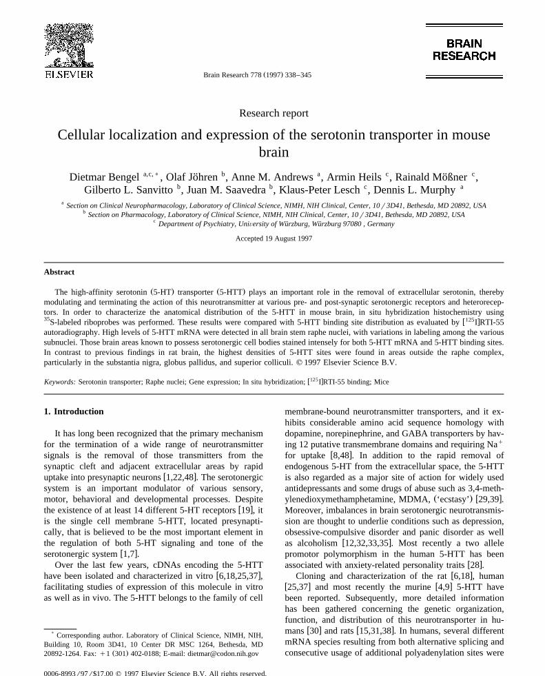
Ž . Ž .Fig. 1. Antisense A and sense B autoradiographies. Dorsal rapheŽ . Ž .nucleus DRN , median raphe nucleus MnR . Note the low diffuse signal
obtained with the sense probe.
ing the manufacturer’s protocol. After transcription, tem-plate DNA was digested with DNAse-1 for 15 min at378C. Labeled riboprobes were separated from unincorpo-
w35 xrated S UTP by centrifugation through spin columnsŽ .Pharmacia, Piscataway, NJ . The integrity of riboprobeswas monitored by polyacrylamide gel electrophoresis and
Ž .subsequent autoradiography see Fig. 1 .
2.3. In situ hybridization histochemistry
In situ hybridization was performed as described earlierw x21 . In brief, after fixation in 4% paraformaldehyde, sec-tions were acetylated for 10 min in 0.1 M triethanolamineHCl, pH 8.0 containing 0.25% acetic anhydride, dehy-drated through graded concentrations of ethanol, and airdried. Hybridization buffer containing 50% formamide, 0.3M NaCl, 2 mM EDTA, 20 mM Tris pH 8.0, 1= Den-hardt’s solution, 10% dextran sulfate, 100 mg mly1 salmonsperm DNA, 250 mg mly1 yeast tRNA, 150 mM DDT,0.1% SDS, and 4=107 c.p.m. mly1 sense or antisenseprobe was applied to each slide. After hybridization for 18h at 548C, sections were rinsed in 4= standard saline
Žcitrate 1= SSC 0.15 M sodium chloride and 0.015 M

( )D. Bengel et al.rBrain Research 778 1997 338–345340
.sodium citrate . Non-hybridized probes were digested byy1 Žincubation with 40 mg ml RNAse A Sigma Chemical,
.St. Louis, MO for 30 min. After a final high stringencywash in 0.1=SSC at 658C for 60 min, sections weredehydrated in graded concentrations of ethanol containing0.3 M ammonium acetate and air dried. Sections were
3 Žexposed to Hyperfilm- H Amersham, Arlington Heights,.IL for 2–3 days. Positive hybridization signals were
evaluated by the cellular localization of silver grains ob-tained by emulsion autoradiography. Semiquantitativeanalyses were performed on digitized images from X-rayfilms in the linear range of the grey values using an imageanalysis system equipped with a computer-based cameraŽ .software NIH image v. 1.62 . Subnuclei of the raphecomplex were measured ten times in four mice. Meandensities "S.E.M. are shown in Table 1.
2.4. Autoradiographic determination of 5-HT uptake sites
Ž .Brains were sectioned sagittally and coronally 20 mmat y208C and thaw-mounted on gelatin coated slides.w125 xI RTI-55 was initially diluted in a 50 mM sodiumphosphate buffer, pH 7.4, containing 1 mgrml bovineserum albumin, 25 mgrml chymostatin, 25 mgrml leu-peptin, 100 mM EDTA and 100 mM EGTA to 150 000c.p.m.rml. Sections were subsequently incubated for 90min at 48C in a 1:10 dilution of radioligand in 50 mM
w Ž . x Žphosphate buffer. 1- 2- diphenylmethoxy ethyl -4- 3-phen-. Ž .ylpropyl homopiperazine LR 1111, 10 mM was used to
w125 xinhibit binding of I RTI-55 to dopamine uptake sites.Nonspecific binding was determined in the presence of 10mM paroxetine and represented -10% of the total bind-ing. Following incubation, slides were rinsed twice in cold
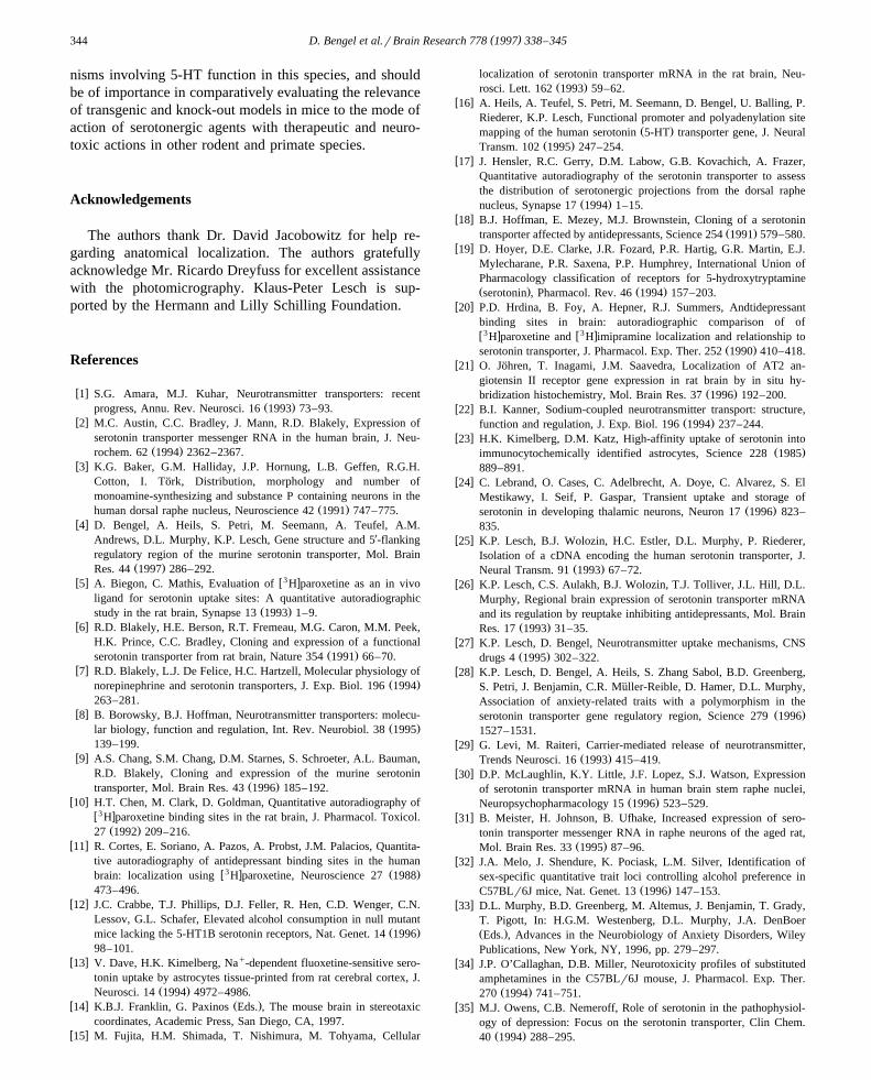
Ž . Ž .Fig. 2. Dark B, D and bright field photomicrographs A, C, E , showing neurons containing serotonin transporter mRNA in the adult mouse brain stem.Ž . Ž . Ž .Rostral linear nucleus raphe RLi , dorsal raphe nucleus, dorsal part DRD , dorsal raphe nucleus, ventral part DRV , dorsal raphe nucleus, ventrolateral
Ž . Ž . Ž . Ž .part DRVL , dorsal raphe nucleus, inferior part DRI , median raphe nucleus MnR , caudal linear raphe nucleus CLi . All images are 50-fold magnified.Ž .Reproduced at 80%.

( )D. Bengel et al.rBrain Research 778 1997 338–345 341
Ž .phosphate buffer, dipped in distilled water 48C and driedunder a stream of cool air. The slides were apposed to
Žradiosensitive films Hyperfilm, Amersham, Arlington. Žw125 xHeights, IL with plastic standard I microscales,
. w125 xAmersham for 3 days at 48C. I RTI-55 binding wasquantitated by measuring each region of interest threetimes in four animals using a standard curve generated
w125 xfrom the I microscales using the NIH Image programŽand are expressed as nCirmg tissue mean"S.E.M., ns
.4 .
3. Results
Hybridization to coronal sections of mouse brain takenat various levels of the neuraxis showed a distinct distribu-tion typically expected for serotonergic neurons. Expres-sion of the 5-HTT mRNA was confined to the midbrainand hindbrain. It was found in neurons of the raphe nuclei,with the densest hybridization signals in the dorsal and
Ž .median raphe nuclei Fig. 2 C–E, Table 1 . 5-HTT mRNAwas also expressed in the raphe magnus, pontis and in the
Žrostral and caudal linear raphe nuclei Fig. 2 A, B, E and.Fig. 3 A–C .
Outside the midbrain raphe complex, laterally scatteredcells expressing 5-HTT mRNA were detected throughoutthe B9 area, in the supralemniscal cell group and in theregion surrounding the decussation of the superior cerebel-
Ž .lar peduncles Fig. 2 A and B . No additional mRNAexpression was found in any other regions of the adultmouse brain.
Using emulsion autoradiography, we were able todemonstrate the expression of 5-HTT mRNA at the cellu-
Table 1Serotonin transporter mRNA mean densities in subnuclei of the murinemidbrain raphe complex
a Ž .Brain regions Mean relative density"S.E.M. % of DRV
DRV 100.0"1.4DRVL 95.5"2.7MnR 95.2"2.5DRC 87.8"1.6DRD 87.2"3.5DRI 80.6"2.9RLi 69.3"3.0CLi 58.0"2.1PnR 57.6"2.0RMg 48.8"2.1
Mean"S.E.M. of 10 separate measurements per region from a total offour mice.a Mean relative densities shown as percentage of mean DRV which wasfound to have the highest relative density.DRV, dorsal raphe nucleus, ventral part; DRVL, dorsal raphe nucleus,ventrolateral part; MnR, median raphe nucleus; DRC, dorsal raphe nu-cleus, caudal part; DRD, dorsal raphe nucleus, dorsal part; DRI, dorsalraphe nucleus, inferior part; RLi, rostral linear nucleus raphe; CLi, caudallinear raphe nucleus; PnR, pontine raphe nucleus; RMg, raphe magnusnucleus.
lar level in Nissl-stained sections and found mRNA ex-Ž .pression in neurons rather than in glial cells Fig. 4 .
The regional distribution of serotonin uptake sites wasw125 xstudied using the specific ligand I RTI-55 in the pres-
ence of LR 1111. Autoradiographic results are shown inFig. 5 and are quantitatively summarized in Table 2.Binding ranged more than 6-fold between various brainregions from a high of 10–12 nCirmg in substantia nigrawhich is richly innervated by 5-HT terminals, and the
Ž . Ž .Fig. 3. Dark A, C and bright field photomicrographs B , showing neurons containing serotonin transporter mRNA. Doral raphe nucleus, caudal partŽ . Ž . Ž . Ž .DRC , dorsal raphe nucleus, inferior part DRI , pontine raphe nucleus PnR , Raphe magnus nucleus RMg . Magnification of A and B is 50-fold, C
Ž .200-fold. All images are reproduced at 80%.

( )D. Bengel et al.rBrain Research 778 1997 338–345342
Fig. 4. Bright field image, showing the cellular localization of serotonintransporter mRNA in the rostral linear raphe nucleus. Most of the neurons
Ž .are labeled intensely arrow ; However, no glial cells were labeledŽ . Ž .arrowhead . Magnification 400-fold. Reproduced at 80%.
raphe nuclei, to a low of 1.6 nCirmg in occipital cortex,Ž .and non-quantifiable binding in the cerebellum Table 2 .
As expected, the midbrain dorsal and median raphe areaswere strongly labeled, most likely representing 5-HTT
Table 2w125 xQuantitative autoradiographic analysis of specific I RTI-55 binding to
serotonin uptake sites in the mouse brain125w xBrain regions I RTI-55 binding
aŽ .nCirmg tissue, mean"S.E.M.
Substantia nigra 12"1.1Globus pallidus 10"0.5Superior colliculus 10"0.5Lateral geniculate nucleus 9.9"0.4Dorsal raphe nucleus 9.4"0.5
Ž .Thalamus dorsolateral 8.0"0.4Accumbens nucleus 7.4"0.4Caudate putamen 6.1"0.3Inferior colliculus 5.6"0.2Hippocampus
CA1 3.4"0.1CA2 3.6"0.2CA3 3.8"0.2
Dentate gyrus 3.9"0.2Frontal cortex 3.0"0.2Occipital cortex 1.6"0.1Cerebellum not detectable
a Values represent the mean binding density measured in triplicate fromfour mice.
w125 x Ž .Fig. 5. Autoradiograms of I RTI-55 binding to coronal sections A–D of the mouse brain. Coronal brain sections are shown in a rostro-caudalŽ . Ž . Ž . Ž . Ž .sequence. Caudate putamen CPu , accumbens nucleus Acb , lateral septum LS , anterior commissure, anterior part aca , corpus callosum cc , thalamus
Ž . Ž . Ž . Ž . Ž . Ž . Ž .TH , globus pallidus GP , hippocampus HI , lateral geniculate nucleus LG , posterior hypothalamus PH , superior colliculus SC aqueduct Aq ,Ž . Ž . Ž . Ž .dorsal raphe nucleus DR , median raphe nucleus MnR , inferior colliculus IC , suprachiasmatic nucleus, SCh .

( )D. Bengel et al.rBrain Research 778 1997 338–345 343
sites on serotonergic cell bodies and adjacent dendritesw x17 . Binding in the cerebral cortex was heterogenous, with
Žthe highest density found in the superficial layers Fig. 5.A–D . Intermediate to moderately high concentrations of
5-HTT binding sites were uniformly distributed throughoutthe basal ganglia.
4. Discussion
In the present study, we localized the cellular distribu-tion of 5-HTT mRNA in discrete nuclei of the mousemidbrain and hindbrain raphe complex using in situ hy-bridization histochemistry. The mRNA expression patternwas compared to the distribution of serotonin transportersites, as reflected by autoradiographic studies usingw125 xI RTI-55. Specificity of in situ hybridization was en-sured by selecting a 3X-UTR-sequence that has less than
Ž .50% homology to other cloned neurotransporters Fig. 1 .The presynaptic location of the 5-HTT is well estab-
lished in vivo, although some 5-HTT binding sites havealso been reported in astroglial cells in culture preparations
w xin vitro 13,23 .We found the vast majority of mRNA for the 5-HTT
associated with 5-HT cell bodies in serotonergic brainregions, which is in good agreement with previous studies
w xin rats and humans 15,30,31,38 . 5-HTT mRNA wasexclusively expressed in neurons of the midbrain andhindbrain raphe complex. The expression pattern of 5-HTTmRNA was associated with the subdivisions of the raphenuclei, where tryptophan hydroxylase immunoreactiveneurons are most abundant, particularly in the ventral andventrolateral subnuclei of the dorsal raphe complex. It alsocoincides with that of 5-HT-immunoreactive cell bodies in
w xthe midbrain 3 . The dorsal and median raphe nucleicontained intensely labeled neurons, whereas other raphe
Ž .subnuclei e.g. CLi, PnR, RMg showed weaker hybridiza-Ž .tion signals Table 1 . This variation of signal intensity is
considered to result from differences in the levels of5-HTT mRNA in individual neurons and may reflect somereported differences in sensitivity to antidepressants and
w xneurotoxins 27 .Using emulsion autoradiography, we were able to
demonstrate the expression of 5-HTT mRNA at the cellu-lar level in Nissl-stained sections and found mRNA ex-
Ž .pression in neurons rather than in glial cells Fig. 4 . Thisfinding fits with previous binding experiments in ratsw x5,10,11,17 as well as with recent studies using specificpolyclonal antibodies which react with the 5-HTTw x36,46,49 . While expression of the 5-HTT in adult animalsseems to be restricted to raphe neurons, some studies havereported fluoxetine-sensitive and sodium-dependent sero-tonin uptake sites in astroglial cells in vitro as well as thetransient expression of these sites in early postnatal periods
w xof thalamus in rodents 13,23,24 .In a previous study in rats, low levels of 5-HTT mRNA
were detected in frontal cortex, hippocampus and neostria-w xtum by RT-PCR analysis 26 . The fact that we failed to
detect 5-HTT mRNA in any other brain regions outside ofthe midbrain raphe complex might be a result of somewhatlower sensitivity of in situ hybridization compared toRT-PCR. However, as RT-PCR is a highly sensitivemethod, even able to amplify single molecules of mRNA,it is not yet clear whether this mRNA expression results ina 5-HTT protein and has functional relevance.
A direct comparison between the 5-HTT mRNA expres-sion and the pattern of the corresponding protein revealedthat not all brain regions express both mRNA and trans-porter binding. Several brain areas, especially projectionareas of the ascending serotonergic pathways, were identi-
w125 xfied by I RTI-55 binding but without any 5-HTT mRNAexpression. Serotonin transporter binding labeled byw125 xI RTI-55 was abundantly expressed throughout the brainwith highest densities in regions of the midbrain and basalganglia. Beside the serotonergic cell bodies of the dorsalraphe complex, similar or even higher levels of 5-HTTbinding were found in the substantia nigra, superior col-liculus, globus pallidus and in the dorsolateral geniculate
Ž .nucleus Table 2 . These findings are in partial contrast toprevious studies in rats, in which the highest 5-HTTbinding was found in the midbrain raphe complex withsomewhat lower binding densities found in substantia ni-
w xgra and other subcortical areas 10,11,17,20 . This mayrepresent a result of differential innervation of variousbrain regions by the dorsal and median raphe nuclei inmice, which subsequently could lead to the reported differ-ential sensitivity found in rats versus mice to certainneurotoxic amphetamine congeners including MDMA andPCA, as well as other substituted amphetamines and sero-
w xtonin uptake inhibiting antidepressants 27,34,42,44 .For many brain proteins, as indicated by specific ligand
binding studies, the protein binding site is located inaxonal projection areas, whereas the mRNA is found in thecytoplasm of the neuronal cell bodies. Our observation ofw125 xI RTI-55-binding in terminal projection areas in theabsence of mRNA expression most likely represents thiswell-established mismatch rather than a limitation in sensi-tivity of the in situ hybridization technique. Therefore the5-HTT is presumably a classic example for intracellularmRNA trafficking in neurons as described previouslyw x43,45 . Co-expression of mRNA and protein suggests thatserotonergic neurons express the 5-HTT on adjacent den-drites and possibly cell bodies, or that some of the cells areinterneurons with local projections.
Overall, our results indicate that the primary sites of5-HTT mRNA localization in the mouse brain are neuronalrather than glial, and demonstrate that neuronal 5-HTTmRNA expression is limited to a few highly discrete areasof the central nervous system. This neuroanatomical local-ization of 5-HTT mRNA and the more widespread local-ization of uptake sites in serotonin neuronal projectionareas in mouse brain provides insights into brain mecha-
( )D. Bengel et al.rBrain Research 778 1997 338–345344
nisms involving 5-HT function in this species, and shouldbe of importance in comparatively evaluating the relevanceof transgenic and knock-out models in mice to the mode ofaction of serotonergic agents with therapeutic and neuro-toxic actions in other rodent and primate species.
Acknowledgements
The authors thank Dr. David Jacobowitz for help re-garding anatomical localization. The authors gratefullyacknowledge Mr. Ricardo Dreyfuss for excellent assistancewith the photomicrography. Klaus-Peter Lesch is sup-ported by the Hermann and Lilly Schilling Foundation.
References
w x1 S.G. Amara, M.J. Kuhar, Neurotransmitter transporters: recentŽ .progress, Annu. Rev. Neurosci. 16 1993 73–93.
w x2 M.C. Austin, C.C. Bradley, J. Mann, R.D. Blakely, Expression ofserotonin transporter messenger RNA in the human brain, J. Neu-
Ž .rochem. 62 1994 2362–2367.w x3 K.G. Baker, G.M. Halliday, J.P. Hornung, L.B. Geffen, R.G.H.
Cotton, I. Tork, Distribution, morphology and number of¨monoamine-synthesizing and substance P containing neurons in the
Ž .human dorsal raphe nucleus, Neuroscience 42 1991 747–775.w x4 D. Bengel, A. Heils, S. Petri, M. Seemann, A. Teufel, A.M.
Andrews, D.L. Murphy, K.P. Lesch, Gene structure and 5X-flankingregulatory region of the murine serotonin transporter, Mol. Brain
Ž .Res. 44 1997 286–292.w x w3 x5 A. Biegon, C. Mathis, Evaluation of H paroxetine as an in vivo
ligand for serotonin uptake sites: A quantitative autoradiographicŽ .study in the rat brain, Synapse 13 1993 1–9.
w x6 R.D. Blakely, H.E. Berson, R.T. Fremeau, M.G. Caron, M.M. Peek,H.K. Prince, C.C. Bradley, Cloning and expression of a functional
Ž .serotonin transporter from rat brain, Nature 354 1991 66–70.w x7 R.D. Blakely, L.J. De Felice, H.C. Hartzell, Molecular physiology of
Ž .norepinephrine and serotonin transporters, J. Exp. Biol. 196 1994263–281.
w x8 B. Borowsky, B.J. Hoffman, Neurotransmitter transporters: molecu-Ž .lar biology, function and regulation, Int. Rev. Neurobiol. 38 1995
139–199.w x9 A.S. Chang, S.M. Chang, D.M. Starnes, S. Schroeter, A.L. Bauman,
R.D. Blakely, Cloning and expression of the murine serotoninŽ .transporter, Mol. Brain Res. 43 1996 185–192.
w x10 H.T. Chen, M. Clark, D. Goldman, Quantitative autoradiography ofw3 xH paroxetine binding sites in the rat brain, J. Pharmacol. Toxicol.
Ž .27 1992 209–216.w x11 R. Cortes, E. Soriano, A. Pazos, A. Probst, J.M. Palacios, Quantita-
tive autoradiography of antidepressant binding sites in the humanw3 x Ž .brain: localization using H paroxetine, Neuroscience 27 1988
473–496.w x12 J.C. Crabbe, T.J. Phillips, D.J. Feller, R. Hen, C.D. Wenger, C.N.
Lessov, G.L. Schafer, Elevated alcohol consumption in null mutantŽ .mice lacking the 5-HT1B serotonin receptors, Nat. Genet. 14 1996
98–101.w x q13 V. Dave, H.K. Kimelberg, Na -dependent fluoxetine-sensitive sero-
tonin uptake by astrocytes tissue-printed from rat cerebral cortex, J.Ž .Neurosci. 14 1994 4972–4986.
w x Ž .14 K.B.J. Franklin, G. Paxinos Eds. , The mouse brain in stereotaxiccoordinates, Academic Press, San Diego, CA, 1997.
w x15 M. Fujita, H.M. Shimada, T. Nishimura, M. Tohyama, Cellular
localization of serotonin transporter mRNA in the rat brain, Neu-Ž .rosci. Lett. 162 1993 59–62.
w x16 A. Heils, A. Teufel, S. Petri, M. Seemann, D. Bengel, U. Balling, P.Riederer, K.P. Lesch, Functional promoter and polyadenylation site
Ž .mapping of the human serotonin 5-HT transporter gene, J. NeuralŽ .Transm. 102 1995 247–254.
w x17 J. Hensler, R.C. Gerry, D.M. Labow, G.B. Kovachich, A. Frazer,Quantitative autoradiography of the serotonin transporter to assessthe distribution of serotonergic projections from the dorsal raphe
Ž .nucleus, Synapse 17 1994 1–15.w x18 B.J. Hoffman, E. Mezey, M.J. Brownstein, Cloning of a serotonin
Ž .transporter affected by antidepressants, Science 254 1991 579–580.w x19 D. Hoyer, D.E. Clarke, J.R. Fozard, P.R. Hartig, G.R. Martin, E.J.
Mylecharane, P.R. Saxena, P.P. Humphrey, International Union ofPharmacology classification of receptors for 5-hydroxytryptamineŽ . Ž .serotonin , Pharmacol. Rev. 46 1994 157–203.
w x20 P.D. Hrdina, B. Foy, A. Hepner, R.J. Summers, Andtidepressantbinding sites in brain: autoradiographic comparison of ofw3 x w3 xH paroxetine and H imipramine localization and relationship to
Ž .serotonin transporter, J. Pharmacol. Exp. Ther. 252 1990 410–418.w x21 O. Johren, T. Inagami, J.M. Saavedra, Localization of AT2 an-¨
giotensin II receptor gene expression in rat brain by in situ hy-Ž .bridization histochemistry, Mol. Brain Res. 37 1996 192–200.
w x22 B.I. Kanner, Sodium-coupled neurotransmitter transport: structure,Ž .function and regulation, J. Exp. Biol. 196 1994 237–244.
w x23 H.K. Kimelberg, D.M. Katz, High-affinity uptake of serotonin intoŽ .immunocytochemically identified astrocytes, Science 228 1985
889–891.w x24 C. Lebrand, O. Cases, C. Adelbrecht, A. Doye, C. Alvarez, S. El
Mestikawy, I. Seif, P. Gaspar, Transient uptake and storage ofŽ .serotonin in developing thalamic neurons, Neuron 17 1996 823–
835.w x25 K.P. Lesch, B.J. Wolozin, H.C. Estler, D.L. Murphy, P. Riederer,
Isolation of a cDNA encoding the human serotonin transporter, J.Ž .Neural Transm. 91 1993 67–72.
w x26 K.P. Lesch, C.S. Aulakh, B.J. Wolozin, T.J. Tolliver, J.L. Hill, D.L.Murphy, Regional brain expression of serotonin transporter mRNAand its regulation by reuptake inhibiting antidepressants, Mol. Brain
Ž .Res. 17 1993 31–35.w x27 K.P. Lesch, D. Bengel, Neurotransmitter uptake mechanisms, CNS
Ž .drugs 4 1995 302–322.w x28 K.P. Lesch, D. Bengel, A. Heils, S. Zhang Sabol, B.D. Greenberg,
S. Petri, J. Benjamin, C.R. Muller-Reible, D. Hamer, D.L. Murphy,¨Association of anxiety-related traits with a polymorphism in the
Ž .serotonin transporter gene regulatory region, Science 279 19961527–1531.
w x29 G. Levi, M. Raiteri, Carrier-mediated release of neurotransmitter,Ž .Trends Neurosci. 16 1993 415–419.
w x30 D.P. McLaughlin, K.Y. Little, J.F. Lopez, S.J. Watson, Expressionof serotonin transporter mRNA in human brain stem raphe nuclei,
Ž .Neuropsychopharmacology 15 1996 523–529.w x31 B. Meister, H. Johnson, B. Ufhake, Increased expression of sero-
tonin transporter messenger RNA in raphe neurons of the aged rat,Ž .Mol. Brain Res. 33 1995 87–96.
w x32 J.A. Melo, J. Shendure, K. Pociask, L.M. Silver, Identification ofsex-specific quantitative trait loci controlling alcohol preference in
Ž .C57BLr6J mice, Nat. Genet. 13 1996 147–153.w x33 D.L. Murphy, B.D. Greenberg, M. Altemus, J. Benjamin, T. Grady,
T. Pigott, In: H.G.M. Westenberg, D.L. Murphy, J.A. DenBoerŽ .Eds. , Advances in the Neurobiology of Anxiety Disorders, WileyPublications, New York, NY, 1996, pp. 279–297.
w x34 J.P. O’Callaghan, D.B. Miller, Neurotoxicity profiles of substitutedamphetamines in the C57BLr6J mouse, J. Pharmacol. Exp. Ther.
Ž .270 1994 741–751.w x35 M.J. Owens, C.B. Nemeroff, Role of serotonin in the pathophysiol-
ogy of depression: Focus on the serotonin transporter, Clin Chem.Ž .40 1994 288–295.

( )D. Bengel et al.rBrain Research 778 1997 338–345 345
w x36 Y. Qian, H.E. Melikian, D.B. Rye, A.I. Levey, R.D. Blakely,Identification and characterization of antidepressant-sensitive sero-tonin transporter proteins using site-specific antibodies, J. Neurosci.
Ž .15 1995 1261–1274.w x37 S. Ramamoorthy, A.L. Bauman, K.R. Moore, H. Han, T. Yang-Feng,
A.S. Chang, V. Ganapathy, R.D. Blakely, Antidepressant and co-caine-sensitive human serotonin transporter: molecular cloning ex-pression and chromosomal localization, Proc. Natl. Acad. Sci. USA
Ž .90 1993 2542–2546.w x38 M. Rattray, G. Wotherspoon, D. Svery, S. Baldessari, C. Marden,
J.V. Priestley, C. Bendotti, Chronic d-fenfluramine decreases sero-tonin transporter messenger RNA expression in dorsal raphe nu-
Ž .cleus, Eur. J. Pharmacol. 268 1994 439–442.w x39 G. Rudnick, S.C. Wall, The molecular mechanism of ‘ecstasy’
Ž Ž ..3,4-methylenedioxymethamphetamine MDMA : serotonin trans-porters are targets for MDMA-induced serotonin release, Proc. Natl.
Ž .Acad. Sci. USA 98 1992 1817–1821.w x40 F. Saudou, D.A. Amara, A. Dierich, M. LeMeur, S. Ramboz, L.
Segu, M.C. Buhot, R. Hen, Enhanced aggressive behavior in miceŽ .lacking 5-HT1B receptor, Science 265 1994 1875–1878.
w x Ž .41 B.M. Slotnick, C.M. Leonard Eds. , A stereotaxic Atlas of thealbino mouse forebrain, Dhew Publikation, 1975.
w x42 T.D. Steele, U.D. McCann, G.A. Ricaurte, 3,4-Methylenedioxy-Ž .methamphetamine MDMA, ‘ecstasy’ : pharmacology and toxicol-
Ž .ogy in animals and humans, Addiction 89 1994 539–551.w x43 O. Steward, G.A. Banker, Getting the message from the gene to the
synapse: sorting an intracellular transport of RNA in neurons, TrendsŽ .Neurosci. 15 1992 180–186.
w x44 D.M. Stone, G.R. Hanson, J.W. Gibb, Differences in the centralŽ .serotonergic effects of methylenedioxymethamphetamine MDMA
Ž .in mice and rats, Neuropharmacology 26 1987 1657–1661.w x45 C.T. Sundell, R.H. Singer, Requirement of microfilaments in sorting
Ž .of actin messenger RNA, Science 253 1991 1275–1277.w x46 C. Sur, H. Betz, P. Schloss, Immunocytochemical detection of the
Ž .serotonin transporter in the rat brain, Neuroscience 73 1996 217–231.
w x47 L.H. Tecott, L.M. Sun, S.F. Akana, A.M. Strack, D.H. Lowenstein,D.F. Dallman, D. Julius, Eating disorder and epilepsy in mice
Ž .lacking 5-HT2C serotonin receptors, Nature 374 1995 542–546.w x48 G.R. Uhl, Neurotransmitter transporters: a promising new gene
Ž .family, Trends Neurosci. 15 1992 265–268.w x49 F.C. Zhou, Y. Xu, S. Bledsoe, R. Lin, M.R. Kelley, Serotonin
transporter antibodies: characterization and localization in the brain,Ž .Mol. Brain Res. 43 1996 267–278.