research open access impact of precise modulation of ... · species levels on spermatozoa proteins...
TRANSCRIPT

CLINICALPROTEOMICS
Ayaz et al. Clinical Proteomics 2015, 12:4http://www.clinicalproteomicsjournal.com/content/12/1/4
RESEARCH Open Access
Impact of precise modulation of reactive oxygenspecies levels on spermatozoa proteins in infertilemenAhmet Ayaz1, Ashok Agarwal1*, Rakesh Sharma1, Mohamed Arafa2, Haitham Elbardisi2 and Zhihong Cui1
Abstract
Background: Elevated levels of reactive oxygen species (ROS) are detected in 25% to 80% of infertile men. They areinvolved in the pathology of male infertility. Understanding the effect of increasing levels of ROS on the differentialexpression of sperm proteins is important to understand the cellular processes and or/pathways that may beimplicated in male infertility. The aim of this study was to examine differentially expressed proteins (DEPs) inspermatozoa from patients with low, medium and high ROS levels.
Methods: A total of 42 infertile men presenting for infertility and 17 proven fertile men were enrolled in the study.ROS levels were measured by chemiluminescence assay. Infertile men were divided into Low (0- < 93 RLU/s/106 sperm)(n = 11), Medium (>93-500 RLU/s/106 sperm) (n = 17) and High ROS (>500 RLU/s/106 sperm) group (n = 14). All fertilemen had ROS levels between 4-50 RLU/s/106 sperm. 4 subjects from fertile group and 4 each from the Low, Mediumand High ROS were pooled. Protein extraction, protein estimation, gel separation of the proteins, in-gel digestion,LTQ-orbitrap elite hybrid mass spectrometry system was conducted. The DEPs, the cellular localization and pathways ofDEPs involved were examined utilizing bioinformatics tools.
Results: 1035 proteins were identified in the 3 groups by global proteomic analysis. Of these, 305 were DEPs. 51 wereunique to the Low ROS group, 47 Medium ROS group and 104 were unique to the High ROS group. 6 DEPs wereidentified by Uniprot and DAVID that had distinct reproductive functions and they were expressed only in 3 ROSgroups but not in the control.
Conclusions: We have for the first time demonstrated the presence of 6 DEPs with distinct reproductive functions onlyin men with low, medium or high ROS levels. These DEPs can serve as potential biomarkers of oxidative stress inducedmale infertility.
Keywords: Spermatozoa, Reactive oxygen species, Male infertility, Spermatozoa proteins, Proteomics, Bioinformatics
BackgroundInfertility is defined as the inability to achieve a preg-nancy after one year of unprotected intercourse [1].Male factor infertility accounts for almost 40% of infer-tility problems, and another 40% is attributed to the fe-male, while in about 20%, it is due to both male andfemale factors [2]. The etiology of male infertility ismultifactorial, and in about 20% of the cases, the eti-ology is unknown or idiopathic [3]. Varicocele is present
* Correspondence: [email protected] for Reproductive Medicine, Glickman Urological & Kidney Institute,Cleveland Clinic, Cleveland, OH 44195, USAFull list of author information is available at the end of the article
© 2015 Ayaz et al.; licensee BioMed Central. TCommons Attribution License (http://creativecreproduction in any medium, provided the orDedication waiver (http://creativecommons.orunless otherwise stated.
in about 15% of the male population, and in about 40%of men presenting with male infertility, which makes itthe most common cause of male infertility [4]. The ma-jority of the causes of male infertility are treatable orpreventable, so a keen understanding of these conditionsis paramount [5].Oxidative stress results from the imbalance between
reactive oxygen species (ROS) and the antioxidants thatneutralize these excessive free radicals [6]. Oxidativestress results in abnormal semen parameters and [7] isstrongly correlated with male infertility [8,9]. When usedin conjunction with conventional semen analysis, oxida-tive stress can help differentiate between fertile and
his is an Open Access article distributed under the terms of the Creativeommons.org/licenses/by/4.0), which permits unrestricted use, distribution, andiginal work is properly credited. The Creative Commons Public Domaing/publicdomain/zero/1.0/) applies to the data made available in this article,

Ayaz et al. Clinical Proteomics 2015, 12:4 Page 2 of 16http://www.clinicalproteomicsjournal.com/content/12/1/4
infertile men, as well as help to identify a subgroup ofinfertile men who may benefit from antioxidant sup-plementation [7]. Factors such as lifestyle (smoking),environmental (pesticide, air pollution, electromagneticradiation), and health (chemotherapy, urogenital infec-tion, prostatitis) can significantly alter the balance be-tween ROS levels and total antioxidant capacity, disruptsperm plasma membrane fluidity, impair sperm motilityand induce sperm DNA damage [3,7].Elevated levels of ROS are detected in 25% to 80% of in-
fertile men [10]. Infertile men are also reported to havesignificantly lower reserves of antioxidants in their seminalplasma compared to their fertile counterparts [11-13].Men with elevated ROS are also less likely to achieve aspontaneous pregnancy. Using chemiluminescence, themost common method used to measure ROS levels inthe seminal ejaculates [14], we have identified that ROSlevels <90 Relative Light Units per second per millionspermatozoa (RLU/s/ X 106 sperm) in the ejaculate areconsidered normal in fertile men (unpublished work). Wealso found that infertile men and those with unexplained oridiopathic infertility have ROS levels that are significantlyhigher than 90 RLU/s/106 sperm (unpublished work).Proteomics is developing rapidly as a promising field
in the assessment of male infertility. Novel tools devel-oped in the last decade have helped identify more than6000 different sperm proteins. In addition to identifica-tion of unique sperm proteins that may contribute tomale infertility, quantitative proteomics can help to de-termine the physiological and pathological function ofthese proteins with respect to their cellular localizationand biological processes [15]. Differential expression of aspecific protein may be used as a biomarker acting as anon-invasive diagnostic tool [16]. We have recently identi-fied differentially expressed proteins (DEPs) in the sperm-atozoa and seminal plasma from subjects exhibiting highlevels of ROS in the seminal ejaculates [17,18]. Under-standing the differential expression of sperm proteins isthe key to understanding the cellular processes and or/pathways that may be implicated in male infertility [19].The goal of this study is to determine the effect of in-
creasing presence of ROS in the seminal ejaculate on thehuman spermatozoa proteome in male infertility patients.ROS levels in the seminal ejaculates of infertile men, cate-gorized as low, medium and high were compared to healthyfertile men with physiological levels of ROS. To our know-ledge, this is the first study that is aimed at understandingthe impact of ROS levels on sperm protein alterations, bio-logical functions and signaling pathways in infertile men.
MethodsSample collectionFollowing approval from the Institutional Review Boardof Cleveland Clinic, semen samples were collected from
male infertility patients with different oxidative stresslevels (n = 42) as well as healthy men of proven fertility(n =17). All subjects provided written consent to be en-rolled in this prospective study.
Inclusion/exclusion criteriaMale infertility patients 20-40 years of age who pre-sented for infertility were enrolled in the study fromMarch 2012 to March 2014. All female partners of theinfertile men had undergone gynecologic evaluation andhad normal results on fertility assessment. Patients wereexcluded if they had a recurring fever in the 90-dayperiod prior to semen analysis. Patients with leukocytos-permia, azoospermia and oligozoospermia were not in-cluded in the study.
Semen analysisAll specimens were collected by masturbation at theAndrology Laboratory after 2-3 days of sexual abstin-ence. Samples were allowed to liquefy completely for15-30 minutes at 37°C and manual semen analysis was per-formed using a MicroCell counting chamber (Vitrolife, SanDiego, CA) to determine sperm concentration and motil-ity. Smears of the raw semen were stained with a Diff-Quikkit (Baxter Healthcare Corporation, Inc., McGaw Park, IL)for assessment of sperm morphology according to WHOcriteria [20]. Sample was tested for Leukoctyospermia,i.e. >1 X 10⁶ WBC/mL when the round cell concentrationwas >1 X 10⁶m/mL. This was confirmed by the peroxidaseor the Endtz test [17].
Reactive oxygen species (ROS) measurementROS formation was measured by chemiluminescence assayusing luminol (5-amino-2, 3-dihydro-1, 4-phthalazinedione)as the probe. Chemiluminescence was measured for15 min using a Berthold luminometer (Autolumat Plus953, Oakridge, TN). Results were expressed as relativelight units (RLU)/sec/X106 sperm [14]. ROS concentrationwas divided into three categories:
Low ROS group: ROS levels 0- < 93 RLU/sec/X 106
spermMedium ROS: ROS levels >93-500 RLU/sec/X 106
spermHigh ROS group: ROS concentration > 500 RLU/sec/X 106
Pooling of samples and protein extractionPooling of samples is common in proteomic studies.However, it is important to have equal contribution ofsperm from each pool. The number is based on theamount of proteins necessary to conduct the proposedmeasurement of sperm concentration and running ofthe gel for proteomic analysis. For normalizing thesperm and protein concentration needed for proteomic

Ayaz et al. Clinical Proteomics 2015, 12:4 Page 3 of 16http://www.clinicalproteomicsjournal.com/content/12/1/4
analysis, a minimum total sperm concentration of about100 X 106 sperm are required to give about 1.5 mg/mLof protein. 20 μL of protein are loaded in triplicate foreach run. Therefore about 75 -100 μL of protein aliquotwith a protein concentration of about 1.5 mg/mL is ad-equate. To accomplish this, we pooled 4 samples fromeach group.For proteomic analysis, we pooled 4 subjects from the
fertile group and 4 subjects each from the low, mediumand High ROS group in the infertile men. Equal numberof spermatozoa were pooled from patients in each groupand washed with PBS three times. Once the super-natant was removed, the spermatozoa were solubilizedin radio-immunoprecipitation assay (RIPA) lysis buffer(Sigma-Aldrich, St. Louis, MO) containing the proteinaseinhibitor cocktail (Roche, Indianapolis, IN). After com-plete lysis of the spermatozoa, protein concentration wasdetermined using a bicinchoninic acid (BCA) kit (Thermo,Rockford, IL) and equal amounts of proteins was fraction-ated using SDS-Page 1D gel electrophoresis.Twelve bands were cut from a single Coomassie blue
stained 1D gel and analyzed in triplicate. The bands werewashed, reduced, alkylated, and digested with trypsin. Thecompletely digested extracts were eluted on LC-MS system,and the CID spectra searched against the human referencesequence database. These samples were analyzed using anLC gradient from 2 to 70% acetonitrile in 120 minutes. Allof these chromatograms contained several peaks indicatingefficient digestion of the samples.
Global proteomics analysisSamples were mixed with SDS Page buffer and separatedon a 1D gel. For the protein digestion, the bands werecut to minimize excess polyacrylamide, divided into anumber of smaller pieces. The gel pieces were washedwith water and dehydrated in acetonitrile. The bandswere then reduced with dithiothreitol (DTT) and alky-lated with iodoacetamide prior to the in-gel digestion.All bands were digested in-gel using trypsin, by adding5 μL 10 ng/μL trypsin in 50 mM ammonium bicar-bonate and incubated overnight at room temperatureto achieve complete digestion. The peptides that wereformed were extracted from the polyacrylamide intwo aliquots of 30 μL 50% acetonitrile with 5% formicacid. These extracts were combined and evaporatedto <10 μL in Speedvac and then resuspended in 1% aceticacid to make up a final volume of ~30 μL for LC-MSanalysis [21].The LC-MS system was a Finnigan LTQ-Obitrap Elite
hybrid mass spectrometer system. The HPLC columnwas a Dionex 15 cm × 75 μm internal diameter AcclaimPepmap C18, 2 μm, 100 Å reverse phase capillary chro-matography column. Five μL volumes of the extract wereinjected and the peptides eluted from the column by an
acetonitrile/0.1% formic acid gradient at a flow rate of0.3 μL/min were introduced into the source of the massspectrometer on-line. The microelectrospray ion sourceis operated at 1.9 kV. The digest was analyzed usingthe data dependent multitask capability of the instru-ment acquiring full scan mass spectra to determinepeptide molecular weights and product ion spectra todetermine amino acid sequence in successive instrumentscans.
Data analysisFor semen parameters, comparison was made betweenfertile men and patients as well as fertile men and pa-tients in each ROS group by Wilcoxon–rank sum test.
Database searchingTandem mass spectra were extracted by ProteomeDiscoverer version 1.4.1.288. Charge state deconvolu-tion and de-isotoping were not performed. All MS/MS samples were analyzed using Mascot (Matrix Science,London, UK; version 2.3.02), SEQUEST (Thermo FisherScientific, San Jose, CA, USA; version 1.4.0.288) and X!Tandem (The GPM, thegpm.org; version CYCLONE(2010.12.01.1). Mascot, Sequest and X! Tandem were setup to search the human reference with database (33,292entries) assuming trypsin as the digestion enzyme. Thesesearches were performed with a fragment ion mass toler-ance of 0.8 Da, and a parent ion tolerance of 10 parts permillion (PPM). Carbamidomethylation of cysteine wasspecified as a fixed modification, and oxidation of methio-nine was specified as a variable modification.
Criteria for protein identificationTo validate MS/MS-based peptide and protein identifi-cations Scaffold (version 4.0.6.1, Proteome Software Inc.,Portland, OR) was used. Peptide identifications were ac-cepted if they could be established at >95.0% probabilityby the Peptide Prophet algorithm [22] with Scaffolddelta-mass correction. Protein identifications were ac-cepted if they could be established at > 99.0% probabilityto achieve a false discovery rate (FDR) of <1.0% and con-tained at least 2 identified peptides. Protein probabilitieswere assigned by the Protein Prophet algorithm [23].Proteins that contained similar peptides and could notbe differentiated based on MS/MS analysis alone weregrouped to satisfy the principles of parsimony. Proteinswere annotated with gene ontology (GO) terms fromNational Center for Biotechnology Information (NCBI)(downloaded Oct 21, 2013) [24].
Quantitative proteomicsFor proteomic analysis, the relative quantity of the pro-teins was determined by comparing the number of spec-tra, termed spectral counts (SpCs), used to identify each

Table 1 Semen parameters in fertile men and infertilepatients
Parameter Fertile men(n = 17)
Patients(n = 42)
P value(t-test)
Age (y) 34.2 ± 7.5 34.6 ± 6.9 0.76
Volume (mL) 3.6 ± 1.4 3.2 ± 1.5 0.68
Concentration (106/mL) 53.60 ± 46.98 29.77 ± 33.09 0.005
Motility (%) 47.7 ± 13.7 39.6 ± 19.2 0.13
Normal morphology(Strict criteria %)
7.7 ± 2.6 2.1 ± 1.5 <0.001
ROS (RLU/s/106 sperm)* 27.7 (4.6, 45.9) 174.4 (0, 982.9) 0.024
*Values are median (25th, 75th percentile). P<0.05 was considered significantby Wilcoxon-rank sum test.
Ayaz et al. Clinical Proteomics 2015, 12:4 Page 4 of 16http://www.clinicalproteomicsjournal.com/content/12/1/4
protein. The total number of mass spectra (SpC) thatmatched peptides to a particular protein was used tomeasure the abundance of proteins in the complex mix-ture. Normalization of spectral counts using the NSAF(normalized spectral abundance factor) approach wasapplied prior to relative protein quantification. DEPswere obtained by applying different constraints for sig-nificance tests and/or fold-change cutoffs based on theaverage SpC of the protein from multiple runs.Appropriate filters were used to identify DEPs that
were dependent on the overall abundance of the pro-teins. It has been reported [25] that accurate quantifica-tion and determination of real biological change isdependent on the number of SpCs and hence differentconstraints have to be applied to SpC levels in order tocircumvent the biases and maintain a constant falsepositive ratio (FPR) for all proteins. The abundance ofthe proteins was classified as High (H), Medium (M),Low (L), or Very Low (VL) based on their average spectralcounts amongst the 3 replicate runs. Different constraintsfor significance tests (p-value) and/or fold change cutoffs(or NSAF ratio) were applied for these 4 abundance cat-egories, as shown below:
Very Low abundance: spectral count range 1.7-7;p ≤ 0.001 and NSAF ratio ≥ 2.5 for Upregulated, ≤ 0.4for Downregulated proteinsLow abundance: spectral count range 8-19; p ≤ 0.01and NSAF ratio ≥2.5 for Upregulated, ≤ 0.4 forDownregulated proteinsMedium abundance: spectral count range between20-79; p ≤ 0.05 and NSAF ratio ≥ 2.0 forUpregulated, ≤ 0.5 for Downregulated proteinsHigh abundance: spectral counts >80; p ≤ 0.05 andNSAF ratio ≥ 1.5 for Upregulated, ≤ 0.67 forDownregulated proteins
Bioinformatics analysisFunctional annotation and enrichment analysis were per-formed using publicly available bioinformatics annotationtools and databases such as GO Term Finder [26], GOTerm Mapper, UniProt, Software Tools for ResearchingAnnotations of Proteins (STRAP) [27], Database for An-notation, Visualization and Integrated Discovery (DAVID)(http://david.niaid.nih.gov), and proprietary software pack-age such as IPA (Ingenuity Pathway Analysis) fromIngenuity® Systems, used to obtain consensus-based, com-prehensive functional context for the large list of proteinsderived from proteomic study.
ResultsSemen analysisSemen analysis results for fertile men, infertile patientgroup, and the ROS (low, medium, high) groups are
shown in Tables 1 and 2. Sperm concentration, morph-ology and ROS levels were significantly different amongthe fertile men and infertile men. Among the 17 fertilemen, ROS levels were between 4-50 RLU (physiologicallevels). Of the 42 infertile men, 11 men had Low ROSlevels between 0 - <93 RLU/sec/106 sperm, 17 had MediumROS group levels >93 - 500 RLU/sec/106 sperm and 14 hadhigh ROS levels >500 RLU/sec/106 sperm.Among the different ROS groups, sperm concentra-
tion, motility, and morphology were significantly re-duced in the high ROS group compared to the fertilegroup. Semen parameters in the other two ROS groupswere comparable with the fertile group.
Analysis of spermatozoa proteinsThe number of proteins identified from the LC-MS/MSanalysis of spermatozoa from fertile men was 1337, frompatients with Low ROS was 1297, Medium ROS was1280 and High ROS was 1331 respectively (Additionalfile 1: Table S2a, Additional file 2: Table S2b, Additionalfile 3: Table S2c, and Additional file 4: Table S2d). Fromthe total proteins identified in each group, the numberof proteins identified in:
� all three replicates were 1205 (90%) infertile men and 1132 (87%), 1094 (85%), 1133(85%) in the Low, Medium and High ROS groupsrespectively.
� two of the three replicates were 72 (5%) in fertilemen and 107 (8%), 114 (9%), 125 (9%) in the Low,Medium and High ROS groups respectively.
� one of three replicates was 60 (4%) in fertile menand 61 (5%), 72 (6%), 73 (5%) in the Low, Mediumand High ROS groups respectively.
Some of the more abundant proteins found in the fer-tile group (Additional file 1: Table S2a) as well as in the3 ROS groups (Additional file 2: Table S2b, Additionalfile 3: Table S2c and Additional file 4: Table S2d) were

Table 2 Semen parameters in fertile men and low medium and high ROS in infertile patients
Parameter Fertile men (n = 17) Low ROS (n = 17) Medium ROS (n = 11) High ROS (n = 14) P valuea P valueb P valuec
Age (y) 34.2 ± 7.5 34.3 ± 4.8 33.8 ± 6.8 35.7 ± 9.0 0.73 0.96 0.71
Volume (mL) 3.6 ± 1.4 2.7 ± 1.8 3.3 ± 1.4 3.8 ± 1.1 0.21 0.79 0.51
Concentration (106/mL) 53.60 ± 46.98 33.49 ± 33.25 36.43 ± 32.56 20.02 ± 33.45 0.07 0.17 0.001
Motility (%) 47.7 ± 13.7 40.1 ± 23.9 45.9 ± 11.3 34.0 ± 17.1 0.41 0.94 0.006
Normal morphology(Strict criteria %)
7.7 ± 2.6 2.0 ± 1.4 1.6 ± 1.1 2.5 ± 1.8 <0.001 <0.001 <0.001
ROS (RLU/s/106 sperm)* 27.7 (4.6, 45.9) 0 (0, 12.7) 189.2 (131.5, 320.1) 2003.2 (924.2, 9395) 0.02 <0.001 <0.001aP value comparison between donors and low ROS patients.bP value comparison between donors and medium ROS patients.cP value comparison between donors and high ROS patients by Wilcoxon-rank sum test.
Ayaz et al. Clinical Proteomics 2015, 12:4 Page 5 of 16http://www.clinicalproteomicsjournal.com/content/12/1/4
lactotransferrin isoform 1; dynein heavy chain 8, axo-nemal isoform X1 and fibronectin isoform 3.
Common proteins in low, medium and high ROS groupsA total of 1035 proteins were commonly expressed inthe 3 groups. 102 proteins were only identified in theLow ROS group; 101 proteins in the medium ROS groupand 145 in the High ROS group (Figure 1A).
Differentially expressed proteinsBased on the filtering criteria described in the Methodssection, the total number of DEPs obtained were 305.Proteins that were unique, similar, or common to the
3 (Low, Medium, and High) ROS groups are shown inFigure 1B:
� 33 DEPs were identified as common to all 3(Low, Medium and High) ROS groups whencompared to the control group.
� In the Low ROS group: 132 DEPs were observed. Ofthese, 51 were unique, 29 were similar to MediumROS and 19 were similar to High ROS.
Figure 1 Venn diagram showing A: global proteomic analysis and B:and High ROS group.
� In the Medium ROS group: 131 DEPs wereobtained. Of these, 47 were unique, 29 were similarto Low ROS and 22 were similar to High ROS.
� In the High ROS group: a higher number of DEPs(178) were expressed compared to the other twogroups. Of the 178 DEPs, 104 were unique, 19 weresimilar to Low ROS, and 22 were similar to MediumROS.
The distribution of overexpressed (OE), underexpressed(UE), and unique proteins in each of the three categories(Low, Medium and High ROS) and the control group isshown in Figure 2A:
� Of the 132 DEPs in the Low ROS group: 37 wereOE and 84 were UE respectively compared to thecontrol group. 4 DEP were expressed only in theLow ROS group and 7 DEP were expressed only inthe control group.
� Of the 131 DEPs in the Medium ROS group: 44were OE and 66 were UE respectively compared tothe control group. 9 DEP were expressed only in the
distribution of differentially expressed proteins in Low, Medium
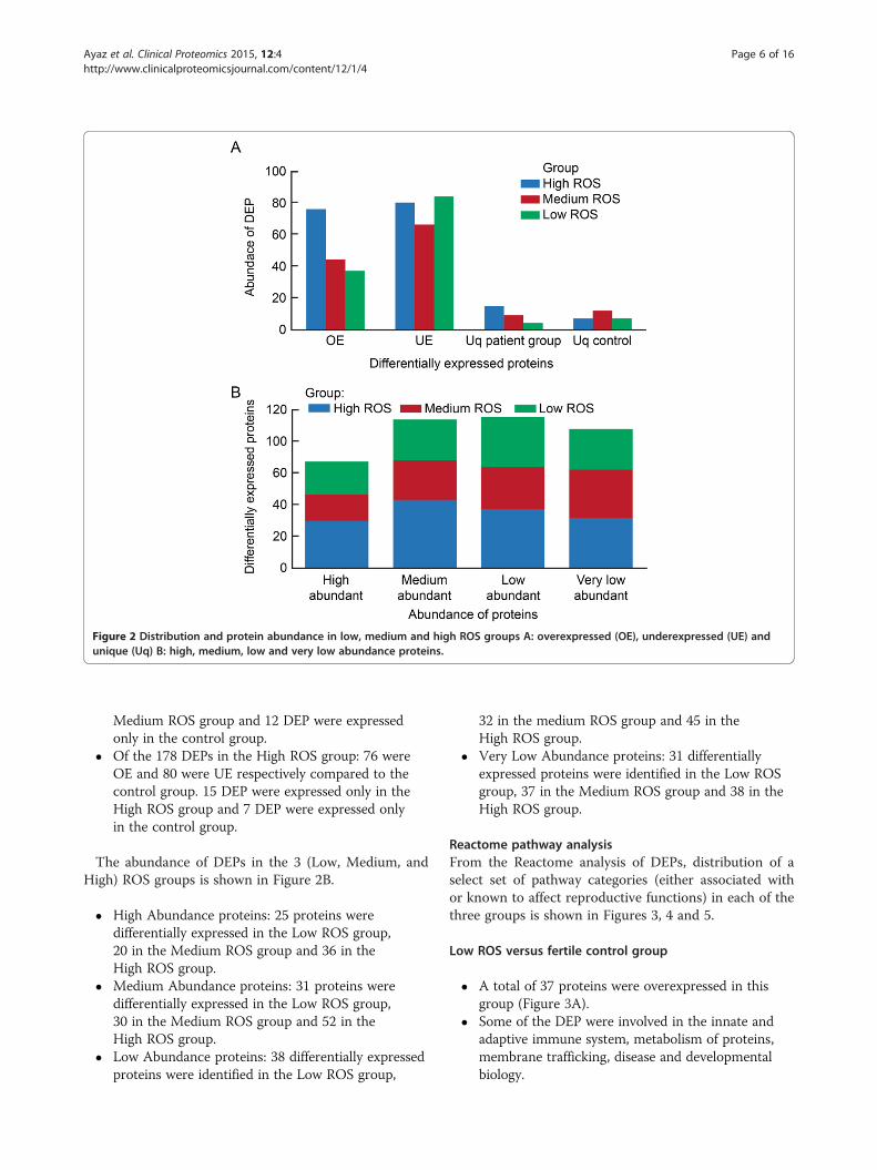
Figure 2 Distribution and protein abundance in low, medium and high ROS groups A: overexpressed (OE), underexpressed (UE) andunique (Uq) B: high, medium, low and very low abundance proteins.
Ayaz et al. Clinical Proteomics 2015, 12:4 Page 6 of 16http://www.clinicalproteomicsjournal.com/content/12/1/4
Medium ROS group and 12 DEP were expressedonly in the control group.
� Of the 178 DEPs in the High ROS group: 76 wereOE and 80 were UE respectively compared to thecontrol group. 15 DEP were expressed only in theHigh ROS group and 7 DEP were expressed onlyin the control group.
The abundance of DEPs in the 3 (Low, Medium, andHigh) ROS groups is shown in Figure 2B.
� High Abundance proteins: 25 proteins weredifferentially expressed in the Low ROS group,20 in the Medium ROS group and 36 in theHigh ROS group.
� Medium Abundance proteins: 31 proteins weredifferentially expressed in the Low ROS group,30 in the Medium ROS group and 52 in theHigh ROS group.
� Low Abundance proteins: 38 differentially expressedproteins were identified in the Low ROS group,
32 in the medium ROS group and 45 in theHigh ROS group.
� Very Low Abundance proteins: 31 differentiallyexpressed proteins were identified in the Low ROSgroup, 37 in the Medium ROS group and 38 in theHigh ROS group.
Reactome pathway analysisFrom the Reactome analysis of DEPs, distribution of aselect set of pathway categories (either associated withor known to affect reproductive functions) in each of thethree groups is shown in Figures 3, 4 and 5.
Low ROS versus fertile control group
� A total of 37 proteins were overexpressed in thisgroup (Figure 3A).
� Some of the DEP were involved in the innate andadaptive immune system, metabolism of proteins,membrane trafficking, disease and developmentalbiology.
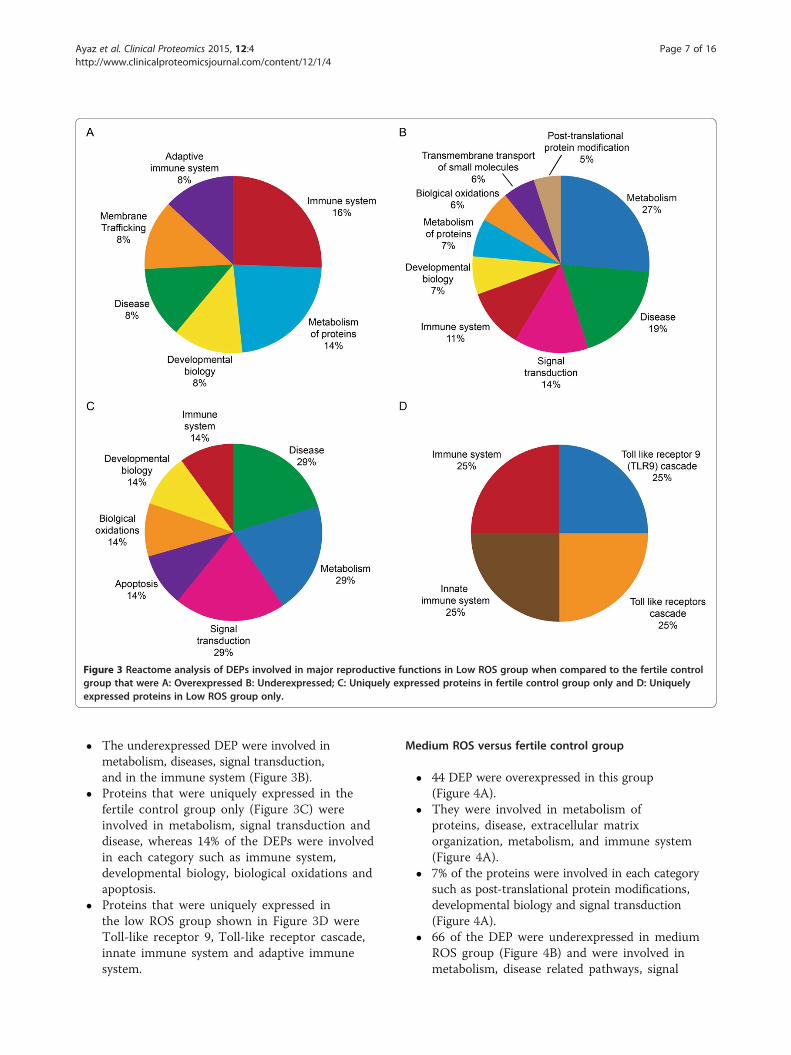
Figure 3 Reactome analysis of DEPs involved in major reproductive functions in Low ROS group when compared to the fertile controlgroup that were A: Overexpressed B: Underexpressed; C: Uniquely expressed proteins in fertile control group only and D: Uniquelyexpressed proteins in Low ROS group only.
Ayaz et al. Clinical Proteomics 2015, 12:4 Page 7 of 16http://www.clinicalproteomicsjournal.com/content/12/1/4
� The underexpressed DEP were involved inmetabolism, diseases, signal transduction,and in the immune system (Figure 3B).
� Proteins that were uniquely expressed in thefertile control group only (Figure 3C) wereinvolved in metabolism, signal transduction anddisease, whereas 14% of the DEPs were involvedin each category such as immune system,developmental biology, biological oxidations andapoptosis.
� Proteins that were uniquely expressed inthe low ROS group shown in Figure 3D wereToll-like receptor 9, Toll-like receptor cascade,innate immune system and adaptive immunesystem.
Medium ROS versus fertile control group
� 44 DEP were overexpressed in this group(Figure 4A).
� They were involved in metabolism ofproteins, disease, extracellular matrixorganization, metabolism, and immune system(Figure 4A).
� 7% of the proteins were involved in each categorysuch as post-translational protein modifications,developmental biology and signal transduction(Figure 4A).
� 66 of the DEP were underexpressed in mediumROS group (Figure 4B) and were involved inmetabolism, disease related pathways, signal

Figure 4 Reactome analysis of DEPs involved in major reproductive functions in Medium ROS group when compared to the fertilecontrol group that were A: Overexpressed B: Underexpressed; C: Uniquely expressed proteins in fertile control group only andD: Uniquely expressed proteins in Low ROS group only.
Ayaz et al. Clinical Proteomics 2015, 12:4 Page 8 of 16http://www.clinicalproteomicsjournal.com/content/12/1/4
transduction, and metabolism of proteins,and a small percentage in immune system,apoptosis and post-translational proteinmodifications.
� Uniquely expressed proteins in the fertile controlgroup only that were differentially expressed wereinvolved in metabolism, signaling by Robo receptor,developmental biology and disease as shown inFigure 4C.
� 9 DEP were uniquely expressed in the medium ROSgroup were involved in metabolism, while 11% ofthe proteins were uniquely involved in each of thecategories such as toll-like receptor cascades, cellcycle, and immune system and gene expression asshown in Figure 4D.
High ROS versus fertile control group
� 76 of the DEP were overexpressed in the High ROSgroup compared to the fertile control group(Figure 5A).
� The overexpressed proteins were involved inmetabolism, immune system, disease, andextracellular matrix organization, developmentalbiology, and signal transduction, membranetrafficking and cellular responses to stress (Figure 5A).
� 80 DEPs were underexpressed and were involvedin metabolism, disease, metabolism of lipids andlipoproteins, immune system, cell cycle, signaltransduction, and cellular responses to stress(Figure 5B).

Figure 5 Reactome analysis of DEPs involved in major reproductive functions in High ROS group when compared to the fertile controlgroup that were A: Overexpressed B: Underexpressed; C: Uniquely expressed proteins in fertile control group only and D: Uniquelyexpressed proteins in Low ROS group only.
Ayaz et al. Clinical Proteomics 2015, 12:4 Page 9 of 16http://www.clinicalproteomicsjournal.com/content/12/1/4
� 7 DEP were uniquely expressed only in thefertile control group and were involved inelectron transport, mitochondrial iron-sulphurcluster biogenesis, and synthesis of PIPs at theGolgi membrane, phospholipid metabolism,metabolism of lipids and lipoproteins and generalmetabolism (Figure 5C).
� 15 DEP were uniquely expressed in theHigh ROS group (Figure 5D) and involved inmetabolism, disease, metabolism of lipidsand lipoproteins, immune system, cell cycle,signal transduction and cellular responseto stress.
DAVID software’s functional annotations in low, mediumand high ROS groupThe results of the functional annotation analysis ofDEPs using DAVID software is shown in Table 3. Inthe Low ROS compared to the control group, 12DEPs involved in reproduction and fertilization wereunder-expressed. In the Medium ROS group, 109 DEPwere under-expressed. Some of the functions wererelated to sexual reproduction, gamete generation andreproductive process. 44 DEPs participating in reproduc-tion, spermatogenesis, spermatid development and dif-ferentiation were under-expressed in the High ROSgroup.

Table 3 Functional Annotations analysis of DEPs by DAVIDS analysis in low, medium and high ROS group
Functionalannotations
Low ROS Medium ROS High ROS
Functionalcategories relatedto reproduction/spermatogenesis
Single fertilization (4), sexual reproduction (8) Sexual reproduction (7), gametegeneration (96), reproductive process inmulticellular organism (6)
Sexual reproduction (11), multicellularorganism reproduction (10), gametegeneration (9), spermatogenesis (8),spermatid development/differentiation(3), germ cell development (3)
Enrichedfunctionalcategories
Cell cortex (5), actin binding (5),myofibril (3), protease (4), peptidaseactivity (4), metal ion binding (12),protein targeting (3), IC proteintransport and localization (3), cell cyclephase (3), nucleotide binding (5),regulation of transcription (3); lysosome(7), influence lipid concentrations and riskof CAD (4), plasma lipoprotein particleremodeling (3), heparin binding (5),protein-lipid complex (3), glucosemetabolic process (5), cytoskeletonorganization (6), sterol transport andhomeostasis (3), EGF-like region (5), cellularresponse to stress (3), negative regulation ofapoptosis (6), cell migration (5), cell cycle (3)
Basement membrane (6), EC matrix (6),ECM-receptor interaction (5), EGFdomain (5), cell motility and localization(6), cytoplasmic vesicle (6), regulation ofneurological system process (3), motorprotein (3), calmodulin binding (3),metal ion binding (16) cellular ionhomeostasis (3), cell morphogenesis (3),protein targeting and localization (3),synaptic transmission (3), ATP binding(5); Dynein (6), microtubule associatedcomplex (6), motor activity (6), negativeregulation of cellular metabolic process(5), Ca binding region (4), cytoskeletonorganization (5), gamete generation (6),reproductive process in multicellularorganism (6), nucleotide biosyntheticprocess (4), nucleotide binding (12),metalloendopeptidase activity (3),glycosaminoglycan binding (3),phospholipid metabolic process (3),regulation of apoptosis (6), cytoskeleton(11), metal ion binding (12)
Oxidoreductase (12), Protease (11),cofactor binding (8), metallopeptidaseactivity (7), metalloprotease (7), zincion binding (13), cytoskeleton (14), celljunction (6), regulation of apoptosis(7), intracellular protein transport andlocalization (5), proteasome complex(3), vasculature development (4), focaladhesion (4); nucleotide binding (24),sexual reproduction (11), multicellularorganism reproduction (10), gametegeneration (9), spermatogenesis (8),spermatid development/differentiation(3), germ cell development (3),glutathione S-transferase (3), peroxisome(4), fatty acid oxidation (3), lipidmodification (3), glycerolipid metabolicprocess (4), protein targeting (3), metalion binding (13), apoptosis (3)
Majority ofproteinsassociated withfunctions
Polymorphism (29), phosphoprotein(22), acetylation (15), glycoprotein (13),Golgi (9); polymorphism (62), signalpeptide (38), glycoprotein (36), disulfidebond (34), extracellular (30), acetylation (26),secreted (23), oxidoreductase (11), lipidbinding (11)
Phosphoprotein (26), glycoprotein(20), signal peptide (18), disulfidebond (15), acetylation (13), cytoplasm(13), Golgi (10), EC region (11),secreted (10), Ca ion binding (8), celladhesion (7); Polymorphism (50), Signalpeptide (18), EC (17), secreted (15),mitochondrion (13), nucleotide binding(15), cell fraction (10), cytoskeleton (9),sexual reproduction (7)
Polymorphism (57), phosphoprotein(41), ion binding (34), glycoprotein(27), cytoplasm (26), signal peptide(22), cell membrane (16), proteolysis(14), hydrolase (15), oxidoreductase(12), defense response (9),metalloprotease (7), protein transportand localization (8), exopeptidaseactivity (6), adherens junction (6), cellmigration (6); purine nucleotide binding(24), ribonucleotide binding (22), sexualreproduction (11), gamete generation(9), spermatogenesis (8), mitochondrion(15), reproductive cellular process (5),protein folding (5), spermatiddevelopment (3)
Cellulardistribution
Cell cortex (5), Golgi (9), cytoskeleton (9),cytosol (8), ER (6), organelle envelope (5),vesicle (5), sarcomere (3); extracellularregion (30), cytosol (13), lysosome (7),vesicle (11), secretory granule (4),protein-lipid complex (3), cell surface (6),golgi (10)
Golgi (10), EC region (9), organellemembrane (9), ER (8), proteinaceousECM (6), cytoplasmic vesicle (6),intrinsic to plasma membrane (8);dynein complex (6), mitochondrion (13),EC region (17), microtubule cytoskeleton(8), cell projection (8), cytoskeleton (11),cilium (4)
Actin cytoskeleton (10), cell cortex (7),cortical cytoskeleton (5), cell-celladherens junction (4), vesicle (11), Golgi(12), EC region (19), mitochondriallumen (4); mitochondrion (15), cytosol(15), dynein complex (3), microbody (4),peroxisome (4), cell fraction (11),secretory granule (4), organelle envelope(7), pore complex (3)
Activatedprocesses/functions
Intracellular transport (7), proteinlocalization (7), cytoskeletalorganization (5), regulation of proteincomplex assembly (3), microtubulebased process (4), calmodulin binding(4), motor activity (4), actin binding (5),serine hydrolase activity (3),phospholipid binding (3)
Cell motility and localization (6), cellulariron ion hemostasis (3), cell migration(5), cell motion (6), cell adhesion (7),integrin-mediated signaling pathway(3), macromolecular complex assembly(6), response to wounding (5),regulation of neurological systemprocess (3), Ca ion binding (8),peptidase activity (5), carbohydratebinding (4), microfilament motoractivity (2), structural moleculeactivity (5)
Actin filament based process (9),oxidation reduction (12), integrinmediated signaling pathway (5),cytoskeleton organization (9), proteolysis(14), cell motion (9), defense response(9), intracellular transport (8), cell-celladhesion (5), oxygen and ROS metabolicprocess (3), actin binding (10), exo- orendo-peptidase activity (6), cytoskeletalprotein binding (11), calcium ion binding(11), cofactor binding (8), peptidaseactivity (11), antioxidant activity (3)
Ayaz et al. Clinical Proteomics 2015, 12:4 Page 10 of 16http://www.clinicalproteomicsjournal.com/content/12/1/4
Table 3 Functional Annotations analysis of DEPs by DAVIDS analysis in low, medium and high ROS group (Continued)
Downregulatedprocesses/functions
Glycerolipid metabolic process (6), lipidhemostasis (4), oxidation reduction (11),single fertilization (4), protein-lipid complexremodeling (3), sexual reproduction (8),protein folding (5), sterol transport (3),response to oxidative stress (5), lipidbinding (11), carbohydrate binding (8),glycosaminoglycan binding (5), oxidoreductaseactivity (3), antioxidase activity (3)
Actin filament organization (4), proteinfolding (5), negative regulation ofcellular metabolic process (5), sexualreproduction (7), protein tetramerization(3), oxidation reduction (8), gametegeneration (6), cell redox homeostasis(3), protein oligomerization (4),generation of precursor metabolites andenergy (5); motor activity (6), enzymeinhibitor activity (6), proteasomeregulator activity 92), protein binding (7),nucleoside binding (12), nucleotidebinding (15), protein homodimerizationactivity (5)
Sexual reproduction (11), reproductiveprocess in a multicellular organism (10),gamete generation (9), spermatogenesis(8), protein folding 95), glycerophospholipidmetabolic process (4), spermatiddevelopment/differentiation (3); purinenucleotide binding (24), ATP binding(18), microtubule motor activity (3),sterol transporter activity (2), cAMPdependent protein kinase regulator activity(2), glutathione transferase activity (2)
Activatedpathways
Ether lipid metabolism (2), Synapticproteins at the synaptic junction (2);
ECM-receptor interaction (5), Integrincell surface interactions (4)
Leukocyte transendothelial migration(5), focal adhesion (8), ECM-receptorinteraction (4), Adherens junction (4),Hemostasis (7), Apoptosis (5),Metabolism of lipids and lipoproteins(5), metabolism of amino acids (5),Wntsignaling (3), Integrin signalingpathway (3), Tight junction (4),Proteasome (3)
Downregulatedpathways
Gluconeogenesis (3), metabolism ofcarbohydrates (4), hemostasis (5)
Fructose and mannose metabolism (3),apoptosis (4), metabolism of amino acids(4)
Glycolysis/gluconeogenesis (3),adipocytokine signaling pathway (3),PPAR signaling pathway (3), metabolismof lipids and lipoproteins (5), Integrationof energy metabolism (5), mechanism ofprotein import into the nucleus (2)
Processes/Functionsunique to eachROS group
Regulation of cellular protein metabolicprocess (2), endocytosis (1), PTM (1)
Organelle fusion (2), proteinhomodimerization activity (2),anti-apoptosis (2), ribosomalsubunit (2), cytosol (3), membrane (4),polymorphism (5)
Disulfide bond (7), signal peptide (6),alternative splicing (8), metal ion binding(7), glycoprotein (8), secreted (4),endocytosis (2), aging (2), plasmamembrane (5), calcium ion binding (5)
Processes/Functionsunique toControl group
MAPK signaling pathway (2), Regulation ofactin cytoskeleton (2), metabolism ofvitamins and cofactors (2)
Tissue morphogenesis (3), tubedevelopment (3), embryonicmorphogenesis (3), leucine rich repeat (3),extracellular region (5), glycoprotein (5),membrane (3), urogenital systemdevelopment (2)
Transmembrane (3), alternative splicing(3), transport (4), metabolism of vitaminsand cofactors (1)
Bold text = Overexpressed proteins; regular text = Underexpressed proteins; italics text = unique processes or functions; number in parenthesis = number of proteins.
Ayaz et al. Clinical Proteomics 2015, 12:4 Page 11 of 16http://www.clinicalproteomicsjournal.com/content/12/1/4
The other enriched functional categories, cellular func-tion and distribution, as well as processes and functionsthat were either activated or suppressed in each of thethree groups are all summarized in Table 3. The numberof proteins associated with each category is shown inparentheses. The fertile control group as well as in eachROS group is also summarized in Table 3.
STRAP analysis of DEP in low, medium and high ROS groupSTRAP analysis identified 6 DEPs (CLGN, TPP2, DNAI2,HSPA4L, EEA1 and SERPINA5) associated with key re-productive related functions. The differential expressionof these proteins in the three ROS groups is shown inTable 4.
DiscussionMale infertility is a multifactorial condition emerging froma wide variety of etiologies including gene mutations, mal-formation of reproductive organs, infectious disease, or
environmental exposure to toxicants [42]. In the presentstudy, the age of men with oxidative stress was compar-able to that of fertile donors. The majority of the patients(73.2%) presented with either Low ROS, Medium ROS, orHigh ROS. Semen analysis was performed according tothe new WHO 2010 criteria [20]. No significant differ-ences were observed in sperm concentration, motility orpresence of round cells between men presenting with LowROS, Medium ROS and High ROS. Poor sperm morph-ology was seen in men in the high ROS group when com-pared to Medium ROS and Low ROS group.Some abnormal sperm morphologies are genetically-
determined while others are caused as a result of re-peated physiological and environmental stresses. Theseabnormal morphologies are reversible; however, afterrepeated stress attacks, the testis may fail to fully re-cuperate and could result in a permanent decrease innumber of sperm with normal morphology. These ab-normalities can be explained by the effect of elevated

Table 4 Differentially expressed proteins as a potential biomarker for patients exhibiting high, medium and low ROSlevels
Uniprotnumber
Proteinname
Genename
Function Expression/ROSlevel
Possible reasons for proteinexpression and relation to infertility
Reference
O14967 Calmegin CLGN Essential for formation of normalspermatozoa. Important role inspermatogenesis, acts as achaperone for a range of clientproteins that are important for spermadhesion onto the egg zonapellucida and for subsequentpenetration of the zona pellucida.
Overexpressed/High ROS group,Medium ROSgroup, Low ROSgroup
Its expression is triggered in case ofelevated oxidative stress. Thus, inmen with ROS generation abovephysiological levels (oxidative stress),calmegin overexpression may impairthe ability of spermatozoa to bind tothe zona pellucida. Hence, it may be acause of infertility in these men.
[28-30]
P29144 Tripeptidyl-peptidase 2
TPPII Tripeptidyl peptidase II is a‘multi-purpose peptidase’ withhouse-keeping function in intracellularprotein degradation and plays a rolein several vital cellular processes suchas cell division, apoptosis or antigenprocessing. TPPII regulates spermfunction by modifying the levelsof tyrosine phosphorylation. It isinvolved in the fertilization process,and regulates sperm maturation.
Overexpressed/High ROS group,Medium ROSgroup, Low ROSgroup
Overexpression of TPPII in men withROS generation above physiologicallevels may modify sperm proteintyrosine phosphorylation levels,such that spermatozoa is unableto undergo protein tyrosinephosphorylation-associated processessuch as capacitation, hyperactivation,and acrosome reaction.
[31-33]
Q9GZS0 Dyneinintermediatechain 2,axonemal
DNAI2 Part of the dynein complex ofrespiratory cilia. DNAI2 can result inreduced fertility due to sperm tailabnormalities.
Underexpressed/High ROS group,Medium ROSgroup, Low ROSgroup
The underexpression of DHAI2 in menwith ROS above physiological levelsmay contribute to the negative effectof oxidative stress on sperm motility.
[34]
Q15075 Earlyendosomeantigen 1
EEA1,ZFYVE2
Binds phospholipid vesiclescontaining phosphatidylinositol3-phosphate and participates inendosomal trafficking.
Uniquelyexpressed/HighROS group,Medium ROSgroup, Low ROSgroup
Its unique expression in the 3 ROSgroups suggests that EEA1 may beinvolved in the failure of acrosomebiogenesis, that results in maleinfertility.
[35-37]
O95757 Heat shock70 kDaprotein 4L
HSPA4L,APG1,OSP94
Apg-1 encodes a heat shock proteinbelonging to the Hsp110 family andis inducible by a 32 degrees C to 39degrees C heat shock in somaticcells. In mouse testicular germ cellsApg-1 mRNA is constitutivelyexpresseddepending on the developmentalstage. It belongs to the HSP110 heatshock gene family and is producedubiquitously and predominantly inthe testis. It is highly expressed inthe spermatogenic cells, from latepachytene spermatocytes to postmeiotic spermatids. It is required fornormal spermatogenesis.
Underexpressed/High ROS group,Medium ROSgroup, Low ROSgroup
The underexpression of HSPA4L inmen with ROS above physiologicallevels may disrupt the normalspermatogenesis process which maycontribute to infertility seen in thesemen.
[38]
P05154 Plasmaserineproteaseinhibitor
SERPINA5,PCI,PLANH3,PROCI
SERPINA5 is a heparin-dependentserine protease inhibitor that acts onbody fluids and secretions. Serineprotease with lys and arg ester bondspecificity is involved in the controlof sperm motility. SERPINA5 inhibitsthe serpin acrosin and indirectlyprotects the component of the malegenital tract from being degradedby excessive released acrosin. It alsoinhibits tissue-and urinary-typeplasminogen activator, prostate-specificantigen and kallikrein activities and hasa control on sperm motility andfertilization.
Underexpressed/High ROS group,Medium ROSgroup, Low ROSgroup
Underexpression of SERPINA5 inmen with ROS generation abovephysiological levels suggests thatthe reduced motility seen in theseinfertile patients may be due to theserine protease inhibiting action ofSERPINA 5.
[39-41]
Ayaz et al. Clinical Proteomics 2015, 12:4 Page 12 of 16http://www.clinicalproteomicsjournal.com/content/12/1/4

Ayaz et al. Clinical Proteomics 2015, 12:4 Page 13 of 16http://www.clinicalproteomicsjournal.com/content/12/1/4
level of oxidative stress in high ROS group. We have re-cently demonstrated the association of teratozoospermiawith increased levels of ROS [43]. Basic semen analysis isnot adequate to reflect all the parameters of semen qualityand function that are required for an optimum fertilitystatus especially in cases of idiopathic male factor infertil-ity. Mature spermatozoa are particularly susceptible toROS damage because of the polyunsaturated nature of thesperm plasma membrane and the absence of any tran-scriptional activity [3,6].ROS has been shown to be an independent marker of
oxidative stress. ROS levels can predict male factor infer-tility (MFI) with an accuracy of ≥80% [30]. ROS-inducedoxidative stress has been shown to be a significant riskfactor associated with male factor infertility [11]. Wepostulated the mechanisms for infertility among non-leukocytospermic patients with normal semen parame-ters and high or normal ROS levels. These were (1) dir-ect generation of oxygen radicals by low concentrationsof leukocytes; (2) significant DNA damage in samplescontaining abnormal levels of ROS-producing spermato-zoa; (3) diminished levels of antioxidants in the seminalplasma; or (4) the presence of immature sperm in sub-stantial quantities in semen [3,44,45].High ROS levels are known to significantly affect pro-
teins and lipids, induce apoptosis and cause DNA dam-age, leading to male infertility [46]. Cells that are in astate of oxidative stress are more likely to have alteredprotein expression. Utilizing novel proteomic tools andbioinformatics we have recently demonstrated the effectof increased ROS levels on protein alterations seen inthe spermatozoa and seminal plasma of subjects demon-strating high ROS levels when compared to those withphysiological levels [17,18].The proteome provides a dynamic understanding of
post-genomic events and characterizing these events willfurther our understanding of the underlying functionalchanges in men diagnosed with various male-infertility-associated etiologies [47,48]. Current literature suggeststhat while causes of elevated oxidative stress levels aremultifactorial, current management of oxidative stressis unable to accurately predict the benefit for maleinfertility.In the present study, the Reactome software showed
that DEPs in Low, Medium or High ROS group were in-volved in the metabolism of proteins, immune system,disease, transmembrane (TM) transport, extracellularmatrix organization, signal transduction, post-translationalmodifications, and cellular response to stress. (Figures 4and 5). DAVID analysis indicated that DEPs were involvedin the cellular functions of lipid metabolism, small mol-ecule biochemistry, and nucleic acid metabolism (Table 3).The overexpression of these proteins increased with in-creasing ROS levels indicating an overactivity of protein
function in response to stress. In an excellent review byAmaral et al. [19], the authors demonstrated that thepathways involved in metabolism and energy productionhave been recognized as the most significant cellular path-way in the human spermatozoa. These authors furthershowed that 26% of the proteins contributing to the reac-tome belong to the Metabolism and energy productiongroup. The contribution by the metabolism and energygroups comprised of the following: Glycolysis and gluco-neogenesis (8%), Krebs cycle (13%), Mitochondrial electrontransport chain/oxidative phosphorylation (20%), lipid me-tabolism (24%), amino acid metabolism (9%), nucleotidemetabolism (7%) and other carbohydrate pathways (9%).Human sperm can utilize carbohydrate, lipid as well asnucleotide metabolism as an energy source. An increasingbody of data suggests that mitochondrial activity is intim-ately related to sperm function such as sperm motility andfertilization. The sperm proteome is also enriched inproteins related to protein metabolism which includepathways implicated in protein translation, folding,post-translational modifications and protein degradation.The overexpressed proteins were seen in all ROS groupswith varying distribution of the cellular pathways asshown by the reactome analysis.The role of membrane trafficking is well known in
spermiogenesis. The sperm proteome is enriched in pro-teins that mediate membrane fusion and promotes therelease of acrosomal contents. The proteins involved inthe final sperm-oocyte fusion event are also present inthe sperm proteome. Many of the overexpressed proteinsare also involved in signal transduction, extracellularmatrix organization and cellular response to stress.Many of the DEP that were underexpressed in the 3
ROS groups were also involved in the important path-ways such as metabolism of proteins and lipids, signaltransduction, biological oxidations, post-translational pro-tein modifications, metabolism of proteins, transmem-brane transport of small molecules, signal transduction,apoptosis, and cellular response to stress.Compared to the Low ROS group, there were proteins
that were uniquely expressed in the fertile control group.These were involved in disease, apoptosis, biological oxi-dation, signal transduction and immune system. In themedium ROS group, the proteins that were uniquelyexpressed in the fertile group were involved in eventssuch as metabolism, metabolism of lipids and lipoproteins,signaling by Wnt and by Robo receptor, hemostasis andsignal transduction. Compared to the High ROS group,DEP that were uniquely expressed in the fertile groupwere involved in metabolism, electron transport fromNADPH to ferrodoxin, synthesis of phosphatidylinositolphosphate (PIP) at the Golgi membrane, phospholipidmetabolism, mitochondrial iron-sulfur cluster biogen-esis, metabolism of lipids and lipoproteins as well as

Ayaz et al. Clinical Proteomics 2015, 12:4 Page 14 of 16http://www.clinicalproteomicsjournal.com/content/12/1/4
phosphoionositide (PI) metabolism that play a role in lipidsignaling, cell signaling and membrane trafficking.The Reactome analysis also identified proteins that
were uniquely expressed in each ROS group. Proteinsthat were unique to the Low ROS group were involvedin Toll-like Receptor 9 (TLR9) cascade, toll-like receptorcascades, and innate immune system. These receptors me-diate response to unmethylated CpG dinucleotides in bac-terial DNA to trigger an immune response. The presenceof these receptors may be indicative of leukocyte contam-ination in ROS generating semen samples. Proteins thatwere uniquely expressed in the Medium ROS group wereinvolved in disease, gene expression, metabolism of pro-teins, toll-like receptors cascades, nuclear import of Revproteins and both innate and adaptive immune system. In-nate immunity is an inherent immunity (defense) that isalready present at birth.Similarly in the High ROS group, a significantly high
percentage of uniquely expressed proteins were involvedin metabolism, electron transport from NADPH to fer-redoxin, and metabolism of the phospholipids, lipids,lipoprotein synthesis of PIP at the Golgi membrane sug-gesting that these proteins were playing a key role in opti-mal spermatozoa function. On the other hand, in thepatients within the High ROS group, proteins were uniqueand involved in cellular response to stress, signal trans-duction, metabolism of proteins, metabolism of lipids andlipoproteins suggesting that these proteins were operatingunder high oxidative stress conditions that was affectingthe metabolic activity of the sperm and in signal transduc-tion and response to stress.The most significant pathways in the male gamete
are those involved in protein metabolism, membranetrafficking, apoptosis, cell cycle, hemostasis and mei-osis. Developmental biology and extracellular matrixorganization were also detected as putative spermpathways but with lower probability. Signaling trans-duction is not a major pathway but some signalingpathways that are seen to be active include signalingby Wnt proteins, a family of secreted lipid-modifiedsignaling glycoproteins [19]. The overexpression/underexpression or the unique presence of the proteins inthe ROS groups only compared to the fertile group isindicative of the problems in the sperm functions thatare affected or altered as a result of increased pres-ence of ROS in these men. These alterations may beresponsible for poor fertility seen in these men com-pared to the fertile men.We also demonstrated that while oxidative phosphoryl-
ation is important for sperm function, the major pathwayfor energy metabolism was the glycolytic pathway. Ma-jority of the proteins were involved in cellular metabolicand regulatory processes. We demonstrated in our earlierwork that a number of proteins that were overexpressed
or underexpressed in ROS+ group were involved in cellularprocesses, metabolic process, response to stress andtransport. Furthermore, a small number of proteinswere involved in post-translational processes and proteinfolding [18].DAVID software also identified a number of proteins
that were involved in key functions in the 3 ROSgroups. In the functional categories related to reproduc-tion and/or spermatogenic events, 12 proteins wereidentified in the low ROS group, 109 in the mediumROS and 44 in the high ROS group that may bemodified and were underexpressed compared to thefertile group. The various functional annotations areshown in Table 3.The majority of the overexpressed proteins were asso-
ciated with various functions such as acetylation, phos-phoproteins, Ca2+ ion binding, oxidoreductase, defenseresponse, protein transport, etc. whereas other proteinsinvolved in signal peptide, nucleotide, binding, gametegeneration, spermatogenesis, protein folding and sperm-atid development were underexpressed especially in thehigh ROS group. This is the category with the largestdistribution of proteins that were differentially expressedhighlighting the deviation of the various functions fromthe fertile group.We further identified the DEPs in all 3 ROS groups
that are known to play a role in reproduction andspermatogenesis that were underexpressed, over or wereuniquely expressed to each of the 3 ROS groups com-pared to the fertile controls (Table 4). These proteinshighlight the deviation from the normal functions that areseen in fertile men in the presence of physiological levelsof ROS. We identified 6 DEP (Calmegin, Tripeptidyl pep-tidase II, Dynein intermediate chain 2, axonemal, Heatshock 70 kDa protein 4L, Early endosome antigen 1, andPlasma serine protease inhibitor) that were present in allthe 3 ROS groups with varying expression levels andtherefore may serve as potential candidates of oxidativestress (Table 4).In conclusion, we have for the first time demonstrated
poor sperm quality that is associated with elevated oxi-dative stress level by categorizing the patient semensamples into low, medium and high ROS groups. Theseabnormalities can be explained by the altered expres-sion of specifically DEPs Calmegin, tripeptidyl synthe-sase, dynein intermediate chain 2, heat shock 70 kDaprotein 4L, early endosome antigen 1, plasma serineprotease inhibitor in patient groups. These DEPs mayserve as potential biomarkers in the identification ofthe effect of increasing ROS level on protein profileof spermatozoa in infertile men. Further validation ofDEPs is necessary to establish the role of these pro-teins as biomarkers of oxidative stress induced malefactor infertility.

Ayaz et al. Clinical Proteomics 2015, 12:4 Page 15 of 16http://www.clinicalproteomicsjournal.com/content/12/1/4
Additional files
Additional file 1: Table S2a. Spermatozoa proteins in fertile men.
Additional file 2: Table S2b. Spermatozoa proteins in Low ROS group.
Additional file 3: Table S2c. Spermatozoa proteins in Medium ROS group.
Additional file 4: Table S2d. Spermatozoa proteins in High ROS group.
Competing interestsThe authors declare that they have no competing interests.
Authors’ contributionsAA and ZC conducted the study and helped with the data collection andmanagement of this study. AA conceived the idea, supervised the study, andedited the article for submission. MA and HE helped with reviewing andediting of the manuscript. RKS helped with the writing, reviewing andediting of the manuscript. All authors read and approved the finalmanuscript.
AcknowledgementsThe authors are grateful to the Andrology Center technologists forscheduling the study subjects and Jeff Hammel, senior biostatistician, for hiscontribution to data analysis. Belinda Willard, Director, Proteomic Core Lab,Lerner Research Institute for providing assistance with proteomic analysis andBanu Gopalan, Lerner Research Institute with Bioinformatics data analysis. TheOrbitrap Elite mass spectrometer used in this study was purchased with fundsfrom an NIH shared instrument grant 1S10RR031537-01 to Belinda Willard.Financial support was provided by the Center For Reproductive Medicine,Cleveland Clinic.
Author details1Center for Reproductive Medicine, Glickman Urological & Kidney Institute,Cleveland Clinic, Cleveland, OH 44195, USA. 2Male Infertility Unit, Departmentof Urology, Hamad Hospital, Doha, Qatar.
Received: 7 November 2014 Accepted: 15 January 2015Published: 9 February 2015
References1. Rowe PJ, Comhaire FH, Hargreave TB. WHO manual for the standardized
investigation of the infertile couple. Cambridge, UK: Cambridge UniversityPress; 1993.
2. Louis JF, Thoma ME, Sorensen DN, McLain AC, King RB, Sundaram R, et al.The prevalence of couple infertility in the United States from a maleperspective: evidence from a nationally representative sample. Androl.2013;1:741–8.
3. Agarwal A, Durairajanayagam D, Halabi J, Peng J, Vazquez-Levin M.Proteomics, oxidative stress and male infertility. Reprod Biomed Online.2014;29:32–58.
4. Report on varicocele and infertility: a committee opinion. Fertil Steril.2014;102:1556–60.
5. Sigman M. Male infertility. Med Health Rhode Island. 1997;80:406–9.6. Sharma RK, Agarwal A. Role of reactive oxygen species in male infertility.
Urology. 1996;48:835–50.7. Sabanegh E, Agarwal A. Male infertility. In: Wein AJ, editor. Campbell’s
Urology. 10th ed. Philadelphia, PA: Elsevier Saunders; 2011.8. Hamada AJ, Montgomery B, Agarwal A. Male infertility: a critical review of
pharmacologic management. Expert Opin Pharmacother. 2012;13:2511–31.9. Tremellen K. Oxidative stress and male infertility–a clinical perspective.
Hum Reprod Update. 2008;14:243–58.10. Sharma RK, Pasqualotto FF, Nelson DR, Thomas Jr AJ, Agarwal A. The
reactive oxygen species-total antioxidant capacity score is a new measureof oxidative stress to predict male infertility. Hum Reprod. 1999;14:2801–7.
11. Pasqualotto FF, Sharma RK, Nelson DR, Thomas AJ, Agarwal A. Relationshipbetween oxidative stress, semen characteristics, and clinical diagnosis inmen undergoing infertility investigation. Fertil Steril. 2000;73:459–64.
12. Pahune PP, Choudhari AR, Muley PA. The total antioxidant power of semenand its correlation with the fertility potential of human male subjects. J ClinDiagn Res. 2013;7:991–5.
13. Lewis SE, Boyle PM, McKinney KA, Young IS, Thompson W. Total antioxidantcapacity of seminal plasma is different in fertile and infertile men. FertilSteril. 1995;64:868–70.
14. Kashou AH, Sharma R, Agarwal A. Assessment of oxidative stress in spermand semen. Methods Mol Biol. 2013;927:351–61.
15. Zhou T, Zhou ZM, Guo XJ. Bioinformatics for spermatogenesis: annotationof male reproduction based on proteomics. Asian J Androl.2013;15:594–602.
16. Milardi D, Grande G, Vincenzoni F, Castagnola M, Marana R. Proteomics ofhuman seminal plasma: identification of biomarker candidates for fertilityand infertility and the evolution of technology. Mol Reprod Dev.2013;80:350–7.
17. Sharma R, Agarwal A, Mohanty G, Du Plessis SS, Gopalan B, Willard B, et al.Proteomic analysis of seminal fluid from men exhibiting oxidative stress.Reprod Biol Endocrinol. 2013;11:85.
18. Sharma R, Agarwal A, Mohanty G, Hamada AJ, Gopalan B, Willard B, et al.Proteomic analysis of human spermatozoa proteins with oxidative stress.Reprod Biol Endocrinol. 2013;11:48.
19. Amaral A, Castillo J, Ramalho-Santos J, Oliva R. The combined human spermproteome: cellular pathways and implications for basic and clinical science.Hum Reprod Update. 2014;20:40–62.
20. World Health Organization. WHO laboratory manual for the examinationand processing of human semen. 5th ed. Geneva, Switzerland: WHO Press;2010.
21. Sharma RK, Sabanegh E, Mahfouz R, Gupta S, Thiyagarajan A, Agarwal A.TUNEL as a test for sperm DNA damage in the evaluation of male infertility.Urology. 2010;76:1380–6.
22. Keller A, Nesvizhskii AI, Kolker E, Aebersold R. Empirical statistical model toestimate the accuracy of peptide identifications made by MS/MS anddatabase search. Anal Chem. 2002;74:5383–92.
23. Nesvizhskii AI, Keller A, Kolker E, Aebersold R. A statistical model foridentifying proteins by tandem mass spectrometry. Anal Chem.2003;75:4646–58.
24. Ashburner M. A biologist's view of the Drosophila genome annotationassessment project. Genome Res. 2000;10:391–3.
25. Gokce E, Shuford CM, Franck WL, Dean RA, Muddiman DC. Evaluation ofnormalization methods on GeLC-MS/MS label-free spectral counting data tocorrect for variation during proteomic workflows. J Am Soc Mass Spectrom.2011;22:2199–208.
26. Boyle EI, Weng S, Gollub J, Jin H, Botstein D, Cherry JM, et al. GO::TermFinder–open source software for accessing gene ontology informationand finding significantly enriched gene ontology terms associated with alist of genes. Bioinformatics. 2004;20:3710–5.
27. Bhatia VN, Perlman DH, Costello CE, McComb ME. Software tool forresearching annotations of proteins: open-source protein annotationsoftware with data visualization. Anal Chem. 2009;81:9819–23.
28. Choi YC, Gu W, Hecht NB, Feinberg AP, Chae CB. Molecular cloning ofmouse somatic and testis-specific H2B histone genes containing amethylated CpG island. DNA Cell Biol. 1996;15:495–504.
29. Ikawa M, Nakanishi T, Yamada S, Wada I, Kominami K, Tanaka H, et al.Calmegin is required for fertilin alpha/beta heterodimerization and spermfertility. Dev Biol. 2001;240:254–61.
30. Ikawa M, Wada I, Kominami K, Watanabe D, Toshimori K, Nishimune Y, et al.The putative chaperone calmegin is required for sperm fertility. Nature.1997;387:607–11.
31. Geier E, Pfeifer G, Wilm M, Lucchiari-Hartz M, Baumeister W, Eichmann K,et al. A giant protease with potential to substitute for some functions of theproteasome. Science. 1999;283:978–81.
32. Baker MA, Hetherington L, Ecroyd H, Roman SD, Aitken RJ. Analysis of themechanism by which calcium negatively regulates the tyrosinephosphorylation cascade associated with sperm capacitation. J Cell Sci.2004;117:211–22.
33. Zhou Y, Ru Y, Wang C, Wang S, Zhou Z, Zhang Y. Tripeptidyl peptidase IIregulates sperm function by modulating intracellular Ca(2+) stores via theryanodine receptor. PLoS One. 2013;8:e66634.
34. Loges NT, Olbrich H, Fenske L, Mussaffi H, Horvath J, Fliegauf M, et al.DNAI2 mutations cause primary ciliary dyskinesia with defects in the outerdynein arm. Am J Hum Genet. 2008;83:547–58.
35. Tang XM, Lalli MF, Clermont Y. A cytochemical study of the Golgi apparatusof the spermatid during spermiogenesis in the rat. Am J Anat.1982;163:283–94.

Ayaz et al. Clinical Proteomics 2015, 12:4 Page 16 of 16http://www.clinicalproteomicsjournal.com/content/12/1/4
36. Martinez-Menarguez JA, Geuze HJ, Ballesta J. Evidence for a nonlysosomalorigin of the acrosome. J Histochem Cytochem. 1996;44:313–20.
37. Moreno RD, Alvarado CP. The mammalian acrosome as a secretorylysosome: new and old evidence. Mol Reprod Dev. 2006;73:1430–4.
38. Held T, Paprotta I, Khulan J, Hemmerlein B, Binder L, Wolf S, et al.Hspa4l-deficient mice display increased incidence of male infertility andhydronephrosis development. Mol Cell Biol. 2006;26:8099–108.
39. de Lamirande E, Sherins RJ, Gagnon C. The presence of a motility inhibitorwithin spermatozoa may explain the poor sperm motility of some infertilemen. J Androl. 1986;7:215–9.
40. de Lamirande E, Bardin CW, Gagnon C. Aprotinin and a seminal plasmafactor inhibit the motility of demembranated reactivated rabbitspermatozoa. Biol Reprod. 1983;28:788–96.
41. de Lamirande E, Gagnon C. Aprotinin and a seminal plasma factor providetwo new tools to study the regulation of sperm motility. J SubmicroscCytol. 1983;15:83–7.
42. Sherins RJ. Are semen quality and male fertility changing? N Engl J Med.1995;332:327–8.
43. Agarwal A, Tvrda E, Sharma R. Relationship amongst teratozoospermia,seminal oxidative stress and male infertility. Reprod Biol Endocrinol.2014;12:45.
44. Agarwal A, Sharma RK, Nallella KP, Thomas Jr AJ, Alvarez JG, Sikka SC.Reactive oxygen species as an independent marker of male factor infertility.Fertil Steril. 2006;86:878–85.
45. Aitken RJ, Buckingham D, West K, Wu FC, Zikopoulos K, Richardson DW.Differential contribution of leucocytes and spermatozoa to the generationof reactive oxygen species in the ejaculates of oligozoospermic patientsand fertile donors. J Reprod Fertil. 1992;94:451–62.
46. Agarwal A, Virk G, Ong C, du Plessis SS. Effect of oxidative stress on malereproduction. World J Men's Health. 2014;32:1–17.
47. Martinez-Heredia J, Estanyol JM, Ballesca JL, Oliva R. Proteomic identificationof human sperm proteins. Proteomics. 2006;6:4356–69.
48. Baker MA, Aitken RJ. Proteomic insights into spermatozoa: critiques,comments and concerns. Expert Rev Proteomics. 2009;6:691–705.
doi:10.1186/1559-0275-12-4Cite this article as: Ayaz et al.: Impact of precise modulation of reactiveoxygen species levels on spermatozoa proteins in infertile men. ClinicalProteomics 2015 12:4.
Submit your next manuscript to BioMed Centraland take full advantage of:
• Convenient online submission
• Thorough peer review
• No space constraints or color figure charges
• Immediate publication on acceptance
• Inclusion in PubMed, CAS, Scopus and Google Scholar
• Research which is freely available for redistribution
Submit your manuscript at www.biomedcentral.com/submit