research open access hiv-1 tat c-mediated regulation of tumor
TRANSCRIPT
RESEARCH Open Access
HIV-1 Tat C-mediated regulation of tumornecrosis factor receptor-associated factor-3 bymicroRNA 32 in human microgliaRitu Mishra, Chintan Chhatbar and Sunit Kumar Singh*
Abstract
Background: HIV-1 Tat protein is known to be associated with neuroinflammation, a condition that develops inalmost half of patients infected with HIV-1. HIV-1 Tat can alter glial neuroprotective functions, leading toneurotoxicity within the CNS. HIV-1 Tat is known to be secreted from productively infected cells and can affectneighboring uninfected cells by modulating cellular gene expression in a bystander fashion.
Methods: We were interested to study whether exogenous exposure to HIV-1 Tat-C protein perturbs the microRNA(miRNA) expression profile of human microglial cells, leading to altered protein expression. We used proteinexpression and purification, miRNA overexpression, miRNA knockdown, transfection, site-directed mutagenesis,real-time PCR, luciferase assay and western blotting techniques to perform our study.
Results: HIV-1 Tat-C treatment of human microglial cells resulted in a dose-dependent increase in miR-32expression. We found that tumor necrosis factor-receptor–associated factor 3 TRAF3) is a direct target for miR-32,and overexpression of miR-32 in CHME3 cells decreased TRAF3 both at the mRNA and the protein level. Recoveryof TRAF3 protein expression after transfection of anti-miR-32 and the results of the luciferase reporter assayprovided direct evidence of TRAF3 regulation by miR-32. We found that the regulation of interferon regulatoryfactor 3 (IRF3) and IRF7 is controlled by cellular levels of TRAF3 protein in microglial cells, as after overexpression ofmiR-32 and application of anti-miR-32, expression levels of IRF3 and IRF7 were inversely regulated by expressionlevels of TRAF3. Thus, our results suggest a novel miRNA mediated mechanism for regulation of TRAF3 in humanmicroglial cells exposed to HIV-1 Tat C protein. These results may help to elucidate the detrimentalneuroinflammatory consequences of HIV-1 Tat C protein in bystander fashion.
Conclusion: HIV-1 Tat protein can modulate TRAF3 expression through miRNA mediated pathway and can changethe downstream expression of IRF3 and IRF7. This study demonstrates a novel mechanism of HIV-1 Tat Cprotein-mediated perturbation of miRNA, resulting in dysregulation of cellular TRAF3.
Keywords: HIV Tat protein, microRNA, HIV Neuroinflammation, HIV and miRNA, Tat and miRNA, HIV Tat andbystander effects, HIV Tat and gene regulation
* Correspondence: [email protected] of Neurovirology and Inflammation Biology, Centre for Cellularand Molecular Biology (CCMB), Council of Scientific and Industrial Research(CSIR), Uppal Road, Hyderabad 500007, India
JOURNAL OF NEUROINFLAMMATION
© 2012 Mishra et al.; licensee BioMed Central Ltd. This is an Open Access article distributed under the terms of the CreativeCommons Attribution License (http://creativecommons.org/licenses/by/2.0), which permits unrestricted use, distribution, andreproduction in any medium, provided the original work is properly cited.
Mishra et al. Journal of Neuroinflammation 2012, 9:131http://www.jneuroinflammation.com/content/9/1/131

BackgroundAIDS (acquired immunodeficiency syndrome) is causedby human immunodeficiency virus (HIV), which resultsin compromised immunity in the host. Although sys-temic immune cells such as CD4 positive T cells andmacrophages are the prime targets for HIV infection,other cells, such as microglia and astrocytes, the residentcells of the CNS, are also reported to be productivelyinfected by HIV [1]. Neuroinflammatory consequencesof HIV infection into the CNS lead to complex neuro-psychological and behavioral changes, collectively knownas HIV-associated neurological disorder (HAND) [2].Recent reports show that the prevalence of neurocogni-tive disorders has risen from 30% to 50% of patientsinfected with HIV [3]. HIV entry into the CNS isreported to take place during early acute infection [4]via infected macrophages, which cross the blood–brainbarrier and transport the virus inside the CNS [5].Antibodies against the HIV-1 transactivator of transcrip-
tion (Tat) protein have been reported in the brains ofpatients with HIV encephalitis [6]. The HIV-1 Tat proteinis essential for transactivation of viral and cellular genes [7],and has been reported to have a role in neuroinflammation,which ultimately leads to HIV-associated neurocognitivedisorders [8].HIV Tat has been shown to be the first protein expressed
during HIV infection [9], and is capable of being activelyreleased from the infected cells. Released Tat protein canbe taken up by nearby infected and uninfected cells, thusaffecting them in a bystander fashion [9]. In the CNS,microglia and astrocytes are often the first cells to respondto viral infection [10]. Microglia are resident macrophagesof the CNS, and comprise about 10% of the total cell popu-lation of the brain [11]. Microglia protect the brain frompathogens, but overactivation of microglia can also resultin inflammation, with subsequent damage to the neuronsand hampering of brain functions [12,13]. The inflamma-tory mediators released by microglia strongly influenceneurons and their ability to process information. Exogen-ous exposure of Tat mimics the extracellular release ofHIV Tat protein from productively infected cells duringHIV infection and acts as a model of the pathophysiologicalchanges induced by Tat in bystander fashion. Neuropatho-logical changes in the brains of patients infected with HIVhave been attributed to various factors, including HIV Tatprotein, but the exact mechanism of HIV Tat-mediatedneuroinflammation is not well understood [14].MicroRNAs (miRNAs) belong to a class of small non-
coding RNAs ranging from 19 to 21 nucleotides inlength, which are capable of regulating almost all cellu-lar processes by suppressing translation of their targetmRNAs [15,16]. Mature miRNAs are generated fromlonger primary RNA transcripts (pri-miRNAs), whichare processed into shorter transcripts by the enzymes
Drosha in the nucleus and Dicer in the cytoplasm [15,17].Dysregulation of miRNA expression and function has beenshown to be correlated with the altered levels of proteinexpression [18]. miRNA-mediated modulation of proteinexpression has been reported in various types of cancersand neurodegenerative diseases [19], and dysregulation ofmiRNAs has been reported in various neurological diseases.The CYP2E1 gene, a cytochrome p450 isoform, is asso-ciated with Parkinson disease, and is regulated via miR-378[20].Changes in miRNA expression patterns have also been
reported in HIV infection [19]. The HIV Tat protein hasbeen reported to modulate neuronal functions via pertur-bations in the miRNA expression. Tat-mediated inductionof miR-34a has been shown to downregulate specificgenes [21], and this in turn leads to physiological changesin neurons, resulting in neuronal deregulation, neuronalloss, and consequently the development of HAND.In this study, we examined whether HIV Tat protein
can affect the levels of cellular proteins in uninfected cellsin a bystander fashion by modulating miRNA expressionpatterns.Tumor necrosis factor (TNF) receptor-associated factors
(TRAFs) are intracellular adaptor proteins that bind to thecytoplasmic domain of TNF receptors and mediate down-stream signaling [22]. The TRAF family is comprised of sixproteins having a regulatory role in immune signaling.TRAF2 and TRAF5 have been reported to have positivefunctions, and TRAF3 and TRAF6 have both positive andnegative regulatory functions in activation of the canonicalnuclear factor kappa B (NF-κB) pathway [23,24]. TRAF3 isa major regulator of type I interferon (IFN) production andthe innate antiviral response [25]. TRAF3 regulates bothinnate and adaptive immunity by modulating signalingmediated by various receptors such as Toll-like receptors,TNF receptors, and lymphotoxin-β receptor (LTBR) [12],and deficiency of TRAF3 has also been implicated in pri-mary immunodeficiency diseases [26]. TRAF3 is a molecu-lar switch that inhibits the LTBR-dependent activation ofNF-κB1 [27], and has been reported to be capable of sup-pressing canonical NF-κB activation and downstream geneexpression both in vitro and in vivo [28].Degradative ubiquitination of TRAF3 after viral infec-
tion has been reported to be essential for virus-triggeredinterferon regulatory factor (IRF)3 activation and IFN in-duction [29]. Various studies have shown that inhibition ofTRAF3 results in activation of both the canonical andnon-canonical NF-κB activation pathways [27]. Collect-ively, these reports suggest that TRAF3 is an importantsuppressor of inflammatory responses through negativeregulation of the canonical and noncanonical NF-κBpathways.The mechanism of TRAF3 regulation in bystander
cells through extracellularly secreted HIV Tat protein
Mishra et al. Journal of Neuroinflammation 2012, 9:131 Page 2 of 15http://www.jneuroinflammation.com/content/9/1/131

has not been reported to date. This study is the first, to ourknowledge, to report that TRAF3 is targeted by miR-32.HIV-1 Tat C exposure leads to the upregulation of miR-32,which targets TRAF3 and regulates it post-transcriptionally. This finding demonstrates how extracellularly secretedviral proteins might modulate expression of their targetgenes through miRNA-mediated pathway in a bystanderfashion.
Experimental proceduresEukaryotic cell cultureHuman microglial cells (CHME3) and HeLa cells weregrown in DMEM (catlogue number 12100-046; Gibco-BRL, Gaithersburg, MD, USA) supplemented with 10%fetal bovine serum and 100 U of penicillin and 100 μg/mlstreptomycin (10378016; Gibco-BRL). CEM-GFP cells(NIH-AIDS Reagent Program, Germantown, MD, USA)were cultured in RPMI 1640 (23400-021; Gibco-BRL)supplemented with 10% fetal bovine serum (16000-044;Gibco BRL), 2 mmol/l glutamine, 100 U/ml penicillin,and 100 U/ml streptomycin. All cells were incubated at37°C in a humidified chamber supplemented with 5%CO2.
Expression and purification of HIV tat proteinThe tat-c gene was amplified from HIV-1 infected cellsusing the primer set listed in Table 1. The tat-c gene wascloned into the pET-21b bacterial expression vector. TheHis-tag was attached to Tat at the C terminal. To expressrecombinant Tat C protein, the Escherichia coli BL21(DE3) strain was used. Protein production was induced for3 hours, by adding isopropyl-β-D-thio-galactoside to give afinal concentration of 1 mmol/l. Cell pellets were resus-pended in 20 ml of lysis buffer (50 mmol/l phosphate buffer
pH 7.9, 300 mmol/l KCl, 0.4 mmol/l EDTA, 10 mmol/limidazole, 0.2 mmol/l phenylmethanesulfonyl fluoride,1 mmol/l dithiothreitol, 10% glycerol, and 0.1% Triton-X100). Cells were lysed by sonication, and proteins werepurified by affinity chromatography using nickel-nitrilotriacetic acid (Ni-NTA) columns. Finally, Tat proteinwas eluted with phosphate buffer (pH 8.0) and 300 mmol/limidazole, and was further purified and concentrated bycentrifugal concentrators (Amicon MWCO 3 kDa; Milli-pore Corp., Billerica, MA, USA) and centrifugation wasdone at 4000 rpm, then resuspended in 30 mmol/l phos-phate buffer, 70 mmol/l KCl, and 1 mmol/l DTT, andstored at −70°C. The identity of the recombinant Tatproteins was confirmed by western blot analysis using anti-Tat antibody (4138; NIH AIDS Research and ReferenceReagent Program), and the level of endotoxin in recom-binant Tat-C protein was checked by LAL assay (Lonza50–647 U) in accordance with the manufacturer’sinstructions.
Transactivation assay of HIV Tat proteinTo test the functionality of the HIV Tat protein, we per-formed a transactivation assay using CEM-GFP cells (3655;NIH AIDS Research and Reference Reagent Program) con-taining a stably integrated green fluorescent protein (GFP)gene under the control of the HIV subtype B long terminalrepeat (LTR). Cells were transfected with 5 μg/ml of puri-fied Tat-C protein with a commercial transfection reagent(71281–3; ProteoJuice; Novagen/Millipore Corp., Billerica,MA, USA). GFP expression was visualized by fluorescencemicroscopy (Axio Imager; Carl Zeiss, Jena, Germany).
Immunostaining and nuclear localization of HIV-1 Tat CproteinTo track the localization of HIV-1 Tat C protein, CHME3cells were seeded onto sterilized coverslips in six-well cul-ture plates. After cells had achieved confluency, they weretreated with 5 μg/ml Tat C protein. After 12 hours oftreatment, cells were washed with PBS and fixed in 4%paraformaldehyde. Cells were permeabilized with 0.25%Triton X-100, and blocked with 3% BSA for 30 minutes,then incubated with the monoclonal Tat antibody (1:1000)overnight. Cells were washed with PBS and incubated withthe secondary antibody conjugated to Alexa 488 (A-11008;Invitrogen Corp., Carlsbad, CA, USA). Cells were visualizedunder a fluorescence microscope (Axio Imager; Carl Zeiss,Jena, Germany).
MicroRNA targets predictionsBioinformatic prediction tools PicTar (http://pictar.mdc-berlin.de), Target Scan (version 5.2; http://www.targetscan.org) were used to identify the potential targets of miR-32.The miR-32 target binding sites in the 3′ untranslated
Table 1 List of primers
Gene Direction Primer sequence 5′!3′
Tat –C Forward GGAATTCCATATGATGGAGCCAGTAGATCC
Reverse CCGCTCGAGATCGAATGGATCTGTCTTTG.
IFNB1 Forward GCTCTCCTGTTGTGCTTCTCCAC
Reverse CAATAGTCTCATTCCAGCCAGTGC
IRF7 Forward GCTGGACGTGACCATCATGTA
Reverse GGGCCGTATAGGAACGTGC
Beta-actin Forward TCATGAAGTGTGACGTGGAC
Reverse CAGGAGGAGCAATGATCTTGAT
TRAF3 Forward GCGTGTCAAGAGAGCATCGTT
Reverse GCAGATGTCCCAGCATTAACT-3
IRF3 Forward GTGGCCTGGGTGAACAAGAG
Reverse TGGAAGATTCCGAAATCCTCCT
MUT TRAF3 3′ UTR Forward CAACAAGATAAATGCTGTCAGAGAAGG3′
Reverse CCTTCTCTGACAGCATTTATCTTGTTG 3′
Mishra et al. Journal of Neuroinflammation 2012, 9:131 Page 3 of 15http://www.jneuroinflammation.com/content/9/1/131

region (UTR) of human TRAF3 transcripts were identifiedwith TargetScan Human software as above.
Tat treatment on human microglial cellsThe human microglial cell line (CHME3) was growntill 75% confluency, and exposed to HIV Tat proteinat 500 ng/ml concentration in serum-free media.After 24 hours of treatment, CHME3 cells were har-vested for RNA isolation and protein studies.
RNA isolation and microRNA assayRNA isolation was performed with a commercial kit(217004; miRNeasy Mini Kit; Qiagen Inc., Valencia, CA,USA). The cDNA synthesis for miRNA was performedusing miRNA specific primers and a commercial kit(4366596; TaqMan Reverse Transcription Kit; AppliedBiosystems, Foster City, CA, USA), with the followingsettings: 16°C for 30 minutes, 42°C for 30 minutes, and85°C for 5 minutes. miRNA assays were performed usingquantitative (q)PCR with miRNA-specific Taqmanprobes and a master mix (4324018; Universal PCR Mas-ter Mix Applied Biosystems), using settings of 95°C for10 minutes, followed by 40 cycles of 95°C for 15 secondsand 60°C for 60 seconds in a thermal cycler (ABI 7900;Applied Biosystems).
Cell lysates and western blot analysisRIPA buffer (150 mmol/l NaCl, 50 mmol/l Tris. HCLpH 7.0, 1% NP-40, 0.5% sodium deoxycholate, 0.1% SDS,and 1X protease inhibitor cocktail) was used to lysecells. Protein concentrations were determined by theBradford assay (500-0006; Bio-Rad Laboratories, Inc.,Hercules, CA, USA). Equal amount of proteins wereseparated in a 12% polyacrylamide gel, and transferredonto polyvinylidene fluoride membrane at 100 V for 2hours. The membrane was blocked in 5% skimmed milkpowder prepared in Tris-buffered saline with Triton X-100 (TBS-T). Membranes were incubated overnight at 4°C with primary antibody (1:1000). After three washes of10 minutes each with TBST, the horse-radishperoxidase-conjugated secondary antibody was appliedfor 45 minutes. Membranes were again washed in TBS-T three times and developed (34095; Super Signal Devel-oping Reagent; Pierce Biotechnology Inc., Rockford, Illi-nois, USA).
MicroRNA overexpressionCells were seeded into six-well plates at 60% confluency1 day before transfection. Transfection mixtures were pre-pared in commercial medium (11058–021; Opti-MEM;Invitrogen Corp.), and cells were kept in antibiotic-freemedium during transfection. miR-32 overexpression wasperformed by transfecting the miRNA expression plasmid(SC400329; Origene Technologies, Rockville, MD, USA)
using transfection reagent (11668–019; Lipofectamine2000; Invitrogen Corp.). The empty vector was used asvehicle control. miR-32 overexpression was confirmed byqPCR using TaqMan probes specific to miR-32.
Reverse transcription and real-time PCRRNA was subjected to DNAse treatment (M0303L; NewEngland Biolabs, Beverly, MA, USA) for 30 minutes at37°C. cDNA synthesis was performed with reverse tran-scriptase (11904–18; Superscript II; Invitrogen) in accord-ance with the manufacturer’s protocol. PCR conditionswere: 65°C for 5 minutes, 25°C for 10 minute, 42°C for 50minutes, and 70°C for 10 minutes, and finally RNAse Htreatment for 20 minutes at 37°C. SYBR Green (4309155;Applied Biosystems) was used for qPCR. The primersequences used in the study for qPCR are listed in Table 1.
Transfection with anti-microRNA (microRNA inhibitor)CHME3 cells were transfected (Lipofectamine 2000; Invi-trogen Corp.) with 100 pmol of anti-miR-32 (AM12584;Ambion, Foster City, CA, USA) and Cy3-labeled controlanti-miR (AM17011; Ambion). After 48 hours of transfec-tion, cells were pelleted for RNA isolation and protein lys-ate preparation. Transfection efficiency was assessed byvisualizing the fluorescence of Cy3-labeled control anti-miR. Knockdown of miR-32 in anti-miR transfected cellswas assessed using an miR-32 assay. TRAF3 protein ex-pression in cells transfected with anti-miR-32 was ana-lyzed using western blotting with anti-TRAF3 antibody(ab76147; Abcam, Cambridge, MA, USA).
Luciferase reporter assayHeLa cells were seeded in six-well plates and co-transfected (Lipofectamine 2000; Invitrogen Corp.)with luciferase reporter clones of TRAF3 3′ UTR and amiR-32-expressing plasmid. The TRAF3 3′ UTR con-struct in pMirTarget (SC206836; Origene Technolo-gies) and miR-32 construct as pCMV-Mir (SC400329;Origene Technologies) were used. A mutation in the3′ UTR of TRAF3 in miR-32 binding sites was createdby deleting the TATT sequence at position 463 to 466of the 3′ UTR using the primer set listed in Table 1. Asite-directed mutagenesis kit (200518; Stratagene, LaJolla, CA, USA) was used for generating the deletionmutations. Both the wild-type (WT) and mutant 3′UTR of TRAF3 were transfected along with miR-32 ex-pression clones in HeLa cells. Cells were harvestedafter 24 hours of transfection for luciferase assays(E4030; Luciferase Assay Kit; Promega Corp., Madison,WI, USA) in accordance with the manufacturer’s proto-col. A β-galactosidase assay (E2000; Promega Corp.)was used for normalization.
Mishra et al. Journal of Neuroinflammation 2012, 9:131 Page 4 of 15http://www.jneuroinflammation.com/content/9/1/131

Statistical analysisResults are shown as the mean and standard error of themean from three independent repeated experiments.Results are shown relative to controls in miR-32 assay.The level of significance (P values) between treated anduntreated (control) groups was analyzed using the Stu-dent’s t-test, and P< 0.05 was considered significant.
ResultsExpression and purification of tat proteinWe could express the recombinant HIV-1 Tat C proteinsuccessfully by using the standard procedures as describedin material and methods section. After the purification ofHIV-1 Tat C protein; we used LAL assay to determine theendotoxin levels in the purified protein. The concentra-tion of endotoxin was found to be 0.04 EU/μg of protein,which is greatly below the acceptable limits. To confirmthe biological activity of the recombinant Tat protein, thetransactivation assay was performed in CEM-GFP cellswith stably integrated HIV-1 LTR. These cells showedenhanced transcription after 24 hours of Tat C transfec-tion, as visualized by GFP expression (data not shown).Purified Tat C protein was found to be transcriptionallyactive as it significantly enhanced GFP expression throughthe transcription of HIV LTR.
HIV-1 Tat C upregulates the expression level of microRNA32HIV-1 Tat protein is known to activate the cellular genes,including expression of small RNAs [30]. CHME3 cellswere treated with increasing dose of Tat-C protein tostudy the dose-dependent effect of Tat protein on miR-32expression levels. Expression levels of miR-32 wereassessed by qPCR using miR-32 specific TaqMan probes.A gradual increase in miR-32 expression levels was seenafter Tat C treatment in a dose-dependent manner, with a2.2-fold change in miR-32 expression seen in cells treatedwith 100 ng/ml of Tat C protein, and a 4.4-fold increase incells treated 2.5 μg/ml of Tat C (Figure 1a).
HIV-1 Tat C decreases tumor necrosis factorreceptor-associated factor 3 levels in CHME3 cellsUsing bioinformatic prediction tools (Pictar, TargetScan),we found that TRAF3 is a putative target of miR-32. To in-vestigate whether changes in miR-32 expression affect thelevels of TRAF3 (Figure 1A), we assessed the expressionlevel of cellular TRAF3 protein in HIV-1 Tat C-treatedCHME3 cells. TRAF3 protein level decreased sharply withthe increase in Tat-C mediated upregulation of miR-32(Figure 1b), suggesting that TRAF3 can be a direct target ofmiR-32 and that its expression can be modulated bychanges in miR-32 expression.
Figure 1 Expression of miR-32 increases with Tat C treatment in a dose-dependent manner. (a) CHME3 cells were treated with anincreasing dose of HIV-1 Tat C protein. After 24 hours, cells were harvested for RNA isolation and protein lysate preparation. miR-32 assays wereperformed by quantitative PCR with TaqMan probes and primers specific for human miR-32. Data was normalized to the expression level of thesmall RNA, RNU24, and results are shown as fold change compared with untreated control. Changes in miR-32 expression level were significant(P≤ 0.05). (b), Western blot analysis for tumor necrosis factor receptor-associated factor 3(TRAF3) of the same samples treated with increasingconcentrations of Tat C, showing a gradual reduction in TRAF3 protein expression. (c) Western blot image intensity was normalized to β-tubulin.All experiments were performed three times and are presented as mean± SE. Changes in the level of expression of TRAF3 in response toincreasing dose of Tat C were significant (**P≤ 0.005, *P≤ 0.05) compared with the untreated group.
Mishra et al. Journal of Neuroinflammation 2012, 9:131 Page 5 of 15http://www.jneuroinflammation.com/content/9/1/131

Figure 2 HIV-1 Tat C protein downregulates tumor necrosis factor receptor-associated factor 3 (TRAF3) protein expression. (a) Westernblot analysis of TRAF3 in CHME3 cells exposed to HIV-1 Tat C protein. Treating CHME3 cells with 500 ng/ml Tat C significantly reduced the cellularTRAF3 protein level. (b) Densitometry analysis of TRAF3, normalized to β-tubulin image density. The change in TRAF3 expression level in thetreated group versus the untreated control group was significant (*P≤ 0.05). (c) Quantitative PCR analysis of TRAF3 in CHME3 cells exposed to HIVTat C protein. The graph is representative of three independent experiments. All experiments were performed at least three times and data arepresented as mean± SE. **P≤ 0.005.
Figure 3 HIV-1 Tat protein exerts no significant effect on the RNA interference machinery. (a) Nuclear localization of Tat C protein inCHME3 cells exposed to recombinant HIV-1 Tat C protein. CHME3 cells were treated with Tat C protein at 5 μg per ml of culture media for 24hours. Cells were then washed with PBS, fixed and stained with anti-Tat antibody, and visualized with Alexa 488-conjugated fluorescent antibody.(b) Western blot analysis for Dicer and Drosha in CHME 3 cells exposed to Tat C for 24 hours showed no significant change in the expression ofeither protein. β-tubulin was used as loading control for normalizing the image density. (c) Densitometry quantification for Drosha and Dicerenzyme normalized to β-tubulin. All experiments were performed three times, and data are presented as mean± SE.
Mishra et al. Journal of Neuroinflammation 2012, 9:131 Page 6 of 15http://www.jneuroinflammation.com/content/9/1/131

In another experiment, CHME3 cells were exposed toTat C (500 ng/ml) for 24 hours, and both mRNA andprotein expression of TRAF3 decreased significantly(P< 0.05)(Figure 2A,C).
No significant change in drosha and dicer levels after TatC treatmentAfter exposing the CHME3 cells to the HIV-1 Tat Cprotein, the cellular localization of HIV-1 Tat C proteinin CHME3 cells was visualized by immunostaining(Figure 3A). The localization study confirmed that re-combinant HIV Tat C protein was internalized by cellsand entered the nucleus (Figure 3a).Viruses or viral proteins are known to influence the
cellular RNA interference machinery. To assess whetherchanges in miR-32 expression levels after Tat C treat-ment involve the miRNA biogenesis machinery, weassessed the expression of Dicer and Drosha, and foundno significant changes in their expression (Figure 3b), in-dicating that the Tat protein does not modulate themiRNA biogenesis machinery in CHME3 cells.
microRNA 32 overexpression downregulates expressionlevel of cellular tumor necrosis factor receptor-associatedfactor 3A pCMV-miR-32 construct encoding miR-32 was trans-fected into CHME3 cells, and the expression level of miR-32 was determined after 24 hours of transfection. Thelevel of miR-32 was 7.5-fold higher after transfection(P< 0.05) (Figure 4B). Therefore, we investigated expres-sion of TRAF3 protein in miR-32-transfected CHME3cells to establish a correlation between the expression levelof miR-32 and that of TRAF3 protein. We found a reduc-tion in protein expression and mRNA levels of TRAF3after Tat C treatment, as well as reduction in the expres-sion of TRAF3 protein as a consequence of miR-32 over-expression (Figure 4A). The overexpression of miR-32(7.5-fold higher) led to a 60% reduction in the TRAF3 ex-pression level compared with the cells transfected withempty vector. qPCR analysis confirmed the change inTRAF3 transcription after miR-32 overexpression inCHME3 cells (Figure 4D) Thus, miR-32 was found to tar-get TRAF3, suggesting a direct link between expression ofmiR-32 and TRAF3.
Figure 4 Overexpresssion of miR-32 suppresses tumor necrosis factor receptor-associated factor 3(TRAF3) protein expression. (a)Western blot analysis for TRAF3 in CHME3 cells after miR-32 overexpression. Plasmid pCMV-miR-32 was transfected into CHME3 cells. The emptyvector was used as the negative control. Cell lysates were prepared after 24 hours of transfection, and western blot analysis was performed usinganti-TRAF3 antibody. miR-32 overexpression significantly reduced both mRNA and protein levels of TRAF3 (P≤ 0.05) (indicated by * in thetransfected group) compared with empty vector. (b) Quantitative (q)PCR analysis of miR-32 overexpression in CHME3 cells, using TaqMan miR-32assay. miR-32 expression was found to be 7.5-fold higher in miR-32-overexpressed cells. (c) Densitometry quantification of TRAF3 normalized to β-tubulin. (d) qPCR analysis for detection of changes in transcript level of TRAF3 after miR-32 overexpression in CHME3 cells. All experiments wereperformed at least three times and data are presented as mean± SE.
Mishra et al. Journal of Neuroinflammation 2012, 9:131 Page 7 of 15http://www.jneuroinflammation.com/content/9/1/131

Anti-microRNA 32 rescues the expression level of tumornecrosis factor receptor-associated factor 3 proteinThis experiment was carried out to test the specificity ofmiR-32-mediated downregulation of TRAF3. CHME3 cellswere transfected with anti-miR-32 (an miRNA inhibitor)along with a scrambled Cy3-labeled anti-miR-32 as nega-tive control. The objective was to block the effects of miR-32 expression, which would confirm that these effects weredue solely to the presence of miR-32. Anti-miR-32 wastransfected into CHME3 cells, and this was followed byTat C treatment. This procedure resulted in enhanced ex-pression of TRAF3 (Figure 5c) and recovered the cellularTRAF3 level almost to that of control cells (Figure 5C,D).This demonstrates that cellular TRAF3 level can be recov-ered with the help of an antagonist against miR-32.
microRNA 32 directly targets the 3′ untranslated region oftumor necrosis factor receptor-associated factor 3To test the mode of interaction between miR-32 andTRAF3, a luciferase assay was performed. In this experi-ment, luciferase reporter constructs was co-transfectedwith the miR-32 overexpression plasmid. In this reporter
construct, the TRAF3 3′ UTR is flanked upstream byfirefly luciferase coding sequences. Another constructwas generated having deletions in the predicted bindingsite in the 3′ UTR of TRAF3 complementary to the seedregion of miR-32. A deletion mutant was generated bysite-directed mutageneis to modify the complementarysequence in the 3′ UTR of TRAF3 to abrogate theseven-mer match of the seed region of miR-32 and theTRAF3 3′ UTR. The empty construct without 3′ UTRof TRAF3, was simultaneously transfected with miR-32overexpression plasmids as a negative control. A signifi-cant reduction of up to 80% in the luciferase activity wasseen when the TRAF3 3′ UTR was cotransfected withmiR-32 (P ≤ 0.0005). The TRAF3 3′ UTR mutant did notshow a significant reduction in luciferase activity whentransfected with miR-32. The TRAF3 3′UTR was trans-fected with a irrelevant miR-146 expression construct,which showed a much lower non-specific decrease inthe luciferase activity (Figure 6b). The luciferase expres-sion level of the control cells (transfected with TRAF33′UTR) was considered as 100%, and the reduction bymiRNAs has been shown relative to this.
Figure 5 Anti-miR-32 transfection rescues tumor necrosis factor receptor-associated factor 3 (TRAF3) protein expression in CHME3cells. (a) Transfection efficiency of anti-miR, by using Cy3-labeled anti-miR as negative control. (b) Quantitative (q)PCR analysis of cellular miR-32level after anti-miR-32 transfection, to confirm the suppression of miR-32. The expression level of miR-32 decreased by 40% in cells transfectedwith anti-miR-32; compared to cells transfected with scrambled anti-miR negative control (*P≤ 0.05). (c) Western blot analysis of TRAF3 in CHME3cells after anti-miR-32 transfection, showing the recovery of TRAF3 expression level in cells treated with anti-miR-32 and anti-miR-32 plus Tat.Anti-miR-32 transfection was performed at a concentration of 100 pmol/l. After 24 hours of anti-miR-32 transfection, a set of transfected cellswere treated with 500 ng/ml Tat C protein to augment the cellular expression level of miR-32. (d) Densitometry analysis of TRAF3 normalized toβ-tubulin. There was a significant (**P≤ 0.005) recovery of TRAF3 expression level.
Mishra et al. Journal of Neuroinflammation 2012, 9:131 Page 8 of 15http://www.jneuroinflammation.com/content/9/1/131

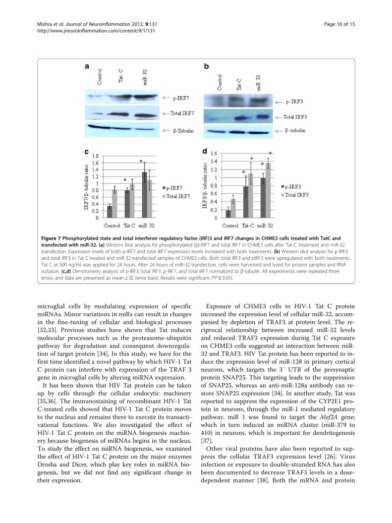
Interferon regulatory factor (IRF)3 and IRF7 are perturbedby tat C treatment and microRNA 32 overexpressionIRF3 and IRF7 are constitutively located in the cytoplasm,and are phosphorylated and translocated to the nucleus inresponse to a stimulatory trigger. After translocation to thenucleus, they induce the expression of IFNs and other in-flammatory cytokines. TRAF3 is regarded as a dual-moderepressor of the NF-κB pathway, having an important rolein regulating the IFN response of cells during infections.Tat C treatment and miR-32 overexpression resulted in thesame trend of activation of IRF3 and IRF7 (Figure 7).Western blot analysis showed higher levels of phosphory-lated IRF3 and IRF7 in Tat C-treated cells (Figure 7A,B)and a similar trend was seen in cells overexpressing miR-32. Total protein levels of IRF3 and IRF7 were also foundto be higher in Tat C treated and miR-32 overexpressingCHME3 cells (Figure 7). Downregulation of TRAF3 afterTat C treatment and miR-32 overexpression, and increasedlevels of phosphorylation of IRF3/7 suggest a negativeregulatory role of TRAF3 in controlling IRF3 and IRF7 ex-pression, which is in accordance with previously shownfunction of TRAF3 [27].The expression levels of total IRF3, IRF7 and pIRF3/7
in CHME3 cells transfected with anti-miR-32 were ana-lyzed by western blotting. Expression levels of total IRF3and IRF7 decreased (Figure 8A,B) in cells transfected
with anti-miR-32, but phosphorylation of IRF3 and IRF7increased compared with the scrambled anti-miR trans-fection (negative control).Both;the Tat C treatment and miR-32 overexpression
increases the IRF3 and IRF7 expression at the transcriptlevel (Figure 9A,B). The suppression of cellular miR-32 byusing anti-miR-32, resulted in the reduced expression ofboth IRF3 and IRF7 at the transcript level (Figure 9C).
DiscussionMicroglia are brain resident macrophages that areinvolved in crucial regulatory processes of developmentand surveillance of the neural environment during in-jury, infection, and repair [11]. As the primary sourcefor pro-inflammatory cytokines, microglial cells areimplicated as pivotal mediators of neuroinflammation.Activated microglia leading to an inflammatory re-sponse has been reported in many models of neuroin-flammation and neurotoxicity [11]. Viruses attempt tomodulate the cellular environment using various strat-egies, changing the expression pattern of protein-codinggenes and non-coding genes such as miRNAs [31].Small RNAs have been reported to play an importantrole in neuroviral infections [21]. In the present study,we identified a novel mechanism by which HIV-1 Tat Cprotein can change the expression of specific genes in
Figure 6 miR-32 directly targets the 3′-UTR of tumor necrosis factor receptor-associated factor 3 (TRAF3). (a) Seed sequence in miR-32and complementary sequence in the 3′ UTR of TRAF3 mRNA showing seven-mer binding in wild-type (WT) TRAF3 3′ UTR. A deletion mutation of4 base pairs in the 3′ UTR of TRAF3 was generated by site-directed mutagenesis. This alteration in the 3′ UTR sequence of TRAF3 abrogated theinteraction of miR-32 and the 3′ UTR of TRAF3, resulting in translational derepression. (b) Luciferase assays were performed by transfecting HeLacells with pCMV-β-gal (normalization control), WT TRAF3 3′ UTR and mutated (MUT) TRAF3 3′ UTR plasmids, along with pCMV-miR-32 plasmids.Normalized luciferase light units of control cells are presented as 100 units, and relative light units (RLU) of other treatments are shownaccordingly. All experiments were performed three times and data are presented as mean± SE (error bars). ***P ≤0.0005.
Mishra et al. Journal of Neuroinflammation 2012, 9:131 Page 9 of 15http://www.jneuroinflammation.com/content/9/1/131
microglial cells by modulating expression of specificmiRNAs. Minor variations in miRs can result in changesin the fine-tuning of cellular and biological processes[32,33]. Previous studies have shown that Tat inducesmolecular processes such as the proteasome-ubiquitinpathway for degradation and consequent downregula-tion of target protein [34]. In this study, we have for thefirst time identified a novel pathway by which HIV-1 TatC protein can interfere with expression of the TRAF 3gene in microglial cells by altering miRNA expression.It has been shown that HIV Tat protein can be taken
up by cells through the cellular endocytic machinery[35,36]. The immunostaining of recombinant HIV-1 TatC-treated cells showed that HIV-1 Tat C protein movesto the nucleus and remains there to execute its transacti-vational functions. We also investigated the effect ofHIV-1 Tat C protein on the miRNA biogenesis machin-ery because biogenesis of miRNAs begins in the nucleus.To study the effect on miRNA biogenesis, we examinedthe effect of HIV-1 Tat C protein on the major enzymesDrosha and Dicer, which play key roles in miRNA bio-genesis, but we did not find any significant change intheir expression.
Exposure of CHME3 cells to HIV-1 Tat C proteinincreased the expression level of cellular miR-32, accom-panied by depletion of TRAF3 at protein level. The re-ciprocal relationship between increased miR-32 levelsand reduced TRAF3 expression during Tat C exposureon CHME3 cells suggested an interaction between miR-32 and TRAF3. HIV Tat protein has been reported to in-duce the expression level of miR-128 in primary corticalneurons, which targets the 3′ UTR of the presynapticprotein SNAP25. This targeting leads to the suppressionof SNAP25, whereas an anti-miR-128a antibody can re-store SNAP25 expression [34]. In another study, Tat wasreported to suppress the expression of the CYP2E1 pro-tein in neurons, through the miR-1 mediated regulatorypathway. miR 1 was found to target the Mef2A gene;which in turn induced an miRNA cluster (miR-379 to410) in neurons, which is important for dendritogenesis[37].Other viral proteins have also been reported to sup-
press the cellular TRAF3 expression level [26]. Virusinfection or exposure to double-stranded RNA has alsobeen documented to decrease TRAF3 levels in a dose-dependent manner [38]. Both the mRNA and protein
Figure 7 Phosphorylated state and total interferon regulatory factor (IRF)3 and IRF7 changes in CHME3 cells treated with TatC andtransfected with miR-32. (a) Western blot analysis for phosphorylated (p)-IRF7 and total IRF7 in CHME3 cells after Tat C treatment and miR-32transfection. Expression levels of both p-IRF7 and total IRF7 expression levels increased with both treatments. (b) Western blot analysis for p-IRF3and total IRF3 in Tat C-treated and miR-32-transfected samples of CHME3 cells. Both total IRF3 and pIRF3 were upregulated with both treatments.Tat C at 500 ng/ml was applied for 24 hours. After 24 hours of miR-32 transfection, cells were harvested and lysed for protein samples and RNAisolation. (c,d) Densitometry analysis of p-IRF3, total IRF3, p-IRF7, and total IRF7 normalized to β-tubulin. All experiments were repeated threetimes, and data are presented as mean± SE (error bars). Results were significant (*P≤ 0.05).
Mishra et al. Journal of Neuroinflammation 2012, 9:131 Page 10 of 15http://www.jneuroinflammation.com/content/9/1/131

levels of the TRAF3 adaptor molecule have beenreported to be downregulated in herpes simplex virus(HSV) infection [26]. Therefore, we designed this studyto understand the mechanism by which extracellularlysecreted HIV protein can affect gene expression in un-infected cells.Bioinformatic databases predicted that a conserved rec-
ognition sequence for miR-32 was present in the 3′ UTRof TRAF3 (Figure 6A). miR-32 can regulate TRAF3 at thepost-transcriptional level, through direct targeting of theTRAF3 3′ UTR. Target validation was performed using areporter construct, having a firefly luciferase coding regionfused with the 3′ UTR of TRAF3. By using a luciferase re-porter assay, we showed that TRAF3 is indeed a directtarget for miR-32. The targeting of miR-32 for the 3′ UTRof TRAF3 was specific as shown by the parallel experi-ment, in which irrelevant miR-146 was not able to affectthe luciferase level.The complementary sequence in the TRAF3 3′ UTR
was modified by deleting four bases (TATT) at positions463 to 466 to remove the binding site for miR-32. Luci-ferase expression was unaffected by using a mutated
TRAF3 3′ UTR construct co-transfected with miR-32.This observation clearly supports the specificity of miR-32 binding to the TRAF3 3′ UTR in microglial cells.The complementary interaction between miR-32 andTRAF3 mRNA results in translational inhibition, leadingto reduced expression of TRAF3 protein in CHME3 cellsexposed to HIV-1 Tat C protein.When miR-32 was overexpressed, the levels of TRAF3
mRNA and protein were downregulated in CHME3 cells(Figure 4). This effect can be simply attributed to enhancedcellular level of miR-32. The increase in miR-32 expres-sion was confirmed by qPCR of miR-32, compared withCHME3 cells transfected with the empty vector(Figure 4b). The presence of complementary bindingsites in the 3′ UTR of TRAF3 mRNA for miR-32 is mostlikely the way in which overexpression of miR-32resulted in reduced expression of TRAF3.To verify the specificity of miR-32 targeting to TRAF3
mRNA and blockage of translation, the level of cellularmiR-32 was blocked by transfecting cells with anti-miR-32.This rescued the expression of TRAF3 protein and resultedin an enhanced level of TRAF 3, compared with control
Figure 8 Recovery of tumor necrosis factor receptor-associated factor 3 (TRAF3) expression by anti-miR-32 transfection suppressesexpression levels of total interferon regulatory factor (IRF)3 and IRF7. (a) CHME3 cells were transfected with Cy3-labeled control anti-miR,anti-miR-32 and anti-miR-32 plus Tat C treatment. Phosphorylated (p)IRF3 level increased after anti-miR-32 treatment, while the total IRF3 levelwas downregulated in anti-miR-32-transfected cells, showing a positive relationship between cellular TRAF3 level and activation of IRF3. (b) pIRF7was increased after anti-miR-32 treatment and anti-miR-32 plus Tat C treatment, again showing a positive role of TRAF3 in IRF7 activation. TotalIRF7 level was decreased in anti-miR-32-transfected cells showing that recovery of TRAF3 could modulate the transcription of IRF3and IRF7. (c,d)Densitometry analysis of pIRF3, pIRF7, total IRF3 and total IRF7 normalized to β-tubulin. Experiments were performed three times and data arepresented as mean± SE. Results were significant (*P≤ 0.05).
Mishra et al. Journal of Neuroinflammation 2012, 9:131 Page 11 of 15http://www.jneuroinflammation.com/content/9/1/131

cells (Figure 5). This observation particularly revealed theinhibitory function of miR-32 on the expression of TRAF3.Blockage of cellular miR-32 by application of anti-miR-32would have occupied the binding sites of mature miR-32,leaving the 3′ UTR of TRAF3 unrestricted, which resultedin enhanced protein expression. Additionally, when anti-miR-32-transfected cells were incubated with Tat C, theTRAF3 protein level was maintained at a higher level(Figure 5c). This suggests that anti-miR-32 could nullifythe effect of Tat C-mediated upregulation of miR-32, andsubsequent suppression of TRAF3 expression.Modulation in TRAF3 expression could affect the im-
mune functions of microglial cells. To study the down-stream effect of TRAF3 reduction in altering the immuneresponse, we studied the cellular expression levels of IRF3and IRF7. IRFs are a family of transcription factors thatplay important roles in host defense systems [39]. In TatC-treated and miR-32 transfected CHME3 cells, levels ofphosphorylated IRF3 and IRF7 were higher. These resultsare in agreement with earlier reported functions of TRAF3as a potent suppressor of inflammatory responses during
overactivation in cells caused by any pathogenic insult[28]. In the present study, we found that the expression ofTRAF3 decreased along with the increase in phosphory-lated forms of IRF3 and IRF7 and levels of total IRF3 andtotal IRF7 after Tat C treatment or miR-32 overexpression.These findings show that changes in TRAF3 level affectexpression levels of both pIRF3 and pIRF7, which can dir-ectly trigger the immune response, as well as the expres-sion of total IRF3/7.In another gain-of-function study of TRAF3, anti-miR-32
was transfected into CHME3 cells to assess the expressionlevels of phosphorylated forms of IRF3/7 and total IRF3/7.The phosphorylated IRF3/7 levels were found to be signifi-cantly higher, than those of the control (Figure 8), but totalIRF3 and total IRF7 protein levels were decreased in anti-miR-32 transfected cells (Figure 8). These results are in ac-cordance with the previously reported dual-mode repressorfunction of TRAF3, as adequate TRAF3 expression isrequired for the immune response [25] and for derepres-sion of the alternative NF-κB pathway [27]. When TRAF3protein levels are reduced in HIV-1 Tat C treated and miR-
Figure 9 Modulation of tumor necrosis factor receptor-associated factor 3 (TRAF3) protein level alters the interferon regulatory factor(IRF)3 and IRF7 mRNA level. (a,b) CHME3 cells were treated with Tat C protein for 24 hours and transfected with miR-32, respectively. Relativefold changes in mRNA levels were determined for IRF3 and IRF7 using quantitative(q)PCR with SYBR green. As a consequence of Tat C treatmentand miR-32 overexpression, the transcript expression levels of IRF3 and IRF7 increased. (c) After inhibiting the cellular miR-32 via application ofanti-miR, the transcript level of both IRF3 and IRF7 was reduced. In cells treated with anti-miR-32 plus Tat C, the transcript levels of IRF3 and IRF7were lower than those in control CHME3 cells. All experiments were repeated three times, and data are presented as mean± SE. Relative changein IRF3 transcript in miR-32 transfected cells compared with empty vector were significant ***P≤ 0.0005, **P≤ 0.005 and *P ≤0.05 in respectivegraphs.
Mishra et al. Journal of Neuroinflammation 2012, 9:131 Page 12 of 15http://www.jneuroinflammation.com/content/9/1/131

32-transfected cells, the repressive function of TRAF3 isremoved, leading to activation of the non-canonical NF-κBpathway. As documented in previous studies, TRAF3 hasbeen shown to bind constitutively to NIK (an essential acti-vator of the alternative NFκB pathway) in unstimulatedcells, and to block the activation of the non-canonical NF-κB pathway [40,41]. Our data suggest that reduction inTRAF3 level could release the NIK, which might lead toactivation of the non-canonical NF-κB pathway. Constitu-tive activation of the NF-κB2 pathway has been reported asa consistent attribute of TRAF3 deficiency in multiple celltypes [42]. However, TRAF3-deficient cells display only apartial reduction in IFN production after RNA virus infec-tion and NF-κB activation [43].Virus-triggered ubiquitylation of TRAF3 and TRAF6 by
cIAP1 and cIAP2 has been reported as a necessary step fortype I IFN induction and cellular antiviral response [29].Degradative ubiquitination of TRAF3 has been reported tobe necessary for the activation of mitogen-activated proteinkinases and production of inflammatory cytokines [44].A small interfering RNA-mediated depletion study of
TRAF3 in DLD1 cells also showed a suppressive func-tion of TRAF3 [27]. Decreased TRAF3 association withLTBR enhanced the recruitment of TRAF2 and IKK1 to
LTBR-induced signaling complexes [27]. A reduced levelof TRAF3 causes specific accumulation of a particularsubset of NF-κB regulators, including key componentsof NF-κB2, such as p100, RelB, and NIK [27]. Anotherstudy showed that excess TRAF3 prevents recruitmentof components (TRAF2 and IKK1) to receptor com-plexes necessary for NF-κB1 activation [45].The changes in TRAF3 protein affected the total protein
level of both IRF3 and IRF7 (Figure 5, Figure 7, Figure 8).Tat C-mediated miR-32 overexpression induced the expres-sion levels of IRF3 and IRF7, indicating the correlation ofreduced TRAF3 with increase in IRF3/7. Further, recoveryof TRAF3 via anti-miR32 transfection resulted in decreasedIRF3/7 at both at the mRNA and (Figure 9) and proteinlevels (Figure 8). Induction of type I IFNs requires coordi-nated and cooperative activation of the transcription factorsIRF3/7 and NF-κB [46]. IFN-β is the early-phase IFN,which is primarily regulated by IRF3, whereas IFN-α is thelate-phase IFN, activated by IRF7 through the STAT1 path-way [47]. Thus, HIV-1 Tat C protein can modulate theinnate immune response by affecting the expression levelof IRF3/7 operating via the TRAF3 expression level underthe control of miR-32. In the present study,we suggest anew model (Figure 10) of miRNA-mediated regulation of
Figure 10 Proposed model for HIV-1 Tat C-induced, miR-32-mediated post-transcriptional regulation of tumor necrosis factorreceptor-associated factor 3 (TRAF3). In response to HIV-1 Tat C exposure of human microglial cells, miR-32 was upregulated, consequentlydownregulating the protein level of TRAF3 post-transcriptionally by binding to its 3′ untranslated region. The miRNA inhibitor against miR-32,ant-miR-32, reduced the cellular level of miR-32 and rescued the expression level of TRAF3 protein. The cellular expression level of TRAF3 proteinhad an inverse relationship to the expression level of interferon regulatory factor (IRF)3/7 and this could perturb the expression of inflammatorygenes in microglial cells after exposure to HIV-1 Tat C protein.
Mishra et al. Journal of Neuroinflammation 2012, 9:131 Page 13 of 15http://www.jneuroinflammation.com/content/9/1/131

the TRAF3 adaptor molecule in response to HIV-1 Tat Cprotein in microglial cells. Such miRNA-mediated dysregu-lation in TRAF3 expression might affect the immune acti-vation of microglial cells, and thus might be one of severalfactors affecting neuroinflammation in patients infectedwith HIV. Further studies are required to understand themolecular basis of this regulation.
ConclusionIn this study, we found that the expression of cellularTRAF3 protein in human microglial cells exposed toHIV-1 Tat C protein was regulated by cellular miR-32.The changes in expression levels of TRAF3 mediated bymiR-32 resulted in changes in the expression pattern ofcellular IRF3 and IRF7, which might lead to changes ininterferon stimulatory genes. A well-orchestrated regula-tion of the innate immune response is very important toprevent damage caused by excessive or dysregulated ac-tivation of immune signaling factors. This study demon-strates the plausible mechanism of Tat-induced miRNAmediated dysregulation of the immune adaptor moleculeTRAF3 in human microglial cells.
AbbreviationsAIDS: Acquired immunodeficiency syndrome; BSA: Bovine serum albumin;CNS: Central nervous system; EDTA: ethylenediamene tetraacetic acid;GFP: Green fluorescent protein; HAND: Human immunodeficiencyvirus-associated neurological disorder; HIV: Human immunodeficiency virus;IFN: interferon; IRF: Interferon regulatory factor; LTBR: lymphotoxin-βreceptor; LTR: Long terminal repeat; NF-κB: Nuclear factor kappa B;Ni-NTA: Nickel-nitrilotriacetic acid; PBS: Phosphate-buffered saline;qPCR: Quantitative polymerase chain reaction; SDS: sodium dodecyl sulfate;TBS-T: Tris-buffered saline with Triton X-100; TNF: Tumor necrosis factor;TRAF: Tumor necrosis factor receptor-associated factor; UTR: Untranslatedregion; WT: Wild type.
Competing interestsThe authors declare that they have no competing interests.
AcknowledgementsWe are grateful to Dr.Udaykumar Ranga, JNCASR, Bangalore, for his supportduring preparation of this manuscript and to Dr. Anirban Basu, National Brainresearch Centre (NBRC), Gurgaon, Haryana, for providing the human CHME3cells. This work was supported by funding from the Deptartment ofBiotechnology (DBT), Government of India (Grant Number BT/PR10709/AGR/36/598/2008).
Authors’ contributionsRM completed most of the experimental work related to cell culture,cloning, western blotting, transfection, quantitative real-time PCR and dataanalysis presented in this manuscript. CC helped in preparation ofmanuscript. SKS conceived the idea for the study, helped in designing theexperiments, and critically supervised the complete study. All the authorsread and approved the final revised manuscript.
Authors’ informationRM is a recipient of a Senior Research Fellowship of the Council of Scientificand Industrial Research (CSIR), Government of India, and is pursuing her PhDstudies at the Centre for Cellular and Molecular Biology (CCMB), Hyderabad.CC is a recipient of a Senior Research Fellowship of CSIR, Government of ofIndia and is pursuing his PhD work at CCMB, Hyderabad. SKS is aneurovirologist, currently working as a scientist at the CCMB, Hyderabad. Hisresearch group is involved in research areas of HIV and vector-borne viralinfections.
Received: 3 February 2012 Accepted: 18 June 2012Published: 18 June 2012
References1. Thompson KA, Cherry CL, Bell JE, McLean CA: Brain cell reservoirs of latent
virus in presymptomatic HIV-infected individuals. Am J Pathol 2011,179:1623–1629.
2. Heaton RK, Clifford DB, Franklin DR Jr, Woods SP, Ake C, Vaida F, Ellis RJ,Letendre SL, Marcotte TD, Atkinson JH, et al: HIV-associatedneurocognitive disorders persist in the era of potent antiretroviraltherapy: CHARTER Study. Neurology 2010, 75:2087–2096.
3. Sharma D, Bhattacharya J: Cellular & molecular basis of HIV-associatedneuropathogenesis. Indian J Med Res 2009, 129:637–651.
4. Kaul M, Zheng J, Okamoto S, Gendelman HE, Lipton SA: HIV-1 infectionand AIDS: consequences for the central nervous system. Cell Death Differ2005, 12(Suppl 1):878–892.
5. Sanders VJ, Mehta AP, White MG, Achim CL: A murine model of HIVencephalitis: xenotransplantation of HIV-infected human neuroglia intoSCID mouse brain. Neuropathol Appl Neurobiol 1998, 24:461–467.
6. Del Valle L, Croul S, Morgello S, Amini S, Rappaport J, Khalili K: Detection ofHIV-1 Tat and JCV capsid protein, VP1, in AIDS brain with progressivemultifocal leukoencephalopathy. J Neurovirol 2000, 6:221–228.
7. Ju SM, Song HY, Lee JA, Lee SJ, Choi SY, Park J: Extracellular HIV-1 Tat up-regulates expression of matrix metalloproteinase-9 via a MAPK-NF-kappaB dependent pathway in human astrocytes. Exp Mol Med 2009,41:86–93.
8. Deshmane SL, Mukerjee R, Fan S, Sawaya BE: High-performance capillaryelectrophoresis for determining HIV-1 Tat protein in neurons. PLoS One2011, 6:e16148.
9. Li W, Li G, Steiner J, Nath A: Role of Tat protein in HIV neuropathogenesis.Neurotox Res 2009, 16:205–220.
10. Lewis SD, Butchi NB, Khaleduzzaman M, Morgan TW, Du M, Pourciau S,Baker DG, Akira S, Peterson KE: Toll-like receptor 7 is not necessary forretroviral neuropathogenesis but does contribute to virus-inducedneuroinflammation. J Neurovirol 2008, 14:492–502.
11. Kraft AD, Harry GJ: Features of microglia and neuroinflammation relevantto environmental exposure and neurotoxicity. Int J Environ Res PublicHealth 2011, 8:2980–3018.
12. Antony JM: Grooming and growing with microglia. Sci Signal 2010, 3:jc8.13. Chugh P, Fan S, Planelles V, Maggirwar SB, Dewhurst S, Kim B: Infection of
human immunodeficiency virus and intracellular viral Tat protein exert apro-survival effect in a human microglial cell line. J Mol Biol 2007, 366:67–81.
14. Hudson L, Liu J, Nath A, Jones M, Raghavan R, Narayan O, Male D, Everall I:Detection of the human immunodeficiency virus regulatory protein tatin CNS tissues. J Neurovirol 2000, 6:145–155.
15. Singh SK: miRNAs: from neurogeneration to neurodegeneration.Pharmacogenomics 2007, 8:971–978.
16. Fang JH, Zhou HC, Zeng C, Yang J, Liu Y, Huang X, Zhang JP, Guan XY,Zhuang SM: MicroRNA-29b suppresses tumor angiogenesis, invasion, andmetastasis by regulating matrix metalloproteinase 2 expression.Hepatology 2011, 54:1729–1740.
17. Lee Y, Ahn C, Han J, Choi H, Kim J, Yim J, Lee J, Provost P, Radmark O, KimS, Kim VN: The nuclear RNase III Drosha initiates microRNA processing.Nature 2003, 425:415–419.
18. O’Connell RM, Rao DS, Chaudhuri AA, Baltimore D: Physiological andpathological roles for microRNAs in the immune system. Nat RevImmunol 2010, 10:111–122.
19. Eacker SM, Dawson TM, Dawson VL: Understanding microRNAs inneurodegeneration. Nat Rev Neurosci 2009, 10:837–841.
20. Shahabi HN, Westberg L, Melke J, Hakansson A, Belin AC, Sydow O, Olson L,Holmberg B, Nissbrandt H: Cytochrome P450 2E1 gene polymorphisms/haplotypes and Parkinson’s disease in a Swedish population. J NeuralTransm 2009, 116:567–573.
21. Chang JR, Mukerjee R, Bagashev A, Del Valle L, Chabrashvili T, Hawkins BJ,He JJ, Sawaya BE: HIV-1 Tat protein promotes neuronal dysfunctionthrough disruption of microRNAs. J Biol Chem 2011, 286:41125–41134.
22. Ely KR, Kodandapani R, Wu S: Protein-protein interactions in TRAF3. AdvExp Med Biol 2007, 597:114–121.
23. Saitoh T, Nakayama M, Nakano H, Yagita H, Yamamoto N, Yamaoka S:TWEAK induces NF-kappaB2 p100 processing and long lasting NF-kappaB activation. J Biol Chem 2003, 278:36005–36012.
Mishra et al. Journal of Neuroinflammation 2012, 9:131 Page 14 of 15http://www.jneuroinflammation.com/content/9/1/131

24. Hacker H, Tseng PH, Karin M: Expanding TRAF function: TRAF3 as atri-faced immune regulator. Nat Rev Immunol 2011, 11:457–468.
25. Oganesyan G, Saha SK, Guo B, He JQ, Shahangian A, Zarnegar B, Perry A,Cheng G: Critical role of TRAF3 in the Toll-like receptor-dependent and-independent antiviral response. Nature 2006, 439:208–211.
26. Perez de Diego R, Sancho-Shimizu V, Lorenzo L, Puel A, Plancoulaine S,Picard C, Herman M, Cardon A, Durandy A, Bustamante J, et al: HumanTRAF3 adaptor molecule deficiency leads to impaired Toll-like receptor 3response and susceptibility to herpes simplex encephalitis. Immunity2010, 33:400–411.
27. Bista P, Zeng W, Ryan S, Bailly V, Browning JL, Lukashev ME: TRAF3 controlsactivation of the canonical and alternative NFkappaB by thelymphotoxin beta receptor. J Biol Chem 2010, 285:12971–12978.
28. Zarnegar B, Yamazaki S, He JQ, Cheng G: Control of canonical NF-kappaBactivation through the NIK-IKK complex pathway. Proc Natl Acad Sci U S A2008, 105:3503–3508.
29. Mao AP, Li S, Zhong B, Li Y, Yan J, Li Q, Teng C, Shu HB: Virus-triggeredubiquitination of TRAF3/6 by cIAP1/2 is essential for induction ofinterferon-beta (IFN-beta) and cellular antiviral response. J Biol Chem2010, 285:9470–9476.
30. Romani B, Engelbrecht S, Glashoff RH: Functions of Tat: the versatile proteinof human immunodeficiency virus type 1. J Gen Virol 2010, 91:1–12.
31. Boss IW, Renne R: Viral miRNAs and immune evasion. Biochim Biophys Acta2011, 1809:708–714.
32. Hornstein E, Shomron N: Canalization of development by microRNAs. NatGenet 2006, 38(Suppl):S20–S24.
33. Cao X, Pfaff SL, Gage FH: A functional study of miR-124 in the developingneural tube. Genes Dev 2007, 21:531–536.
34. Eletto D, Russo G, Passiatore G, Del Valle L, Giordano A, Khalili K, Gualco E,Peruzzi F: Inhibition of SNAP25 expression by HIV-1 Tat involves theactivity of mir-128a. J Cell Physiol 2008, 216:764–770.
35. Frankel AD, Pabo CO: Cellular uptake of the tat protein from humanimmunodeficiency virus. Cell 1988, 55:1189–1193.
36. Cardarelli F, Serresi M, Albanese A, Bizzarri R, Beltram F: Quantitativeanalysis of Tat peptide binding to import carriers reveals unconventionalnuclear transport properties. J Biol Chem 2011, 286:12292–12299.
37. Fiore R, Khudayberdiev S, Christensen M, Siegel G, Flavell SW, Kim TK,Greenberg ME, Schratt G: Mef2-mediated transcription of the miR379-410cluster regulates activity-dependent dendritogenesis by fine-tuningPumilio2 protein levels. EMBO J 2009, 28:697–710.
38. Nakhaei P, Mesplede T, Solis M, Sun Q, Zhao T, Yang L, Chuang TH, WareCF, Lin R, Hiscott J: The E3 ubiquitin ligase Triad3A negatively regulatesthe RIG-I/MAVS signaling pathway by targeting TRAF3 for degradation.PLoS Pathog 2009, 5:e1000650.
39. Wang L, Toomey NL, Diaz LA, Walker G, Ramos JC, Barber GN, Ning S:Oncogenic IRFs Provide a Survival Advantage for Epstein-Barr Virus- orHuman T-Cell Leukemia Virus Type 1-Transformed Cells throughInduction of BIC Expression. J Virol 2011, 85:8328–8337.
40. Xiao G, Fong A, Sun SC: Induction of p100 processing byNF-kappaB-inducing kinase involves docking IkappaB kinase alpha(IKKalpha) to p100 and IKKalpha-mediated phosphorylation. J BiolChem 2004, 279:30099–30105.
41. Xiao G, Harhaj EW, Sun SC: NF-kappaB-inducing kinase regulates theprocessing of NF-kappaB2 p100. Mol Cell 2001, 7:401–409.
42. Hildebrand JM, Yi Z, Buchta CM, Poovassery J, Stunz LL, Bishop GA: Roles oftumor necrosis factor receptor associated factor 3 (TRAF3) and TRAF5 inimmune cell functions. Immunol Rev 2011, 244:55–74.
43. Tang ED, Wang CY: TRAF5 is a downstream target of MAVS in antiviralinnate immune signaling. PLoS One 2010, 5:e9172.
44. Tseng PH, Matsuzawa A, Zhang W, Mino T, Vignali DA, Karin M: Different modesof ubiquitination of the adaptor TRAF3 selectively activate the expression oftype I interferons and proinflammatory cytokines. Nat Immunol 2010, 11:70–75.
45. He L, Grammer AC, Wu X, Lipsky PE: TRAF3 forms heterotrimers withTRAF2 and modulates its ability to mediate NF-{kappa}B activation. J BiolChem 2004, 279:55855–55865.
46. Matsuzawa A, Tseng PH, Vallabhapurapu S, Luo JL, Zhang W, Wang H, VignaliDA, Gallagher E, Karin M: Essential cytoplasmic translocation of a cytokinereceptor-assembled signaling complex. Science 2008, 321:663–668.
47. Sun F, Zhang YB, Liu TK, Shi J, Wang B, Gui JF: Fish MITA serves as amediator for distinct fish IFN gene activation dependent on IRF3 or IRF7.J Immunol 2011, 187:2531–2539.
doi:10.1186/1742-2094-9-131Cite this article as: Mishra et al.: HIV-1 Tat C-mediated regulation oftumor necrosis factor receptor-associated factor-3 by microRNA 32 inhuman microglia. Journal of Neuroinflammation 2012 9:131.
Submit your next manuscript to BioMed Centraland take full advantage of:
• Convenient online submission
• Thorough peer review
• No space constraints or color figure charges
• Immediate publication on acceptance
• Inclusion in PubMed, CAS, Scopus and Google Scholar
• Research which is freely available for redistribution
Submit your manuscript at www.biomedcentral.com/submit
Mishra et al. Journal of Neuroinflammation 2012, 9:131 Page 15 of 15http://www.jneuroinflammation.com/content/9/1/131