research open access downstream signaling and genome … · genome-wide regulatory effects of ptk7...
TRANSCRIPT

Golubkov and Strongin Cell Communication and Signaling 2014, 12:15http://www.biosignaling.com/content/12/1/15
RESEARCH Open Access
Downstream signaling and genome-wideregulatory effects of PTK7 pseudokinase and itsproteolytic fragments in cancer cellsVladislav S Golubkov* and Alex Y Strongin*
Abstract
Background: The full-length membrane protein tyrosine kinase 7 (PTK7) pseudokinase, an important component ofthe planar cell polarity and the Wnt canonical and non-canonical pathways, is a subject of step-wise proteolysis incells and tissues. The proteolysis of PTK7 involves membrane type-matrix metalloproteinase (MT1-MMP), membersof the Disintegrin Domain and Metalloproteinase (ADAM) family, and γ-secretase. This multi-step proteolysis resultsin the generation of the digest fragments of PTK7. These fragments may be either liberated into the extracellularmilieu or retained on the plasma membrane or released into the cytoplasm and then transported into the nucleus.
Results: We employed the genome-wide transcriptional and kinome array analyses to determine the role of thefull-length membrane PTK7 and its proteolytic fragments in the downstream regulatory mechanisms, with anemphasis on the cell migration-related genes and proteins. Using fibrosarcoma HT1080 cells stably expressing PTK7and its mutant and truncated species, the structure of which corresponded to the major PTK7 digest fragments, wedemonstrated that the full-length membrane 1–1070 PTK7, the N-terminal 1–694 soluble ectodomain fragment,and the C-terminal 622–1070 and 726–1070 fragments differentially regulate multiple genes and signaling pathwaysin our highly invasive cancer cell model. Immunoblotting of the selected proteins were used to validate the resultsof our high throughput assays.
Conclusions: Our results suggest that PTK7 levels need to be tightly controlled to enable migration and thatthe anti-migratory effect of the full-length membrane PTK7 is linked to the down-regulation of multiplemigration-related genes and to the activation of the Akt and c-Jun pathway. In turn, the C-terminal fragments ofPTK7 act predominantly via the RAS-ERK and CREB/ATF1 pathway and through the up-regulation of cadherin-11.In general, our data correlate well with the distinct functionality of the full-length receptor tyrosine kinases and theirrespective intracellular domain (ICD) proteolytic fragments.
Keywords: Protein tyrosine kinase 7, PTK7, Cell migration, Cadherin-11, Metalloprotease, CREB, ATF1
BackgroundPseudokinase PTK7 is a Wnt co-receptor and an essen-tial regulator of planar cell polarity (PCP) and directionalcell motility in vertebrate development and embryogen-esis [1,2]. In humans, the full-length membrane PTK7consists of seven extracellular immunoglobulin-like (Ig)domains, a transmembrane region, a juxtamembrane re-gion and a catalytically inert cytoplasmic tyrosine kinasedomain [3]. The earlier experimental data suggest that
* Correspondence: [email protected];[email protected] Research Center, Sanford-Burnham Institute for Medical Research, LaJolla, CA 92037, USA
© 2014 Golubkov and Strongin; licensee BioMCreative Commons Attribution License (http:/distribution, and reproduction in any mediumDomain Dedication waiver (http://creativecomarticle, unless otherwise stated.
PTK7 directly interacts with β-catenin [4], Wnt proteins[5,6], plexins and semaphorins [7,8], RACK1 and PKCδ1,recruits Dishevelled [9,10] and regulates both the non-canonical Wnt/PCP and canonical Wnt pathways. Be-cause of severe defects in neural tube closure, mice withPTK7 truncation died during embryogenesis [1]. SolublePTK7 species inhibited angiogenesis by competing withthe full-length PTK7 [11].In contrast to its well-appreciated, crucial role in ver-
tebrate development, the functional importance of theintact full-length PTK7 in malignancy is still a matter ofdebate. The expression levels of the PTK7 mRNA are
ed Central Ltd. This is an Open Access article distributed under the terms of the/creativecommons.org/licenses/by/2.0), which permits unrestricted use,, provided the original work is properly credited. The Creative Commons Publicmons.org/publicdomain/zero/1.0/) applies to the data made available in this

Golubkov and Strongin Cell Communication and Signaling 2014, 12:15 Page 2 of 12http://www.biosignaling.com/content/12/1/15
highly elevated in colon, gastric, lung cancer and acutemyeloid leukemia [12-16]. On the contrary, the levels ofPTK7 are decreased in metastatic melanoma [17] andbreast carcinoma [18]. Deletions of the chromosomearm 6p, in which the PTK7 gene is localized in humans,were reported in breast cancer [19] and melanoma [20].Because pericellular proteolysis plays a primary role incell migration, especially in the directional locomotionof tumor cells, it is likely that proteolysis and PCP con-verge to promote directed cancer cell migration. Inagreement, the functionality of PTK7 is directly regu-lated by proteolysis. Ubiquitous membrane type-1 matrixmetalloproteinase (MT1-MMP), arguably the primaryenzyme in pericellular proteolysis and cancer cell migra-tion, cleaves the PKP621↓LI sequence in the seventh Ig-like domain of membrane PTK7 and this cleavage resultsin the liberation of the N-terminal soluble PTK7 frag-ment (sPTK7) [21]. MT1-MMP proteolysis is followedby cleavage of the PEE↓S690 site in the C-terminal re-sidual portion of PTK7 by ADAMs, including ADAM17.The ectodomain shedding by MT1-MMP and ADAMsis a prerequisite for the intramembrane cleavage ofPTK7 by γ-secretase. This cleavage releases the C-terminal cytoplasmic tail fragment of PTK7 (ICD), whichis then either degraded by the proteasome or trans-ported to the nucleus [11,22].The limited pre-existing data suggest that the full-
length membrane PTK7 and its proteolytic productscause a contrasting effect on the efficiency of cell migra-tion. Thus, the continuing presence of the full-lengthmembrane PTK7 on the plasma membrane down-regulated the myosin light chain (MLC) phosphorylation(a downstream event of the Wnt/PCP pathway) and alsoreduced migration efficiency of fibrosarcoma HT1080cells [21]. MT1-MMP proteolysis of PTK7 reversed theinhibitory effect of the full-length membrane PTK7, andresulted in the accumulation of the Stable N-terminalsPTK7 fragment in the extracellular milieu and pro-moted cell invasion of HT1080 cells [11,21,23]. Expres-sion of the Chz PTK7 mutant that exhibited anadditional PEK↓LK503 MT1-MMP cleavage site in thejunction region between the fifth and the sixth Ig-likedomains stimulated cell migration even further [23].These effects suggest the existence of the intriguing andspecific downstream mechanisms by which intact PTK7,its digest fragments and the homo- and heterodimericcomplexes between the PTK7 membrane, soluble andintracellular portions control cell function. These mech-anisms, however, have not been precisely investigated inthe earlier works by us and others [11,21-23]. Under-standing these mechanisms will shed additional light onthe role that PTK7, alone as well as in combination withMT1-MMP, ADAMs and γ-secretase, plays in cancer cellmigration.
In this study, we applied the genome-wide transcrip-tional and kinome profiling of HT1080 cells to gaininsight into the role of the full-length membrane PTK7,and its mutant forms and proteolytic fragments in thedownstream signaling and transcriptional events thatdirectly control the efficiency of cell invasion.
ResultsPTK7 in cell invasionTo evaluate in detail the effects of PTK7 and its proteo-lytic fragments on cell invasion, downstream signalingand genome-wide transcriptional regulation, we specific-ally employed fibrosarcoma HT1080 cells. These highlyinvasive cells express low endogenous levels of PTK7,but high levels of active MT1-MMP and ADAMs [11].Because of these parameters, we could manipulate thispseudokinase functionality using HT1080 cells trans-fected with the recombinant PTK7 constructs. The cellswe employed also included the PTK7 knock-out cells(shPTK7 cells), cells with the enforced overexpression ofthe original membrane PTK7 (PTK7 cells) and theChuzhoi (Chz) mutant (Chz cells), and, lastly, cells,which overexpressed multiple truncated species ofPTK7. These species represented the soluble, the mem-brane and the cytoplasmic digest fragments that werethe result of PTK7 cleavage by MT1-MMP, ADAMs andγ–secretase (sPTK7, cPTK7/622-1070 and cPTK7/726-1070 cells) (Figure 1). In addition, because PTK7 func-tionality is directly linked to MT1-MMP, we isolatedcells in which MT1-MMP was silenced by shRNA(shMT1 cells). We then co-expressed the shMT1 con-struct with the Chz mutant (shMT1-Chz cells). Expres-sion of these constructs in the same cell system allowedus to determine their downstream effect more precisely.Our studies revealed that both transcriptional silencing
and overexpression of PTK7 inhibited cell invasion by~50% relative to the parental cells. In turn, Chz stimu-lated cell invasion by ~50% (Figure 2). Transcriptionalsilencing of MT1-MMP in shMT1-Chz cells reversedthe stimulatory effect of Chz and suppressed cell inva-sion by ~90%, while MT1-MMP silencing alone (shMT1cells) was less potent. The effects of sPTK7, cPTK7/622-1070 and cPTK7/726-1070 were less significant.
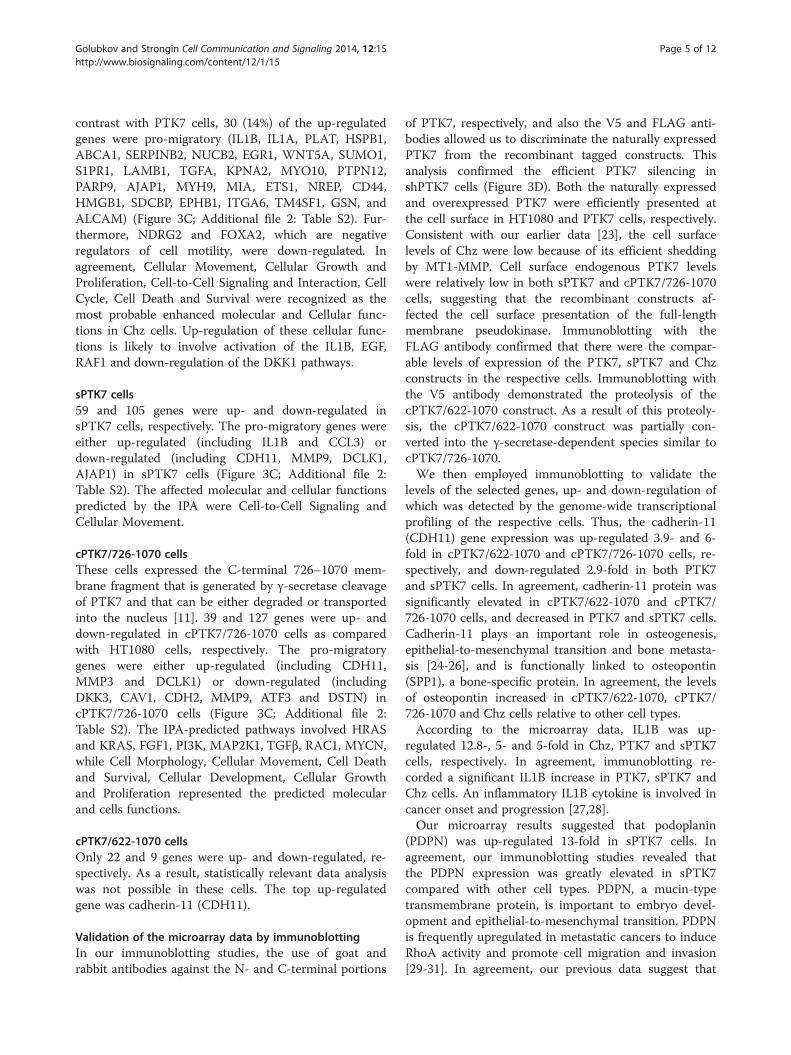
Cell microarraysTo elucidate the genome-wide effects of PTK7 in itsmutant and truncated forms, we performed a whole gen-ome transcriptional analysis of the cells. The differen-tially expressed genes that exhibited at least a 2-folddifference relative to the parental cells are shown inAdditional file 1: Table S1. The hierarchical gene cluster-ing clearly indicated a high similarity among the sPTK7,Chz and PTK7 samples and, on the other hand, amongthe cPTK7/726-1070, cPTK7/622-1070 and shPTK7
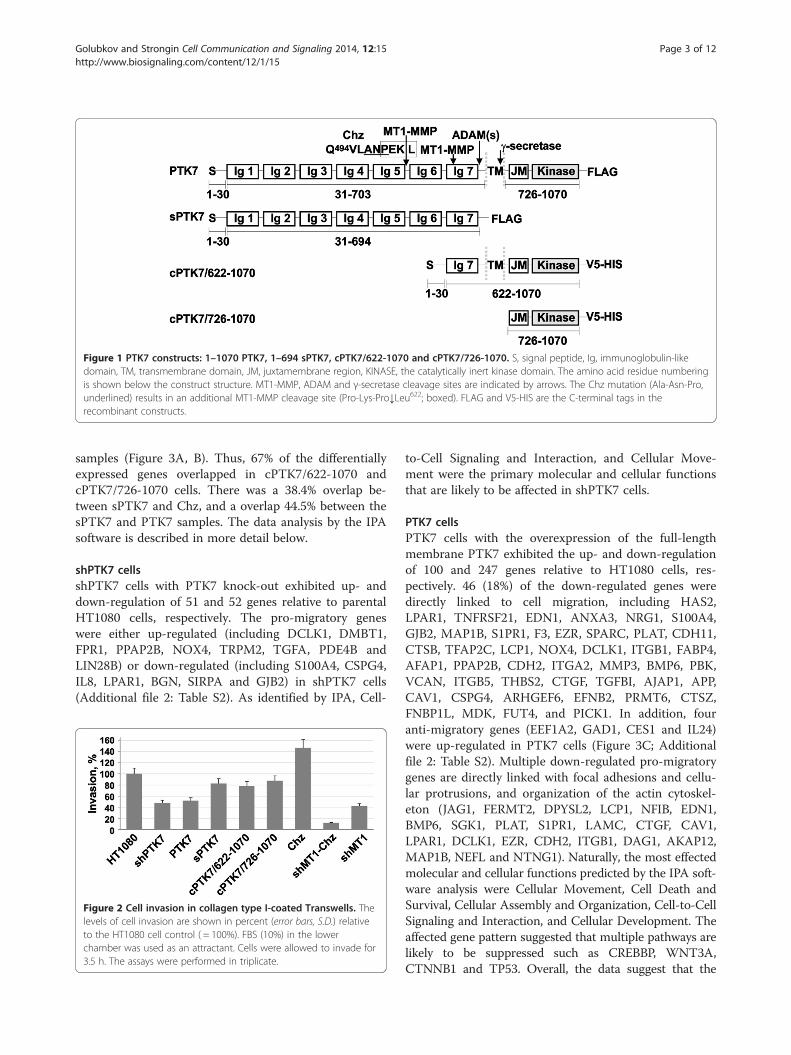
Figure 1 PTK7 constructs: 1–1070 PTK7, 1–694 sPTK7, cPTK7/622-1070 and cPTK7/726-1070. S, signal peptide, Ig, immunoglobulin-likedomain, TM, transmembrane domain, JM, juxtamembrane region, KINASE, the catalytically inert kinase domain. The amino acid residue numberingis shown below the construct structure. MT1-MMP, ADAM and γ-secretase cleavage sites are indicated by arrows. The Chz mutation (Ala-Asn-Pro,underlined) results in an additional MT1-MMP cleavage site (Pro-Lys-Pro↓Leu622; boxed). FLAG and V5-HIS are the C-terminal tags in therecombinant constructs.
Golubkov and Strongin Cell Communication and Signaling 2014, 12:15 Page 3 of 12http://www.biosignaling.com/content/12/1/15
samples (Figure 3A, B). Thus, 67% of the differentiallyexpressed genes overlapped in cPTK7/622-1070 andcPTK7/726-1070 cells. There was a 38.4% overlap be-tween sPTK7 and Chz, and a overlap 44.5% between thesPTK7 and PTK7 samples. The data analysis by the IPAsoftware is described in more detail below.
shPTK7 cellsshPTK7 cells with PTK7 knock-out exhibited up- anddown-regulation of 51 and 52 genes relative to parentalHT1080 cells, respectively. The pro-migratory geneswere either up-regulated (including DCLK1, DMBT1,FPR1, PPAP2B, NOX4, TRPM2, TGFA, PDE4B andLIN28B) or down-regulated (including S100A4, CSPG4,IL8, LPAR1, BGN, SIRPA and GJB2) in shPTK7 cells(Additional file 2: Table S2). As identified by IPA, Cell-
Figure 2 Cell invasion in collagen type I-coated Transwells. Thelevels of cell invasion are shown in percent (error bars, S.D.) relativeto the HT1080 cell control ( = 100%). FBS (10%) in the lowerchamber was used as an attractant. Cells were allowed to invade for3.5 h. The assays were performed in triplicate.
to-Cell Signaling and Interaction, and Cellular Move-ment were the primary molecular and cellular functionsthat are likely to be affected in shPTK7 cells.
PTK7 cellsPTK7 cells with the overexpression of the full-lengthmembrane PTK7 exhibited the up- and down-regulationof 100 and 247 genes relative to HT1080 cells, res-pectively. 46 (18%) of the down-regulated genes weredirectly linked to cell migration, including HAS2,LPAR1, TNFRSF21, EDN1, ANXA3, NRG1, S100A4,GJB2, MAP1B, S1PR1, F3, EZR, SPARC, PLAT, CDH11,CTSB, TFAP2C, LCP1, NOX4, DCLK1, ITGB1, FABP4,AFAP1, PPAP2B, CDH2, ITGA2, MMP3, BMP6, PBK,VCAN, ITGB5, THBS2, CTGF, TGFBI, AJAP1, APP,CAV1, CSPG4, ARHGEF6, EFNB2, PRMT6, CTSZ,FNBP1L, MDK, FUT4, and PICK1. In addition, fouranti-migratory genes (EEF1A2, GAD1, CES1 and IL24)were up-regulated in PTK7 cells (Figure 3C; Additionalfile 2: Table S2). Multiple down-regulated pro-migratorygenes are directly linked with focal adhesions and cellu-lar protrusions, and organization of the actin cytoskel-eton (JAG1, FERMT2, DPYSL2, LCP1, NFIB, EDN1,BMP6, SGK1, PLAT, S1PR1, LAMC, CTGF, CAV1,LPAR1, DCLK1, EZR, CDH2, ITGB1, DAG1, AKAP12,MAP1B, NEFL and NTNG1). Naturally, the most effectedmolecular and cellular functions predicted by the IPA soft-ware analysis were Cellular Movement, Cell Death andSurvival, Cellular Assembly and Organization, Cell-to-CellSignaling and Interaction, and Cellular Development. Theaffected gene pattern suggested that multiple pathways arelikely to be suppressed such as CREBBP, WNT3A,CTNNB1 and TP53. Overall, the data suggest that the
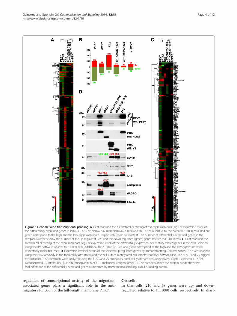
Figure 3 Genome-wide transcriptional profiling. A. Heat map and the hierarchical clustering of the expression data (log2 of expression level) ofthe differentially expressed genes in PTK7, sPTK7, Chz, cPTK7/726-1070, cPTK7/622-1070 and shPTK7 cells relative to the parental HT1080 cells. Red andgreen correspond to the high and the low expression levels, respectively (color bar inset). B. The number of differentially expressed genes in thesamples. Numbers show the number of the up-regulated (red) and the down-regulated (green) genes relative to HT1080 cells. C. Heat map and thehierarchical clustering of the expression data (log2 of expression level) of the differentially expressed, cell motility-related genes in the cells (selectedusing the IPA software) relative to HT1080 cells (Additional file 2: Table S2). Red and green correspond to the high and the low expression levels,respectively (color bar inset). D. Expression level validation of the selected up-regulated genes by immunoblotting. Top two panels, PTK7 was analyzedusing the PTK7 antibody in the total cell lysates (total) and the cell surface-biotinylated cell samples (surface). Bottom panel, The FLAG- and V5-taggedrecombinant PTK7 constructs were analyzed using the FLAG and V5 antibodies (total cell lysate samples), respectively. CDH11, cadherin-11; SPP1,osteopontin; IL1B, interleukin 1β; PDPN, podoplanin; MAGEC1, melanoma antigen family C1. The numbers above the protein bands show thefold-difference of the differentially expressed genes as detected by transcriptional profiling. Tubulin, loading control.
Golubkov and Strongin Cell Communication and Signaling 2014, 12:15 Page 4 of 12http://www.biosignaling.com/content/12/1/15
regulation of transcriptional activity of the migration-associated genes plays a significant role in the anti-migratory function of the full-length membrane PTK7.
Chz cellsIn Chz cells, 210 and 58 genes were up- and down-regulated relative to HT1080 cells, respectively. In sharp
Golubkov and Strongin Cell Communication and Signaling 2014, 12:15 Page 5 of 12http://www.biosignaling.com/content/12/1/15
contrast with PTK7 cells, 30 (14%) of the up-regulatedgenes were pro-migratory (IL1B, IL1A, PLAT, HSPB1,ABCA1, SERPINB2, NUCB2, EGR1, WNT5A, SUMO1,S1PR1, LAMB1, TGFA, KPNA2, MYO10, PTPN12,PARP9, AJAP1, MYH9, MIA, ETS1, NREP, CD44,HMGB1, SDCBP, EPHB1, ITGA6, TM4SF1, GSN, andALCAM) (Figure 3C; Additional file 2: Table S2). Fur-thermore, NDRG2 and FOXA2, which are negativeregulators of cell motility, were down-regulated. Inagreement, Cellular Movement, Cellular Growth andProliferation, Cell-to-Cell Signaling and Interaction, CellCycle, Cell Death and Survival were recognized as themost probable enhanced molecular and Cellular func-tions in Chz cells. Up-regulation of these cellular func-tions is likely to involve activation of the IL1B, EGF,RAF1 and down-regulation of the DKK1 pathways.
sPTK7 cells59 and 105 genes were up- and down-regulated insPTK7 cells, respectively. The pro-migratory genes wereeither up-regulated (including IL1B and CCL3) ordown-regulated (including CDH11, MMP9, DCLK1,AJAP1) in sPTK7 cells (Figure 3C; Additional file 2:Table S2). The affected molecular and cellular functionspredicted by the IPA were Cell-to-Cell Signaling andCellular Movement.
cPTK7/726-1070 cellsThese cells expressed the C-terminal 726–1070 mem-brane fragment that is generated by γ-secretase cleavageof PTK7 and that can be either degraded or transportedinto the nucleus [11]. 39 and 127 genes were up- anddown-regulated in cPTK7/726-1070 cells as comparedwith HT1080 cells, respectively. The pro-migratorygenes were either up-regulated (including CDH11,MMP3 and DCLK1) or down-regulated (includingDKK3, CAV1, CDH2, MMP9, ATF3 and DSTN) incPTK7/726-1070 cells (Figure 3C; Additional file 2:Table S2). The IPA-predicted pathways involved HRASand KRAS, FGF1, PI3K, MAP2K1, TGFβ, RAC1, MYCN,while Cell Morphology, Cellular Movement, Cell Deathand Survival, Cellular Development, Cellular Growthand Proliferation represented the predicted molecularand cells functions.
cPTK7/622-1070 cellsOnly 22 and 9 genes were up- and down-regulated, re-spectively. As a result, statistically relevant data analysiswas not possible in these cells. The top up-regulatedgene was cadherin-11 (CDH11).
Validation of the microarray data by immunoblottingIn our immunoblotting studies, the use of goat andrabbit antibodies against the N- and C-terminal portions
of PTK7, respectively, and also the V5 and FLAG anti-bodies allowed us to discriminate the naturally expressedPTK7 from the recombinant tagged constructs. Thisanalysis confirmed the efficient PTK7 silencing inshPTK7 cells (Figure 3D). Both the naturally expressedand overexpressed PTK7 were efficiently presented atthe cell surface in HT1080 and PTK7 cells, respectively.Consistent with our earlier data [23], the cell surfacelevels of Chz were low because of its efficient sheddingby MT1-MMP. Cell surface endogenous PTK7 levelswere relatively low in both sPTK7 and cPTK7/726-1070cells, suggesting that the recombinant constructs af-fected the cell surface presentation of the full-lengthmembrane pseudokinase. Immunoblotting with theFLAG antibody confirmed that there were the compar-able levels of expression of the PTK7, sPTK7 and Chzconstructs in the respective cells. Immunoblotting withthe V5 antibody demonstrated the proteolysis of thecPTK7/622-1070 construct. As a result of this proteoly-sis, the cPTK7/622-1070 construct was partially con-verted into the γ-secretase-dependent species similar tocPTK7/726-1070.We then employed immunoblotting to validate the
levels of the selected genes, up- and down-regulation ofwhich was detected by the genome-wide transcriptionalprofiling of the respective cells. Thus, the cadherin-11(CDH11) gene expression was up-regulated 3.9- and 6-fold in cPTK7/622-1070 and cPTK7/726-1070 cells, re-spectively, and down-regulated 2.9-fold in both PTK7and sPTK7 cells. In agreement, cadherin-11 protein wassignificantly elevated in cPTK7/622-1070 and cPTK7/726-1070 cells, and decreased in PTK7 and sPTK7 cells.Cadherin-11 plays an important role in osteogenesis,epithelial-to-mesenchymal transition and bone metasta-sis [24-26], and is functionally linked to osteopontin(SPP1), a bone-specific protein. In agreement, the levelsof osteopontin increased in cPTK7/622-1070, cPTK7/726-1070 and Chz cells relative to other cell types.According to the microarray data, IL1B was up-
regulated 12.8-, 5- and 5-fold in Chz, PTK7 and sPTK7cells, respectively. In agreement, immunoblotting re-corded a significant IL1B increase in PTK7, sPTK7 andChz cells. An inflammatory IL1B cytokine is involved incancer onset and progression [27,28].Our microarray results suggested that podoplanin
(PDPN) was up-regulated 13-fold in sPTK7 cells. Inagreement, our immunoblotting studies revealed thatthe PDPN expression was greatly elevated in sPTK7compared with other cell types. PDPN, a mucin-typetransmembrane protein, is important to embryo devel-opment and epithelial-to-mesenchymal transition. PDPNis frequently upregulated in metastatic cancers to induceRhoA activity and promote cell migration and invasion[29-31]. In agreement, our previous data suggest that

Golubkov and Strongin Cell Communication and Signaling 2014, 12:15 Page 6 of 12http://www.biosignaling.com/content/12/1/15
RhoA is significantly activated in sPTK7 cells, poten-tially, via the PDPN-dependent mechanism [21].As the microarray data demonstrated, MAGEC1 (mel-
anoma antigen family C1) expression was up-regulated14-fold in PTK7 cells. Consistently, the MAGEC1 levelswere elevated in PTK7 cells. MAGEC1 belongs to cancer/testis (CT) antigen family. The expression of MAGEC1 isfrequently elevated in a variety of cancers [32,33].
Phospho-kinase arrayTo get a deeper insight into the PTK7-dependent regula-tion of cell signaling, we employed the Proteome ProfilerHuman Phospho-Kinase Array. The use of this arraypermits a comparative analysis of 43 kinase phosphoryl-ation sites.Our array data suggested that the PTK7 transcriptional
silencing decreased phosphorylation of p38a (T180/Y182),ERK1/2 (T202/Y204, T185/Y187), Akt (S473, T308), c-Jun(S63) and CREB (S133) in shPTK7 cells and increased
Figure 4 Protein phosphorylation profiling. A. Top panel, Protein phosp(S473), CREB (S133), β-catenin protein, p53 (S15) and c-Jun (S63) in HT1080shMT1-Chz and shMT1 cells. The spots were scanned and digitized. Pixel dB. A representative array of the HT1080 cell sample. The spots (boxed) of pβ-catenin protein, p53 (S15) and c-Jun (S63) are numbered. C. Western bloshPTK7, PTK7, sPTK7, cPTK7/622-1070, cPTK7/726-1070, Chz, shMT1-Chz and
phosphorylation of p53 (S15) relative to the HT1080 cellcontrol transfected with the scrambled shRNA construct(Figure 4). The levels of β-catenin were also decreased. Adecrease in phosphorylation of c-Jun (S63) and in the β-catenin protein levels were recorded in sPTK7 cells.A significant increase in phosphorylation of CREB
(S133) and a decrease in p38a (T180/Y182) and c-Jun(S63) were recorded in cPTK7/726-1070 cells. In a waythat is similar with cPTK7/726-1070 cells, enhanced andrepressed phosphorylation of CREB (S133) and c-Jun(S63) were the characteristic features of cPTK7/622-1070 cells, respectively. In shMT1-Chz cells, the mostevident was the reduced phosphorylation of ERK1/2(T202/Y204, T185/Y187) and p53 (S15) relative to Chzand shMT1 cells. On the contrary, the levels of phos-phorylated Akt (S473, T308) and c-Jun (S63) and of thetotal β-catenin protein increased in shMT1-Chz cells.To corroborate the kinome array data, we analyzed the
levels of p-c-Jun (S63) and p-CREB (S133) in the total
horylation profiling of p38a (T180/Y182), ERK1/2 (T202/Y204), Akt 1/2/3-scr, shPTK7, PTK7, sPTK7, cPTK7/622-1070, cPTK7/726-1070, Chz,ensity of the spots is shown by the graphs in the bottom panel.38a (T180/Y182), ERK1/2 (T202/Y204), Akt 1/2/3 (S473), CREB (S133),tting of phosphorylated and total cell c-Jun, CREB and ATF1 in HT1080,shMT1 cells.

Golubkov and Strongin Cell Communication and Signaling 2014, 12:15 Page 7 of 12http://www.biosignaling.com/content/12/1/15
cell lysate of HT1080, shPTK7, PTK7, sPTK7, cPTK7/622-1070, cPTK7/726-1070, Chz, shMT1-Chz andshMT1 cells (Figure 4C). We also determined levels ofATF1, a transcription factor closely related to CREB. Inagreement with the kinome array data, p-c-Jun phos-phorylation was enhanced in PTK7 cells as comparedwith HT1080 cells, and also in shMT1-Chz cells relativeto shMT1 cells. In addition, we recorded an increase inp-CREB (S133) and p-ATF1 (S63) in cPTK7/622-1070and cPTK7/726-1070 cells as compared with other cells.To further support the increased levels of β-catenin weobserved in the kinome array in PTK7 cells, we per-formed immunostaining of HT1080 and PTK7 cells. Asexpected, the increased β-catenin immunoreactivity atthe cell-to-cell contacts was readily observed in PTK7cells but not in HT1080 cells (Figure 5).The data, especially when combined, led us to con-
clude that the N-terminal ectodomain and the C-terminal cytoplasmic portions differentially regulatemultiple signaling pathways. The summary of our dataand the predicted regulatory signaling pathways are pre-sented in Figure 6.
DiscussionPseudokinase PTK7, a functionally important regulatorof Wnt pathways [1-6], is a subject of the multi-step pro-teolysis in multiple cell/tissue types [11,21-23]. PTK7levels need to be tightly controlled to enable migration ofHT1080 cancer cells. The full-length membrane PTK7has a pronounced anti-migratory effect in HT1080 cellswhile its proteolysis reverses this anti-migratory effectand facilitates cell locomotion. The recent data suggestthat proteolysis of the full-length membrane PTK7 in-volves MT1-MMP, ADAMs and γ-secretase [11,21-23].The resulting multiple and stable digest fragments ofPTK7 may be either liberated into the extracellular mi-lieu or retained on the plasma membrane or released into
Figure 5 Immunostaining of β-catenin in HT1080 and PTK7 cells. Cellscytoskeleton was stained using Alexa Fluor 594-conjugated phalloidin. DAPaccumulation in the cell-to-cell contacts in PTK7 cells.
the cytoplasm and then transported into the nucleus. Wehypothesized that certain PTK7 fragments display a dis-tinct, albeit currently unknown, function relative to intactPTK7. To test this hypothesis, we employed fibrosarcomaHT1080 cells, which expressed the full-length membranePTK7 or its truncated and mutant forms that corre-sponded to the PTK7 digest fragments. We also used thecells in which PTK7 and MT1-MMP were transcription-ally silenced. The analysis of PTK7 expression suggeststhat the PTK7 proteolytic fragments modulate the full-length PTK7 cell surface presentation and function.We demonstrated that MT1-MMP silencing in
shMT1-Chz cells inhibited proteolytic processing of theChz mutant and both induced accumulation of the full-length Chz at the cell surface and markedly reduced cellinvasion [11]. Thus, we used the shMT1-Chz cells to re-veal the signaling by the stabilized full-length PTK7.Based on the phospho-kinase array analysis of shMT1-Chz cells, we now suggest that the stabilized full-lengthPTK7 up-regulates Akt and c-Jun signaling. It is welldocumented that Akt regulates cellular survival path-ways, inhibits GSK3 and modulates Wnt signaling[34,35]. c-Jun is a proto-oncogene that represses p53transcription, protects cells from apoptosis and inducescell cycle progression [36]. In agreement, the levels ofp53 (S15) were reduced while the levels of β-cateninwere elevated in shMT1-Chz cells. It was also demon-strated by others that PTK7 silencing induced apoptosisin colon cancer HCT-116 cells, reinforcing that PTK7regulates cell survival [37].Furthermore, our analysis of the signaling pathways
predicted that WNT3A and CTNNB1 (β-catenin) wereinhibited in PTK7 cells, suggesting that β-catenin wasstabilized by PTK7 and was not efficiently translocatedinto the cell nucleus. The recent study that documenteddirect interactions between PTK7 and β-catenin sup-ports our data [4]. In agreement, our results indicated
were stained using the β-catenin monoclonal antibody. The actinI, the nuclei. Arrows indicate the β-catenin immunoreactivity
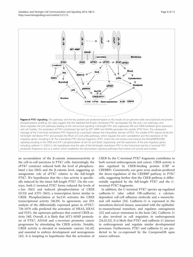
Figure 6 PTK7 signaling. The pathways and the key proteins are predicted based on the results of our genome-wide transcriptional and proteinphosphorylation profiling. Our data suggest that the stabilized full-length membrane PTK7 up-regulates the Akt and c-Jun pathways, anddown-regulates the p53 pathways leading to the cell survival signaling. Full-length PTK7 also suppresses ERK and CREB-mediated gene expressionand cell motility. The proteolysis of PTK7 ectodomain (Ig1-Ig7) by MT1-MMP and ADAMs generates the soluble sPTK7 form. The subsequentcleavage of the C-terminal membrane PTK7 fragment by γ-secretase releases the intracellular domain (cPTK7). The soluble sPTK7 species binds thefull-length membrane PTK7 and activates the RhoA [21] and p38a pathways, which regulate the actin cytoskeleton and the expression of themigratory genes, including IL1B. The intracellular PTK7 domain fragment, cPTK7, enters the cell nucleus and induces the RAS/p90RSK/CREBsignaling resulting in the CREB and ATF1 phosphorylation at Ser133 and Ser63, respectively, and the expression of the pro-migratory genes,including cadherin-11 (CDH11). We hypothesize that the ratio of the full-length membrane PTK7 to the N-terminal and the C-terminal PTK7proteolytic fragments acts as a switch, which establishes the downstream signaling pathways that control cell survival and motility.
Golubkov and Strongin Cell Communication and Signaling 2014, 12:15 Page 8 of 12http://www.biosignaling.com/content/12/1/15
an accumulation of the β-catenin immunoreactivity atthe cell-to-cell junctions in PTK7 cells. Interestingly, thesPTK7 construct reduced both the level of phosphory-lated c-Jun (S63) and the β-catenin level, suggesting anantagonistic role of sPTK7 relative to the full-lengthPTK7. We hypothesize that the c-Jun activity is specific-ally induced by the intact full-length PTK7. On the con-trary, both C-terminal PTK7 forms reduced the levels ofc-Jun (S63) and induced phosphorylation of CREB(S133) and ATF1 (S63), a transcription factor similar toCREB. Phosphorylation at Ser133 activates the CREBtranscriptional activity [38,39]. In agreement, our IPAanalysis of the differentially expressed genes in cPTK7/726-1070 cells predicted the activation of HRAS, KRASand FGF1, the upstream pathways that control CREB ac-tivity [40]. Overall, it is likely that MT1-MMP proteoly-sis of PTK7, ADAM and γ-secretase promotes cancerprogression by stimulating the CREB activity. Notably,CREB activity is elevated in metastatic cancers [41,42]and essential to embryo development and neurogenesis[43]. It is tempting to hypothesize that the activation of
CREB by the C-terminal PTK7 fragments contributes toboth normal embryogenesis and cancer. CREB activity isalso regulated by CREB-binding protein (CBP orCREBBP). Consistently, our gene array analysis predictedthe down-regulation of the CREBBP pathway in PTK7cells, suggesting further that the CREB pathway is differ-entially regulated by the full-length PTK7 and the C-terminal PTK7 fragments.In addition, the C-terminal PTK7 species up-regulated
cadherin-11 (also called OB-cadherin), a calcium-dependent cell-cell adhesion molecule and a mesenchy-mal cell marker [24]. Cadherin-11 is expressed in themesoderm-derived tissues, associated with the epithelial-to-mesenchymal transition, and regulates osteogenesis[25] and cancer metastasis to the bone [26]. Cadherin-11is also involved in cell migration in embryogenesis[24,25,32]. It is likely that PTK7 and cadherin-11 interactin embryogenesis and regulate similar developmentalprocesses. Furthermore, PTK7 and cadherin-11 are pre-dicted to be co-expressed by the CoexpressDB opensource software.

Golubkov and Strongin Cell Communication and Signaling 2014, 12:15 Page 9 of 12http://www.biosignaling.com/content/12/1/15
ConclusionsOverall, by using genome-wide transcriptional andphospho-kinase profiling of the cell samples, we deter-mined that the full-length membrane PTK7 and itsN-terminal and C-terminal proteolytic fragments differ-entially regulate multiple genes and proteins involved incell motility. These differences were most evident in thephosphorylation levels of p38a, ERK, Akt, c-Jun, CREB,ATF1, p53, and in the β-catenin and cadherin-11 proteinlevels. Our data correlate well with the results in otherreceptor tyrosine kinases and support the general ideaabout the distinct functions of the full-length receptorand its intracellular domain (ICD) fragment [44]. The re-sults generated from our model system will shed morelight on the PTK7 functionality in both physiologicaland pathological conditions.
MethodsAntibodies and reagentsGeneral reagents were purchased from Sigma-Aldrich(Saint Louis, MO, USA). A goat polyclonal AF4499 anti-body against the N-terminal 31–199 portion of PTK7, agoat polyclonal antibody to IL1B and a rabbit polyclonalantibody to pCREB (S133) were from R&D Systems(Minneapolis, MN, USA). A mouse monoclonal antibodyto the V5 tag was from Invitrogen (Carlsbad, CA, USA)A mouse monoclonal M2 antibody to the FLAG tag wasfrom Sigma-Aldrich. The rabbit polyclonal antibodies tocadherin-11 (CDH11, OB-cadherin) and α-tubulin,rabbit monoclonal antibodies to CREB, podoplanin andp-c-Jun (S63), and a mouse monoclonal antibody to c-Jun were from Cell Signaling Technology (Danvers, MA,USA). A rabbit monoclonal antibody to p-ATF1 (S63)was from Abcam (Cambridge, MA, USA). The mousemonoclonal antibodies to osteopontin (SPP1) andMAGEC1 were from EMD Millipore (Temecula, CA,USA) and Santa Cruz Biotechnology (Dallas, TX, USA),respectively. The species-specific HRP-conjugated second-ary antibodies were from Fitzgerald Industries (Acton,MA, USA).
Cells, cloning and mutagenesisHuman fibrosarcoma HT1080 cells (HT1080 cells) werefrom ATCC (Manassas, VA, USA). HT1080 cells trans-fected with the full-length 1–1070 PTK7 containing theC-terminal FLAG tag (PTK7 cells), the N-terminal 1–694fragment with the C-terminal FLAG tag (sPTK7cells) and the C-terminal 726–1070 fragment with theV5-HIS tag (cPTK7/726-1070 cells) were described earl-ier [11,21,23]. Cells in which the transcription of theMT1-MMP gene was silenced using the shRNA con-struct (shMT1 cells) were characterized earlier [11,45].The C-terminal 622–1070 membrane fragment (cPTK7/622-1070 cells) that corresponded to the MT1-MMP
cleavage fragment of the full-length membrane PTK7was generated by PCR using the full-length wild-typePTK7-1 cDNA (OriGene, Rockville, MD, USA) as a tem-plate, and 5′-GGTACCCAGACAGCCCTGATTCAGTGGAAAGG-3′ and 5′-CGGCTTGCTGTCCAC-3′ as theforward and reverse primers, respectively. The 1–30PTK7 signal peptide sequence was amplified using 5′-CACCATGGGAGCTGCGCGGGGATC-3′ and 5′-CCTTTCCACTGAATCAGGGCTGTCTGGGTACC-3′ as theforward and reverse primers, respectively, and then thesignal peptide sequence was inserted at the N-terminusto ensure delivery of the cPTK7/622-1070 construct tothe plasma membrane. The resulting secretory cPTK7/622-1070 construct was sub-cloned into the pcDNA3.1D/V5-His-TOPO vector (Invitrogen) and then used to sta-bly transfect HT1080 cells using Lipofectamine LTX(Invitrogen). Stably transfected cells were selected in thepresence of G418 (200 μg/ml). Cell expression of thecPTK7/622-1070 construct was confirmed using Westernblotting with the V5 antibody.The 29-mer shRNA constructs in the retroviral RFP vec-
tor (pRFP-C-RS) (Origene; catalog number TF200451)were used to silence the endogenous PTK7 in HT1080cells. Following Lipofectamine LTX transfection of thecells with the vector, the RFP-positive cells were selectedusing a cell sorter. Selected cells were then grown in thepresence of puromycin (2 μg/ml). Immunoblotting withthe goat polyclonal PTK7 antibody was used to select thesilenced clones. Three selected clones with the silencedPTK7 were pooled together (shPTK7 cells). Similarly, thescrambled shRNA constructs were used to generate theHT1080-scr cell control.
Invasion assayThe invasion assay was performed in triplicate in wells ofa 24-well Transwell plate with an 8-μm pore size mem-brane [11]. The membranes of the Transwell inserts werecoated with type I collagen (30 μg/well, BD Biosciences,San Jose, CA, USA). Cells (1 × 10⁵/well) in serum-freeDMEM (0.1 ml) were placed into the upper chamber. The10% fetal bovine serum (FBS)-containing DMEM (used asa chemoattractant, 0.6 ml) was placed in the lower cham-ber. Serum-free DMEM (0.6 ml) was used as a control.Cells were allowed to invade for 3.5 h. The cells werestained for 10 min with 0.2% crystal violet/20% methanol(0.3 ml). The cells on the upper membrane surface wereremoved with a cotton swab. The dye from the cells thatmigrated onto the lower surface of the membrane was ex-tracted with 1% SDS (0.25 ml). The resulting A570 nmwas measured using a plate reader.
Genome-wide transcriptional profiling and data analysisCells (1 × 10⁴/ml) were plated in DMEM-10% FBSin a 100-mm dish and grown for 72 h to produce a

Golubkov and Strongin Cell Communication and Signaling 2014, 12:15 Page 10 of 12http://www.biosignaling.com/content/12/1/15
subconfluent culture. Total cellular RNA was extractedusing a Direct-zol RNA MiniPrep kit (Zymo Research,Irvine, CA, USA). Biotin-labeled cRNA samples wereprepared using an RNA Amplification Kit (Life Tech-nologies, Grand Island, NY, USA). The labeled cRNA(750 ng) was hybridized for 18 h at 58°C to theHumanHT-12 v4 Expression BeadChip with over 46,000gene transcripts (Illumina, San Diego, CA, USA). Bead-Chips were then developed using fluorolink streptavidin-Cy3 (GE Healthcare, Piscataway, NJ, USA). Array chipswere scanned using an Illumina BeadArray Reader. Theinitial data extraction and normalization were performedusing the BeadArray Reader and GeneSpring GX soft-ware (Agilent, Santa Clara, CA, USA). The differentiallyexpressed genes (compared to parental HT1080 cells)were identified based on the Welch’s t test and -fold dif-ference of the expression level (cutoff >2-fold difference,p-value <0.05). Heatmaps and hierarchical clusteringwere generated using the GenePattern open-source soft-ware package (Broad Institute, Cambridge, MA, USA).The pathway and functional analyses were conductedusing the Ingenuity Pathway Analysis (IPA) software (In-genuity Systems, Redwood City, CA, USA). The micro-array data were deposited to the Gene ExpressionOmnibus (GEO) data base with the accession numberGSE53340.
Human phospho-kinase arrayThe phosphorylation profile was analyzed in the cellsusing the Proteome Profiler Human Phospho-KinaseArray (R&D Systems). Cells (1 × 10⁴/ml) were plated inDMAM-10% FBS in a 100-mm dish and grown for 72 hto produce a subconfluent culture. Cell lysate samples(0.5 mg; 1.5 mg/ml each) were applied per array setcomprised of two nitrocellulose membranes with thespotted capture antibodies. The bound material was de-tected using the biotinylated antibodies followed bystreptavidin conjugated with horseradish peroxidase.The chemiluminescent signal was acquired using theHyBlot CL autoradiography film (Denville, South Plain-field, NJ, USA). The film was scanned and digitized.Pixel density of the spots was quantified using the Ima-geJ software.
Cell surface biotinylation and total cell lysatesCell surface proteins were biotinylated by incubatingcells for 1 h on ice in PBS containing 0.1 mg/ml EZ-Link sulfosuccinimidyl 2-(biotinamido)-ethyl-1,3-dithio-propionate (Thermo Fisher Scientific, Rockford, IL,USA). Cells were lysed in 20 mM Tris–HCl buffer,pH 7.4, containing 150 mM NaCl, 1% deoxycholate, 1%octylphenoxypolyethoxyethanol (IGEPAL), protease in-hibitor mixture set III, 1 mM phenylmethylsulfonylfluoride and 10 mM EDTA. Biotinylated proteins were
then captured using streptavidin-agarose beads. Biotinyl-ated proteins were eluted from the beads using 2X SDSsample loading buffer (125 mM Tris–HCl, pH 6.8, con-taining 4% SDS, 0.005% bromophenol blue, 20% glyceroland 20 mM DTT).For preparation of the total cell lysate samples, cells
were lysed using the cell lysis buffer of the Proteome Pro-filer Human Phospho-Kinase Array kit (R&D Systems).The samples were centrifuged at 4°C (14,000 rpm,15 min). The protein concentration in the supernatantsamples was adjusted to 1.5 mg/ml. The sample aliquots(30 μg total protein each) were separated by SDS-gel elec-trophoresis in the 4-12% gels and analyzed by immuno-blotting with the specific primary antibodies followed bythe horseradish peroxidase-conjugated species-specificsecondary antibody and the SuperSignal West Dura ex-tended duration substrate (Thermo Fisher Scientific). Thechemiluminescent signal was acquired using the HyBlotCL autoradiography film.
Cell immunostainingCells grown on a microscope coverglass (Thermo FisherScientific) were fixed using 4% p-formaldehyde, perme-abilized using 0.1% Triton X-100, and blocked in 1% ca-sein. Cells were stained using the primary monoclonalmouse antibody to β-catenin (1:1,000 dilution) (BectonDickinson, Franklin Lakes, NJ, USA) for 16 h at 4°Cfollowed by Alexa Fluor 488-conjugated anti-mouse sec-ondary antibody (1:500 dilution). The Alexa Fluor 594-conjugated phalloidin (1:500 dilution) (Thermo FisherScientific) was used to visualize the actin cytoskeleton.The specimens were mounted in the Vectashield mount-ing medium with 4′,6-diamidino-2-phenylindole (DAPI)(Vector Laboratories, Burlingame, CA, USA). Imageswere acquired using an Olympus BX51 fluorescencemicroscope equipped with a MagnaFire digital cameraand MagnaFire 2.1C software (Olympus, Center Valley,PA, USA).
Additional files
Additional file 1: Table S1. Genome-wide transcriptional profiling ofChz, shPTK7, cPTK7/622-1070, cPTK7/726-1070, sPTK7 and PTK7 cellsrelative to the parental fibrosarcoma HTR1080 cells.
Additional file 2: Table S2. Genome-wide transcriptional profilingof the cell migration-related genes in Chz, shPTK7, cPTK7/622-1070,cPTK7/726-1070, sPTK7 and PTK7 cells relative to the parentalfibrosarcoma HTR1080 cells.
AbbreviationsADAM: A disintegrin and metalloprotease; Chz: Chuzhoi mutant of PTK7;ICD: Intracellular domain; MT1-MMP: Membrane type-1 matrixmetalloproteinase; PCP: Planar cell polarity; PTK7: Protein tyrosine kinase 7;cPTK7/622-1070 and cPTK7/726-1070: The C-terminal, cytoplasmic 622–1070and 726–1070 fragments of PTK7; Respectively; sPTK7: The N-terminal solublePTK7 fragment; shMT1: MT1-MMP knockout; shPTK7: PTK7 knockout.

Golubkov and Strongin Cell Communication and Signaling 2014, 12:15 Page 11 of 12http://www.biosignaling.com/content/12/1/15
Competing interestsAuthors declare that there are no competing interests.
Authors’ contributionsVSG carried out the studies. VSG and AYS wrote the manuscript. All authorsread and approved the final manuscript.
AcknowledgmentsThe work reported here was supported by NIH Grants CA077470, CA083017and CA157328 (to AYS).
Received: 23 December 2013 Accepted: 25 February 2014Published: 11 March 2014
References1. Lu X, Borchers AG, Jolicoeur C, Rayburn H, Baker JC, Tessier-Lavigne M:
PTK7/CCK-4 is a novel regulator of planar cell polarity in vertebrates.Nature 2004, 430:93–98.
2. Yen WW, Williams M, Periasamy A, Conaway M, Burdsal C, Keller R, Lu X,Sutherland A: PTK7 is essential for polarized cell motility and convergentextension during mouse gastrulation. Development 2009, 136:2039–2048.
3. Jung JW, Ji AR, Lee J, Kim UJ, Lee ST: Organization of the human PTK7 geneencoding a receptor protein tyrosine kinase-like molecule andalternative splicing of its mRNA. Biochim Biophys Acta 2002, 1579:153–163.
4. Puppo F, Thome V, Lhoumeau AC, Cibois M, Gangar A, Lembo F, Belotti E,Marchetto S, Lecine P, Prebet T, Sebbagh M, Shin WS, Lee ST, KodjabachianL, Borg JP: Protein tyrosine kinase 7 has a conserved role inWnt/beta-catenin canonical signalling. EMBO Rep 2011, 12:43–49.
5. Peradziryi H, Kaplan NA, Podleschny M, Liu X, Wehner P, Borchers A,Tolwinski NS: PTK7/Otk interacts with Wnts and inhibits canonical Wntsignalling. EMBO J 2011, 30:3729–3740.
6. Lhoumeau AC, Puppo F, Prebet T, Kodjabachian L, Borg JP: PTK7: a cellpolarity receptor with multiple facets. Cell Cycle 2011, 10:1233–1236.
7. Whitford KL, Ghosh A: Plexin signaling via off-track and rho familyGTPases. Neuron 2001, 32:1–3.
8. Toyofuku T, Zhang H, Kumanogoh A, Takegahara N, Suto F, Kamei J, Aoki K,Yabuki M, Hori M, Fujisawa H, Kikutani H: Dual roles of Sema6D in cardiacmorphogenesis through region-specific association of its receptor,Plexin-A1, with off-track and vascular endothelial growth factor receptortype 2. Genes Dev 2004, 18:435–447.
9. Shnitsar I, Borchers A: PTK7 recruits dsh to regulate neural crestmigration. Development 2008, 135:4015–4024.
10. Wehner P, Shnitsar I, Urlaub H, Borchers A: RACK1 is a novel interactionpartner of PTK7 that is required for neural tube closure. Development2011, 138:1321–1327.
11. Golubkov VS, Strongin AY: Insights into ectodomain shedding andprocessing of protein-tyrosine pseudokinase 7 (PTK7). J Biol Chem 2012,287:42009–42018.
12. Mossie K, Jallal B, Alves F, Sures I, Plowman GD, Ullrich A: Colon carcinomakinase-4 defines a new subclass of the receptor tyrosine kinase family.Oncogene 1995, 11:2179–2184.
13. Saha S, Bardelli A, Buckhaults P, Velculescu VE, Rago C, St Croix B, RomansKE, Choti MA, Lengauer C, Kinzler KW, Vogelstein B: A phosphataseassociated with metastasis of colorectal cancer. Science 2001,294:1343–1346.
14. Gorringe KL, Boussioutas A, Bowtell DD: Novel regions of chromosomalamplification at 6p21, 5p13, and 12q14 in gastric cancer identified byarray comparative genomic hybridization. Genes Chromosomes Cancer2005, 42:247–259.
15. Endoh H, Tomida S, Yatabe Y, Konishi H, Osada H, Tajima K, Kuwano H,Takahashi T, Mitsudomi T: Prognostic model of pulmonaryadenocarcinoma by expression profiling of eight genes as determinedby quantitative real-time reverse transcriptase polymerase chainreaction. J Clin Oncol 2004, 22:811–819.
16. Muller-Tidow C, Schwable J, Steffen B, Tidow N, Brandt B, Becker K,Schulze-Bahr E, Halfter H, Vogt U, Metzger R, Schneider PM, Buchner T,Brandts C, Berdel WE, Serve H: High-throughput analysis of genome-widereceptor tyrosine kinase expression in human cancers identifies potentialnovel drug targets. Clin Cancer Res 2004, 10:1241–1249.
17. Easty DJ, Mitchell PJ, Patel K, Florenes VA, Spritz RA, Bennett DC: Loss ofexpression of receptor tyrosine kinase family genes PTK7 and SEK inmetastatic melanoma. Int J Cancer 1997, 71:1061–1065.
18. Su YA, Yang J, Tao L, Nguyen H, He P: Undetectable and decreasedexpression of KIAA1949 (Phostensin) encoded on chromosome 6p21.33in human breast cancers revealed by transcriptome analysis. J CancerEduc 2010, 1:38–50.
19. Piao Z, Lee KS, Kim H, Perucho M, Malkhosyan S: Identification of noveldeletion regions on chromosome arms 2q and 6p in breast carcinomasby amplotype analysis. Genes Chromosomes Cancer 2001, 30:113–122.
20. Baudrier-Regnier A, Bodenant C, Proust F, Delangre T, Hemet J, LaquerriereA: An isochromosome 6p in a primary meningeal malignant melanoma.Cancer Genet Cytogenet 2000, 119:80–82.
21. Golubkov VS, Chekanov AV, Cieplak P, Aleshin AE, Chernov AV, Zhu W,Radichev IA, Zhang D, Dong PD, Strongin AY: The Wnt/planar cellpolarity protein-tyrosine kinase-7 (PTK7) is a highly efficientproteolytic target of membrane type-1 matrix metalloproteinase:implications in cancer and embryogenesis. J Biol Chem 2010,285:35740–35749.
22. Na HW, Shin WS, Ludwig A, Lee ST: The cytosolic domain ofprotein-tyrosine kinase 7 (PTK7), generated from sequential cleavage bya disintegrin and metalloprotease 17 (ADAM17) and gamma-secretase,enhances cell proliferation and migration in colon cancer cells.J Biol Chem 2012, 287:25001–25009.
23. Golubkov VS, Aleshin AE, Strongin AY: Potential relation of aberrantproteolysis of human protein tyrosine kinase 7 (PTK7) chuzhoi bymembrane type 1 matrix metalloproteinase (MT1-MMP) to congenitaldefects. J Biol Chem 2011, 286:20970–20976.
24. Borchers A, David R, Wedlich D: Xenopus cadherin-11 restrains cranialneural crest migration and influences neural crest specification.Development 2001, 128:3049–3060.
25. Koehler A, Schlupf J, Schneider M, Kraft B, Winter C, Kashef J: Loss ofXenopus cadherin-11 leads to increased Wnt/beta-catenin signaling andup-regulation of target genes c-myc and cyclin D1 in neural crest.Dev Biol 2013, 383:132–145.
26. Huang CF, Lira C, Chu K, Bilen MA, Lee YC, Ye X, Kim SM, Ortiz A, Wu FL,Logothetis CJ, Yu-Lee LY, Lin SH: Cadherin-11 increases migration andinvasion of prostate cancer cells and enhances their interaction withosteoblasts. Cancer Res 2010, 70:4580–4589.
27. Shigematsu Y, Niwa T, Rehnberg E, Toyoda T, Yoshida S, Mori A,Wakabayashi M, Iwakura Y, Ichinose M, Kim YJ, Ushijima T: Interleukin-1betainduced by Helicobacter pylori infection enhances mouse gastriccarcinogenesis. Cancer Lett 2013, 340:141–147.
28. Landvik NE, Hart K, Haugen A, Zienolddiny S: Functional analysis of a lungcancer risk haplotype in the IL1B gene regulatory region. J Hum Genet2012, 57:747–752.
29. Krishnan H, Ochoa-Alvarez JA, Shen Y, Nevel E, Lakshminarayanan M,Williams MC, Ramirez MI, Miller WT, Goldberg GS: Serines in theintracellular tail of podoplanin (PDPN) regulate cell motility. J Biol Chem2013, 288:12215–12221.
30. Nakashima Y, Yoshinaga K, Kitao H, Ando K, Kimura Y, Saeki H, Oki E, MoritaM, Kakeji Y, Hirahashi M, Oda Y, Maehara Y: Podoplanin is expressed at theinvasive front of esophageal squamous cell carcinomas and is involvedin collective cell invasion. Cancer Sci 2013, 104:1718–1725.
31. Wicki A, Lehembre F, Wick N, Hantusch B, Kerjaschki D, Christofori G: Tumorinvasion in the absence of epithelial-mesenchymal transition:podoplanin-mediated remodeling of the actin cytoskeleton. Cancer Cell2006, 9:261–272.
32. Kashef J, Kohler A, Kuriyama S, Alfandari D, Mayor R, Wedlich D:Cadherin-11 regulates protrusive activity in Xenopus cranial neural crestcells upstream of Trio and the small GTPases. Genes Dev 2009,23:1393–1398.
33. de Carvalho F, Alves VL, Braga WM, Xavier CV Jr, Colleoni GW: MAGE-C1/CT7 and MAGE-C2/CT10 are frequently expressed in multiple myelomaand can be explored in combined immunotherapy for this malignancy.Cancer Immunol Immunother 2013, 62:191–195.
34. Cassinelli G, Zuco V, Gatti L, Lanzi C, Zaffaroni N, Colombo D, Perego P:Targeting the Akt kinase to modulate survival, invasiveness and drugresistance of cancer cells. Curr Med Chem 2013, 20:1923–1945.
35. Fukumoto S, Hsieh CM, Maemura K, Layne MD, Yet SF, Lee KH, Matsui T,Rosenzweig A, Taylor WG, Rubin JS, Perrella MA, Lee ME: Akt participation
Golubkov and Strongin Cell Communication and Signaling 2014, 12:15 Page 12 of 12http://www.biosignaling.com/content/12/1/15
in the Wnt signaling pathway through Dishevelled. J Biol Chem 2001,276:17479–17483.
36. Schreiber M, Kolbus A, Piu F, Szabowski A, Mohle-Steinlein U, Tian J, Karin M,Angel P, Wagner EF: Control of cell cycle progression by c-Jun is p53dependent. Genes Dev 1999, 13:607–619.
37. Meng L, Sefah K, O’Donoghue MB, Zhu G, Shangguan D, Noorali A,Chen Y, Zhou L, Tan W: Silencing of PTK7 in colon cancer cells:caspase-10-dependent apoptosis via mitochondrial pathway. PLoS One2010, 5:e14018.
38. Ghosh A, Ginty DD, Bading H, Greenberg ME: Calcium regulation of geneexpression in neuronal cells. J Neurobiol 1994, 25:294–303.
39. Andrisani OM: CREB-mediated transcriptional control. Crit Rev EukaryotGene Expr 1999, 9:19–32.
40. Tan Y, Rouse J, Zhang A, Cariati S, Cohen P, Comb MJ: FGF and stressregulate CREB and ATF-1 via a pathway involving p38 MAP kinase andMAPKAP kinase-2. Embo J 1996, 15:4629–4642.
41. Jean D, Bar-Eli M: Regulation of tumor growth and metastasis of humanmelanoma by the CREB transcription factor family. Mol Cell Biochem 2000,212:19–28.
42. Braeuer RR, Zigler M, Villares GJ, Dobroff AS, Bar-Eli M: Transcriptionalcontrol of melanoma metastasis: the importance of the tumormicroenvironment. Semin Cancer Biol 2011, 21:83–88.
43. Dworkin S, Malaterre J, Hollande F, Darcy PK, Ramsay RG, Mantamadiotis T:cAMP response element binding protein is required for mouse neuralprogenitor cell survival and expansion. Stem Cells 2009, 27:1347–1357.
44. Carpenter G, Liao HJ: Receptor tyrosine kinases in the nucleus. Cold SpringHarb Perspect Biol 2013, 5:a008979.
45. Golubkov VS, Boyd S, Savinov AY, Chekanov AV, Osterman AL, Remacle A,Rozanov DV, Doxsey SJ, Strongin AY: Membrane type-1 matrixmetalloproteinase (MT1-MMP) exhibits an important intracellularcleavage function and causes chromosome instability. J Biol Chem 2005,280:25079–25086.
doi:10.1186/1478-811X-12-15Cite this article as: Golubkov and Strongin: Downstream signaling andgenome-wide regulatory effects of PTK7 pseudokinase and itsproteolytic fragments in cancer cells. Cell Communication and Signaling2014 12:15.
Submit your next manuscript to BioMed Centraland take full advantage of:
• Convenient online submission
• Thorough peer review
• No space constraints or color figure charges
• Immediate publication on acceptance
• Inclusion in PubMed, CAS, Scopus and Google Scholar
• Research which is freely available for redistribution
Submit your manuscript at www.biomedcentral.com/submit