research open access autocrine transforming growth factor
TRANSCRIPT

RESEARCH Open Access
Autocrine transforming growth factor b signalingregulates extracellular signal-regulated kinase 1/2phosphorylation via modulation of proteinphosphatase 2A expression in sclerodermafibroblastsGlady H Samuel1,2, Andreea M Bujor1, Sashidhar S Nakerakanti1, Faye N Hant2, Maria Trojanowska1*
Abstract
Background: During scleroderma (SSc) pathogenesis, fibroblasts acquire an activated phenotype characterized byenhanced production of extracellular matrix (ECM) and constitutive activation of several major signaling pathwaysincluding extracellular signal-related kinase (ERK1/2). Several studies have addressed the role of ERK1/2 in SScfibrosis however the mechanism of its prolonged activation in SSc fibroblasts is still unknown. Protein phosphatase2A (PP2A) is a key serine threonine phosphatase responsible for dephosphorylation of a wide array of signalingmolecules. Recently published microarray data from cultured SSc fibroblasts suggests that the catalytic subunit(C-subunit) of PP2A is downregulated in SSc. In this study we examined the role and regulation of PP2A in SScfibroblasts in the context of ERK1/2 phosphorylation and matrix production.
Results: We show for the first time that PP2A mRNA and protein expression are significantly reduced in SScfibroblasts and correlate with an increase in ERK1/2 phosphorylation and collagen expression. Furthermore,transforming growth factor b (TGFb), a major profibrotic cytokine implicated in SSc fibrosis, downregulates PP2Aexpression in healthy fibroblasts. PP2A-specific small interfering RNA (siRNA) was utilized to confirm the role ofPP2A in ERK1/2 dephosphorylation in dermal fibroblasts. Accordingly, blockade of autocrine TGFb signaling in SScfibroblasts using soluble recombinant TGFb receptor II (SRII) restored PP2A levels and decreased ERK1/2phosphorylation and collagen expression. In addition, we observed that inhibition of ERK1/2 in SSc fibroblastsincreased PP2A expression suggesting that ERK1/2 phosphorylation also contributes to maintaining low levels ofPP2A, leading to an even further amplification of ERK1/2 phosphorylation.
Conclusions: Taken together, these studies suggest that decreased PP2A levels in SSc is a result of constitutivelyactivated autocrine TGFb signaling and could contribute to enhanced phosphorylation of ERK1/2 and matrixproduction in SSc fibroblasts.
IntroductionScleroderma (SSc) is an autoimmune connective tissuedisease characterized by excess production and deposi-tion of extracellular matrix proteins leading to fibrosisof the tissue. During this process, normal fibroblastsbecome ‘activated’ and acquire a fibrotic phenotype.
Transforming growth factor b (TGFb) is a major profi-brotic cytokine that plays important roles in a variety ofphysiological processes including cell proliferation, dif-ferentiation and survival. Although the mechanism ofSSc fibrosis is not fully understood, there is strong evi-dence to suggest that TGFb is central to the develop-ment and maintenance of the SSc phenotype [1-3].Normal healthy dermal fibroblasts treated with TGFbreproduce characteristics of SSc fibroblasts, further
* Correspondence: [email protected] Center, Division of Rheumatology, Boston University MedicalCampus, Boston, MA, USAFull list of author information is available at the end of the article
Samuel et al. Fibrogenesis & Tissue Repair 2010, 3:25http://www.fibrogenesis.com/content/3/1/25
© 2010 Samuel et al; licensee BioMed Central Ltd. This is an Open Access article distributed under the terms of the Creative CommonsAttribution License (http://creativecommons.org/licenses/by/2.0), which permits unrestricted use, distribution, and reproduction inany medium, provided the original work is properly cited.
supporting the notion that TGFb is a major mediator ofSSc fibrosis [4].During tissue injury, rapid release of TGFb attracts
inflammatory cells and fibroblasts to the site of injury,resulting in extracellular matrix production/remodelingand myofibroblast differentiation [5]. In normal tissue,following the injury response, coordinated apoptosis offibroblasts and myofibroblasts prevents scarring andexcessive fibrosis [6]. Published data suggests that nor-mal and SSc dermal fibroblasts in culture secrete simi-lar levels of TGFb ligand [7,8] However, there isevidence of increased TGFb signaling in SSc fibroblastswhen compared to normal fibroblasts. Several studieshave shown elevated levels of TGFb receptors in SScfibroblasts, which contribute to an autocrine TGFb sig-naling cascade that is maintained in culture even in theabsence of exogenous ligand [9-11]. The chronic acti-vation of the TGFb pathway in SSc produces fibro-blasts with constitutively activated Akt and ERK1/2pathways that are resistant to apoptosis [12-14]. TheERK1/2 pathway regulates numerous cellular processesand more recently has also been implicated in the pro-cess of fibrosis. Several papers have reported the func-tion of the activated ERK1/2 pathway in fibrosis. Forexample, it has been demonstrated that the ERK1/2pathway is required for Smad1 phosphorylation inresponse to overexpression of TGFbRI and for subse-quent upregulation of connective tissue growth factor(CCN2) and other profibrotic genes [15]. Activation ofmitogen-activated protein kinase kinase 1(MEK1)/ERK1/2 pathway was also shown to be a primarymechanism responsible for the TGFb-induced upregu-lation of early growth response factor 1 (Egr-1) [16]. Inaddition, Chen et al. recently reported that activationof the ERK1/2 pathway contributes to the enhancedfibrosis and contractile ability of scleroderma fibro-blasts [12]. The ERK1/2 pathway also induces up regu-lation of avb3 integrin, which contributes to theautocrine TGFb signaling in scleroderma fibroblasts[17]. However, although constitutively phosphorylatedERK1/2 may play important roles in SSc pathogenesis,the mechanism of prolonged activation of this pathwayis largely unknown.Protein phosphatase 2A (PP2A) is a member of the
PPP family and one of the most abundant serine-threo-nine phosphatases, accounting for a substantial part ofthe total phosphatase activity. PP2A plays an importantrole in signal transduction pathways, regulation of cellcycle and transcriptional and translational regulation[18]. PP2A has a complex structure, comprising of threesubunits: the catalytic (C), regulatory (B) and structuralsubunit (A). The catalytic subunit (C) and structuralsubunit (A) have two isoforms: a and b. The regulatorysubunit (B) consists of four families with many isoforms
that confer specificity of location and function [18]. Thephosphatase activity of PP2A is present in the C-subunitand its effects include dephosphorylation of varioustranscription factors and protein kinases includingMEK, ERK1/2, Akt, and sphingosine kinase (SK)[19-21]. Recently published microarray data from cul-tured early passage SSc fibroblasts suggests that the bisoform of the catalytic subunit of PP2A is downregu-lated in SSc [22]. Based on the evidence of constitutiveactivation of ERK1/2 pathways in SSc fibroblasts andrecent microarray data suggesting that PP2A may alsobe altered in SSc, we wished to further study themechanism and significance of dysregulated PP2A inSSc fibroblasts.
ResultsTGFb stimulates prolonged phosphorylation of ERK1/2 indermal fibroblastsBecause of the central role of TGFb in the pathogenesisof SSc, we first examined the regulation of ERK1/2phosphorylation by TGFb treatment in healthy fibro-blasts. To investigate the kinetics of ERK1/2 activation,time course experiments were performed. Near conflu-ent cells were serum starved and then treated withTGFb for increasing time periods ranging from 0-24 h.Using western blot analysis we observed that stimulationof cells with TGFb resulted in rapid phosphorylation ofERK1/2 as early as 15 min and sustained ERK1/2 phos-phorylation up to 24 h (Figure 1a). This suggests thatTGFb can activate both early and prolonged phosphory-lation of ERK1/2 in dermal fibroblasts.
PP2A expression is decreased upon treatment with TGFbSince PP2A has been previously described as a majorERK1/2 phosphatase we next sought to determinewhether TGFb could also be involved in the regulationof PP2A expression in dermal fibroblasts. Confluent der-mal fibroblasts were serum starved and then treatedwith TGFb for different time periods. The mRNA levelsof a and b isoforms of the PP2A catalytic subunit wereanalyzed by quantitative reverse transcription (qRT)-PCR. As shown in Figure 1b, the mRNA levels of PP2Awere decreased as early as 6 h after TGFb addition andthe lower levels persisted up to 48 h. Treatment of cellswith TGFb affected both catalytic subunit isoforms, butthe b isoform showed a greater overall decrease. Tofurther validate the effects of TGFb on PP2A geneexpression we measured the protein levels of PP2A (Fig-ure 1c,d) after 24 h of TGFb treatment. PP2A proteinlevels were decreased at 24 h, correlating with and con-firming the mRNA data. These observations show thatTGFb negatively regulates PP2A expression, suggestingthat PP2A may be involved in TGFb-mediated ERK1/2phosphorylation.
Samuel et al. Fibrogenesis & Tissue Repair 2010, 3:25http://www.fibrogenesis.com/content/3/1/25
Page 2 of 9

PP2A inhibition contributes to increased ERK1/2phosphorylationTo further confirm the role of PP2A in ERK1/2 phos-phorylation in dermal fibroblasts, experiments were per-formed using okadaic acid (OA), a pharmacologicalinhibitor of PP2A activity, and PP2A-specific smallinterfering RNA (siRNA). Upon treatment of normaldermal fibroblasts with OA (2 nM) for 1 h, increasedERK1/2 phosphorylation was observed (Figure 2a). Con-sistent with this data, siRNA against the catalytic subu-nit of PP2A also increased phosphorylation levels ofERK1/2, suggesting that PP2A is involved in ERK1/2dephosphorylation (Figure 2b,c). From these experi-ments we can conclude that PP2A downregulation inSSc fibroblasts may contribute to enhanced ERK1/2phosphorylation. This data is in accordance with pre-viously published reports that PP2A is an ERK1/2 phos-phatase in several different cell types.
PP2A expression is decreased and correlates withincreased ERK1/2 phosphorylation in SSc dermalfibroblastsTo further study the relationship of PP2A and ERK1/2phosphorylation in the pathological context, age, raceand gender matched SSc and normal dermal fibroblastsobtained from patient biopsy were analyzed for PP2Aexpression and ERK1/2 activation. ERK1/2 phosphoryla-tion was increased in SSc fibroblasts, consistent withdata from previous reports [12] (Figure 3a). The mRNA
levels of both isoforms of the PP2A C-subunit weresignificantly decreased in SSc fibroblasts when comparedto normal controls (Figure 3b). Consistent with themRNA data, the protein levels of the catalytic subunit ofPP2A were significantly lower in SSc fibroblasts com-pared to normal controls (Figure 3c). The observationsfrom SSc fibroblasts are consistent with the results fromnormal fibroblasts treated with TGFb that show PP2Adownregulation, suggesting a role for TGFb in mediat-ing these changes in SSc fibroblasts.
Autocrine TGFb signaling regulates PP2A expression inSSc fibroblastsAutocrine TGFb signaling has been reported to play amajor role in the pathogenesis of SSc. and blockade ofendogenous TGFb signaling has been shown to attenu-ate the scleroderma fibrotic phenotype [8]. Recombinantsoluble TGFb receptor II (SRII) has been successfullyused as a TGFb antagonist to block the effects of TGFbsignaling such as upregulated collagen production. SRIIbinds TGFb ligand and prevents its interaction with sur-face receptors thereby neutralizing its activity [23,24].To further investigate whether TGFb signaling isresponsible for the decreased levels of PP2A in SSc, weblocked autocrine TGFb signaling using SRII. As a con-trol experiment to confirm the effectiveness of SRII,normal cells were pretreated with SRII for 1 h and thentreated with TGFb for 24 h. Pretreatment with SRII effi-ciently blocked downregulation of PP2A by TGFb
Figure 1 Transforming growth factor (TGF)b regulates extracellular signal-regulated kinase (ERK)1/2 phosphorylation and proteinphosphatase 2A (PP2A) expression in dermal fibroblasts. (a) Adult dermal fibroblasts were treated with TGFb (5 ng/ml) for 0 h, 15 min, 1 h,3 h, 6 h, 12 h and 24 h after serum starvation. Western blot analysis was performed using phospho-ERK1/2 and ERK1/2 primary antibodies andrabbit horseradish peroxidase-conjugated secondary antibody. (b) Adult dermal fibroblasts were serum starved overnight and treated with 5 ng/ml of TGFb for 0 h, 6 h, 12 h, 24 h and 48 h. RNA was then extracted and cDNA synthesized for quantitative PCR analysis; n = 3, *P < 0.05, **P <0.01. (c) Dermal fibroblasts were treated for 24 h with 5 ng/ml of TGFb after 24 h of serum starvation, and western blot analysis was performedusing a PP2A antibody directed against the catalytic subunit; n = 5, representative blot of three experiments. (d) Bar graphs representquantification of western blot analysis from (c); n = 5, *P < 0.05 versus normal control.
Samuel et al. Fibrogenesis & Tissue Repair 2010, 3:25http://www.fibrogenesis.com/content/3/1/25
Page 3 of 9
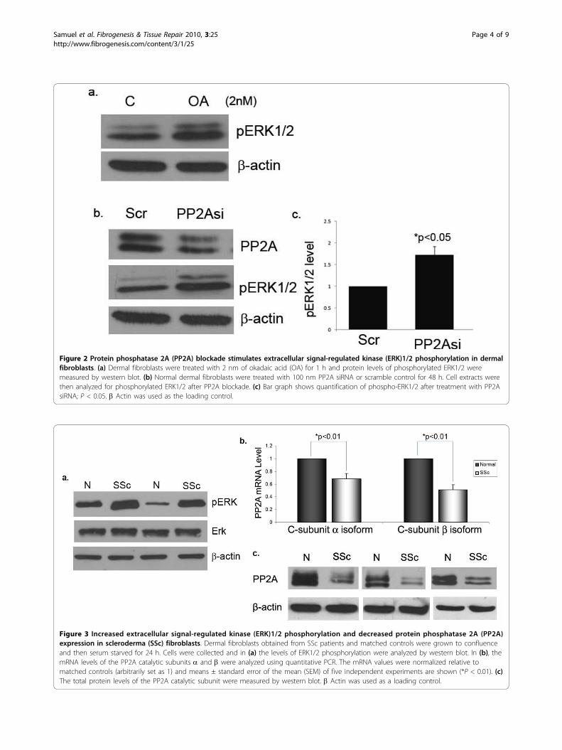
Figure 3 Increased extracellular signal-regulated kinase (ERK)1/2 phosphorylation and decreased protein phosphatase 2A (PP2A)expression in scleroderma (SSc) fibroblasts. Dermal fibroblasts obtained from SSc patients and matched controls were grown to confluenceand then serum starved for 24 h. Cells were collected and in (a) the levels of ERK1/2 phosphorylation were analyzed by western blot. In (b), themRNA levels of the PP2A catalytic subunits a and b were analyzed using quantitative PCR. The mRNA values were normalized relative tomatched controls (arbitrarily set as 1) and means ± standard error of the mean (SEM) of five independent experiments are shown (*P < 0.01). (c)The total protein levels of the PP2A catalytic subunit were measured by western blot. b Actin was used as a loading control.
Figure 2 Protein phosphatase 2A (PP2A) blockade stimulates extracellular signal-regulated kinase (ERK)1/2 phosphorylation in dermalfibroblasts. (a) Dermal fibroblasts were treated with 2 nm of okadaic acid (OA) for 1 h and protein levels of phosphorylated ERK1/2 weremeasured by western blot. (b) Normal dermal fibroblasts were treated with 100 nm PP2A siRNA or scramble control for 48 h. Cell extracts werethen analyzed for phosphorylated ERK1/2 after PP2A blockade. (c) Bar graph shows quantification of phospho-ERK1/2 after treatment with PP2AsiRNA; P < 0.05. b Actin was used as the loading control.
Samuel et al. Fibrogenesis & Tissue Repair 2010, 3:25http://www.fibrogenesis.com/content/3/1/25
Page 4 of 9
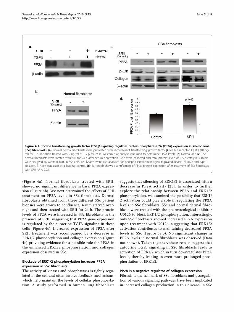
(Figure 4a). Normal fibroblasts treated with SRII,showed no significant difference in basal PP2A expres-sion (Figure 4b). We next determined the effects of SRIItreatment on PP2A levels in SSc fibroblasts. Dermalfibroblasts obtained from three different SSc patientbiopsies were grown to confluence, serum starved over-night and then treated with SRII for 24 h. The proteinlevels of PP2A were increased in SSc fibroblasts in thepresence of SRII, suggesting that PP2A gene expressionis regulated by the autocrine TGFb signaling in thesecells (Figure 4c). Increased expression of PP2A afterSRII treatment was accompanied by a decrease inERK1/2 phosphorylation and collagen expression (Figure4c) providing evidence for a possible role for PP2A inthe enhanced ERK1/2 phosphorylation and collagenexpression observed in SSc.
Blockade of ERK1/2 phosphorylation increases PP2Aexpression in SSc fibroblastsThe activity of kinases and phosphatases is tightly regu-lated in the cell and often involve feedback mechanisms,which help maintain the levels of cellular phosphoryla-tion. A study performed in human lung fibroblasts
suggests that silencing of ERK1/2 is associated with adecrease in PP2A activity [25]. In order to furtherexplore the relationship between PP2A and ERK1/2phosphorylation, we examined the possibility that ERK1/2 activation could play a role in regulating the PP2Alevels in SSc fibroblasts. SSc and normal dermal fibro-blasts were treated with the pharmacological inhibitorU0126 to block ERK1/2 phosphorylation. Interestingly,only SSc fibroblasts showed increased PP2A expressionupon treatment with U0126, suggesting that ERK1/2activation contributes to maintaining decreased PP2Alevels in SSc (Figure 5a,b). No significant change inPP2A levels in normal fibroblasts was observed (Datanot shown). Taken together, these results suggest thatautocrine TGFb signaling in SSc fibroblasts leads toactivation of ERK1/2 which in turn downregulates PP2Alevels, thereby leading to even more prolonged phos-phorylation of ERK1/2.
PP2A is a negative regulator of collagen expressionFibrosis is the hallmark of SSc fibroblasts and dysregula-tion of various signaling pathways have been implicatedin increased collagen production in this disease. In SSc
Figure 4 Autocrine transforming growth factor (TGF)b signaling regulates protein phosphatase 2A (PP2A) expression in scleroderma(SSc) fibroblasts. (a) Normal dermal fibroblasts were pretreated with recombinant transforming growth factor b soluble receptor II (SRII) (10 ng/ml) for 1 h and then treated with 5 ng/ml of TGFb for 24 h. Western blot analysis was used to determine PP2A levels. (b) Normal and (c) SScdermal fibroblasts were treated with SRII for 24 h after serum deprivation. Cells were collected and total protein levels of PP2A catalytic subunitwere analyzed by western blot. In SSc cells, cell lysates were also analyzed for phospho-extracellular signal-regulated kinase (ERK)1/2 and type 1collagen. b Actin was used as a loading control. (d) Bar graph shows quantification of PP2A protein expression after treatment of SSc fibroblastswith SRII; *P < 0.05.
Samuel et al. Fibrogenesis & Tissue Repair 2010, 3:25http://www.fibrogenesis.com/content/3/1/25
Page 5 of 9
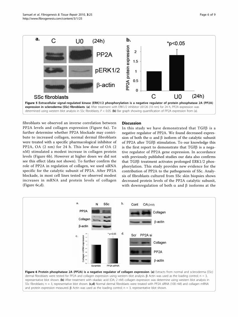
fibroblasts we observed an inverse correlation betweenPP2A levels and collagen expression (Figure 6a). Tofurther determine whether PP2A blockade may contri-bute to increased collagen, normal dermal fibroblastswere treated with a specific pharmacological inhibitor ofPP2A, OA (2 nm) for 24 h. This low dose of OA (2nM) stimulated a modest increase in collagen proteinlevels (Figure 6b). However at higher doses we did notsee this effect (data not shown). To further confirm therole of PP2A in regulation of collagen, we used siRNAspecific for the catalytic subunit of PP2A. After PP2Ablockade, in most cell lines tested we observed modestincreases in mRNA and protein levels of collagen(Figure 6c,d).
DiscussionIn this study we have demonstrated that TGFb is anegative regulator of PP2A. We found decreased expres-sion of both the a and b isoform of the catalytic subunitof PP2A after TGFb stimulation. To our knowledge thisis the first report to demonstrate that TGFb is a nega-tive regulator of PP2A gene expression. In accordancewith previously published studies our data also confirmsthat TGFb treatment activates prolonged ERK1/2 phos-phorylation. This study provides new evidence for thecontribution of PP2A to the pathogenesis of SSc. Analy-sis of fibroblasts cultured from SSc skin biopsies showsdecreased protein levels of the PP2A catalytic subunit,with downregulation of both a and b isoforms at the
Figure 6 Protein phosphatase 2A (PP2A) is a negative regulator of collagen expression. (a) Extracts from normal and scleroderma (SSc)dermal fibroblasts were tested for PP2A and collagen expression using western blot analysis. b Actin was used as the loading control; n = 3,representative blot shown. (b) After treatment with okadaic acid (OA; 2 nM) collagen expression was determine using western blot analysis inSSc fibroblasts; n = 3, representative blot shown. (c,d) Normal dermal fibroblasts were treated with PP2A siRNA (100 nM) and collagen mRNAand protein expression measured. b Actin was used as the loading control; n = 3, representative blot shown.
Figure 5 Extracellular signal-regulated kinase (ERK)1/2 phosphorylation is a negative regulator of protein phosphatase 2A (PP2A)expression in scleroderma (SSc) fibroblasts. (a) After treatment with ERK1/2 inhibitor U0126 (10 nm) for 24 h, PP2A expression wasdetermined using western blot analysis in SSc fibroblasts; P < 0.05. (b) Bar graph showing quantification of PP2A expression from (a).
Samuel et al. Fibrogenesis & Tissue Repair 2010, 3:25http://www.fibrogenesis.com/content/3/1/25
Page 6 of 9

mRNA levels, reproducing the effects of TGFb in nor-mal dermal fibroblasts. These data validate a previousgene array study, which showed decreased levels of theb isoform in cultured SSc fibroblasts. However, downre-gulation of the a isoform, which is the most abundantisoform in vivo, has not been described in SSc fibro-blasts. Previous reports indicated that an autocrineTGFb signaling pathway contributes to the SSc pheno-type [4]. We hypothesized that PP2A downregulation inSSc could be the result of constitutive TGFb signaling.This hypothesis was supported by our data showing thatrecombinant soluble TGFb receptor II, an antagonist ofTGFb signaling, was able to block the downregulationof PP2A and to reverse the constitutive phosphorylationof ERK1/2 in cultured SSc fibroblasts. This suggests thatautocrine TGFb signaling in SSc induces prolongedERK1/2 phosphorylation, possibly via modulation ofPP2A expression. Furthermore, in our study weobserved that activated ERK1/2 can suppress PP2Aexpression in SSc fibroblasts but not in normal controlfibroblasts. This suggested the presence of a self-sus-tained signaling loop between PP2A and ERK1/2 in SScfibroblasts, whereby increased ERK1/2 phosphorylationin response to TGFb downregulates PP2A expressionand in turn results in a further increase in ERK1/2phosphorylation. ERK1/2 phosphorylation has been pre-viously implicated in fibrosis [12,26,27]. In this study,we observe that PP2A is also involved in regulation ofcollagen. The modest increase in collagen upon PP2Ablockade suggests that the collagen production in SScfibroblasts is a cumulative result of many dysregulatedpathways present in SSc fibroblasts.Reversible protein phosphorylation plays a central role
in the regulation of vast majority of the biological pro-cesses. This process is tightly controlled by the proteinkinases and phosphatases that together regulate the levelsof cellular phosphoproteins. The balance between theactivities of kinases and phosphatases is often disruptedduring pathological conditions including neurodegenera-tive diseases and cancer [18,28]. Persistent downregulationof PP2A in SSc fibroblasts strongly suggests that this path-way is involved is the pathogenesis of SSc. It is noteworthythat the study of Tan and colleagues [22], who firstreported on the aberrant expression of PP2A, was per-formed using fibroblasts from uninvolved skin. This sug-gests that this defect is present in the early stages of thedisease. The constitutive activation of the ERK1/2 pathwayin SSc may play a critical role in the development andmaintenance of fibrosis and the activated status ofexplanted SSc fibroblasts. In addition to its role as a majorERK1/2 phosphatase, PP2A has been also implicated inthe regulation of sphingosine kinase (SK), a profibrogenicsphingolipid enzyme induced by TGFb [20,29]. SK cata-lyzes the conversion of sphingosine to sphingosine1
phosphate, which mimics some of the profibrotic effects ofTGFb [30,31]. Additionally, SK is a major prosurvivalmolecule and may also indirectly contribute to fibrosis byinducing resistance to apoptosis in activated fibroblasts[31]. Further experiments using animal models of PP2Aknockout or transgenic mice would be essential to studyand dissect the pathways involved in PP2A downregula-tion in vivo and its role in fibrosis. However there are sev-eral limitations to this approach considering the vastnumber of subunits and splice variants present for thismolecule as well as the numerous substrates and methodsof posttranslational regulation. Several experimentalmouse models have been generated including the PP2ACknockout mouse and transgenic models of various otherPP2A subunits [32]. The PP2Aca knockout mouse isembryonic lethal and results in degeneration of theembryo and lack of formation of the mesoderm. Interest-ingly, in these embryos, the two highly homologous cataly-tic subunits are found in different subcellular locations,the Ca in the plasma membrane and Cb in the cytosol,making it unlikely that Cb can compensate for Ca in thesemice [32]. However, since these mice are embryonic lethal,a tissue-specific knockout of PP2Aca in fibroblasts wouldprovide key insights into the role of PP2A in fibrosis.
ConclusionsIn conclusion, this study describes a novel role forTGFb in the regulation of PP2A gene expression. Whileour study focused on ERK1/2, PP2A dephosphorylatesnumerous signaling molecules, many of them with apotential role in fibrosis, and it is likely that such globaldownregulation of PP2A activity would modulate addi-tional cellular pathways. We also show that SSc fibro-blasts have decreased levels of PP2A and that this couldbe restored by blockade of autocrine TGFb signaling,suggesting that negative regulation of the PP2A catalyticsubunit gene expression may be a physiological mechan-ism by which sustained ERK1/2 phosphorylation occursin SSc. This study highlights an unanticipated regulatoryfunction for TGFb in modulating PP2A activity and pro-vides support for an essential role of PP2A in the patho-genesis of SSc. Further studies are necessary to gaininsight into the role of PP2A and ERK1/2 activation inthe modulation of ECM components in SSc fibroblasts.
MethodsReagentsThe following antibodies were used: anti-PP2A (Upstate,Temecula, CA, USA), anti-phospho-ERK1/2, anti-ERK1/2, anti-Akt (Cell Signaling, Beverly, MA, USA), anti-phospho-Akt, Ser 473 (Santa Cruz Biotechnology, SantaCruz, CA, USA), monoclonal b actin (Sigma Aldrich, StLouis, MO, USA), anti-type 1 collagen (Southern Bio-tech, Birmingham, AL, USA).
Samuel et al. Fibrogenesis & Tissue Repair 2010, 3:25http://www.fibrogenesis.com/content/3/1/25
Page 7 of 9
Recombinant human TGFb1 was obtained from R&DSystems (Minneapolis, MN, USA). OA was purchasedfrom Sigma Aldrich. Tissue culture reagents, Dulbecco’smodified Eagle medium (DMEM) and 100× antibioticantimycotic solution (penicillin streptomycin andamphotericin B) were obtained from Gibco BRL (GrandIsland, NY, USA) and fetal bovine serum was purchasedfrom HyClone (Logan, UT, USA). Enhanced chemilumi-nescence reagent and bovine serum albumin (BSA) pro-tein assay reagent were obtained from Pierce (Rockford,IL, USA). TriReagent was purchased from the MolecularResearch Center (Cincinnati, OH, USA). Primers werepurchased from Operon (Huntsville, AL, USA).SMARTpool siRNA against PP2A C-subunit was pur-chased from Dharmacon RNA Technologies (Lafayette,CO, USA) and Hiperfect siRNA transfection reagentfrom Qiagen (Germantown, MD, USA).
Cell cultureHuman dermal fibroblast cultures were established frombiopsy specimens obtained from the dorsal forearms ofSSc patients with diffuse cutaneous disease and fromage, race and gender matched healthy donors, uponinformed consent and in compliance with the Institu-tional Review Board. Dermal fibroblasts were culturedfrom the biopsy specimens as described previously [15].Normal and SSc skin fibroblasts were cultured inDMEM supplemented with 10% FBS and 1% antibioticantimycotic solution. For experiments cells were pre-treated with serum-free media for 24 h. Cells were trea-ted with TGFb, 5 ng/ml.
Real-time PCRTotal RNA was isolated from dermal fibroblasts usingTriReagent (Molecular Research Center) according tothe manufacturer’s instructions. RNA (2 μg) was reversetranscribed in a 20-μl reaction using random primersand Transcriptor First Strand synthesis kit (RocheApplied Sciences Indianapolis, IN. Quantitative (q)PCRwas carried out using IQ SYBR Green mixture (Bio-Rad,Hercules, CA) on an iCycler PCR machine (Bio-Rad)using 1 μl of cDNA in triplicate with b actin as theinternal control. The primers used are as follows. PP2AC-subunit a isoform: forward, 5’-GCACTTGATCGCC-TACAAGA-3’ and reverse, 5’-GAAATATCTTGCC-CAAAGGTGT-3’. PP2A C-subunit b isoform: forward,5’-TTCTTGTAGCATTAAAGGTGCGT-3’ and reverse,5’-CATTCCCATACTTCGCAGACA-3’.
ImmunoblottingWhole cell protein extracts were prepared according tothe manufacturer’s recommendations (Pierce). Immuno-blotting was performed as previously described [33].
RNA interferenceSMARTpool siRNA directed against human PP2A cata-lytic subunit was purchased from Dharmacon RNATechnologies. Negative-control siRNA was purchasedfrom Qiagen (Chatsworth, CA, USA) and Hiperfecttransfection reagent (Qiagen) was used for transfectionof dermal fibroblasts according to the manufacturer’srecommendations.
AcknowledgementsThis study was supported by NIH grant AR-44883.
Author details1Arthritis Center, Division of Rheumatology, Boston University MedicalCampus, Boston, MA, USA. 2Division of Rheumatology and Immunology,Medical University of South Carolina, Charleston, South Carolina, USA.
Authors’ contributionsGHS was involved in the development/experimental design of the project,performed the majority of the experiments, performed data acquisition andanalysis, and wrote the manuscript. AMB isolated SSc and normal fibroblastsfrom patient biopsies used for experiments, and was involved withexperimental design. SSN performed some inhibitor experiments and wasinvolved with experimental design. FH provided biopsies from patients andwas involved with experimental design. MT was involved with theconception, experimental design and supervision of the study. All authorshave read and approved the final manuscript.
Received: 7 July 2010 Accepted: 6 December 2010Published: 6 December 2010
References1. Varga JA, Trojanowska M: Fibrosis in systemic sclerosis. Rheum Dis Clin
North Am 2008, 34:115-143.2. Ihn H: Pathogenesis of fibrosis: role of TGF-beta and CTGF. Curr Opin
Rheumatol 2002, 14:681-685.3. Ihn H: The role of TGF-beta signaling in the pathogenesis of fibrosis in
scleroderma. Arch Immunol Ther Exp (Warsz) 2002, 50:325-331.4. Ihn H: Autocrine TGF-beta signaling in the pathogenesis of systemic
sclerosis. J Dermatol Sci 2008, 49:103-113.5. Tomasek JJ, Gabbiani G, Hinz B, Chaponnier C, Brown RA: Myofibroblasts
and mechano-regulation of connective tissue remodelling. Nat Rev MolCell Biol 2002, 3:349-363.
6. Desmouliere A, Redard M, Darby I, Gabbiani G: Apoptosis mediates thedecrease in cellularity during the transition between granulation tissueand scar. Am J Pathol 1995, 146:56-66.
7. Needleman BW, Choi J, Burrows-Mezu A, Fontana JA: Secretion andbinding of transforming growth factor beta by scleroderma and normaldermal fibroblasts. Arthritis Rheum 1990, 33:650-656.
8. Ihn H, Yamane K, Kubo M, Tamaki K: Blockade of endogenoustransforming growth factor beta signaling prevents up-regulatedcollagen synthesis in scleroderma fibroblasts: association with increasedexpression of transforming growth factor beta receptors. Arthritis Rheum2001, 44:474-480.
9. Kawakami T, Ihn H, Xu W, Smith E, LeRoy C, Trojanowska M: Increasedexpression of TGF-beta receptors by scleroderma fibroblasts: evidencefor contribution of autocrine TGF-beta signaling to sclerodermaphenotype. J Invest Dermatol 1998, 110:47-51.
10. Kubo M, Ihn H, Yamane K, Tamaki K: Upregulated expression oftransforming growth factor-beta receptors in dermal fibroblasts of skinsections from patients with systemic sclerosis. J Rheumatol 2002,29:2558-2564.
11. Yamane K, Ihn H, Kubo M, Tamaki K: Increased transcriptional activities oftransforming growth factor beta receptors in scleroderma fibroblasts.Arthritis Rheum 2002, 46:2421-2428.
12. Chen Y, Leask A, Abraham DJ, Pala D, Shiwen X, Khan K, Liu S, Carter DE,Wilcox-Adelman S, Goetinck P, Denton CP, Black CM, Pitsillides AA,Sarraf CE, Eastwood M: Heparan sulfate-dependent ERK activation
Samuel et al. Fibrogenesis & Tissue Repair 2010, 3:25http://www.fibrogenesis.com/content/3/1/25
Page 8 of 9

contributes to the overexpression of fibrotic proteins and enhancedcontraction by scleroderma fibroblasts. Arthritis Rheum 2008, 58:577-585.
13. Jelaska A, Korn JH: Role of apoptosis and transforming growth factorbeta1 in fibroblast selection and activation in systemic sclerosis. ArthritisRheum 2000, 43:2230-2239.
14. Jun JB, Kuechle M, Min J, Shim SC, Kim G, Montenegro V, Korn JH, Elkon KB:Scleroderma fibroblasts demonstrate enhanced activation of Akt(protein kinase B) in situ. J Invest Dermatol 2005, 124:298-303.
15. Pannu J, Nakerakanti S, Smith E, ten Dijke P, Trojanowska M: Transforminggrowth factor-beta receptor type I-dependent fibrogenic gene programis mediated via activation of Smad1 and ERK1/2 pathways. J Biol Chem2007, 282:10405-10413.
16. Bhattacharyya S, Chen SJ, Wu M, Warner-Blankenship M, Ning H, Lakos G,Mori Y, Chang E, Nihijima C, Takehara K, Feghali-Bostwick C, Varga J: Smad-independent transforming growth factor-beta regulation of early growthresponse-1 and sustained expression in fibrosis: implications forscleroderma. Am J Pathol 2008, 173:1085-1099.
17. Asano Y, Ihn H, Yamane K, Jinnin M, Mimura Y, Tamaki K: Increasedexpression of integrin alpha(v)beta3 contributes to the establishment ofautocrine TGF-beta signaling in scleroderma fibroblasts. J Immunol 2005,175:7708-7718.
18. Janssens V, Goris J: Protein phosphatase 2A: a highly regulated family ofserine/threonine phosphatases implicated in cell growth and signalling.Biochem J 2001, 353:417-439.
19. Junttila MR, Li SP, Westermarck J: Phosphatase-mediated crosstalkbetween MAPK signaling pathways in the regulation of cell survival.FASEB J 2008, 22:954-965.
20. Barr RK, Lynn HE, Moretti PA, Khew-Goodall Y, Pitson SM: Deactivation ofsphingosine kinase 1 by protein phosphatase 2A. J Biol Chem 2008,283:34994-35002.
21. Millward TA, Zolnierowicz S, Hemmings BA: Regulation of protein kinasecascades by protein phosphatase 2A. Trends Biochem Sci 1999, 24:186-191.
22. Tan FK, Hildebrand BA, Lester MS, Stivers DN, Pounds S, Zhou X, Wallis DD,Milewicz DM, Reveille JD, Mayes MD, Jin L, Arnett FC Jr: Classificationanalysis of the transcriptosome of nonlesional cultured dermalfibroblasts from systemic sclerosis patients with early disease. ArthritisRheum 2005, 52:865-876.
23. Komesli S, Vivien D, Dutartre P: Chimeric extracellular domain type IItransforming growth factor (TGF)-beta receptor fused to the Fc regionof human immunoglobulin as a TGF-beta antagonist. Eur J Biochem 1998,254:505-513.
24. Russo LM, Brown D, Lin HY: The soluble transforming growth factor-betareceptor: advantages and applications. Int J Biochem Cell Biol 2009,41:472-476.
25. Bae D, Ceryak S: Raf-independent, PP2A-dependent MEK activation inresponse to ERK silencing. Biochem Biophys Res Commun 2009,385:523-527.
26. Li F, Zeng B, Chai Y, Cai P, Fan C, Cheng T: The linker region of Smad2mediates TGF-beta-dependent ERK2-induced collagen synthesis. BiochemBiophys Res Commun 2009, 386:289-293.
27. Leask A, Holmes A, Black CM, Abraham DJ: Connective tissue growthfactor gene regulation. Requirements for its induction by transforminggrowth factor-beta 2 in fibroblasts. J Biol Chem 2003, 278:13008-13015.
28. Eichhorn PJ, Creyghton MP, Bernards R: Protein phosphatase 2Aregulatory subunits and cancer. Biochim Biophys Acta 2009, 1795:1-15.
29. Yamanaka M, Shegogue D, Pei H, Bu S, Bielawska A, Bielawski J, Pettus B,Hannun YA, Obeid L, Trojanowska M: Sphingosine kinase 1 (SPHK1) isinduced by transforming growth factor-beta and mediates TIMP-1 up-regulation. J Biol Chem 2004, 279:53994-54001.
30. Bu S, Kapanadze B, Hsu T, Trojanowska M: Opposite effects ofdihydrosphingosine 1-phosphate and sphingosine 1-phosphate ontransforming growth factor-beta/Smad signaling are mediated throughthe PTEN/PPM1A-dependent pathway. J Biol Chem 2008, 283:19593-19602.
31. Hait NC, Oskeritzian CA, Paugh SW, Milstien S, Spiegel S: Sphingosinekinases, sphingosine 1-phosphate, apoptosis and diseases. BiochimBiophys Acta 2006, 1758:2016-2026.
32. Gotz J, Schild A: Transgenic and knockout models of PP2A. MethodsEnzymol 2003, 366:390-403.
33. Pannu J, Gore-Hyer E, Yamanaka M, Smith EA, Rubinchik S, Dong JY,Jablonska S, Blaszczyk M, Trojanowska M: An increased transforminggrowth factor beta receptor type I:type II ratio contributes to elevated
collagen protein synthesis that is resistant to inhibition via a kinase-deficient transforming growth factor beta receptor type II inscleroderma. Arthritis Rheum 2004, 50:1566-1577.
doi:10.1186/1755-1536-3-25Cite this article as: Samuel et al.: Autocrine transforming growth factorb signaling regulates extracellular signal-regulated kinase 1/2phosphorylation via modulation of protein phosphatase 2A expressionin scleroderma fibroblasts. Fibrogenesis & Tissue Repair 2010 3:25.
Submit your next manuscript to BioMed Centraland take full advantage of:
• Convenient online submission
• Thorough peer review
• No space constraints or color figure charges
• Immediate publication on acceptance
• Inclusion in PubMed, CAS, Scopus and Google Scholar
• Research which is freely available for redistribution
Submit your manuscript at www.biomedcentral.com/submit
Samuel et al. Fibrogenesis & Tissue Repair 2010, 3:25http://www.fibrogenesis.com/content/3/1/25
Page 9 of 9