research note open access rapidepeionandpicaion
TRANSCRIPT

Cartwright et al. BMC Res Notes (2017) 10:340 DOI 10.1186/s13104-017-2692-8
RESEARCH NOTE
Rapid expression and purification of the hepatitis delta virus antigen using the methylotropic yeast Pichia pastorisStephanie P. Cartwright1, Roslyn M. Bill1, Bui Tien Sy2, Hieu Tran‑Van3 and Hung Minh Nguyen4*
Abstract
Objective: Patients with dual hepatitis B (HBV) and hepatitis D (HDV) virus infection are at an increased risk of pro‑gression to liver cirrhosis and hepatocellular carcinoma than patients with a single viral infection. Treatment of viral hepatitis due to dual HBV/HDV infection represents a challenge. Currently there is no vaccine against HDV. Recombi‑nant production of HDV antigen (HDAg) is the first step towards a potential vaccine candidate and the development of assays for HDV detection.
Results: This study demonstrates the expression of one HDAg isoform, S‑HDAg, in Pichia pastoris. A recombinant vector carrying a tagged gene encoding S‑HDAg under the control of the methanol‑inducible promoter AOX1 was designed and integrated into P. pastoris X33. The protein, which was purified using a Ni2+ affinity column and eluted at 100–150 mM imidazole, has potential as a recombinant antigen for further study.
Keywords: Hepatitis delta virus, HDAg, Pichia pastoris, Protein expression
© The Author(s) 2017. This article is distributed under the terms of the Creative Commons Attribution 4.0 International License (http://creativecommons.org/licenses/by/4.0/), which permits unrestricted use, distribution, and reproduction in any medium, provided you give appropriate credit to the original author(s) and the source, provide a link to the Creative Commons license, and indicate if changes were made. The Creative Commons Public Domain Dedication waiver (http://creativecommons.org/publicdomain/zero/1.0/) applies to the data made available in this article, unless otherwise stated.
IntroductionIt is estimated that ~240 million people are chronic hepatitis B surface antigen (HBsAg) carriers, of which ~15–20 million are also infected with hepatitis delta virus (HDV) [1–3]. The HDV virion comprises an RNA genome, a single HDV-encoded antigen (HDAg) and a lipoprotein envelope provided by HBsAg [4–7]. HDAg comprises two isoforms, small HDAg (S-HDAg) and large HDAg (L-HDAg) [8, 9]. These two isoforms share the same core sequence, but L-HDAg is extended by an additional 19 amino acids at the carboxyl terminus of S-HDAg. S-HDAg may represent a candidate for human vaccine development. Protection induced by immuniza-tion of adjuvanted S-HDAg (p24) was evaluated in wood-chucks challenged with HDV by measuring humoral- and T cell-mediated responses to HDAg [10]. In another study, a DNA vaccine expressing S-HDAg generated a higher titer of anti-HDV antibodies than one expressing
L-HDAg [11]. However, efforts to characterize and evalu-ate the immunological properties of S-HDAg have been limited due to the lack of proper methods for efficient expression and purification of S-HDAg. In this work, we present a short procedure to express and detect S-HDAg in Pichia pastoris culture medium.
Main textMethodsPCR amplification of the S‑HDAg geneTwo primers, HDAg-F: 5′-GCTCTAGATTTGG GAATCCCTGGTTTCC-3′ and HDAg-R: 5′-GCGG TACCATGAGCCGGTCCGAATCG-3′ (XbaI and KpnI sites underlined, respectively), were used to amplify the S-HDAg gene. The volume of the PCR reaction was 50 µL including: 1× Phusion buffer, 0.2 mM dNTP (NEB, N0446S), 0.5 mM each primer (IDT) and 5 ng pHDV3 plasmid as a template, 1U Phusion High-Fidelity DNA polymerase (NEB, M0530S). The PCR reaction was per-formed by using the following program: 98 °C for 30 s; 30 cycles of (98 °C for 10 s, 55 °C for 30 s, 72 °C for 30 s) and final extension at 72 °C for 5 min. The PCR product was analyzed by electrophoresis using a 1% (w/v) agarose
Open Access
BMC Research Notes
*Correspondence: [email protected] 4 Center for Molecular Biology, Institute of Research and Development, Duy Tan University, K7/25 Quang Trung, Da Nang City, Viet NamFull list of author information is available at the end of the article

Page 2 of 6Cartwright et al. BMC Res Notes (2017) 10:340
gel (BioBasic, D0012) and visualized by Red-Safe Solution (iNtRON, 21141) on a Blue LED Illuminator. The desired DNA band ~590 bp was excised from the gel and purified by QIAquick Gel Extraction Kit (Qiagen, 28706) follow-ing manufacturer’s instructions.
Cloning of the S‑HDAg gene into pPICZαAEnzymatic digestion and ligation The purified S-HDAg gene and vector pPICZαA (TFS, V19520) were digested with XbaI and KpnI (NEB, R0145S and R0142S, respec-tively) and purified by QIAquick PCR Purification Kit (Qiagen, 28106) following the manufacturer’s instruc-tions. The digested HDAg gene was ligated into the lin-earized vector pPICZαA using T4 DNA ligase (NEB, M0202S). The reaction was performed in a 20 µL volume including 2 µL 10× Rapid Ligation Buffer, 8 µL DNA (~100 ng), 1 µL 5 U/µL T4 DNA ligase and incubated at 22 °C for 2 h.
Transformation and screening of E. coli 10 μL of the ligation mixture was transformed into competent E. coli DH5α cells by heat shock at 42 °C for 30 s. The cells were then recovered by adding 500 µL liquid LB medium and incubating at 37 °C for 1 h and then plated on LB plates supplemented with 25 μg/mL Zeocin (TFS, R25001). After incubated at 37 °C overnight, ten colonies were cultured in 3 mL liquid LB medium supplemented with 25 µg/mL Zeocin at 37 °C overnight. The recombinant plasmids were isolated from the cell pellets using a GeneJET Plas-mid Miniprep kit (TFS, K0503) following the manufac-turer’s instructions and digested using XbaI and KpnI for screening positive plasmids carrying the S-HDAg gene.
Sequencing and analysisIn order to confirm positive clones, purified plasmid was used for nucleotide sequencing. 5 µL of eluted plas-mid was subjected to cycle sequencing with 1.0 µL of the ABI Prism BigDye terminator cycle sequencing ready reaction kit (ABI) using 0.5 µL of 5′AOX-F: 5′-GACTG GTTCCAATTGACAAGC-3′ on the AOX1 promoter and 3′AOX1-R: 5′-GCAAATGGCATTCTGACATCC-3′ on the AOX1 terminator. Consensus sequences were gen-erated by alignment of both sequenced strands after vali-dation using DNAstar software V7.
Expression of recombinant S‑HDAg in P. pastorisPlasmid preparation A positive colony of E. coli was cultured at 37 °C overnight in 50 mL liquid LB medium supplemented with 25 μg/mL Zeocin. The recombinant plasmid (pPICAαA-S-HDAg) was then isolated by Gen-Elute Plasmid Midiprep kit (Sigma-Aldrich, NA0200) following the manufacturer’s instructions and linearized
using PmeI (NEB, R0560S). The linearized plasmid was then separated on a 1.5% agarose gel and purified by Wiz-ard SVGel and PCR Clean-Up System (Promega, A9281) following the manufacturer’s instructions.
Transformation and screening of yeastTransformation 5 μg of the linearized recombinant vec-tor was transformed into 50 µL of competent P. pastoris X33 or SMD1163 cells using a Gene-pulser electroporator (Bio-Rad) at 1800 V (25 µF, 600 Ω) in a 10 mm gap elec-troporator cuvette. After adding 1 mL 1 M ice-cold sorbi-tol, the cells were recovered at 30 °C for 2 h. 100 µL of the transformation mixture were then plated on YDPS plates supplemented with 100, 500 and 1000 µg/mL Zeocin and then incubated at 30 °C for 2–3 days until colonies appeared.
Induction on a small scale For expression screening, 24 colonies of each parent strain, X33 or SMD1163, were cultured at 30 °C in 2.5 mL liquid BMGY media with-out Zeocin in a Micro-24 plate (Corning) to A600 around 15–20. Cells were centrifuged and transferred to 10 mL induction BMMY medium. Cells and culture media were harvested every 24-h post-induction. 1% methanol was added every 24-h post-induction.
Secreted protein preparation Cultures were centrifuged at 5000 rpm for 3 min and 20 µL of the supernatant was taken forward for immunoblot analysis.
Intracellular protein preparation Cell pellets were used to determine total, intracellular protein. 1 mL break-ing buffer (50 mM Na2HPO4, 50 mM NaH2PO4, 2.0 mM EDTA, pH 7.4, 100 mM NaCl and 5% glycerol; pH 7.4), 2.0 µL protease inhibitor (Calbiochem) and 200 mg glass bead were added to the cell pellets. The cells were then lysed by breaking at 50 Hz for 3 min in a Tissue Lyser LT (Qiagen). The supernatant was transferred to a 1.5 mL Eppendorf and centrifuged at 13,000 rpm for 15 min. The cell lysate was used for immunoblot analysis.
Immunoblot For detection of the HDAg-His-tag fusion protein, immunoblotting was used to detect the His6-tag fused to the HDAg protein in the supernatant (culture medium) or intracellular protein (cell lysate). 20 µL (10 µg) of each sample and 5 µL Protomarker pre-stained protein ladder (National Diagnostics) (10–225 kDa) were applied onto a 12.5% SDS gel and run in 1× Tris/glycine/SDS (GeneFlow) at 100 V for 1 h. The SDS gel was transferred on to a nitrocellulose membrane (Whatman, 09-301-111), blocked in 5% milk in 1× PBS buffer and incubated with primary antibody (6× His monoclonal antibody (Serotec)

Page 3 of 6Cartwright et al. BMC Res Notes (2017) 10:340
at a 1:5000 dilution at room temperature for 1 h). After washing with 1× PBST, the membrane was incubated with secondary antibody against mouse IgG conjugated with HRP (Sigma, A0545) at a 1:5000 dilution for 1 h. After washing with 1× PBST, protein bands on the mem-brane were detected using EZ-ECL chemiluminescence solution (Geneflow, 20-500-120) and visualized using a Uvitec instrument.
Nickel affinity purificationRecombinant protein was purified a using a His-trap col-umn. Total secreted protein from 300 mL culture broth was dialyzed against binding buffer (300 mM NaCl, 10 mM imi-dazole, 50 mM NaH2PO4, pH 8.0; Sigma-Aldrich, 56750) which was also used as the binding and equilibration solu-tion. A 5 mL His-trap HP column (GE Healthcare) was equilibrated with 5 column volumes of binding buffer. All dialyzed protein (5 mg) was loaded into the column with a flow rate of 1 mL/min for 50 min. The column was washed with 5 column volumes of binding buffer followed by 5 col-umn volumes of wash buffer (300 mM NaCl, 30 mM imi-dazole, 50 mM NaH2PO4, pH 8.0). The protein was eluted with elution buffer (300 mM NaCl, 250 mM imidazole, 50 mM NaH2PO4, pH 8.0) at a flow rate of 1 mL/min for 20 min. Each 1 mL fraction was analyzed by SDS-PAGE and visualized using a silver staining kit (Sigma-Aldrich). The protein concentration of the eluted fractions was quan-tified using a Bradford kit (BioBasic).
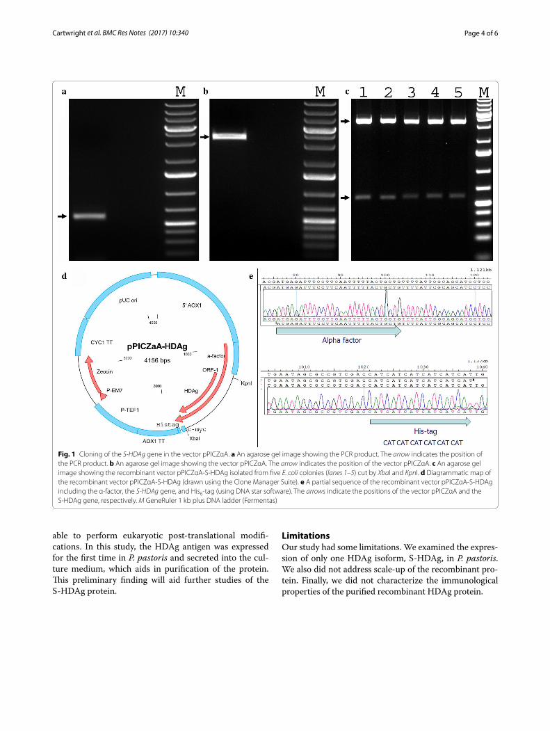
ResultsCloning and sequencingA 589 bp fragment comprising the HDAg gene was amplified by PCR (Fig. 1a). The PCR amplicon was cloned into the vector pPICZαA by enzymatic diges-tion and ligation. To confirm this, the recombinant vec-tor pPICZαA-S-HDAg was digested with XbaI and KpnI. Two bands of 589 and 3567 bp in length were produced as expected (Fig. 1b, c). To ensure the S-HDAg gene was in frame, two primers 5′AOX1-F and 3′AOX1-R were used for sequencing a segment of the recombinant vec-tor pPICZαA-S-HDAg (Fig. 1d). As shown in Fig. 1e, the HDAg gene was cloned into vector pPICZαA and located between the α-factor at the 5′-end and the hexa histidine-tag at the 3′-end. The linearized recombinant vector pPICZαA/S-HDAg was transformed into freshly-pre-pared competent P. pastoris X33 or SMD1163 cells. Posi-tive colonies were screened based on their resistance to Zeocin due to expression of the Zeocin resistance gene.
Protein expression and purificationTo test the expression levels of the HDAg-His-tag fusion protein, 24 positive colonies of each yeast strain were
cultured in 2.5 mL BMGY media in a Micro-24 micro-plate and transferred into 10 mL BMMY supplemented with 1% v/v methanol as an inducer at three time points: 24, 48 and 72 h of induction. Under the control of the promoter AOX1 (methanol inducible promoter), the S-HDAg gene was expressed in P. pastoris X33 but not in SMD1163. For the X33 strain, the expressed protein signal was detected in the culture medium after 48 and 72 h of induction, while no signal was detected in the cell lysate at all three induction time points. A clear band at 25 kDa was observed by immunoblot (Fig. 2). This is the expected size of the recombinant protein includ-ing α-factor, S-HDAg, c-myc epitope, and His6-tag. The recombinant protein was purified exploiting its fused His6-tag. The protein eluted at imidazole concentra-tions from 108–144 mM (wells 4–7, Fig. 3), but not at other concentrations (data not shown). This protein had a molecular weight of 25 kDa which is similar to the pre-dicted molecular weight of the recombinant protein. The yield of purified protein was 115 µg/L culture medium.
DiscussionS-HDAg may present a good candidate for HDV vac-cine development and for diagnostic assays of HDV, but its characterization and immunological evaluation are still limited. One reason is that expression and purifica-tion are not effective [12]. For example, expression of the S-HDAg protein has been performed in several cells including E. coli [13, 14] and baculovirus/insect cells. However, the former lacks the systems for post-transla-tional modifications and the latter results in rapid deg-radation of the HDAg protein after 2 days post-infection [12, 15]. The insertion of the HDAg gene into the chro-mosome of animal cells resulting in a stable cell-line is a good choice, but this is yet to be reported because HDAg is a nuclear protein and the accumulation of this protein results in significant cytotoxicity. In 1990, a number of HDAg-positive HeLa clones were developed, but these cells were lost in culture, whereas a proportion of HDAg-positive HepG2 clones were expanded successfully [16], suggesting that HDAg cytotoxicity may contribute to the cytopathic nature of HDV that was postulated previously [17]. Transient expression in mammalian cells mediated by viral systems (e.g. vaccinia virus) may be possible as well, however these viruses result in cell death and lysis.
Post-translational modifications have been demon-strated to participate in modulating properties and functions of several proteins [18, 19]. HDAg has been identified as being post-translationally modified, which is important for its RNA replication and cellular localiza-tion [20]. Yeast expression systems in general, and in par-ticular P. pastoris, have several advantages such as being
Page 4 of 6Cartwright et al. BMC Res Notes (2017) 10:340
able to perform eukaryotic post-translational modifi-cations. In this study, the HDAg antigen was expressed for the first time in P. pastoris and secreted into the cul-ture medium, which aids in purification of the protein. This preliminary finding will aid further studies of the S-HDAg protein.
LimitationsOur study had some limitations. We examined the expres-sion of only one HDAg isoform, S-HDAg, in P. pastoris. We also did not address scale-up of the recombinant pro-tein. Finally, we did not characterize the immunological properties of the purified recombinant HDAg protein.
Fig. 1 Cloning of the S‑HDAg gene in the vector pPICZαA. a An agarose gel image showing the PCR product. The arrow indicates the position of the PCR product. b An agarose gel image showing the vector pPICZαA. The arrow indicates the position of the vector pPICZαA. c An agarose gel image showing the recombinant vector pPICZαA‑S‑HDAg isolated from five E. coli colonies (lanes 1–5) cut by XbaI and KpnI. d Diagrammatic map of the recombinant vector pPICZαA‑S‑HDAg (drawn using the Clone Manager Suite). e A partial sequence of the recombinant vector pPICZαA‑S‑HDAg including the α‑factor, the S‑HDAg gene, and His6‑tag (using DNA star software). The arrows indicate the positions of the vector pPICZαA and the S‑HDAg gene, respectively. M GeneRuler 1 kb plus DNA ladder (Fermentas)

Page 5 of 6Cartwright et al. BMC Res Notes (2017) 10:340
AbbreviationsBMMY: buffered methanol‑complex medium; BMGY: buffered glycerol‑com‑plex medium; E. coli: Escherichia coli; HDAg: hepatitis delta antigen; LB: Luria Broth; MCS: multiple cloning sites; YPD: yeast extract peptone dextrose.
Authors’ contributionsHMN and RMB designed the research; HMN, SPC, and HTV performed the research; HMN, BTS and HTV analyzed data; and HMN, SPC, BTS, HTV and RMB wrote the paper. All authors read and approved the final manuscript.
Author details1 School of Life & Health Sciences, Aston University, Aston Triangle, Birming‑ham B4 7ET, UK. 2 Department of Molecular Biology, 108 Military Central Hospital, 1 Tran Thanh Tong, Ha Noi City, Viet Nam. 3 Faculty of Biology and Bio‑technology, University of Science, Vietnam National University, Ho Chi Minh City, Viet Nam. 4 Center for Molecular Biology, Institute of Research and Devel‑opment, Duy Tan University, K7/25 Quang Trung, Da Nang City, Viet Nam.
AcknowledgementsWe thank to Prof. Dr. Thomas Bock, Robert Koch Institute, Berlin, Germany for kindly providing pHDV3 plasmid.
Competing interestsThe authors declare that they have no competing interests.
Availability of data and materialsAll data and materials present in the main paper.
Consent for publicationNot applicable.
Ethics approval and consent to participateNot applicable.
FundingThis work was sponsored by Vietnam’s National Foundation for Science and Technology Development (NAFOSTED) Grant No. 106‑YS.02‑2014.03 and Brit‑ish Council for granting a fellowship to H.M.N.
Publisher’s NoteSpringer Nature remains neutral with regard to jurisdictional claims in pub‑lished maps and institutional affiliations.
Received: 1 September 2016 Accepted: 26 July 2017
References 1. Hadziyannis SJ. Review: hepatitis delta. J Gastroenterol Hepatol.
1997;12:289–98. 2. Rizzetto M. Hepatitis D: thirty years after. J Hepatol. 2009;50:1043–50. 3. Abbas Z. Hepatitis D: scenario in the Asia‑Pacific region. World J Gastroen‑
terol. 2010;16:554. 4. Wang KS, Choo QL, Weiner AJ, Ou JH, Najarian RC, et al. Structure,
sequence and expression of the hepatitis delta (delta) viral genome. Nature. 1986;323:508–14.
5. Kuo MY, Sharmeen L, Dinter‑Gottlieb G, Taylor J. Characterization of self‑cleaving RNA sequences on the genome and antigenome of human hepatitis delta virus. J Virol. 1988;62:4439–44.
6. Smedile A, Asey JL, Cote P. Hepatitis D viremia following orthotopic liver transplantation involves a typical HDV virion with a hepatitis B surface antigen envelope. Hepatology. 1998;27:1723–9.
7. Abou‑Jaoude G, Sureau C. Entry of hepatitis delta virus requires the conserved cysteine residues of the hepatitis B virus envelope protein antigenic loop and is blocked by inhibitors of thiol‑disulfide exchange. J Virol. 2007;81:13057–66.
8. Dény P. Hepatitis delta virus genetic variability: from genotypes I, II, III to eight major clades? Curr Top Microbiol Immunol. 2006;307:151–71.
Fig. 2 Expression of the S‑HDAg‑His6‑tag fusion protein. An SDS‑PAGE gel image showing the expression of the S‑HDAg‑His6‑tag fusion protein after a 24‑h, b 48‑h and c 72‑h induction. Lanes 1–3 indicate three colonies, of which lanes 1 and 3 show recombinant protein expressed after 48‑h and 72‑h induction; M are Protomarker Protein Markers (National Diagnostics). Protein was probed with a 6× His monoclonal antibody (Serotec)
Fig. 3 Purification of S‑HDAg‑His6‑tag fusion protein using nickel affinity chromatography. L indicates protein ladder; 1–9 are eluted fractions which have imidazole concentrations ranging from 72 to 156 mM (the concentration interval between 2 consecutive fractions is about 12 mM). The gel was visualized by silver staining

Page 6 of 6Cartwright et al. BMC Res Notes (2017) 10:340
• We accept pre-submission inquiries
• Our selector tool helps you to find the most relevant journal
• We provide round the clock customer support
• Convenient online submission
• Thorough peer review
• Inclusion in PubMed and all major indexing services
• Maximum visibility for your research
Submit your manuscript atwww.biomedcentral.com/submit
Submit your next manuscript to BioMed Central and we will help you at every step:
9. Huang C‑R, Lo SJ. Evolution and diversity of the human hepatitis D virus genome. Adv Bioinform. 2010;2010:1–9.
10. Dugo E, Paroli M, Palmieri G, Giuseppetti R, Argentini C, et al. Immuniza‑tion of woodchucks with adjuvanted sHDAg (p24): immune response and outcome following challenge. Vaccine. 2004;22:457–66.
11. Shiau YT, Huang YH, Wu JC, Tao MH, Syu W, et al. Analysis of humoral immunity of hepatitis D virus DNA vaccine generated in mice by using different dosage, gene gun immunization, and in vivo electroporation. JCMA. 2006;69:7–13.
12. Chiang YW, Wu JC, Wang KC, Lai CW, Chung YC, et al. Efficient expression of histidine‑tagged large hepatitis delta antigen in baculovirus‑trans‑duced baby hamster kidney cells. World J Gastroenterol. 2006;12:1551–7.
13. Calogero R, Barbieri U, Borla M, Osborne S, Poisson F, et al. Purification of recombinant hepatitis delta antigen expressed in E. coli cells. FEBS Lett. 1993;318:322–4.
14. Gt Sheu, Mm Lai. Recombinant hepatitis delta antigen from E. coli pro‑motes hepatitis delta virus RNA replication only from the genomic strand but not the antigenomic strand. Virology. 2000;278:578–86.
15. Hwang SB, Lee CZ, Lai MMC. Hepatitis delta antigen expressed by recombinant baculoviruses: comparison of biochemical properties and post‑translational modifications between the large and small forms. Virol‑ogy. 1992;190:413–22.
16. Li YJ, Macnaughton T, Gao L, Lai MMC. RNA‑templated replication of hepatitis delta virus: genomic and antigenomic RNAs associate with dif‑ferent nuclear bodies. J Virol. 2006;80:6478–86.
17. Popper H, Thung SN, Gerber MA, Hadler SC, De Monzon M, et al. Histologic studies of severe delta agent infection in Venezuelan Indians. Hepatology. 1983;3:906–12.
18. Schwoebel ED, Moore MS. The control of gene expression by regulated nuclear transport. Essays Biochem. 2000;36:105–13.
19. Van Der Geer P. Phosphorylation of LRP1: regulation of transport and signal transduction. Trends Cardiovasc Med. 2002;12:160–5.
20. Hwang SB, Lai MM. Isoprenylation masks a conformational epitope and enhances trans‑dominant inhibitory function of the large hepatitis delta antigen. J Virol. 1994;68:2958–64.