research - environmental health perspectives · pdf fileresearch phthalate esters ......
TRANSCRIPT

472 volume 118 | number 4 | April 2010 • Environmental Health Perspectives
Research
Phthalate esters, ubiquitously used as plasti-cizers in many polyvinyl chloride (PVC) products, have become widespread in the envi-ronment (Peijnenburg et al. 2008). Diisononyl phthalate (DINP) is used in flooring, wire and cable, dip coating, coated fabrics, tubing, shoes, sealants, and artificial leather, and humans may be exposed to DINP by the oral, dermal, and inhalation routes (Kavlock et al. 2002). It has been expected that general population expo-sure to DINP would not exceed levels of di-(2-ethylhexyl)phthalate (DEHP) (Kavlock et al. 2002), which are estimated at 3–30 µg/kg body weight/day (Doull et al. 1999). Plasticizers, including DINP, are not covalently bound to the plastics and can migrate into saliva and be swallowed (Earls et al. 2003; Kavlock et al. 2002). Thus, children may be exposed to higher levels of DINP than are adults because infants and small children mouth toys and other articles containing DINP (Babich et al. 2004; Kavlock et al. 2002).
Several epidemiologic studies have sug-gested that exposure to phthalate esters may be associated with develop ment of asthma, wheezing, and allergic symptoms (Bornehag et al. 2004; Jaakkola et al. 1999, 2004, 2006). Bornehag et al. (2004) revealed the positive association between allergic asthma in children and phthalate esters in house dust. Thus, it is possible that phthalate esters in
the environment may also be associated with development of the other allergic diseases such as atopic dermatitis (AD).
In our previous studies, DEHP enhanced AD-like skin lesions in atopic-prone NC/Nga mice at hundreds-fold lower levels than the no observed adverse effect level (NOAEL) determined from histologic changes in the liver of rodents (Takano et al. 2006; Yanagisawa et al. 2008). The enhancing effects of DEHP paralleled the infiltration of eosinophils, mast cell degranulation, and the expression of pro inflammatory proteins in inflamed skin. Furthermore, we have shown that DEHP enhanced differentiation of bone-marrow– derived dendritic cells (BMDCs) and promoted T-helper 2 (TH2) cell response in spleno cytes from NC/Nga mice in vitro (Koike et al. 2009).
The chronic health effects of DINP, including organ toxicity, carcinogenicity, and reproductive toxicity, have been reviewed in dietary studies (Babich et al. 2004; Kavlock et al. 2002). DINP, as a phthalate plasticizer with specific stereochemical and physico-chemical characteristics, has also been shown to have an adjuvant effect on TH2-dependent immuno globulin (Ig) production in mice (Larsen et al. 2002; Larsen and Nielsen 2008). However, the effects of DINP on allergic dis-eases including AD have remained unclear.
In the present study, we investigated the effects of DINP on AD-like skin lesions in atopic-prone NC/Nga mice in vivo and on the immunologic responses of BMDCs and splenocytes in vitro.
Materials and MethodsAnimals. Six-week-old SPF NC/NgaTndCrlj male mice were purchased from Charles River Japan (Osaka, Japan) and used at 7 weeks of age (body weight, 20–23 g) and 11–15 weeks (body weight, 24–27 g) for in vivo and in vitro study, respectively. Mice were given sterile distilled water and a commercial diet (CE-2; CLEA Japan Inc., Tokyo, Japan) ad libitum, and were housed in an animal facility that was maintained at 22–26°C with 40–69% humidity and a 12/12-hr light/dark cycle under conventional conditions. The pro-cedures for all animal studies were approved by the Institutional Review Board of Japan’s National Institute for Environmental Studies. Animals were treated humanely and with regard for alleviation of suffering.
Protocol for in vivo study. Mice were divided into six groups and were injected intra dermally on the ventral side of their right ears with saline or 5 µg mite extract [Dermatophagoides pteronyssinus (Dp); Cosmo Bio LSL, Tokyo, Japan] dissolved in 10 µL saline on study days 0, 3, 5, 8, 10, 12, 15, and 17 under anesthesia with 4% halo thane (Takeda Pharmaceutical Company, Ltd., Osaka, Japan). DINP (Wako Pure Chemical Industries, Osaka, Japan), at a dose of 0, 0.15, 1.5, 15, or 150 mg/kg/day dissolved in 0.1 mL olive oil (vehicle), was injected intra peritoneally (IP) on days –5, 2, 9, and 16 from the first Dp treatment. Twenty-four hours after each Dp injection, we evaluated ear thickness and clinical scores as described previously (Takano
Address correspondence to E. Koike, Environmental Health Sciences Division, National Institute for Environmental Studies, 16-2 Onogawa, Tsukuba, 305-8506, Japan. Telephone/fax: 81-29-850-2334. E-mail: [email protected]
Supplemental Material is available online (doi:10.1289/ehp.0901255 via http://dx.doi.org/).
We thank M. Sakurai, S. Abe, and N. Ueki for their technical assistance.
This study was supported by the Environmental Technology Development Fund of the Ministry of the Environment and by grants from the National Institute for Environmental Studies.
The authors declare they have no competing financial interests.
Received 27 July 2009; accepted 19 November 2009.
Effects of Diisononyl Phthalate on Atopic Dermatitis in Vivo and Immunologic Responses in VitroEiko Koike,1 Rie Yanagisawa,1 Kaori Sadakane,2 Ken-ichiro Inoue,1 Takamichi Ichinose,2 and Hirohisa Takano1
1Environmental Health Sciences Division, National Institute for Environmental Studies, Tsukuba, Japan; 2Department of Health Sciences, Oita University of Nursing and Health Sciences, Oita, Japan
Background: Diisononyl phthalate (DINP), a principal plasticizer in many polyvinyl chloride products, has been shown to have an adjuvant effect on immunoglobulin (Ig) production in mice. However, the effects of DINP on allergic diseases have not been fully elucidated.
oBjectives: In the present study we investigated the effects of DINP on atopic dermatitis (AD)-like skin lesions induced by Dermatophagoides pteronyssinus (Dp) in atopic-prone NC/Nga mice.
Methods: Mice were injected intradermally with Dp on their ears and were exposed to DINP (0, 0.15, 1.5, 15, or 150 mg/kg/day) intraperitoneally. We evaluated clinical scores, ear thickening, his-tologic findings, protein expression of cytokines/chemokines in the ear, and serum levels of Ig and histamine. Furthermore, we investigated the effects of DINP on bone-marrow–derived dendritic cells (BMDCs) or splenocytes in vitro. After exposure to DINP (0–100 µM), cells were evaluated for phenotype and function.
results: DINP aggravated AD-like skin lesions related to Dp. The aggravation was consistent with eosinophilic inflammation, mast cell degranulation, and thymic stromal lymphopoietin (TSLP) expression in the ear. DINP enhanced the expression of cell surface activation markers on BMDCs and their production of TARC/CCL17 (thymus- and activation-regulated chemokine) and MDC/CCL22 (macrophage-derived chemokine), as well as their capacity to stimulate Dp-specific T-cell proliferation. DINP also enhanced interleukin-4 production and Dp-stimulated proliferation of splenocytes.
conclusions: DINP can aggravate AD-like skin lesions related to Dp. The mechanisms of the aggravation might be mediated, at least partly, through the TSLP-related activation of dendritic cells and by direct or indirect activation of the immune cells.
key words: antigen-presenting activity, atopic dermatitis, bone-marrow–derived dendritic cells, chemokines, diisononyl phthalate, eosinophils, mast cells, splenocytes. Environ Health Perspect 118:472–478 (2010). doi:10.1289/ehp.0901255 [Online 19 November 2009]

Effects of DINP on immune responses
Environmental Health Perspectives • volume 118 | number 4 | April 2010 473
et al. 2006). Twenty-four hours after the last injection of Dp (day 18), the animals were sac-rificed, and histologic findings, protein levels of cytokines and chemokines in the ear tissue supernatants, and the levels of Ig and histamine in serum were evaluated.
Histologic evaluation. Right ears of mice were removed 24 hr after the last Dp injec-tion (day 18) and were fixed in 10% neu-tral phosphate-buffered formalin (pH 7.2) and embedded in paraffin. Sections (3 µm) were routinely stained with hematoxylin and eosin (H&E) or with toluidine blue (pH 4.0). Histologic analy sis was performed using an AX80 microscope (Olympus, Tokyo, Japan). We measured the length of the cartilage and the numbers of infiltrated eosinophils and mast cells in each sample using a video micrometer (VM-30; Olympus). We also evaluated the degranulation of mast cells as non degranulated (0%), mildly degranulated (0–50%), or severely degranulated (> 50%), as described previously (Takano et al. 2006).
Quantitation of cytokines/chemokines in the ear tissue. Right ears of mice were removed 24 hr after the last injection of Dp (day 18) and were homogenized and centrifuged as pre-viously described (Takano et al. 1997). Levels of interferon (IFN)-γ (Endogen, Cambridge, MA, USA), inter leukin (IL)-4 (Amersham, Buckinghamshire, UK), IL-5 (Endogen), IL-13 (R&D Systems, Minneapolis, MN, USA), eotaxin (R&D Systems), eotaxin-2 (R&D Systems), and thymic stromal lympho-poietin (TSLP; R&D Systems) in the ear tissue supernatants were measured by enzyme-linked immuno sorbent assay (ELISA) according to the manufacturers’ instructions. The detection limits of IFN-γ, IL-4, IL-5, IL-13, eotaxin, and TSLP were less than 10, 5, 5, 1.5, 3, and 2.63 pg/mL, respectively. The detection limit of eotaxin-2 was not defined, and the assay range was 15.6–1,000 pg/mL. The total protein level in the ear tissue supernatants was measured by the Bradford method using a protein assay kit (Bio-Rad, Hercules, CA, USA). The values of cytokines/chemokines were compensated with the total protein and were expressed as pico-grams per milligram of total protein.
Quantitation of Ig and histamine in serum. Blood was sampled by cardiac puncture 24 hr after the last injection of Dp (day 18) and serum was collected. Levels of Dp-specific IgG1 were measured by ELISA with solid-phase antigen, as previously described (Sadakane et al. 2002). Levels of total IgE antibodies and histamine in serum were measured by OptELISA Set Mouse IgE (BD Biosciences, San Diego, CA, USA) and Histamine Enzyme Immunoassay Kit (SPI-BIO, Montigny le Bretonneux, France), respectively, according to the manufacturers’ instructions.
Cell preparation for in vitro study. For the in vitro study, bone marrow cells and
splenocytes were prepared as previously described (Koike et al. 2009). Briefly, the mar-row was flushed with Dulbecco’s calcium- and magnesium-free, phosphate-buffered saline (PBS; Takara Bio Inc., Shiga, Japan) and passed through nylon mesh; the red blood cells were then lysed with ammonium chlo-ride. The spleen was pushed through a sterile stainless-steel wire mesh, and the red blood cells were similarly lysed. The cells were cen-trifuged at 400 × g for 5 min at 20°C. After washing with PBS, the cells were resus-pended in culture medium R10, consisting of GIBCO RPMI 1640 medium (Invitrogen, Grand Island, NY, USA) supplemented with 10% heat-inactivated fetal bovine serum (MP Biomedicals Inc., Eschwege, Germany), 100 U/mL penicillin and 100 µg/mL strep-tomycin (Sigma, St. Louis, MO, USA), and 50 µM 2-mercapto ethanol (Invitrogen). The numbers of viable cells were determined by the trypan blue (Invitrogen) exclusion method.
Differentiation of BMDCs. Bone mar-row cells (4 × 10⁵/mL) were cultured in R10 medium containing 20 ng/mL recombinant mouse granulocyte–macrophage colony-stim-ulating factor (GM-CSF; Sigma) at 37°C in a 5% CO2/95% air atmosphere. On day 3, the same volume of medium was added to the culture, and on day 6, half the medium was replaced with fresh medium. On day 8, non-adherent and loosely adherent cells were col-lected by gentle pipetting. The differentiated BMDCs were centrifuged at 400 × g for 5 min at 20°C and resuspended in fresh medium. The numbers of viable cells were determined by the trypan blue exclusion method.
Exposure of immune cells to DINP. DINP was dissolved in dimethyl sulfoxide (DMSO; Sigma) and diluted with R10. The DINP solu-tion was sonicated for 3 min using an ultrasonic disrupter (UD-201; TOMY, Tokyo, Japan). In the presence of GM-CSF (10 ng/mL), BMDCs (1 × 10⁶/mL) were exposed to DINP (0, 0.1, 1, 10, or 100 µM) in R10 containing 0.1% DMSO for 24 hr. Thereafter, their chemokine production, phenotypes, and antigen- presenting activity were evaluated. Next, sple-nocytes (1 × 10⁶/mL) were exposed to DINP (0, 0.1, 1, 10, or 100 µM) in R10 containing 0.1% DMSO for 24 hr, and their cytokine production was examined. Moreover, spleno-cytes (1 × 10⁶/mL) were exposed to DINP (0, 0.001, 0.01, 0.1, 1, or 10 µM) in R10 con-taining 0.1% DMSO in the presence of Dp (10 µg/mL) for 72 hr. Thereafter, antigen-stim-ulated proliferation and cytokine production of the cells were measured. Each experiment was performed using three individual cultures obtained from three animals, and two or three independent experiments were repeated.
Fluorescence-activated cell-sorting (FACS) analysis. For FACS analysis of BMDCs, we used the following mono clonal antibodies:
major histo compatibility complex (MHC) class II I-A/I-E (2G9, rat IgG2a κ, fluorescein isothiocyanate conjugated; BD Biosciences, San Jose, CA, USA); co-stimulatory molecules CD80 [16-10A1, Ar Ham IgG1 κ, phyco-erythrin (PE) conjugated; BD Biosciences] and CD86 (GL1, rat IgG2a κ, PE conjugated; BD Biosciences); and chemokine receptors CCR7 (4B12, rat IgG2a κ, PE conjugated; Biolegend, San Diego, CA, USA) and CXCR4 (2B11/CXCR4, rat IgG2b κ, PE conjugated; BD Biosciences). After DINP exposure, the cells (3–5 × 10⁵) were resuspended in 100 µL PBS with 0.3% bovine serum albumin and 0.05% sodium azide (Wako Pure Chemical Industries) and were incubated with 1 µg of each antibody for 30 min on ice. After incubation, the cells were washed, and the fluorescence was meas-ured by a FACSCalibur (BD, Franklin Lakes, NJ, USA). For each sample, we collected fluo-rescence data from 10,000 cells and positive cells were expressed as percent of total events.
Antigen-presenting activity. We evaluated BMDC function by antigen-presenting activ-ity and stimulating capacity for cytokine pro-duction from responder T cells as previously described (Koike et al. 2009). Briefly, DINP-exposed BMDCs were treated with 50 µg/mL mitomycin C (Kyowa Hakko Kogyo, Tokyo, Japan) for 20 min at 37°C. Splenocytes were prepared from NC/Nga mice sensitized with 50 µg Dp and 1 mg aluminium hydroxide (three mice per experiment). T cells were iso-lated from the splenocytes using a nylon fiber column (Wako Pure Chemical Industries). Thereafter, Dp-sensitized T cells (2 × 10⁵) and BMDCs (5 × 10³) were cocultured in the presence of 2 µg Dp in 200 µL R10 medium in 96-well flat-bottom plates. The cocultures were performed in triplicate at 37°C in a 5% CO2/95% air atmosphere. After 91 hr, we measured T-cell proliferation as an indicator of antigen-presenting activity, and cytokine production from T cells.
Cell proliferation assay. We measured cell proliferation using a Cell-Proliferation ELISA kit (Roche Molecular Biochemicals, Mannheim, Germany) according to the manufacturer’s instructions. This technique is based on the incorporation of the pyrimidine analogue 5-bromo-2´-deoxyuridine (BrdU) instead of thymidine into the DNA of prolif-erating cells. Cell proliferation was measured by adding BrdU to each well 20 hr before the measurement.
Quantitation of cytokines/chemokines in the culture supernatants. The levels of thy-mus- and activation-regulated chemokine (TARC/CCL17; R&D Systems), macrophage-derived chemokine (MDC/CCL22; R&D Systems), and IL-12p40 (Endogen) in the BMDC culture supernatants and the levels of IFN-γ (Endogen), IL-4 (Amersham), IL-10 (Endogen), and IL-17 (R&D Systems) in the
Koike et al.
474 volume 118 | number 4 | April 2010 • Environmental Health Perspectives
supernatants of BMDCs and T-cell co cultures or splenocyte cultures were meas ured by ELISA according to the manufacturers’ instruc-tions. The detection limits of TARC/CCL17, MDC/CCL22, IL-12p40, IFN-γ, IL-4, IL-10, and IL-17 were less than 5, 1.2, 3, 10, 5, 12, and 5 pg/mL, respectively.
Statistical analysis. Data are presented as mean ± SE. The significance of variation among different groups was determined by one-way analysis of variance or Kruskal-Wallis analysis. Differences among groups were analyzed using Dunnett’s or Steel mul-tiple comparison test (Excel Statistics 2006, Social Survey Research Information Co., Ltd., Tokyo, Japan). A p-value < 0.05 is considered statistically significant.
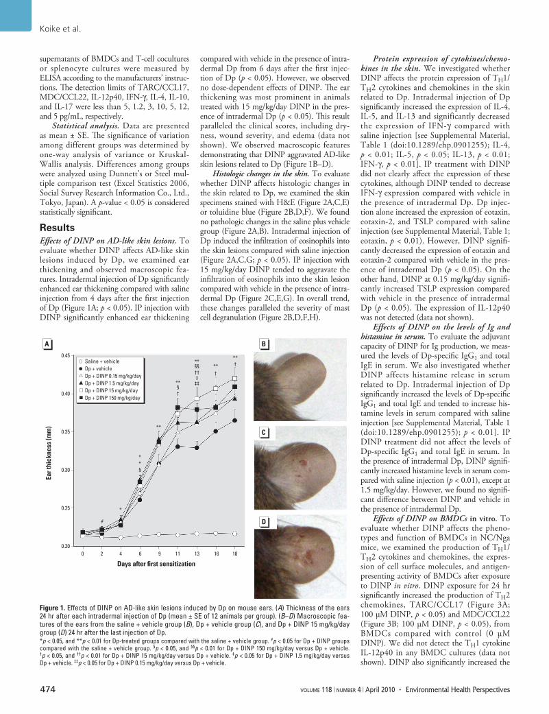
ResultsEffects of DINP on AD-like skin lesions. To evaluate whether DINP affects AD-like skin lesions induced by Dp, we examined ear thickening and observed macroscopic fea-tures. Intradermal injection of Dp significantly enhanced ear thickening compared with saline injection from 4 days after the first injection of Dp (Figure 1A; p < 0.05). IP injection with DINP significantly enhanced ear thickening
compared with vehicle in the presence of intra-dermal Dp from 6 days after the first injec-tion of Dp (p < 0.05). However, we observed no dose-dependent effects of DINP. The ear thickening was most prominent in animals treated with 15 mg/kg/day DINP in the pres-ence of intradermal Dp (p < 0.05). This result paralleled the clinical scores, including dry-ness, wound severity, and edema (data not shown). We observed macroscopic features demonstrating that DINP aggravated AD-like skin lesions related to Dp (Figure 1B–D).
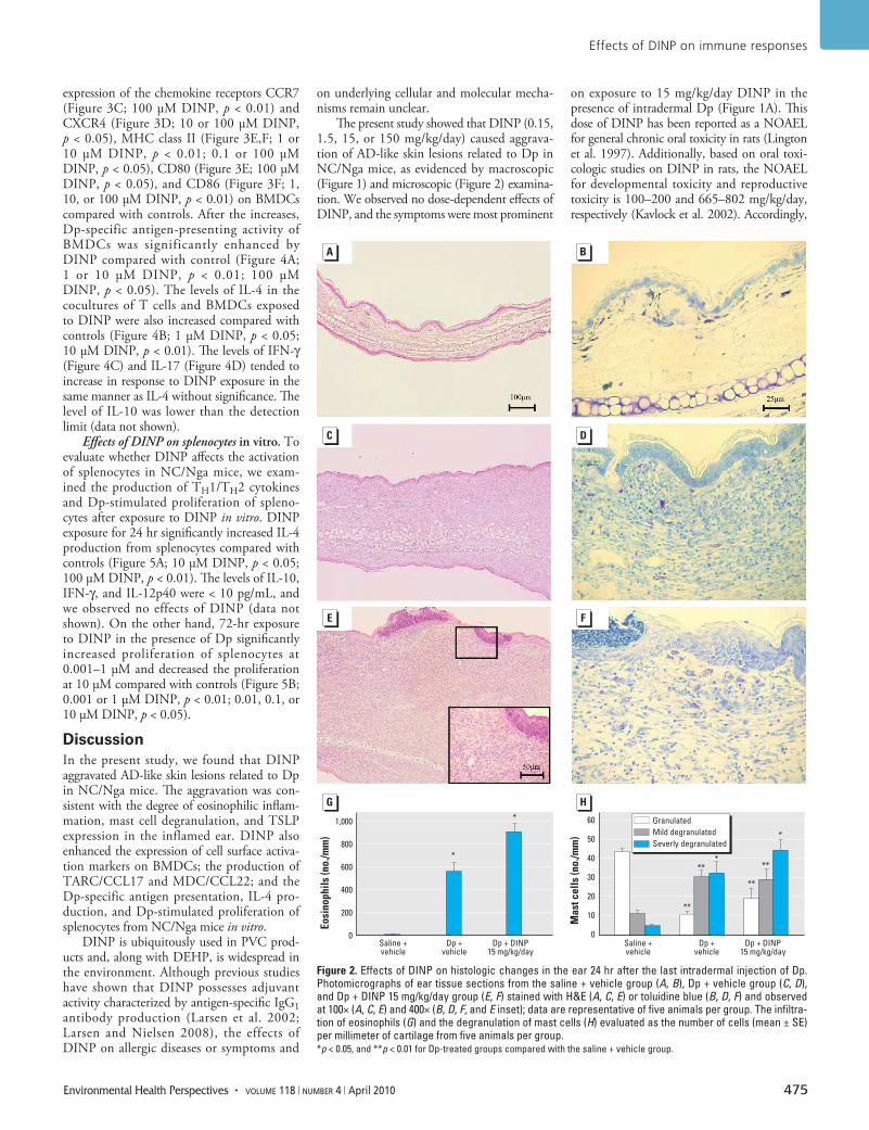
Histologic changes in the skin. To evaluate whether DINP affects histologic changes in the skin related to Dp, we examined the skin specimens stained with H&E (Figure 2A,C,E) or toluidine blue (Figure 2B,D,F). We found no pathologic changes in the saline plus vehicle group (Figure 2A,B). Intradermal injection of Dp induced the infiltration of eosinophils into the skin lesions compared with saline injection (Figure 2A,C,G; p < 0.05). IP injection with 15 mg/kg/day DINP tended to aggravate the infiltration of eosinophils into the skin lesion compared with vehicle in the presence of intra-dermal Dp (Figure 2C,E,G). In overall trend, these changes paralleled the severity of mast cell degranulation (Figure 2B,D,F,H).
Protein expression of cytokines/chemo-kines in the skin. We investigated whether DINP affects the protein expression of TH1/TH2 cytokines and chemokines in the skin related to Dp. Intradermal injection of Dp significantly increased the expression of IL-4, IL-5, and IL-13 and significantly decreased the expression of IFN-γ compared with saline injection [see Supplemental Material, Table 1 (doi:10.1289/ehp.0901255); IL-4, p < 0.01; IL-5, p < 0.05; IL-13, p < 0.01; IFN-γ, p < 0.01]. IP treatment with DINP did not clearly affect the expression of these cytokines, although DINP tended to decrease IFN-γ expression compared with vehicle in the presence of intradermal Dp. Dp injec-tion alone increased the expression of eotaxin, eotaxin-2, and TSLP compared with saline injection (see Supplemental Material, Table 1; eotaxin, p < 0.01). However, DINP signifi-cantly decreased the expression of eotaxin and eotaxin-2 compared with vehicle in the pres-ence of intradermal Dp (p < 0.05). On the other hand, DINP at 0.15 mg/kg/day signifi-cantly increased TSLP expression compared with vehicle in the presence of intradermal Dp (p < 0.05). The expression of IL-12p40 was not detected (data not shown).
Effects of DINP on the levels of Ig and histamine in serum. To evaluate the adjuvant capacity of DINP for Ig production, we meas-ured the levels of Dp-specific IgG1 and total IgE in serum. We also investigated whether DINP affects histamine release in serum related to Dp. Intradermal injection of Dp significantly increased the levels of Dp-specific IgG1 and total IgE and tended to increase his-tamine levels in serum compared with saline injection [see Supplemental Material, Table 1 (doi:10.1289/ehp.0901255); p < 0.01]. IP DINP treatment did not affect the levels of Dp-specific IgG1 and total IgE in serum. In the presence of intradermal Dp, DINP signifi-cantly increased histamine levels in serum com-pared with saline injection (p < 0.01), except at 1.5 mg/kg/day. However, we found no signifi-cant difference between DINP and vehicle in the presence of intra dermal Dp.
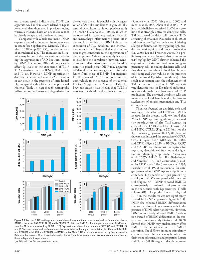
Effects of DINP on BMDCs in vitro. To evaluate whether DINP affects the pheno-types and function of BMDCs in NC/Nga mice, we examined the production of TH1/TH2 cytokines and chemokines, the expres-sion of cell surface molecules, and antigen-presenting activity of BMDCs after exposure to DINP in vitro. DINP exposure for 24 hr significantly increased the production of TH2 chemokines, TARC/CCL17 (Figure 3A; 100 µM DINP, p < 0.05) and MDC/CCL22 (Figure 3B; 100 µM DINP, p < 0.05), from BMDCs compared with control (0 µM DINP). We did not detect the TH1 cytokine IL-12p40 in any BMDC cultures (data not shown). DINP also significantly increased the
Figure 1. Effects of DINP on AD-like skin lesions induced by Dp on mouse ears. (A) Thickness of the ears 24 hr after each intradermal injection of Dp (mean ± SE of 12 animals per group). (B–D) Macroscopic fea-tures of the ears from the saline + vehicle group (B), Dp + vehicle group (C), and Dp + DINP 15 mg/kg/day group (D) 24 hr after the last injection of Dp. *p < 0.05, and **p < 0.01 for Dp-treated groups compared with the saline + vehicle group. #p < 0.05 for Dp + DINP groups compared with the saline + vehicle group. §p < 0.05, and §§p < 0.01 for Dp + DINP 150 mg/kg/day versus Dp + vehicle. †p < 0.05, and ††p < 0.01 for Dp + DINP 15 mg/kg/day versus Dp + vehicle. ‡p < 0.05 for Dp + DINP 1.5 mg/kg/day versus Dp + vehicle. ‡‡p < 0.05 for Dp + DINP 0.15 mg/kg/day versus Dp + vehicle.
0.45
0.40
0.35
0.30
0.25
0.20
Ear t
hick
ness
(mm
)
0 2
#
*
**
**§†
**†
**†**
§§††‡
‡‡
**§
4 6
Days after first sensitization
9 11 13 16 18
Saline + vehicleDp + vehicleDp + DINP 0.15 mg/kg/dayDp + DINP 1.5 mg/kg/dayDp + DINP 15 mg/kg/dayDp + DINP 150 mg/kg/day
Effects of DINP on immune responses
Environmental Health Perspectives • volume 118 | number 4 | April 2010 475
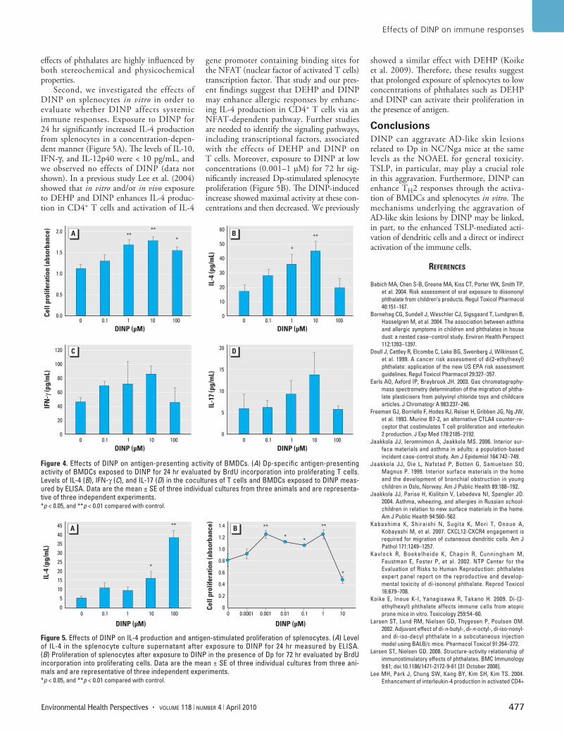
expression of the chemokine receptors CCR7 (Figure 3C; 100 µM DINP, p < 0.01) and CXCR4 (Figure 3D; 10 or 100 µM DINP, p < 0.05), MHC class II (Figure 3E,F; 1 or 10 µM DINP, p < 0.01; 0.1 or 100 µM DINP, p < 0.05), CD80 (Figure 3E; 100 µM DINP, p < 0.05), and CD86 (Figure 3F; 1, 10, or 100 µM DINP, p < 0.01) on BMDCs compared with controls. After the increases, Dp-specific antigen-presenting activity of BMDCs was significantly enhanced by DINP compared with control (Figure 4A; 1 or 10 µM DINP, p < 0.01; 100 µM DINP, p < 0.05). The levels of IL-4 in the cocultures of T cells and BMDCs exposed to DINP were also increased compared with controls (Figure 4B; 1 µM DINP, p < 0.05; 10 µM DINP, p < 0.01). The levels of IFN-γ (Figure 4C) and IL-17 (Figure 4D) tended to increase in response to DINP exposure in the same manner as IL-4 without significance. The level of IL-10 was lower than the detection limit (data not shown).
Effects of DINP on splenocytes in vitro. To evaluate whether DINP affects the activation of splenocytes in NC/Nga mice, we exam-ined the production of TH1/TH2 cytokines and Dp-stimulated proliferation of spleno-cytes after exposure to DINP in vitro. DINP exposure for 24 hr significantly increased IL-4 production from splenocytes compared with controls (Figure 5A; 10 µM DINP, p < 0.05; 100 µM DINP, p < 0.01). The levels of IL-10, IFN-γ, and IL-12p40 were < 10 pg/mL, and we observed no effects of DINP (data not shown). On the other hand, 72-hr exposure to DINP in the presence of Dp significantly increased proliferation of splenocytes at 0.001–1 µM and decreased the proliferation at 10 µM compared with controls (Figure 5B; 0.001 or 1 µM DINP, p < 0.01; 0.01, 0.1, or 10 µM DINP, p < 0.05).
DiscussionIn the present study, we found that DINP aggravated AD-like skin lesions related to Dp in NC/Nga mice. The aggravation was con-sistent with the degree of eosinophilic inflam-mation, mast cell degranulation, and TSLP expression in the inflamed ear. DINP also enhanced the expression of cell surface activa-tion markers on BMDCs; the production of TARC/CCL17 and MDC/CCL22; and the Dp-specific antigen presentation, IL-4 pro-duction, and Dp-stimulated prolifera tion of splenocytes from NC/Nga mice in vitro.
DINP is ubiquitously used in PVC prod-ucts and, along with DEHP, is widespread in the environment. Although previous studies have shown that DINP possesses adjuvant activity characterized by antigen-specific IgG1 antibody production (Larsen et al. 2002; Larsen and Nielsen 2008), the effects of DINP on allergic diseases or symptoms and
on under lying cellular and molecular mecha-nisms remain unclear.
The present study showed that DINP (0.15, 1.5, 15, or 150 mg/kg/day) caused aggrava-tion of AD-like skin lesions related to Dp in NC/Nga mice, as evidenced by macroscopic (Figure 1) and microscopic (Figure 2) examina-tion. We observed no dose-dependent effects of DINP, and the symptoms were most prominent
on exposure to 15 mg/kg/day DINP in the presence of intradermal Dp (Figure 1A). This dose of DINP has been reported as a NOAEL for general chronic oral toxicity in rats (Lington et al. 1997). Additionally, based on oral toxi-cologic studies on DINP in rats, the NOAEL for developmental toxicity and reproductive toxicity is 100–200 and 665–802 mg/kg/day, respectively (Kavlock et al. 2002). Accordingly,
Figure 2. Effects of DINP on histologic changes in the ear 24 hr after the last intradermal injection of Dp. Photomicrographs of ear tissue sections from the saline + vehicle group (A, B), Dp + vehicle group (C, D), and Dp + DINP 15 mg/kg/day group (E, F) stained with H&E (A, C, E) or toluidine blue (B, D, F) and observed at 100× (A, C, E) and 400× (B, D, F, and E inset); data are representative of five animals per group. The infiltra-tion of eosinophils (G) and the degranulation of mast cells (H) evaluated as the number of cells (mean ± SE) per millimeter of cartilage from five animals per group. *p < 0.05, and **p < 0.01 for Dp-treated groups compared with the saline + vehicle group.
1,000
800
600
400
200
0
60
50
40
30
20
10
0
Eosi
noph
ils (n
o./m
m)
Mas
t cel
ls (n
o./m
m)
Saline +vehicle
Dp +vehicle
*
*
Dp + DINP15 mg/kg/day
Saline +vehicle
Dp +vehicle
**
****
**
*
*
Dp + DINP15 mg/kg/day
GranulatedMild degranulatedSeverly degranulated
Koike et al.
476 volume 118 | number 4 | April 2010 • Environmental Health Perspectives
our present results indicate that DINP can aggravate AD-like skin lesions related to Dp at lower levels than those used in previous studies, whereas a NOAEL based on oral intake cannot be directly compared with an injected dose.
Compared with vehicle treatment, DINP exposure tended to increase hista mine release in serum [see Supplemental Material, Table 1 (doi:10.1289/ehp.0901255)] in the presence of intra dermal Dp. The increases in hista-mine may be one of the mechanisms underly-ing the aggravation of AD-like skin lesions by DINP. In contrast, DINP did not clearly affect Ig levels or the expression of TH1/TH2 cytokines such as IFN-γ, IL-4, IL-5, and IL-13. However, DINP signifi cantly decreased eotaxin and eotaxin-2 expression in ear tissue in the presence of intradermal Dp, compared with vehicle (see Supplemental Material, Table 1), even though eosino philic inflammation and mast cell degradation in
the ear were present in parallel with the aggra-vation of AD-like skin lesions (Figure 2). This result differed from that in our previous study on DEHP (Takano et al. 2006), in which we observed increased expression of eotaxin and macrophage inflammatory protein-1α in the ear. It is possible that DINP induced the expression of TH2 cytokines and chemok-ines at an earlier phase and that this induc-tion might contribute to the aggravation of the symptoms. A time-course study is needed to elucidate the correlation between symp-toms and inflammatory mediators. In addi-tion, it is possible that DINP may aggravate AD-like skin lesions through mecha nisms dif-ferent from those of DEHP. For instance, DINP enhanced TSLP expression compared with vehicle in the presence of intradermal Dp (see Supplemental Material, Table 1). Previous studies have shown that TSLP is associated with AD and asthma in humans
(Soumelis et al. 2002; Ying et al. 2005) and mice (Li et al. 2005; Zhou et al. 2005). TSLP is an epithelial-cell–derived IL-7-like cyto-kine that strongly activates dendritic cells. TSLP-activated dendritic cells produce TH2-attracting chemokines (Soumelis et al. 2002) and then induce TH2 cell activation, initiating allergic inflammation by triggering IgE pro-duction, eosinophilia, and mucus production (Liu 2006; Liu and Frederick 2009). In a pre-liminary study, we observed that exposure to 0.15 mg/kg/day DINP further enhanced the expression of activation markers of antigen-presenting cells, including dendritic cells, and that these cell numbers in local lymph node cells compared with vehicle in the presence of intradermal Dp (data not shown). This result is consistent with the enhancement of TSLP expression. Therefore, DINP may acti-vate dendritic cells in Dp-related inflamma-tory sites through the enhancement of TSLP production. The activated dendritic cells can migrate into local lymph nodes, leading to acceleration of antigen presentation and TH2 cell activation.
Thus, we focused on dendritic cells and investigated the effects of DINP on BMDCs in vitro. In the present study we found that 24-hr DINP exposure significantly increased the production of the TH2-attracting chemokines TARC/CCL17 (Figure 3A) and MDC/CCL22 (Figure 3B) but not the TH1-polarizing cytokine IL-12p40 (data not shown), and increased the expression of CCR7, CXCR4 (Figure 3C,D), MHC class II, CD80, and CD86 (Figure 3E,F) in BMDCs. CCR7 and CXCR4 are chemo kine receptors for regulating dendritic cell function and migra-tion into draining lymph nodes (Kabashima et al. 2007). MHC class II (Niederhuber and Shreffler 1977) and costimulatory mol-ecules CD80 and CD86 (Freeman et al. 1993; Lenschow et al. 1993) are essential for anti-gen presentation. DINP exposure significantly enhanced Dp-specific antigen- presenting activity of BMDCs compared with the con-trol (Figure 4A). DINP-exposed BMDCs consequently stimulated IL-4 production in the cocultures with Dp-sensitized T cells (Figure 4B). The production of IFN-γ and IL-17 in the co cultures was not significantly altered by DINP exposure (Figure 4C,D). DINP also enhanced BMDC differentiation after 6-day culture of bone marrow cells in the presence of DINP (data not shown). However, DINP more clearly affected BMDC activa-tion instead of BMDC differentiation. In con-trast, our previous study (Koike et al. 2009) showed that DEHP may predominantly affect BMDC differentiation rather than BMDC activation. The different immune stimulatory effects of these phthalates may be related to their chemi cal structures and properties. Larsen and Nielsen (2008) suggested that the adjuvant
Figure 3. Effects of DINP on the production of chemokines and the expression of cell surface molecules on BMDCs. Levels of TARC/CCL17 (A) and MDC/CCL22 (B) in the BMDC culture supernatant after DINP expo-sure for 24 hr as measured by ELISA. (C,D) Expression of chemokine receptors CCR7 (C) and CXCR4 (D), and (E,F) expression of cell surface molecules associated with antigen presentation, MHC class II (MHC II) and CD80 (E) or MHC II and CD86 (F), on BMDCs after 24-hr DINP exposure as analyzed by flow cytometry. Data are the mean ± SE of three individual cultures from three animals and are representative of two or three independent experiments. *p < 0.05, and **p < 0.01 compared with control.
25,000
20,000
15,000
10,000
5,000
0
20
18
16
14
12
10
8
6
4
2
0
100908070605040302010
0
100908070605040302010
0
18,000
16,000
14,000
12,000
10,000
8,000
6,000
4,000
2,000
0
50
45
40
35
30
25
20
15
10
5
0
TARC
(pg/
mL)
CCR7
-pos
itive
cel
ls (%
)Pe
rcen
t of c
ells
Perc
ent o
f cel
lsCX
CR4-
posi
tive
cells
(%)
MD
C (p
g/m
L)
ControlDINP 0.1 µMDINP 1 µMDINP 10 µMDINP 100 µM
0 10.1 10
**
**
100
DINP (µM)
0
CD80 positive
**
* *** ** **
**
**
** **
****
**
**
CD86 positive
MHC II positive
MHC II positive
Double positive
Double positive
1 10 100 0 1 10 100
**
DINP (µM) DINP (µM)
DINP (µM)0 10.1 10 100
Effects of DINP on immune responses
Environmental Health Perspectives • volume 118 | number 4 | April 2010 477
effects of phthalates are highly influenced by both stereo chemical and physico chemical properties.
Second, we investigated the effects of DINP on splenocytes in vitro in order to evaluate whether DINP affects systemic immune responses. Exposure to DINP for 24 hr significantly increased IL-4 production from splenocytes in a concentration-depen-dent manner (Figure 5A). The levels of IL-10, IFN-γ, and IL-12p40 were < 10 pg/mL, and we observed no effects of DINP (data not shown). In a previous study Lee et al. (2004) showed that in vitro and/or in vivo exposure to DEHP and DINP enhances IL-4 produc-tion in CD4+ T cells and activation of IL-4
gene promoter containing binding sites for the NFAT (nuclear factor of activated T cells) transcription factor. That study and our pres-ent findings suggest that DEHP and DINP may enhance allergic responses by enhanc-ing IL-4 production in CD4+ T cells via an NFAT-dependent pathway. Further studies are needed to identify the signaling pathways, including transcriptional factors, associated with the effects of DEHP and DINP on T cells. Moreover, exposure to DINP at low concentrations (0.001–1 µM) for 72 hr sig-nificantly increased Dp-stimulated splenocyte proliferation (Figure 5B). The DINP-induced increase showed maximal activity at these con-centrations and then decreased. We previously
showed a similar effect with DEHP (Koike et al. 2009). Therefore, these results suggest that prolonged exposure of splenocytes to low concentrations of phthalates such as DEHP and DINP can activate their proliferation in the presence of antigen.
ConclusionsDINP can aggravate AD-like skin lesions related to Dp in NC/Nga mice at the same levels as the NOAEL for general toxicity. TSLP, in particular, may play a crucial role in this aggravation. Furthermore, DINP can enhance TH2 responses through the activa-tion of BMDCs and splenocytes in vitro. The mechanisms underlying the aggravation of AD-like skin lesions by DINP may be linked, in part, to the enhanced TSLP-mediated acti-vation of dendritic cells and a direct or indirect activation of the immune cells.
RefeRences
Babich MA, Chen S-B, Greene MA, Kiss CT, Porter WK, Smith TP, et al. 2004. Risk assessment of oral exposure to diisononyl phthalate from children’s products. Regul Toxicol Pharmacol 40:151–167.
Bornehag CG, Sundell J, Weschler CJ, Sigsgaard T, Lundgren B, Hasselgren M, et al. 2004. The association between asthma and allergic symptoms in children and phthalates in house dust: a nested case–control study. Environ Health Perspect 112:1393–1397.
Doull J, Cattley R, Elcombe C, Lake BG, Swenberg J, Wilkinson C, et al. 1999. A cancer risk assessment of di(2-ethylhexyl)phthalate: application of the new US EPA risk assessment guidelines. Regul Toxicol Pharmacol 29:327–357.
Earls AO, Axford IP, Braybrook JH. 2003. Gas chromatography-mass spectrometry determination of the migration of phtha-late plasticisers from polyvinyl chloride toys and childcare articles. J Chromatogr A 983:237–246.
Freeman GJ, Borriello F, Hodes RJ, Reiser H, Gribben JG, Ng JW, et al. 1993. Murine B7-2, an alternative CTLA4 counter-re-ceptor that costimulates T cell proliferation and inter leukin 2 production. J Exp Med 178:2185–2192.
Jaakkola JJ, Ieromnimon A, Jaakkola MS. 2006. Interior sur-face materials and asthma in adults: a population-based incident case-control study. Am J Epidemiol 164:742–749.
Jaakkola JJ, Oie L, Nafstad P, Botten G, Samuelsen SO, Magnus P. 1999. Interior surface materials in the home and the development of bronchial obstruction in young children in Oslo, Norway. Am J Public Health 89:188–192.
Jaakkola JJ, Parise H, Kislitsin V, Lebedeva NI, Spengler JD. 2004. Asthma, wheezing, and allergies in Russian school-children in relation to new surface materials in the home. Am J Public Health 94:560–562.
Kabashima K, Shiraishi N, Sugita K, Mori T, Onoue A, Kobayashi M, et al. 2007. CXCL12-CXCR4 engagement is required for migration of cutaneous dendritic cells. Am J Pathol 171:1249–1257.
Kavlock R, Boekelheide K, Chapin R, Cunningham M, Faustman E, Foster P, et al. 2002. NTP Center for the Evaluation of Risks to Human Reproduction: phthalates expert panel report on the reproductive and develop-mental toxicity of di-isononyl phthalate. Reprod Toxicol 16:679–708.
Koike E, Inoue K-I, Yanagisawa R, Takano H. 2009. Di-(2-ethylhexyl) phthalate affects immune cells from atopic prone mice in vitro. Toxicology 259:54–60.
Larsen ST, Lund RM, Nielsen GD, Thygesen P, Poulsen OM. 2002. Adjuvant effect of di-n-butyl-, di-n-octyl-, di-iso-nonyl- and di-iso-decyl phthalate in a subcutaneous injection model using BALB/c mice. Pharmacol Toxicol 91:264–272.
Larsen ST, Nielsen GD. 2008. Structure-activity relationship of immunostimulatory effects of phthalates. BMC Immunology 9:61; doi:10.1186/1471-2172-9-61 [31 October 2008].
Lee MH, Park J, Chung SW, Kang BY, Kim SH, Kim TS. 2004. Enhancement of interleukin-4 production in activated CD4+
Figure 4. Effects of DINP on antigen-presenting activity of BMDCs. (A) Dp-specific antigen-presenting activity of BMDCs exposed to DINP for 24 hr evaluated by BrdU incorporation into proliferating T cells. Levels of IL-4 (B), IFN-γ (C), and IL-17 (D) in the cocultures of T cells and BMDCs exposed to DINP meas-ured by ELISA. Data are the mean ± SE of three individual cultures from three animals and are representa-tive of three independent experiments. *p < 0.05, and **p < 0.01 compared with control.
2.0
1.5
1.0
0.5
0.0
20
15
10
5
0
120
100
80
60
40
20
0
60
50
40
30
20
10
0Cell
prol
ifera
tion
(abs
orba
nce)
IL-4
(pg/
mL)
IL-1
7 (p
g/m
L)
IFN
-γ (p
g/m
L)
0 10.1 10
*
****
*
**
100
DINP (µM) DINP (µM)0 10.1 10 100
0 10.1 10 100
DINP (µM) DINP (µM)0 10.1 10 100
Figure 5. Effects of DINP on IL-4 production and antigen-stimulated proliferation of splenocytes. (A) Level of IL-4 in the splenocyte culture supernatant after exposure to DINP for 24 hr measured by ELISA. (B) Proliferation of splenocytes after exposure to DINP in the presence of Dp for 72 hr evaluated by BrdU incorporation into proliferating cells. Data are the mean ± SE of three individual cultures from three ani-mals and are representative of three independent experiments. *p < 0.05, and **p < 0.01 compared with control.
45
40
35
30
25
20
15
10
5
0
1.4
1.2
1.0
0.8
0.6
0.4
0.2
0Cell
prol
ifera
tion
(abs
orba
nce)
IL-4
(pg/
mL)
0 01 10.1 0.10.01
** **
**
*
0.0010.000110 10
*
**
100
DINP (µM) DINP (µM)

Koike et al.
478 volume 118 | number 4 | April 2010 • Environmental Health Perspectives
T cells by diphthalate plasticizers via increased NF-AT binding activity. Int Arch Allergy Immunol 134:213–222.
Lenschow DJ, Su GH, Zuckerman LA, Nabavi N, Jellis CL, Gray GS, et al. 1993. Expression and functional significance of an additional ligand for CTLA-4. Proc Natl Acad Sci USA 90:11054–11058.
Li M, Messaddeq N, Teletin M, Pasquali J-L, Metzger D, Chambon P. 2005. Retinoid X receptor ablation in adult mouse keratinocytes generates an atopic dermatitis trig-gered by thymic stromal lymphopoietin. Proc Natl Acad Sci USA 102:14795–14800.
Lington AW, Bird MG, Plutnick RT, Stubblefield WA, Scala RA. 1997. Chronic toxicity and carcinogenic evaluation of diiso nonyl phthalate in rats. Fundam Appl Toxicol 36:79–89.
Liu Y-J. 2006. Thymic stromal lymphopoietin: master switch for allergic inflammation. J Exp Med 203:269–273.
Liu Y, Frederick WA. 2009. TSLP in epithelial cell and dendritic cell cross talk. Adv Immunol 101:1–25.
Niederhuber JE, Shreffler DC. 1977. Anti-Ia serum blocking of macrophage function in the in vitro humoral response. Transplant Proc 9:875–879.
Peijnenburg WJGM, Sven Erik J, Brian F. 2008. Phthalates. In: Encyclopedia of Ecology. Oxford:Academic Press, 2733–2738.
Sadakane K, Takano H, Ichinose T, Yanagisawa R, Shibamoto T. 2002. Formaldehyde enhances mite allergen-induced eosinophilic inflammation in the murine airway. J Environ Pathol Toxicol Oncol 21:267–276.
Soumelis V, Reche PA, Kanzler H, Yuan W, Edward G, Homey B, et al. 2002. Human epithelial cells trigger dendritic cell mediated allergic inflammation by producing TSLP. Nat Immunol 3:673–680.
Takano H, Yanagisawa R, Inoue K, Ichinose T, Sadakane K, Yoshikawa T. 2006. Di-(2-ethylhexyl) phthalate enhances atopic dermatitis-like skin lesions in mice. Environ Health Perspect 114:1266–1269.
Takano H, Yoshikawa T, Ichinose T, Miyabara Y, Imaoka K, Sagai M. 1997. Diesel exhaust particles enhance antigen-induced airway inflammation and local cytokine expression in mice. Am J Respir Crit Care Med 156:36–42.
Yanagisawa R, Takano H, Inoue K, Koike E, Sadakane K, Ichinose T. 2008. Effects of maternal exposure to di-(2-ethyl-hexyl) phthalate during fetal and/or neonatal periods on atopic dermatitis in male offspring. Environ Health Perspect 116:1136–1141.
Ying S, O’Connor B, Ratoff J, Meng Q, Mallett K, Cousins D, et al. 2005. Thymic stromal lymphopoietin expression is increased in asthmatic airways and correlates with expression of Th2-attracting chemokines and disease severity. J Immunol 174:8183–8190.
Zhou B, Comeau MR, De Smedt T, Liggitt HD, Dahl ME, Lewis DB, et al. 2005. Thymic stromal lymphopoietin as a key initia-tor of allergic airway inflammation in mice. Nat Immunol 6:1047–1053.