research article titanium oxide nanotube surface topography...
TRANSCRIPT

Research ArticleTitanium Oxide Nanotube Surface Topography andMicroRNA-488 Contribute to Modulating Osteogenesis
Yeon-Ho Kang1 Bohm Choi2 Chihyun Ahn1 Seunghan Oh3
Myeung Soo Lee4 and Eun-Jung Jin1
1 Department of Biological Sciences College of Natural Sciences Wonkwang University Iksan Chungbuk 570-749 Republic of Korea2Department of Dentistry Uijeongbu St Maryrsquos Hospital The Catholic University of Korea UijeongbuGyeonggi 480-717 Republic of Korea
3 Department of Dental Biomaterials College of Dentistry Wonkwang University Iksan Chunbuk 570-749 Republic of Korea4Department of Internal Medicine Division of Rheumatology Wonkwang University School of Medicine IksanChunbuk 570-749 Republic of Korea
Correspondence should be addressed to Myeung Soo Lee ckhlmswonkwangackr and Eun-Jung Jin jineunjungwkuackr
Received 13 February 2014 Accepted 31 March 2014 Published 22 April 2014
Academic Editor Young-Bum Park
Copyright copy 2014 Yeon-Ho Kang et alThis is an open access article distributed under the Creative Commons Attribution Licensewhich permits unrestricted use distribution and reproduction in any medium provided the original work is properly cited
Understanding the biocomplexity of cell behavior in relation to the topographical characteristics of implants is essential forsuccessful osseointegration with good longevity and minimum failure Here we investigated whether culture on titanium oxide(TiO2) nanotubes of various diameters could affect the behavior and differentiation of MC3T3-E1 cells Among the tested
nanotubes those of 50 nm in diameter were found to trigger the expression of the osteoblast-specific transcription factors sp7and Dlx5 and upregulate the expression of alkaline phosphatase (ALP) Here we report that miR-488 was significantly induced inosteoblasts cultured on 50 nm nanotubes and continued to increase with the progression of osteoblast differentiation Furthermoredownregulation of miR-488 suppressed the expression levels of ALP and matrix metalloprotease-2 (MMP-2) This suppression ofALP transcription was overcome by treatment with the MMP-2 activator bafilomycin A1 Collectively these results suggest that50 nm is the optimumTiO
2nanotube diameter for implants and thatmodulation ofmiR-488 can change the differentiation activity
of cells on TiO2nanotubes This emphasizes that we must fully understand the physicochemical properties of TiO
2nanotubes and
the endogenous biomolecules that interact with such surfaces in order to fully support their clinical application
1 Introduction
Accumulating experimental evidence suggests that nanoscaletopography is an important factor for cellular recognition ofthe biological microenvironment and biomimetic materialsused for vascular grafts stents and bone implants [1 2]Titanium (Ti) and its alloys have been widely used as implan-tation materials as they provide direct physical bondingwith the adjacent bone surface in orthopedic and dentalsurgery The low success rate of using machined surfacessuch as those first tested as dental implants in 1965 haslong indicated that the materials and particle sizes of implantsurfaces are critical to the initial osseointegration and successof implants [3] Although the current success rate is very highimplants still occasionally loosen and fail due to incomplete
osseointegration between the implantation materials and thesurrounding bone [3]
Several studies have suggested that some cells can showcell-type-specific behaviors and activities on TiO
2nanotubes
For example mesenchymal stem cells showed enhancedactivity on anatase-phase nanotubes (70ndash100 nm diameter)and proliferation of smooth muscle cells was enhancedon TiO
2[4] Furthermore Webster et al [5] reported
that osteoblasts showed significantly greater adhesion to ananophase surface compared to conventional aluminum andtitanium surfaces
MicroRNAs (miRNAs) are short (18ndash25 nucleotides)noncoding RNAs that posttranscriptionally regulate geneexpression by recognizing binding sites in the 31015840-untranslated
Hindawi Publishing CorporationJournal of NanomaterialsVolume 2014 Article ID 589710 8 pageshttpdxdoiorg1011552014589710
2 Journal of Nanomaterials
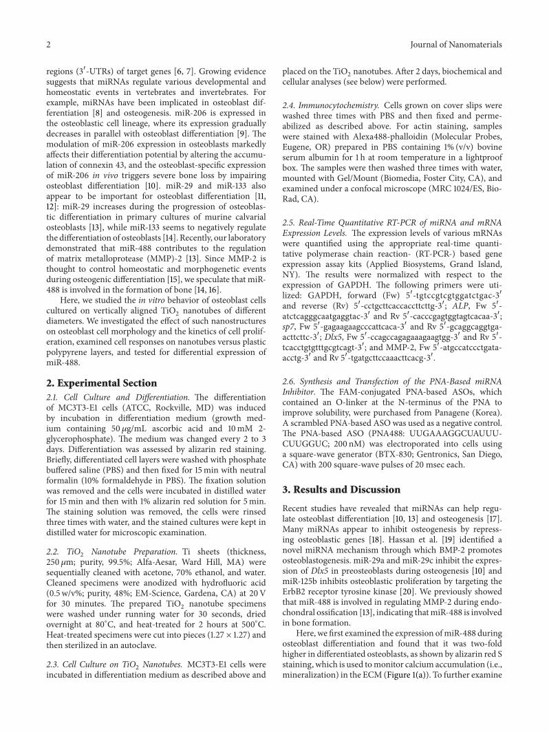
regions (31015840-UTRs) of target genes [6 7] Growing evidencesuggests that miRNAs regulate various developmental andhomeostatic events in vertebrates and invertebrates Forexample miRNAs have been implicated in osteoblast dif-ferentiation [8] and osteogenesis miR-206 is expressed inthe osteoblastic cell lineage where its expression graduallydecreases in parallel with osteoblast differentiation [9] Themodulation of miR-206 expression in osteoblasts markedlyaffects their differentiation potential by altering the accumu-lation of connexin 43 and the osteoblast-specific expressionof miR-206 in vivo triggers severe bone loss by impairingosteoblast differentiation [10] miR-29 and miR-133 alsoappear to be important for osteoblast differentiation [1112] miR-29 increases during the progression of osteoblas-tic differentiation in primary cultures of murine calvarialosteoblasts [13] while miR-133 seems to negatively regulatethe differentiation of osteoblasts [14] Recently our laboratorydemonstrated that miR-488 contributes to the regulationof matrix metalloprotease (MMP)-2 [13] Since MMP-2 isthought to control homeostatic and morphogenetic eventsduring osteogenic differentiation [15] we speculate that miR-488 is involved in the formation of bone [14 16]
Here we studied the in vitro behavior of osteoblast cellscultured on vertically aligned TiO
2nanotubes of different
diameters We investigated the effect of such nanostructureson osteoblast cell morphology and the kinetics of cell prolif-eration examined cell responses on nanotubes versus plasticpolypyrene layers and tested for differential expression ofmiR-488
2 Experimental Section21 Cell Culture and Differentiation The differentiationof MC3T3-E1 cells (ATCC Rockville MD) was inducedby incubation in differentiation medium (growth med-ium containing 50120583gmL ascorbic acid and 10mM 2-glycerophosphate) The medium was changed every 2 to 3days Differentiation was assessed by alizarin red stainingBriefly differentiated cell layers were washed with phosphatebuffered saline (PBS) and then fixed for 15min with neutralformalin (10 formaldehyde in PBS) The fixation solutionwas removed and the cells were incubated in distilled waterfor 15min and then with 1 alizarin red solution for 5minThe staining solution was removed the cells were rinsedthree times with water and the stained cultures were kept indistilled water for microscopic examination
22 TiO2
Nanotube Preparation Ti sheets (thickness250120583m purity 995 Alfa-Aesar Ward Hill MA) weresequentially cleaned with acetone 70 ethanol and waterCleaned specimens were anodized with hydrofluoric acid(05 wv purity 48 EM-Science Gardena CA) at 20Vfor 30 minutes The prepared TiO
2nanotube specimens
were washed under running water for 30 seconds driedovernight at 80∘C and heat-treated for 2 hours at 500∘CHeat-treated specimens were cut into pieces (127 times 127) andthen sterilized in an autoclave
23 Cell Culture on TiO2Nanotubes MC3T3-E1 cells were
incubated in differentiation medium as described above and
placed on the TiO2nanotubes After 2 days biochemical and
cellular analyses (see below) were performed
24 Immunocytochemistry Cells grown on cover slips werewashed three times with PBS and then fixed and perme-abilized as described above For actin staining sampleswere stained with Alexa488-phalloidin (Molecular ProbesEugene OR) prepared in PBS containing 1 (vv) bovineserum albumin for 1 h at room temperature in a lightproofbox The samples were then washed three times with watermounted with GelMount (Biomedia Foster City CA) andexamined under a confocal microscope (MRC 1024ES Bio-Rad CA)
25 Real-Time Quantitative RT-PCR of miRNA and mRNAExpression Levels The expression levels of various mRNAswere quantified using the appropriate real-time quanti-tative polymerase chain reaction- (RT-PCR-) based geneexpression assay kits (Applied Biosystems Grand IslandNY) The results were normalized with respect to theexpression of GAPDH The following primers were uti-lized GAPDH forward (Fw) 51015840-tgtccgtcgtggatctgac-31015840and reverse (Rv) 51015840-cctgcttcaccaccttcttg-31015840 ALP Fw 51015840-atctcagggcaatgaggtac-31015840 and Rv 51015840-cacccgagtggtagtcacaa-31015840sp7 Fw 51015840-gagaagaagcccattcaca-31015840 and Rv 51015840-gcaggcaggtga-acttcttc-31015840 Dlx5 Fw 51015840-ccagccagagaaagaagtgg-31015840 and Rv 51015840-tcacctgtgtttgcgtcagt-31015840 and MMP-2 Fw 51015840-atgccatccctgata-acctg-31015840 and Rv 51015840-tgatgcttccaaacttcacg-31015840
26 Synthesis and Transfection of the PNA-Based miRNAInhibitor The FAM-conjugated PNA-based ASOs whichcontained an O-linker at the N-terminus of the PNA toimprove solubility were purchased from Panagene (Korea)A scrambled PNA-based ASO was used as a negative controlThe PNA-based ASO (PNA488 UUGAAAGGCUAUUU-CUUGGUC 200 nM) was electroporated into cells usinga square-wave generator (BTX-830 Gentronics San DiegoCA) with 200 square-wave pulses of 20 msec each
3 Results and Discussion
Recent studies have revealed that miRNAs can help regu-late osteoblast differentiation [10 13] and osteogenesis [17]Many miRNAs appear to inhibit osteogenesis by repress-ing osteoblastic genes [18] Hassan et al [19] identified anovel miRNA mechanism through which BMP-2 promotesosteoblastogenesis miR-29a and miR-29c inhibit the expres-sion of Dlx5 in preosteoblasts during osteogenesis [10] andmiR-125b inhibits osteoblastic proliferation by targeting theErbB2 receptor tyrosine kinase [20] We previously showedthat miR-488 is involved in regulating MMP-2 during endo-chondral ossification [13] indicating thatmiR-488 is involvedin bone formation
Here we first examined the expression ofmiR-488 duringosteoblast differentiation and found that it was two-foldhigher in differentiated osteoblasts as shown by alizarin red Sstaining which is used tomonitor calcium accumulation (iemineralization) in the ECM (Figure 1(a)) To further examine
Journal of Nanomaterials 3
Relat
ive l
evel
s of m
iR-4
88
0
05
1
15
lowast
1day 3weeks
(a)
Con Anti-miR-488
Rela
tive l
evels
of A
LP(fo
ld o
f Con
)
0
05
1
15
2
25
lowast
(b)
Con
Anti-miR-4883weeks 3weeks6hr
(c)
Figure 1 Induction of miR-488 involved in osteoblastic differentiation of MC3T3-E1 cells grown on culture dish (a)The expression of miR-488 was measured using real-time PCR Alizarin red S stain was used to monitor the mineralization of ECM (inserted photography) Cellswere treated with PNA-based ASOs against miR-488 (anti-miR-488) or a scrambled PNA-based ASO (Con) (b) The expression of ALP wasmeasured using real-time PCR RQ of control culture was used as a control to measure fold change (c) Cells were immunostained for F-actinwith Alexa488-phalloidinThe data shown are representative of at least four independent experimentsThemean is plotted and the error barsrepresent 95 CI (lowerupper limit) lowast119875 lt 0005
the potential involvement of miR-488 in the osteoblasticdifferentiation of MC3T3-E1 cells we suppressed miR-488with PNA-based ASOs MC3T3-E1 cells were treated with200 nM of the anti-miR-488 inhibitor for two days andthe mRNA expression of ALP was determined by real-time PCR The expression of ALP was elevated followingtreatment of cells with the miR-488 inhibitor (Figure 1(b))These data suggest that miR-488 may negatively regulate thedifferentiation of osteoblasts
Cell adhesion is a fundamental process that directlyaffects cell proliferation migration and differentiation and isinvolved in many biological behaviors including the tissueintegration of biomaterials [21] The adhesion of osteoblaststo the implant material surface is essential for the successof any implant that requires osteointegration It has beensuggested that bone cells may produce stress fibers tomaintain their desired morphology in the face of chronicapplied tension Since the fate of osteoblast differentiationmay be determined by cell shapes in vitro we performed
morphological analysis of MC3T3-E1 cells We found that thestress fiber density was higher in cells treated for two dayswith themiR-488 inhibitor compared to cells treated with thescrambled control (Figure 1(c)) A recent study suggested thatRhoA which is known to affect the integrity of actin stressfibers modulates the function and survival of osteoblasts[22] Thus the ability of the miR-488 inhibitor to intensifythe density of stress fibers could suggest that miR-488 isfunctionally relevant in osteoblastic MC3T3-E1 cells
We generated self-assembled amorphous heat-treatedcrystallized anatase-phase TiO
2nanotubes by anodizing Ti
sheets in a phosphate-fluoride electrolyte at 20V with fourdifferent diameters (30 50 70 and 100 nm)Upon their initialformation TiO
2nanotubes typically have an amorphous
structure thereafter the anatase phase was produced byannealing in air at 500∘C for 2 h MC3T3-E1 cells wereinduced to differentiate by incubation with ascorbic acid and2-glycerophosphate and then placed on TiO
2nanotubes or
culture dishes Previously Barthelemi et al [15] demonstrated
4 Journal of Nanomaterials
0
05
1
15
2
25
Cd 705030TiO
Relat
ive l
evel
s of A
LP(fo
ld o
f Cd)
0
2
4
6
8
0
05
1
15
2
25
Relat
ive l
evels
of s
p7(fo
ld o
f Cd)
0
05
1
15
2
25
3
Relat
ive l
evels
of D
lx5
(fold
of C
d)
TiO 705030
Diameter of TiO-nanotube (nm)
Diameter of TiO-nanotube (nm) Diameter of TiO-nanotube (nm)
Diameter of TiO-nanotube (nm)
lowast
lowast
lowast
100nm
100
Cd 705030TiO 100Cd 705030TiO 100
Cd 705030TiO 100
Adhe
rent
cells
(times105)
(a)
TiO
50 nm
70 nm
100 nm
30nm
(b)
Figure 2 Culture on aTiO2nanotube surface affects osteoblastic differentiation ofMC3T3-E1 cellsMC3T3-E1 cells were cultured on titanium
oxide nanotube having various diameters (30 50 70 and 100 nm) titanium oxide (TiO2) or culture dish (Cd) (a)The expression ofALP sp7
and Dlx5 was measured using real-time PCR and cell adhesion was assayed at day 2 RQ of cells on culture dish (Cd) was used as a controlto measure fold change (b) Cells were immunostained for F-actin with Alexa488-phalloidin at day 2 Representative data is shown (119899 = 4)The mean is plotted and the error bars represent 95 CI (lowerupper limit) lowast119875 lt 0005
Journal of Nanomaterials 5
0
1
2
3
Relat
ive l
evel
s of m
iR-4
88
Diameter of TiO-nanotube (nm)
lowast
Cd 705030TiO 100
(a)
The r
elativ
e lev
els o
f ALP
0
05
1
15
2
Con Anti-miR-488
CdTiO
lowast
(b)
0
05
1
15
Diameter of Ti-nanotube (nm)
Anti-miR-488
CdTiO
The r
elativ
e lev
els o
f ALP
lowast
lowast
Sc 705030TiO 100
(c)
Figure 3 miR-488 involved in osteoblastic differentiation of MC3T3-E1 cells on TiO2nanotube surface (a) MC3T3-E1 cells were cultured
on titanium oxide nanotube having various diameters (30 50 70 and 100 nm) titanium oxide (TiO2) or culture dish (Cd) The expression
of miR-488 was measured at day 2 RQ of cells on culture dish (Cd) was used as a control to measure fold change (b) MC3T3-E1 cells werecultured with antisense oligonucleotides of miR-488 (anti-miR-488) or a scrambled PNA-based ASO (Con) ALP expression was measuredusing real-time PCR and cell adhesion was assayed RQ of control cells on culture dish (Cd) was used as a control to measure fold change (c)MC3T3-E1 cells were cultured on titaniumoxide nanotube having various diameters (30 50 70 and 100 nm) titaniumoxide (TiO
2) or culture
dish (Cd) and treated with antisense oligonucleotides of miR-488 (anti-miR-488) or a scrambled PNA-based ASO (Sc) ALP expression wasmeasured using real-time PCR RQ of cells treated with Sc was used as a control to measure fold change Representative data is shown (119899 = 4)The mean is plotted and the error bars represent 95 CI (lowerupper limit) lowast119875 lt 0005
that a TiO2nanotube layer (70 nm diameter) significantly
enhanced osteogenesis-related gene expression bone implantcontact and bone deposition on pure titanium implantsindicating that 70 nm diameter nanotubes are the optimumsize for osseointegration Here we found that the expressionlevels of the osteoblast-differentiation-related transcriptionfactor-encoding genes ALP sp7 and Dlx5 were significantlyincreased on TiO
2nanotubes compared to culture dishes
these levels peaked in cells grown on 50 nm nanotubesand then decreased slightly with increasing tube diameter(Figure 2(a)) The density of stress fibers was also highest incells grown on 50 nm nanotubes (Figure 2(b)) The adhesionof cells was not affected by the nanotube diameter (data notshown)
The expression level of miR-488 was significantly higherin cells grown on 50 nm nanotubes (but not those of
6 Journal of Nanomaterials
The r
elativ
e lev
els o
f MM
P-2
(fold
of C
d)
Diameter (nm)
0
05
1
15
2
25
3
Cd 705030TiO 100
lowast
lowast
lowast
lowast
(a)
Bafilomycin A1Control
GM6001
The r
elat
ive l
evels
of A
LP(fo
ld o
f TiO
)
00
05
10
15
20
25
30
705030 100
lowast
lowast
lowastlowast
lowast
lowast
lowast
(nm)
(b)
Anti-miR-488Con
00
02
04
06
08
10
12
14
16
The r
elativ
e lev
els o
f MM
P-2
(fold
of T
iO)
TiO
lowast
lowast
lowast
lowast
lowast
705030 100
(nm)
(c)
Figure 4 MMP-2 modulation by miR-448 affects osteoblastic differentiation of MC3T3-E1 cells on TiO2nanotube surface (a) MC3T3-E1
cells were cultured on titanium oxide nanotube having various diameters (30 50 70 and 100 nm) titanium oxide (TiO2) or culture dish
(Cd) and the expression of MMP-2 was measured using real-time PCR RQ of cells on culture dish (Cd) was used as a control to measurefold change (b) MC3T3-E1 cells were treated with bafilomycin A1 or GM6001 and the expression of ALP was measured using real-time PCRRQ of cells on flat titanium oxide was used as a control to measure fold change (c) MC3T3-E1 cells were cultured on titanium nanotubehaving various diameters (30 50 70 and 100 nm) and treated with antisense oligonucleotides of miR-488 (anti-miR-488) or a scrambledPNA-based ASO (Con) Expression ofMMP-2was measured RQ of cells on flat titanium oxide was used as a control to measure fold changeRepresentative data is shown (119899 = 4) The mean is plotted and the error bars represent 95 CI (lowerupper limit) lowast119875 lt 0005
Journal of Nanomaterials 7
other diameters) versus those grown on culture dishes(Figure 3(a)) Interestingly cells treated with the anti-miR-488 oligonucleotide inhibitor acted differently on plasticpolypyrene versus TiO
2surfaces the inhibitor induced
ALP transcription in cells grown on culture dishes butslightly reduced ALP transcription in cells grown on TiO
2
(Figure 3(b)) Notably the anti-miR-488 inhibitor suppressedthe transcription of ALP to the greatest degree in cellsplated on 50 nm TiO
2nanotubes (Figure 3(c)) These data
suggest that the nature of the substratum could affect theinternal environment and modulate the differential capacityof osteoblasts
Under normal physiological conditions osteoblasts areable to degrade the organic material of the bone matrixOsteoblast precursors express MMP-2 -8 -13 and -14which may be involved in the degradation of bone matrixmaterials (eg type I collagen) [23] Consistent with thisobservation we found that when the miR-488 inhibitorinduced the differentiation of MC3T3-E1 cells (Figure 1(a))the induction of MMP-2 was also increased (Figure 4(a))However material properties such as surface chemistry andsurface topography are well known to affect cellular behaviorthereby affecting cell fate [24] Here we found that amongcells cultured on TiO
2surfaces of various diameters MMP-
2 expression and ALP activity were significantly increasedon 50 nm nanotubes (Figure 2(a)) In addition we foundthat modulation of MMP-2 activity could affect ALP activityin our system Bafilomycin A1 is known to increase theactivation of proMMP-2 by the abundance of active MT1-MMP on the cell surface [25 26] Here we found thatbafilomycin A1 treatment increased ALP activity in cellsgrown on 50 nm nanotubes In contrast treatment of cellswith 3 120583MGM6001 which has been shown to inhibit MMP-2 activity [27 28] decreased ALP activity among cells grownon various-diameter nanotubes (Figure 4(b)) These datasupport the notion that material properties can affect cellularbehaviors and responses Interestingly the miR-488 inhibitorhad differential effects on MMP-2 expression depending onthe growth substrate it inhibited MMP-2 expression in cellsgrown on 50 nmnanotubes but significantly enhancedMMP-2 expression in cells grown on 70 nmnanotubes (Figure 4(c))
These data collectively suggest that biophysical stimuliand environmental conditions could be important factorsin the clinical use of TiO
2nanotubes Recent studies have
shown that miR-488 is involved in several diseases includingprostate cancer [29] lung cancer [23] and panic disorder[30] For clinical applications of TiO
2nanotubes it may
therefore be necessary to investigate the relationship betweenthe desired implant material and certain patient characteris-tics (eg miR-488 levels)
4 Conclusions
One of the biggest challenges in developing implants withimproved longevity and minimum failure is the control ofsuccessful osseointegration Osteoblasts are critical for theosseointegration of endosseous implants because they syn-thesizeproduce ECM and control its mineralization Here
we explored differences in the osteogenic responses of cellsgrown on 30 nm 50 nm 70 nm and 100 nm TiO
2nanotubes
We found that the 50 nm diameter yielded the best osteoblastdifferentiation (as assessed by ALP expression) and couldtherefore be optimal for implant use However we alsoobtained evidence suggesting that biophysical stimuli andendogenous environmental conditions could also criticallyaffect the behavior of osteoblasts We found that modulationof miR-488 had different effects on cells depending on theirgrowth substrate (ie culture dishes or TiO
2nanotubes of
various diameters) These findings suggest that we need tofully understand the interactions of endogenous biologicalparameters (eg miRNAs) and the physicochemical prop-erties of Ti nanotubes in order to optimize the clinicalapplication of such materials in implant medicine
Abbreviations
ALP Alkaline phosphataseMMP-2 Matrix metalloprotease-2miRNA miR MicroRNATiO2 Titanium oxide
ECM Extracellular matrix
Conflict of Interests
The authors declare that there is no conflict of interestsregarding the publication of this paper
Authorsrsquo Contribution
Yeon-Ho Kang and Bohm Choi contributed equally in thisstudy
Acknowledgments
This study was supported by a Grant fromthe Korean HealthTechnology RampD Project (Ministry of Health amp WelfareRepublic of Korea A120152) and the National ResearchFoundation of Korea (NRF) Grant funded by the Koreangovernment (MSIP) [2011-0030130 and 2013R1A1A2011999]The funders did not play any role in the study design datacollection data analysis decision to publish or preparationof the paper
References
[1] M Lai K Cai L Zhao X Chen Y Hou and Z Yang ldquoSurfacefunctionalization of TiO
2nanotubes with bone morphogenetic
protein 2 and its synergistic effect on the differentiation ofmesenchymal stem cellsrdquo Biomacromolecules vol 12 no 4 pp1097ndash1105 2011
[2] T Miyauchi M Yamada A Yamamoto et al ldquoThe enhancedcharacteristics of osteoblast adhesion to photofunctionalizednanoscale TiO
2layers on biomaterials surfacesrdquo Biomaterials
vol 31 no 14 pp 3827ndash3839 2010[3] R Junker A Dimakis MThoneick and J A Jansen ldquoEffects of
implant surface coatings and composition on bone integration
8 Journal of Nanomaterials
a systematic reviewrdquo Clinical Oral Implants Research vol 20supplement s4 pp 185ndash206 2009
[4] J Park S Bauer P Schmuki and K Von Der Mark ldquoNarrowwindow in nanoscale dependent activation of endothelial cellgrowth and differentiation on TiO
2nanotube surfacesrdquo Nano
Letters vol 9 no 9 pp 3157ndash3164 2009[5] T J Webster R W Siegel and R Bizios ldquoOsteoblast adhesion
on nanophase ceramicsrdquo Biomaterials vol 20 no 13 pp 1221ndash1227 1999
[6] J G Doench C P Petersen and P A Sharp ldquosiRNAs canfunction as miRNAsrdquo Genes and Development vol 17 no 4 pp438ndash442 2003
[7] Y K Kim L Furic L DesGroseillers and L E MaquatldquoMammalian Staufen1 recruits Upf1 to specific mRNA 31015840UTRsso as to elicit mRNA decayrdquo Cell vol 120 no 2 pp 195ndash2082005
[8] K Kapinas and A M Delany ldquoMicroRNA biogenesis andregulation of bone remodelingrdquoArthritis Research andTherapyvol 13 no 3 article 220 2011
[9] Z Q Wang Y Q Lu and J X Han ldquoMicroRNAs importantmediators of ossificationrdquo Chinese Medical Journal (EnglishEdition) vol 125 pp 4111ndash4116 2012
[10] H Inose H Ochi A Kimura et al ldquoA microRNA regulatorymechanism of osteoblast differentiationrdquo Proceedings of theNational Academy of Sciences of the United States of Americavol 106 no 49 pp 20794ndash20799 2009
[11] T Itoh Y Nozawa and Y Akao ldquoMicroRNA-141 and -200a areinvolved in bonemorphogenetic protein-2-inducedmouse pre-osteoblast differentiation by targeting distal-less homeobox 5rdquoJournal of Biological Chemistry vol 284 no 29 pp 19272ndash192792009
[12] Z Li M Q Hassan M Jafferji et al ldquoBiological functionsof miR-29b contribute to positive regulation of osteoblastdifferentiationrdquo Journal of Biological Chemistry vol 284 no 23pp 15676ndash15684 2009
[13] H Li H Xie W Liu et al ldquoA novel microRNA targetingHDAC5 regulates osteoblast differentiation in mice and con-tributes to primary osteoporosis in humansrdquo Journal of ClinicalInvestigation vol 119 no 12 pp 3666ndash3677 2009
[14] J Song D Kim and E-J Jin ldquoMicroRNA-488 suppresses cellmigration through modulation of the focal adhesion activityduring chondrogenic differentiation of chick limb mesenchy-mal cellsrdquo Cell Biology International vol 35 no 2 pp 179ndash1852011
[15] S Barthelemi J Robinet R Garnotel et al ldquoMechanical forces-induced human osteoblasts differentiation involves MMP-2MMP-13MT1-MMP proteolytic cascaderdquo Journal of CellularBiochemistry vol 113 no 3 pp 760ndash772 2012
[16] G S Stein J B Lian and T A Owen ldquoRelationship of cellgrowth to the regulation of tissue-specific gene expressionduring osteoblast differentiationrdquo FASEB Journal vol 4 no 13pp 3111ndash3123 1990
[17] S Gronthos A C W Zannettino S E Graves S Ohta S JHay and P J Simmons ldquoDifferential cell surface expressionof the STRO-1 and alkaline phosphatase antigens on discretedevelopmental stages in primary cultures of human bone cellsrdquoJournal of Bone and Mineral Research vol 14 no 1 pp 47ndash561999
[18] S Dong B Yang H Guo and F Kang ldquoMicroRNAs regulateosteogenesis and chondrogenesisrdquo Biochemical and BiophysicalResearch Communications vol 418 no 4 pp 587ndash591 2012
[19] M Q Hassan J A R Gordon M M Beloti et al ldquoA networkconnecting Runx2 SATB2 and the miR-23asim27asim24-2 clusterregulates the osteoblast differentiation programrdquo Proceedings ofthe National Academy of Sciences of the United States of Americavol 107 no 46 pp 19879ndash19884 2010
[20] Z Li M Q Hassan S Volinia et al ldquoA microRNA signaturefor a BMP2-induced osteoblast lineage commitment programrdquoProceedings of the National Academy of Sciences of the UnitedStates of America vol 105 no 37 pp 13906ndash13911 2008
[21] M Mizuno H Kawamura N Takei and H Nawa ldquoTheanthraquinone derivative Emodin ameliorates neurobehavioraldeficits of a rodent model for schizophreniardquo Journal of NeuralTransmission vol 115 no 3 pp 521ndash530 2008
[22] M Dettin M T Conconi R Gambaretto et al ldquoEffect ofsynthetic peptides on osteoblast adhesionrdquoBiomaterials vol 26no 22 pp 4507ndash4515 2005
[23] N H Kazmers S A Ma T Yoshida and P H Stern ldquoRhoGTPase signaling and PTH 3-34 but not PTH 1-34 maintainthe actin cytoskeleton and antagonize bisphosphonate effects inmouse osteoblasticMC3T3-E1 cellsrdquo Bone vol 45 no 1 pp 52ndash60 2009
[24] K Anselme P Linez M Bigerelle et al ldquoThe relative influenceof the topography and chemistry of TiAl6V4 surfaces onosteoblastic cell behaviourrdquo Biomaterials vol 21 no 15 pp1567ndash1577 2000
[25] S Hernandez-Barrantes M Toth M M Bernardo et alldquoBinding of active (57 kDa) membrane type 1-matrix metallo-proteinase (MT1-MMP) to tissue inhibitor of metalloproteinase(TIMP)-2 regulates MT1-MMP processing and pro-MMP-2activationrdquo Journal of Biological Chemistry vol 275 no 16 pp12080ndash12089 2000
[26] E Maquoi A Noel F Frankenne H Angliker G Murphyand J-M Foidart ldquoInhibition of matrix metalloproteinase2 maturation and HT1080 invasiveness by a synthetic furininhibitorrdquo FEBS Letters vol 424 no 3 pp 262ndash266 1998
[27] W Zhou B O Ibe and J U Raj ldquoPlatelet-activating fac-tor induces ovine fetal pulmonary venous smooth musclecell proliferation role of epidermal growth factor receptortransactivationrdquo American Journal of PhysiologymdashHeart andCirculatory Physiology vol 292 no 6 pp H2773ndashH2781 2007
[28] Y Macotela M B Aguilar J Guzman-Morales et al ldquoMatrixmetalloproteases from chondrocytes generate an antiangio-genic 16 kDa prolactinrdquo Journal of Cell Science vol 119 no 9pp 1790ndash1800 2006
[29] K Sikand J E Slaibi R Singh S D Slane and G C ShuklaldquoMiR 488lowast inhibits androgen receptor expression in prostatecarcinoma cellsrdquo International Journal of Cancer vol 129 no4 pp 810ndash819 2011
[30] M Muinos-Gimeno Y Espinosa-Parrilla M Guidi et alldquoHuman microRNAs miR-22 miR-138-2 miR-148a and miR-488 are associatedwith panic disorder and regulate several anxi-ety candidate genes and related pathwaysrdquo Biological Psychiatryvol 69 no 6 pp 526ndash533 2011
Submit your manuscripts athttpwwwhindawicom
ScientificaHindawi Publishing Corporationhttpwwwhindawicom Volume 2014
CorrosionInternational Journal of
Hindawi Publishing Corporationhttpwwwhindawicom Volume 2014
Polymer ScienceInternational Journal of
Hindawi Publishing Corporationhttpwwwhindawicom Volume 2014
Hindawi Publishing Corporationhttpwwwhindawicom Volume 2014
CeramicsJournal of
Hindawi Publishing Corporationhttpwwwhindawicom Volume 2014
CompositesJournal of
NanoparticlesJournal of
Hindawi Publishing Corporationhttpwwwhindawicom Volume 2014
Hindawi Publishing Corporationhttpwwwhindawicom Volume 2014
International Journal of
Biomaterials
Hindawi Publishing Corporationhttpwwwhindawicom Volume 2014
NanoscienceJournal of
TextilesHindawi Publishing Corporation httpwwwhindawicom Volume 2014
Journal of
NanotechnologyHindawi Publishing Corporationhttpwwwhindawicom Volume 2014
Journal of
CrystallographyJournal of
Hindawi Publishing Corporationhttpwwwhindawicom Volume 2014
The Scientific World JournalHindawi Publishing Corporation httpwwwhindawicom Volume 2014
Hindawi Publishing Corporationhttpwwwhindawicom Volume 2014
CoatingsJournal of
Advances in
Materials Science and EngineeringHindawi Publishing Corporationhttpwwwhindawicom Volume 2014
Smart Materials Research
Hindawi Publishing Corporationhttpwwwhindawicom Volume 2014
Hindawi Publishing Corporationhttpwwwhindawicom Volume 2014
MetallurgyJournal of
Hindawi Publishing Corporationhttpwwwhindawicom Volume 2014
BioMed Research International
MaterialsJournal of
Hindawi Publishing Corporationhttpwwwhindawicom Volume 2014
Nano
materials
Hindawi Publishing Corporationhttpwwwhindawicom Volume 2014
Journal ofNanomaterials
2 Journal of Nanomaterials
regions (31015840-UTRs) of target genes [6 7] Growing evidencesuggests that miRNAs regulate various developmental andhomeostatic events in vertebrates and invertebrates Forexample miRNAs have been implicated in osteoblast dif-ferentiation [8] and osteogenesis miR-206 is expressed inthe osteoblastic cell lineage where its expression graduallydecreases in parallel with osteoblast differentiation [9] Themodulation of miR-206 expression in osteoblasts markedlyaffects their differentiation potential by altering the accumu-lation of connexin 43 and the osteoblast-specific expressionof miR-206 in vivo triggers severe bone loss by impairingosteoblast differentiation [10] miR-29 and miR-133 alsoappear to be important for osteoblast differentiation [1112] miR-29 increases during the progression of osteoblas-tic differentiation in primary cultures of murine calvarialosteoblasts [13] while miR-133 seems to negatively regulatethe differentiation of osteoblasts [14] Recently our laboratorydemonstrated that miR-488 contributes to the regulationof matrix metalloprotease (MMP)-2 [13] Since MMP-2 isthought to control homeostatic and morphogenetic eventsduring osteogenic differentiation [15] we speculate that miR-488 is involved in the formation of bone [14 16]
Here we studied the in vitro behavior of osteoblast cellscultured on vertically aligned TiO
2nanotubes of different
diameters We investigated the effect of such nanostructureson osteoblast cell morphology and the kinetics of cell prolif-eration examined cell responses on nanotubes versus plasticpolypyrene layers and tested for differential expression ofmiR-488
2 Experimental Section21 Cell Culture and Differentiation The differentiationof MC3T3-E1 cells (ATCC Rockville MD) was inducedby incubation in differentiation medium (growth med-ium containing 50120583gmL ascorbic acid and 10mM 2-glycerophosphate) The medium was changed every 2 to 3days Differentiation was assessed by alizarin red stainingBriefly differentiated cell layers were washed with phosphatebuffered saline (PBS) and then fixed for 15min with neutralformalin (10 formaldehyde in PBS) The fixation solutionwas removed and the cells were incubated in distilled waterfor 15min and then with 1 alizarin red solution for 5minThe staining solution was removed the cells were rinsedthree times with water and the stained cultures were kept indistilled water for microscopic examination
22 TiO2
Nanotube Preparation Ti sheets (thickness250120583m purity 995 Alfa-Aesar Ward Hill MA) weresequentially cleaned with acetone 70 ethanol and waterCleaned specimens were anodized with hydrofluoric acid(05 wv purity 48 EM-Science Gardena CA) at 20Vfor 30 minutes The prepared TiO
2nanotube specimens
were washed under running water for 30 seconds driedovernight at 80∘C and heat-treated for 2 hours at 500∘CHeat-treated specimens were cut into pieces (127 times 127) andthen sterilized in an autoclave
23 Cell Culture on TiO2Nanotubes MC3T3-E1 cells were
incubated in differentiation medium as described above and
placed on the TiO2nanotubes After 2 days biochemical and
cellular analyses (see below) were performed
24 Immunocytochemistry Cells grown on cover slips werewashed three times with PBS and then fixed and perme-abilized as described above For actin staining sampleswere stained with Alexa488-phalloidin (Molecular ProbesEugene OR) prepared in PBS containing 1 (vv) bovineserum albumin for 1 h at room temperature in a lightproofbox The samples were then washed three times with watermounted with GelMount (Biomedia Foster City CA) andexamined under a confocal microscope (MRC 1024ES Bio-Rad CA)
25 Real-Time Quantitative RT-PCR of miRNA and mRNAExpression Levels The expression levels of various mRNAswere quantified using the appropriate real-time quanti-tative polymerase chain reaction- (RT-PCR-) based geneexpression assay kits (Applied Biosystems Grand IslandNY) The results were normalized with respect to theexpression of GAPDH The following primers were uti-lized GAPDH forward (Fw) 51015840-tgtccgtcgtggatctgac-31015840and reverse (Rv) 51015840-cctgcttcaccaccttcttg-31015840 ALP Fw 51015840-atctcagggcaatgaggtac-31015840 and Rv 51015840-cacccgagtggtagtcacaa-31015840sp7 Fw 51015840-gagaagaagcccattcaca-31015840 and Rv 51015840-gcaggcaggtga-acttcttc-31015840 Dlx5 Fw 51015840-ccagccagagaaagaagtgg-31015840 and Rv 51015840-tcacctgtgtttgcgtcagt-31015840 and MMP-2 Fw 51015840-atgccatccctgata-acctg-31015840 and Rv 51015840-tgatgcttccaaacttcacg-31015840
26 Synthesis and Transfection of the PNA-Based miRNAInhibitor The FAM-conjugated PNA-based ASOs whichcontained an O-linker at the N-terminus of the PNA toimprove solubility were purchased from Panagene (Korea)A scrambled PNA-based ASO was used as a negative controlThe PNA-based ASO (PNA488 UUGAAAGGCUAUUU-CUUGGUC 200 nM) was electroporated into cells usinga square-wave generator (BTX-830 Gentronics San DiegoCA) with 200 square-wave pulses of 20 msec each
3 Results and Discussion
Recent studies have revealed that miRNAs can help regu-late osteoblast differentiation [10 13] and osteogenesis [17]Many miRNAs appear to inhibit osteogenesis by repress-ing osteoblastic genes [18] Hassan et al [19] identified anovel miRNA mechanism through which BMP-2 promotesosteoblastogenesis miR-29a and miR-29c inhibit the expres-sion of Dlx5 in preosteoblasts during osteogenesis [10] andmiR-125b inhibits osteoblastic proliferation by targeting theErbB2 receptor tyrosine kinase [20] We previously showedthat miR-488 is involved in regulating MMP-2 during endo-chondral ossification [13] indicating thatmiR-488 is involvedin bone formation
Here we first examined the expression ofmiR-488 duringosteoblast differentiation and found that it was two-foldhigher in differentiated osteoblasts as shown by alizarin red Sstaining which is used tomonitor calcium accumulation (iemineralization) in the ECM (Figure 1(a)) To further examine
Journal of Nanomaterials 3
Relat
ive l
evel
s of m
iR-4
88
0
05
1
15
lowast
1day 3weeks
(a)
Con Anti-miR-488
Rela
tive l
evels
of A
LP(fo
ld o
f Con
)
0
05
1
15
2
25
lowast
(b)
Con
Anti-miR-4883weeks 3weeks6hr
(c)
Figure 1 Induction of miR-488 involved in osteoblastic differentiation of MC3T3-E1 cells grown on culture dish (a)The expression of miR-488 was measured using real-time PCR Alizarin red S stain was used to monitor the mineralization of ECM (inserted photography) Cellswere treated with PNA-based ASOs against miR-488 (anti-miR-488) or a scrambled PNA-based ASO (Con) (b) The expression of ALP wasmeasured using real-time PCR RQ of control culture was used as a control to measure fold change (c) Cells were immunostained for F-actinwith Alexa488-phalloidinThe data shown are representative of at least four independent experimentsThemean is plotted and the error barsrepresent 95 CI (lowerupper limit) lowast119875 lt 0005
the potential involvement of miR-488 in the osteoblasticdifferentiation of MC3T3-E1 cells we suppressed miR-488with PNA-based ASOs MC3T3-E1 cells were treated with200 nM of the anti-miR-488 inhibitor for two days andthe mRNA expression of ALP was determined by real-time PCR The expression of ALP was elevated followingtreatment of cells with the miR-488 inhibitor (Figure 1(b))These data suggest that miR-488 may negatively regulate thedifferentiation of osteoblasts
Cell adhesion is a fundamental process that directlyaffects cell proliferation migration and differentiation and isinvolved in many biological behaviors including the tissueintegration of biomaterials [21] The adhesion of osteoblaststo the implant material surface is essential for the successof any implant that requires osteointegration It has beensuggested that bone cells may produce stress fibers tomaintain their desired morphology in the face of chronicapplied tension Since the fate of osteoblast differentiationmay be determined by cell shapes in vitro we performed
morphological analysis of MC3T3-E1 cells We found that thestress fiber density was higher in cells treated for two dayswith themiR-488 inhibitor compared to cells treated with thescrambled control (Figure 1(c)) A recent study suggested thatRhoA which is known to affect the integrity of actin stressfibers modulates the function and survival of osteoblasts[22] Thus the ability of the miR-488 inhibitor to intensifythe density of stress fibers could suggest that miR-488 isfunctionally relevant in osteoblastic MC3T3-E1 cells
We generated self-assembled amorphous heat-treatedcrystallized anatase-phase TiO
2nanotubes by anodizing Ti
sheets in a phosphate-fluoride electrolyte at 20V with fourdifferent diameters (30 50 70 and 100 nm)Upon their initialformation TiO
2nanotubes typically have an amorphous
structure thereafter the anatase phase was produced byannealing in air at 500∘C for 2 h MC3T3-E1 cells wereinduced to differentiate by incubation with ascorbic acid and2-glycerophosphate and then placed on TiO
2nanotubes or
culture dishes Previously Barthelemi et al [15] demonstrated
4 Journal of Nanomaterials
0
05
1
15
2
25
Cd 705030TiO
Relat
ive l
evel
s of A
LP(fo
ld o
f Cd)
0
2
4
6
8
0
05
1
15
2
25
Relat
ive l
evels
of s
p7(fo
ld o
f Cd)
0
05
1
15
2
25
3
Relat
ive l
evels
of D
lx5
(fold
of C
d)
TiO 705030
Diameter of TiO-nanotube (nm)
Diameter of TiO-nanotube (nm) Diameter of TiO-nanotube (nm)
Diameter of TiO-nanotube (nm)
lowast
lowast
lowast
100nm
100
Cd 705030TiO 100Cd 705030TiO 100
Cd 705030TiO 100
Adhe
rent
cells
(times105)
(a)
TiO
50 nm
70 nm
100 nm
30nm
(b)
Figure 2 Culture on aTiO2nanotube surface affects osteoblastic differentiation ofMC3T3-E1 cellsMC3T3-E1 cells were cultured on titanium
oxide nanotube having various diameters (30 50 70 and 100 nm) titanium oxide (TiO2) or culture dish (Cd) (a)The expression ofALP sp7
and Dlx5 was measured using real-time PCR and cell adhesion was assayed at day 2 RQ of cells on culture dish (Cd) was used as a controlto measure fold change (b) Cells were immunostained for F-actin with Alexa488-phalloidin at day 2 Representative data is shown (119899 = 4)The mean is plotted and the error bars represent 95 CI (lowerupper limit) lowast119875 lt 0005
Journal of Nanomaterials 5
0
1
2
3
Relat
ive l
evel
s of m
iR-4
88
Diameter of TiO-nanotube (nm)
lowast
Cd 705030TiO 100
(a)
The r
elativ
e lev
els o
f ALP
0
05
1
15
2
Con Anti-miR-488
CdTiO
lowast
(b)
0
05
1
15
Diameter of Ti-nanotube (nm)
Anti-miR-488
CdTiO
The r
elativ
e lev
els o
f ALP
lowast
lowast
Sc 705030TiO 100
(c)
Figure 3 miR-488 involved in osteoblastic differentiation of MC3T3-E1 cells on TiO2nanotube surface (a) MC3T3-E1 cells were cultured
on titanium oxide nanotube having various diameters (30 50 70 and 100 nm) titanium oxide (TiO2) or culture dish (Cd) The expression
of miR-488 was measured at day 2 RQ of cells on culture dish (Cd) was used as a control to measure fold change (b) MC3T3-E1 cells werecultured with antisense oligonucleotides of miR-488 (anti-miR-488) or a scrambled PNA-based ASO (Con) ALP expression was measuredusing real-time PCR and cell adhesion was assayed RQ of control cells on culture dish (Cd) was used as a control to measure fold change (c)MC3T3-E1 cells were cultured on titaniumoxide nanotube having various diameters (30 50 70 and 100 nm) titaniumoxide (TiO
2) or culture
dish (Cd) and treated with antisense oligonucleotides of miR-488 (anti-miR-488) or a scrambled PNA-based ASO (Sc) ALP expression wasmeasured using real-time PCR RQ of cells treated with Sc was used as a control to measure fold change Representative data is shown (119899 = 4)The mean is plotted and the error bars represent 95 CI (lowerupper limit) lowast119875 lt 0005
that a TiO2nanotube layer (70 nm diameter) significantly
enhanced osteogenesis-related gene expression bone implantcontact and bone deposition on pure titanium implantsindicating that 70 nm diameter nanotubes are the optimumsize for osseointegration Here we found that the expressionlevels of the osteoblast-differentiation-related transcriptionfactor-encoding genes ALP sp7 and Dlx5 were significantlyincreased on TiO
2nanotubes compared to culture dishes
these levels peaked in cells grown on 50 nm nanotubesand then decreased slightly with increasing tube diameter(Figure 2(a)) The density of stress fibers was also highest incells grown on 50 nm nanotubes (Figure 2(b)) The adhesionof cells was not affected by the nanotube diameter (data notshown)
The expression level of miR-488 was significantly higherin cells grown on 50 nm nanotubes (but not those of
6 Journal of Nanomaterials
The r
elativ
e lev
els o
f MM
P-2
(fold
of C
d)
Diameter (nm)
0
05
1
15
2
25
3
Cd 705030TiO 100
lowast
lowast
lowast
lowast
(a)
Bafilomycin A1Control
GM6001
The r
elat
ive l
evels
of A
LP(fo
ld o
f TiO
)
00
05
10
15
20
25
30
705030 100
lowast
lowast
lowastlowast
lowast
lowast
lowast
(nm)
(b)
Anti-miR-488Con
00
02
04
06
08
10
12
14
16
The r
elativ
e lev
els o
f MM
P-2
(fold
of T
iO)
TiO
lowast
lowast
lowast
lowast
lowast
705030 100
(nm)
(c)
Figure 4 MMP-2 modulation by miR-448 affects osteoblastic differentiation of MC3T3-E1 cells on TiO2nanotube surface (a) MC3T3-E1
cells were cultured on titanium oxide nanotube having various diameters (30 50 70 and 100 nm) titanium oxide (TiO2) or culture dish
(Cd) and the expression of MMP-2 was measured using real-time PCR RQ of cells on culture dish (Cd) was used as a control to measurefold change (b) MC3T3-E1 cells were treated with bafilomycin A1 or GM6001 and the expression of ALP was measured using real-time PCRRQ of cells on flat titanium oxide was used as a control to measure fold change (c) MC3T3-E1 cells were cultured on titanium nanotubehaving various diameters (30 50 70 and 100 nm) and treated with antisense oligonucleotides of miR-488 (anti-miR-488) or a scrambledPNA-based ASO (Con) Expression ofMMP-2was measured RQ of cells on flat titanium oxide was used as a control to measure fold changeRepresentative data is shown (119899 = 4) The mean is plotted and the error bars represent 95 CI (lowerupper limit) lowast119875 lt 0005
Journal of Nanomaterials 7
other diameters) versus those grown on culture dishes(Figure 3(a)) Interestingly cells treated with the anti-miR-488 oligonucleotide inhibitor acted differently on plasticpolypyrene versus TiO
2surfaces the inhibitor induced
ALP transcription in cells grown on culture dishes butslightly reduced ALP transcription in cells grown on TiO
2
(Figure 3(b)) Notably the anti-miR-488 inhibitor suppressedthe transcription of ALP to the greatest degree in cellsplated on 50 nm TiO
2nanotubes (Figure 3(c)) These data
suggest that the nature of the substratum could affect theinternal environment and modulate the differential capacityof osteoblasts
Under normal physiological conditions osteoblasts areable to degrade the organic material of the bone matrixOsteoblast precursors express MMP-2 -8 -13 and -14which may be involved in the degradation of bone matrixmaterials (eg type I collagen) [23] Consistent with thisobservation we found that when the miR-488 inhibitorinduced the differentiation of MC3T3-E1 cells (Figure 1(a))the induction of MMP-2 was also increased (Figure 4(a))However material properties such as surface chemistry andsurface topography are well known to affect cellular behaviorthereby affecting cell fate [24] Here we found that amongcells cultured on TiO
2surfaces of various diameters MMP-
2 expression and ALP activity were significantly increasedon 50 nm nanotubes (Figure 2(a)) In addition we foundthat modulation of MMP-2 activity could affect ALP activityin our system Bafilomycin A1 is known to increase theactivation of proMMP-2 by the abundance of active MT1-MMP on the cell surface [25 26] Here we found thatbafilomycin A1 treatment increased ALP activity in cellsgrown on 50 nm nanotubes In contrast treatment of cellswith 3 120583MGM6001 which has been shown to inhibit MMP-2 activity [27 28] decreased ALP activity among cells grownon various-diameter nanotubes (Figure 4(b)) These datasupport the notion that material properties can affect cellularbehaviors and responses Interestingly the miR-488 inhibitorhad differential effects on MMP-2 expression depending onthe growth substrate it inhibited MMP-2 expression in cellsgrown on 50 nmnanotubes but significantly enhancedMMP-2 expression in cells grown on 70 nmnanotubes (Figure 4(c))
These data collectively suggest that biophysical stimuliand environmental conditions could be important factorsin the clinical use of TiO
2nanotubes Recent studies have
shown that miR-488 is involved in several diseases includingprostate cancer [29] lung cancer [23] and panic disorder[30] For clinical applications of TiO
2nanotubes it may
therefore be necessary to investigate the relationship betweenthe desired implant material and certain patient characteris-tics (eg miR-488 levels)
4 Conclusions
One of the biggest challenges in developing implants withimproved longevity and minimum failure is the control ofsuccessful osseointegration Osteoblasts are critical for theosseointegration of endosseous implants because they syn-thesizeproduce ECM and control its mineralization Here
we explored differences in the osteogenic responses of cellsgrown on 30 nm 50 nm 70 nm and 100 nm TiO
2nanotubes
We found that the 50 nm diameter yielded the best osteoblastdifferentiation (as assessed by ALP expression) and couldtherefore be optimal for implant use However we alsoobtained evidence suggesting that biophysical stimuli andendogenous environmental conditions could also criticallyaffect the behavior of osteoblasts We found that modulationof miR-488 had different effects on cells depending on theirgrowth substrate (ie culture dishes or TiO
2nanotubes of
various diameters) These findings suggest that we need tofully understand the interactions of endogenous biologicalparameters (eg miRNAs) and the physicochemical prop-erties of Ti nanotubes in order to optimize the clinicalapplication of such materials in implant medicine
Abbreviations
ALP Alkaline phosphataseMMP-2 Matrix metalloprotease-2miRNA miR MicroRNATiO2 Titanium oxide
ECM Extracellular matrix
Conflict of Interests
The authors declare that there is no conflict of interestsregarding the publication of this paper
Authorsrsquo Contribution
Yeon-Ho Kang and Bohm Choi contributed equally in thisstudy
Acknowledgments
This study was supported by a Grant fromthe Korean HealthTechnology RampD Project (Ministry of Health amp WelfareRepublic of Korea A120152) and the National ResearchFoundation of Korea (NRF) Grant funded by the Koreangovernment (MSIP) [2011-0030130 and 2013R1A1A2011999]The funders did not play any role in the study design datacollection data analysis decision to publish or preparationof the paper
References
[1] M Lai K Cai L Zhao X Chen Y Hou and Z Yang ldquoSurfacefunctionalization of TiO
2nanotubes with bone morphogenetic
protein 2 and its synergistic effect on the differentiation ofmesenchymal stem cellsrdquo Biomacromolecules vol 12 no 4 pp1097ndash1105 2011
[2] T Miyauchi M Yamada A Yamamoto et al ldquoThe enhancedcharacteristics of osteoblast adhesion to photofunctionalizednanoscale TiO
2layers on biomaterials surfacesrdquo Biomaterials
vol 31 no 14 pp 3827ndash3839 2010[3] R Junker A Dimakis MThoneick and J A Jansen ldquoEffects of
implant surface coatings and composition on bone integration
8 Journal of Nanomaterials
a systematic reviewrdquo Clinical Oral Implants Research vol 20supplement s4 pp 185ndash206 2009
[4] J Park S Bauer P Schmuki and K Von Der Mark ldquoNarrowwindow in nanoscale dependent activation of endothelial cellgrowth and differentiation on TiO
2nanotube surfacesrdquo Nano
Letters vol 9 no 9 pp 3157ndash3164 2009[5] T J Webster R W Siegel and R Bizios ldquoOsteoblast adhesion
on nanophase ceramicsrdquo Biomaterials vol 20 no 13 pp 1221ndash1227 1999
[6] J G Doench C P Petersen and P A Sharp ldquosiRNAs canfunction as miRNAsrdquo Genes and Development vol 17 no 4 pp438ndash442 2003
[7] Y K Kim L Furic L DesGroseillers and L E MaquatldquoMammalian Staufen1 recruits Upf1 to specific mRNA 31015840UTRsso as to elicit mRNA decayrdquo Cell vol 120 no 2 pp 195ndash2082005
[8] K Kapinas and A M Delany ldquoMicroRNA biogenesis andregulation of bone remodelingrdquoArthritis Research andTherapyvol 13 no 3 article 220 2011
[9] Z Q Wang Y Q Lu and J X Han ldquoMicroRNAs importantmediators of ossificationrdquo Chinese Medical Journal (EnglishEdition) vol 125 pp 4111ndash4116 2012
[10] H Inose H Ochi A Kimura et al ldquoA microRNA regulatorymechanism of osteoblast differentiationrdquo Proceedings of theNational Academy of Sciences of the United States of Americavol 106 no 49 pp 20794ndash20799 2009
[11] T Itoh Y Nozawa and Y Akao ldquoMicroRNA-141 and -200a areinvolved in bonemorphogenetic protein-2-inducedmouse pre-osteoblast differentiation by targeting distal-less homeobox 5rdquoJournal of Biological Chemistry vol 284 no 29 pp 19272ndash192792009
[12] Z Li M Q Hassan M Jafferji et al ldquoBiological functionsof miR-29b contribute to positive regulation of osteoblastdifferentiationrdquo Journal of Biological Chemistry vol 284 no 23pp 15676ndash15684 2009
[13] H Li H Xie W Liu et al ldquoA novel microRNA targetingHDAC5 regulates osteoblast differentiation in mice and con-tributes to primary osteoporosis in humansrdquo Journal of ClinicalInvestigation vol 119 no 12 pp 3666ndash3677 2009
[14] J Song D Kim and E-J Jin ldquoMicroRNA-488 suppresses cellmigration through modulation of the focal adhesion activityduring chondrogenic differentiation of chick limb mesenchy-mal cellsrdquo Cell Biology International vol 35 no 2 pp 179ndash1852011
[15] S Barthelemi J Robinet R Garnotel et al ldquoMechanical forces-induced human osteoblasts differentiation involves MMP-2MMP-13MT1-MMP proteolytic cascaderdquo Journal of CellularBiochemistry vol 113 no 3 pp 760ndash772 2012
[16] G S Stein J B Lian and T A Owen ldquoRelationship of cellgrowth to the regulation of tissue-specific gene expressionduring osteoblast differentiationrdquo FASEB Journal vol 4 no 13pp 3111ndash3123 1990
[17] S Gronthos A C W Zannettino S E Graves S Ohta S JHay and P J Simmons ldquoDifferential cell surface expressionof the STRO-1 and alkaline phosphatase antigens on discretedevelopmental stages in primary cultures of human bone cellsrdquoJournal of Bone and Mineral Research vol 14 no 1 pp 47ndash561999
[18] S Dong B Yang H Guo and F Kang ldquoMicroRNAs regulateosteogenesis and chondrogenesisrdquo Biochemical and BiophysicalResearch Communications vol 418 no 4 pp 587ndash591 2012
[19] M Q Hassan J A R Gordon M M Beloti et al ldquoA networkconnecting Runx2 SATB2 and the miR-23asim27asim24-2 clusterregulates the osteoblast differentiation programrdquo Proceedings ofthe National Academy of Sciences of the United States of Americavol 107 no 46 pp 19879ndash19884 2010
[20] Z Li M Q Hassan S Volinia et al ldquoA microRNA signaturefor a BMP2-induced osteoblast lineage commitment programrdquoProceedings of the National Academy of Sciences of the UnitedStates of America vol 105 no 37 pp 13906ndash13911 2008
[21] M Mizuno H Kawamura N Takei and H Nawa ldquoTheanthraquinone derivative Emodin ameliorates neurobehavioraldeficits of a rodent model for schizophreniardquo Journal of NeuralTransmission vol 115 no 3 pp 521ndash530 2008
[22] M Dettin M T Conconi R Gambaretto et al ldquoEffect ofsynthetic peptides on osteoblast adhesionrdquoBiomaterials vol 26no 22 pp 4507ndash4515 2005
[23] N H Kazmers S A Ma T Yoshida and P H Stern ldquoRhoGTPase signaling and PTH 3-34 but not PTH 1-34 maintainthe actin cytoskeleton and antagonize bisphosphonate effects inmouse osteoblasticMC3T3-E1 cellsrdquo Bone vol 45 no 1 pp 52ndash60 2009
[24] K Anselme P Linez M Bigerelle et al ldquoThe relative influenceof the topography and chemistry of TiAl6V4 surfaces onosteoblastic cell behaviourrdquo Biomaterials vol 21 no 15 pp1567ndash1577 2000
[25] S Hernandez-Barrantes M Toth M M Bernardo et alldquoBinding of active (57 kDa) membrane type 1-matrix metallo-proteinase (MT1-MMP) to tissue inhibitor of metalloproteinase(TIMP)-2 regulates MT1-MMP processing and pro-MMP-2activationrdquo Journal of Biological Chemistry vol 275 no 16 pp12080ndash12089 2000
[26] E Maquoi A Noel F Frankenne H Angliker G Murphyand J-M Foidart ldquoInhibition of matrix metalloproteinase2 maturation and HT1080 invasiveness by a synthetic furininhibitorrdquo FEBS Letters vol 424 no 3 pp 262ndash266 1998
[27] W Zhou B O Ibe and J U Raj ldquoPlatelet-activating fac-tor induces ovine fetal pulmonary venous smooth musclecell proliferation role of epidermal growth factor receptortransactivationrdquo American Journal of PhysiologymdashHeart andCirculatory Physiology vol 292 no 6 pp H2773ndashH2781 2007
[28] Y Macotela M B Aguilar J Guzman-Morales et al ldquoMatrixmetalloproteases from chondrocytes generate an antiangio-genic 16 kDa prolactinrdquo Journal of Cell Science vol 119 no 9pp 1790ndash1800 2006
[29] K Sikand J E Slaibi R Singh S D Slane and G C ShuklaldquoMiR 488lowast inhibits androgen receptor expression in prostatecarcinoma cellsrdquo International Journal of Cancer vol 129 no4 pp 810ndash819 2011
[30] M Muinos-Gimeno Y Espinosa-Parrilla M Guidi et alldquoHuman microRNAs miR-22 miR-138-2 miR-148a and miR-488 are associatedwith panic disorder and regulate several anxi-ety candidate genes and related pathwaysrdquo Biological Psychiatryvol 69 no 6 pp 526ndash533 2011
Submit your manuscripts athttpwwwhindawicom
ScientificaHindawi Publishing Corporationhttpwwwhindawicom Volume 2014
CorrosionInternational Journal of
Hindawi Publishing Corporationhttpwwwhindawicom Volume 2014
Polymer ScienceInternational Journal of
Hindawi Publishing Corporationhttpwwwhindawicom Volume 2014
Hindawi Publishing Corporationhttpwwwhindawicom Volume 2014
CeramicsJournal of
Hindawi Publishing Corporationhttpwwwhindawicom Volume 2014
CompositesJournal of
NanoparticlesJournal of
Hindawi Publishing Corporationhttpwwwhindawicom Volume 2014
Hindawi Publishing Corporationhttpwwwhindawicom Volume 2014
International Journal of
Biomaterials
Hindawi Publishing Corporationhttpwwwhindawicom Volume 2014
NanoscienceJournal of
TextilesHindawi Publishing Corporation httpwwwhindawicom Volume 2014
Journal of
NanotechnologyHindawi Publishing Corporationhttpwwwhindawicom Volume 2014
Journal of
CrystallographyJournal of
Hindawi Publishing Corporationhttpwwwhindawicom Volume 2014
The Scientific World JournalHindawi Publishing Corporation httpwwwhindawicom Volume 2014
Hindawi Publishing Corporationhttpwwwhindawicom Volume 2014
CoatingsJournal of
Advances in
Materials Science and EngineeringHindawi Publishing Corporationhttpwwwhindawicom Volume 2014
Smart Materials Research
Hindawi Publishing Corporationhttpwwwhindawicom Volume 2014
Hindawi Publishing Corporationhttpwwwhindawicom Volume 2014
MetallurgyJournal of
Hindawi Publishing Corporationhttpwwwhindawicom Volume 2014
BioMed Research International
MaterialsJournal of
Hindawi Publishing Corporationhttpwwwhindawicom Volume 2014
Nano
materials
Hindawi Publishing Corporationhttpwwwhindawicom Volume 2014
Journal ofNanomaterials