research article the potential spread of mycobacterium...
TRANSCRIPT

Research ArticleThe Potential Spread of Mycobacterium tuberculosis into theEnvironment in the Creation of Spondylitis Tuberculosis Rabbit
Ahmad Jabir Rahyussalim1 Tri Kurniawati2 Andriansjah Rukmana3 and Arni Diana Fitri4
1Orthopaedic and Traumatology Department Faculty of Medicine Cipto Mangunkusumo Hospital University of IndonesiaJakarta 10430 Indonesia2Integrated Service Unit of Stem Cell Medical Technology Cipto Mangunkusumo Hospital Jakarta 10430 Indonesia3Microbiology Department Faculty of Medicine University of Indonesia Jakarta 10330 Indonesia4Veterinary Teaching Hospital Bogor Agricultural Institute Bogor West Java 16680 Indonesia
Correspondence should be addressed to Ahmad Jabir Rahyussalim rahyussalim71uiacid
Received 3 July 2015 Revised 17 August 2015 Accepted 31 August 2015
Academic Editor Alistair Bishop
Copyright copy 2015 Ahmad Jabir Rahyussalim et al This is an open access article distributed under the Creative CommonsAttribution License which permits unrestricted use distribution and reproduction in any medium provided the original work isproperly cited
Direct Mycobacterium tuberculosis inoculation on rabbit vertebral body was used in rabbit spinal infection study The potentialspread of Mycobacterium tuberculosis into the environment will be observed in order to create the conditions fulfilling biosafetyaspects Two groups of six New Zealand rabbits were treatment group (119899 = 4) and control group (119899 = 2) The treatment grouphad injection of 01mL (107 cfumL) suspension of Mycobacterium tuberculosis into the vertebral body T12 They were incubatedfor 2 to 14 weeks One rabbit per period of 2 4 6 and 14 weeks was euthanized to collect feces urine saliva and tissue lesionsThe control group had only feces urine and saliva to detect bacteria using AFB staining culture and PCR Both two groups werekept in individual cages They were put together in a large cage for 3 hours every day to interact with each other AFB stainingculture and radiological examination showed negative result but in one rabbit histopathological examination showed positiveresult and PCR examination in another rabbit of the treatment group Spreading score was 105 and infected score was 0 (null)The procedure did not reveal the potential spread ofMycobacterium tuberculosis into the environment
1 Introduction
Spondylitis tuberculosis is an infection disease of the spinecaused byMycobacterium tuberculosis Spondylitis tuberculo-sis results in corpus defects that lead to spinal instability anddisruption of surrounding structuresThe healing of bacterialinfections in cases of spondylitis tuberculosis is influenced bythe severity of the corpus defects and the bacterial infectionin the spine [1 2] All this time the treatment effortsof spondylitis tuberculosis with corpus defects have beendone through the operative approach and the addition ofgraft (scaffold) [2] but the treatment in some cases givesunsatisfactory results because of the failure to achieve fusiondue to the absence of new bone formation therefore newmethods need to be developed to achieve more satisfactorytreatment
Rabbits are models that are often used as experimentalanimals in various studies in terms of the relative merits andits historical context with human diseases A study ofManabeet al [3] Converse et al [4] and Tsenova et al [5] showedthat white rabbits of New Zealand strain can be infected byMycobacterium tuberculosis using aerosol infection systemafter being exposed for 6ndash33 weeks On the other hand asanimal models of spondylitis tuberculosis rabbits are chosenbecause they have the size of the spine that is not too smallso that it will facilitate the induction of infection or otherinterventions [6]
In the experiment of creation of animal models infectedby the Mycobacterium tuberculosis in the lungs (lung tuber-culosis) a closed aerosol pump system is used which requiresall experimental procedures to be performed in the facilitiesof Animal Bio Safety Level 3 (ABSL3) with the aim of
Hindawi Publishing CorporationAdvances in EcologyVolume 2015 Article ID 394593 6 pageshttpdxdoiorg1011552015394593
2 Advances in Ecology
fulfilling aspects of biosafety and biosecurity [5 7] Whilefor the study of the same bacterial infection to the corpusof the spine with direct inoculation method (nonaerosol) itis debatable whether the experimental procedure shall alsobe conducted in facilities of ABSL3 In certain circumstanceswhere the facilities of ABSL3 are limited or not possible touse the opportunity to perform of the primary infection atfacilities other than ABSL3 should be learned In this studythe potential spread of Mycobacterium tuberculosis into theenvironment from a rabbit model of spondylitis tuberculosiswas observed in order to create the conditions and methodsfulfilling aspects of biosafety and biosecurity
2 Material and Methods
The study was an interventional study on rabbits that havemet the ethical review and have obtained the approval fromAnimal Care and Use Committee (ACUC) of PT BimanaIndomedical and Animal Hospital of Bogor AgriculturalInstitute Most of this research used animal facilities at RSHIPB PT Bimana Indomedical and Clinical MicrobiologyLaboratory of Faculty of Medicine University of Indonesia(FMUI)
The selection of rabbit samples was based on the rabbitrsquosbody weight bone maturity sex and clinical radiologicaland laboratory examination where the inclusion criteria werewhite rabbits of New Zealand strains weighing 2500ndash3500grams which were healthy and skeletally mature whereasthe exclusion criteria were rabbits with congenital spineabnormalities andor abnormalities in the spine caused byinfections trauma neoplasm and so forth
Before being used in this study Mycobacterium tubercu-losis was activated by making a suspension in Middlebrookrsquosliquid medium and being incubated in a shaker with a speedof 150 rpm temperature of 37∘C for 18 hours The bacterialsuspension was then diluted using sterile physiological waterto obtain a number of bacteria of 107 cfumL (colony formingunitsmilliliter) or equivalent to 01 A in the measurement ofOptical Density (OD) at wavelength of 600 nm The treat-ment for the bacteria followed standard operating proceduresapplied in Clinical Microbiology Laboratory of FMUI
Six rabbits were divided into two groups treatment group(119899 = 4) and control group (119899 = 2) The induction procedureof bacterial infection of Mycobacterium tuberculosis wasperformed to four rabbits of the treatment group while totwo rabbits of the control group the induction procedure ofinfection was not performed
Four rabbits of treatment group (G3 G4 G5 and G6)were shaved on their back as high as T12 to L1 and werethen given anesthetic mixture of ketamine HCl (44mgkg)and xylazine (5mgkg) intramuscularly After the anestheticworked the rabbits were faced to the side (the left side of theback facing the surgery operator) and then antiseptic andaseptic procedures were performed using 70 alcohol andbetadine on the shaved back of the rabbits and then theyweregiven sterile cloth Identification of thoracal 12was performedby touching the 12th rib and being traced to the transversesprocess transverse incision was then performed starting
from the spinous process of 3ndash5 cm width directing to theleft lateral penetrating cutis and subcutis Paraspinal muscleswere separated reaching the 12th rib the transverses processand lamina thoracal 12 Reidentification was performed todetermine the position of thoracal 12 before a hole wasmade using a drill at the midpoint of the corpus thoracal12 (+5mm from the transverses process) of 6ndash10mm depthusing the drill bit of 15mm One-tenth mL suspension ofMycobacterium tuberculosis (107 cfumL sim 01 absorbance)was injected aseptically into the hole made in the corpusthen the hole was covered by subcutis fat using a rootdissector surgical wound was covered layer by layer andskin was sewn one by one using vycril 30 thread Surgicalwound was then covered by bandages and the rabbits werereturned to the cage and were given ketoprofen (3mgkg)intramuscularly every 12 hours for 3 days Four rabbits werethen maintainedincubated in individual cages for 2 6 10and 14 weeks While the two rabbits of control group (G1 andG2) were also kept in individual cages and were interactedwith the rabbits of treatment group for 3 hours per day
Clinical examinations were performed namely observa-tions of daily activities appetite paralysis signs of infection(the presence of sinus abscess) and measurement of bodyweight every three days on all treatment and control rabbitsSampling of feces urine and saliva was done for Acid FastBacilli (AFB) staining which refers to grading of AFB smearsas per WHO and IUATLD recommendation examination ofPolymerase Chain Reaction (PCR) and culture at the end ofweeks 2 6 10 and 14 after infection induction proceduresfor all rabbits (control and treatment) At the end of weeks2 6 10 and 14 radiological examination was performedto one rabbit from treatment group per each period therabbit was then euthanized using lethal injection at a dose of150mgkg intravenously (IV) Necropsy was then performedto the rabbit and sample of lesion tissue at thoracal 12 wastaken for AFB staining and examinations of PCR cultureand histopathology
PCR amplification for IS6110 gene was performed by setof primers TB15-CTC GCGAGCGTAGGCGTCGG-31015840 andTB251015840-CTCGTCCAGCGCCGCTTCGG-31015840 which amplifya fragment of 130-base pair (bp) of the target gene DNAamplification was performed in a Biorad thermocycler in afinal volume of 20 120583L containing 10x PCR buffer 25mMMgCl2 10mM dNTPs 10 120583M of each primer 012 120583L HotStar
DNA polymerase and 4 120583L of template DNAThe amplifica-tion programwas consisted of initial denaturation at 95∘C for5min followed by 40 cycles of denaturation at 94∘C for 30 sannealing at 60∘C for 30 s extension at 72∘C for 30 s and afinal extension at 72∘C for 5min
PCR examination was performed on liquid (urine saliva)and solid (stools lesion tissues) specimens For urine theprocedure was started by centrifugation at 12000 rpm for5 minutes The supernatant was discarded DNA containedin the pellets was isolated according to the manufacturerrsquosinstructions (QIAmp DNA kit Qiagen) For Saliva becausethe numbers of saliva was a bit then the centrifugationsteps were eliminated So that the isolation of DNA in salivawas done directly on the samples in accordance with themanufacturerrsquos instructions (QIAmp DNA kit Qiagen) For
Advances in Ecology 3
Table 1 Potential spreadinfected score
Potential spread scoreResults Score Results Score Results Score Results Score
PCR
Feces Neg 0 Pos 1 4 Pos 2 7 Pos 3 10Urine Neg 0 Pos 1 4 Pos 2 7 Pos 3 10Saliva Neg 0 Pos 1 4 Pos 2 7 Pos 3 10
Lesion T12 Neg 0 Pos 1 1 Pos 2 3 Pos 3 5
AFB
Feces Neg 0 Pos 1 4 Pos 2 7 Pos 3 10Urine Neg 0 Pos 1 4 Pos 2 7 Pos 3 10Saliva Neg 0 Pos 1 4 Pos 2 7 Pos 3 10
Lesion T12 Neg 0 Pos 1 1 Pos 2 3 Pos 3 5
Culture
Feces Neg 0 Pos 1 12 Pos 2 21 Pos 3 30Urine Neg 0 Pos 1 12 Pos 2 21 Pos 3 30Saliva Neg 0 Pos 1 12 Pos 2 21 Pos 3 30
Lesion T12 Neg 0 Pos 1 8 Pos 2 14 Pos 3 20X-ray AP Vert T12 Neg 0 Pos 1 8 Pos 2 14 Pos 3 20X-ray Lat Vert T12 Neg 0 Pos 1 8 Pos 2 14 Pos 3 20X-ray AP Lung Neg 0 Pos 1 12 Pos 2 21 Pos 3 30X-ray Lat Lung Neg 0 Pos 1 12 Pos 2 21 Pos 3 30Histopathology Lesion T12 Neg 0 Pos 1 1 Pos 2 3 Pos 3 5Total score 285Score from 0 to 9 is not infected score from 10 to 39 is mildly infected score from 40 to 69 is infected score from 70 to 89 is heavily infected score from 90 to100 is very heavily infectedScore from 0 to 9 is not potential score from 10 to 39 is low potential score from 40 to 69 is medium potential score from 70 to 89 is high potential scorefrom 90 to 100 is very high potentialFormula of spreadinfected score Potential spreadInfected = (Mean scoreTotal score) times 100
solid samples such as tissue lesions and feces the stool sam-ples were weighed to obtain 200mg Isolation of DNA wasthen performed according to the manufacturerrsquos instructions(DNeasy Blood And Tissue Kit Qiagen)
The results of the examination on feces urine and salivaand tissue lesion samples were calculated using scoringsystems which were potential spread scoring and infectedscoring Potential spread scoring was calculated from theexamination results obtained from the treatment rabbitswhile infected scoring was calculated from the examinationresults obtained from control rabbits The description on thisscoring system can be seen in Table 1
There are 8 modalities examination in this study namelyPCR AFB culture AP thoracolumbar X-ray lateral tho-racolumbar X-ray AP lung X-ray lateral lung X-ray andhistopathology examination Authors made a rule that anytest results should be reported as fourth grade which weredivided into negative positive 1 positive 2 and positive 3Each grade is given particular weight assigned by authors(Table 1)
Potential and infected scores were developed based on thepremise that more specimens were examined (feces urinesaliva and lesions) and examination modalities (AFB PCRculture imaging and histopathology) used themore accuratethe prediction results of potential spread score and infectedscore We did a weighting to each modality examination andthe type specimens to be examined (Table 2) It was basedon the experience of authors and literature reviews of alldiagnostic methods used in this study
Weprovide a highweight for culture examination of urineand saliva and X-ray examination of lung (score 30) withthe consideration that the potency for disease transmissionfrom urine and saliva specimens larger than the other whilehistological examination and PCR examination of vertebrallesions got the lowest weight (score 5)
3 Results
The results of AFB staining PCR and culture examinationon feces urine and saliva samples were obtained All theseexaminations showed negative In addition the data ofAFB staining PCR culture (Table 3) and histopathology(Figure 1) examination of lesion tissue specimens and theresults of X-ray images (Figure 2) of the rabbitsrsquo spine afterthe incubation period specified were also obtained Sincetreatment rabbit 3 (G3) which should be euthanized at week2 was dead in week 1 then the data of examination resultswere adjusted to the data of weeks 1 6 10 and 14 RabbitG3 was dead in week 1 due to surgical trauma It suffered noappetite after inoculation its activity was reduced and then itlost weight until death PCR examination of lesion tissue fromRabbit G3 showed positive 3 and histopathological examina-tion of lesion tissue from rabbit G4 showed positive 2 whileothers showed negative Negative results indicated that therewas no Mycobacterium tuberculosis found in examinationthrough the method used in this study whereas positiveresults indicated the presence of Mycobacterium tuberculosison the examination through the method used
4 Advances in Ecology
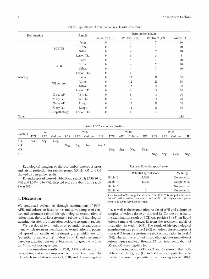
Table 2 Equivalency of examination results with score value
Examination Sample Examination resultsNegative 1 (minus) Positive 1 (+1) Positive 2 (+2) Positive 3 (+3)
Scoring
PCR TB
Feces 0 4 7 10Urine 0 4 7 10Saliva 0 4 7 10
Lesion T12 0 1 3 5
AFB
Feces 0 4 7 10Urine 0 4 7 10Saliva 0 4 7 10
Lesion T12 0 1 3 5
TB culture
Feces 0 12 21 30Urine 0 12 21 30Saliva 0 12 21 30
Lesion T12 0 8 14 20X-ray AP Vert 12 0 8 14 20X-ray Lat Vert 12 0 8 14 20X-ray AP Lungs 0 12 21 30X-ray Lat Lungs 0 12 21 30
Histopathology Lesion T12 0 1 3 5Total 285
Table 3 T12 lesion examination
Rabbits W-1 W-6 W-10 W-14PCR AFB Culture PCR AFB Culture HP PCR AFB Culture HP PCR AFB Culture HP
G3 Pos 3 Neg NegG4 Neg Neg Neg Pos 2G5 Neg Neg Neg NegG6 Neg Neg Neg Neg
Radiological imaging of thoracolumbar anteroposteriorand lateral projection for rabbits groups G3 G4 G5 and G6showed also negative results
Potential spread score of rabbit 3 and rabbit 4 is 175 (0 to9) and 105 (0 to 9) Infected score of rabbit 1 and rabbit2 was 0
4 Discussion
We conducted evaluations through examinations of PCRAFB and culture on feces urine and saliva samples of con-trol and treatment rabbits histopathological examination oflesion tissue thoracal 12 of treatment rabbits and radiologicalexamination after the incubation period to treatment rabbits
We developed two methods of potential spread assess-ment which are assessment based on examinations of poten-tial spread on rabbits of treatment group which we callldquopotential spread scoringrdquo (Tables 1 and 4) and assessmentbased on examinations on rabbits of control group which wecall ldquoinfected scoring systemrdquo
The examination results of PCR AFB and culture onfeces urine and saliva samples of control and treatment rab-bits which were taken in weeks 1 6 10 and 14 were negative
Table 4 Potential spread score
Potential spread score MeaningRabbit 3 175 Not potentialRabbit 4 105 Not potentialRabbit 5 0 Not potentialRabbit 6 0 Not potentialScore from 0 to 9 is not potential score from 10 to 39 is low potential scorefrom40 to 69 ismediumpotential score from 70 to 89 is high potential scorefrom 90 to 100 is very high potential
(minus) as well as the examination results of AFB and culture onsamples of lesions tissue of thoracal 12 On the other handthe examination result of PCR was positive 3 (+3) on liquidlesions sample of thoracal 12 from the treatment rabbit ofincubation in week 1 (G3) The result of histopathologicalexamination was positive 2 (+2) on lesions tissue samples ofthoracal 12 from the treatment rabbit of incubation in week 6(G4) whereas the results of histopathological examination oflesions tissue samples of thoracal 12 from treatment rabbits ofG5 and G6 were negative (minus)
The scoring results (Tables 2 and 5) showed that bothrabbits of control group (G1 and G2) were not potential to beinfected because the potential spread scoring was of 000
Advances in Ecology 5
Figure 1 Histopathology examination of T12 lesion on RabbitG3 There were no changes seen in the bone marrow but therewere inflammatory cells fibrosis tissue and capsule image seencorresponding to bacterial infection byMycobacterium tuberculosisThere were also necrotic tissue and cell formation resembling DatiaLanghans cell (zoom in the picture) Caseous process was not seenclearly but it was incomplete The process of bone remodeling wasclearly seen
Table 5 Infected score
Infected score MeaningRabbit 1 0 Not infectedRabbit 2 0 Not infected
while from the rabbits of treatment group potential spreadscoring of treatment rabbit 3 (G3) was of 175 and treatmentrabbit 4 (G4) was of 105 This means that although therewere positive results obtained from the examination therewas no potential to spread to the environment
These results indicated that using the inoculationmethodof Mycobacterium tuberculosis suspension of 106 cfumLdirectly into the corpus and incubating the rabbit untilweek 14 there was not any potential spread of Mycobac-terium tuberculosis to the environment The environmentassumed here was the area around the location of rabbitstreatmentincubation including living creatures contactingdirectly (animal care workers veterinarians operator doc-tors and rabbit control) or indirectly (the place of feces andurine disposal) with treatment rabbits
This could mean that the method used did not revealthe potential spread of Mycobacterium tuberculosis into theenvironment or perhaps the rabbit bodyrsquos immune reactionwas able to eliminate the infection of Mycobacterium tuber-culosis completely especially in the first weeks of inoculationwhere there was no direct or hematogenous spread on thearea around infected corpus
In the inoculationmethodwe usedMycobacterium tuber-culosis of 106 cfumL (01mL suspension with a quantity of107 cfumL) It was based on a study of Cremieux and Carbon[8] which had successfully created a skin tuberculosis modelon New Zealand rabbits through a liquefaction methodon skin Bacteria used were Mycobacterium tuberculosis ofH37RV strain from the collection of the Laboratory ofClinical Microbiology FKUI This is a pure strain of bacteria
so that the virulence was probably not strong enough tofight against the rabbitsrsquo immune systemThe use of bacterialstrains with stronger virulence of course can be considered infuture studies
The difference between the procedure of infection induc-tion performed to rabbit models of spondylitis tuberculosisand the procedure performed to rabbit models of tubercu-losis infection in the lungs is on the bacterial inoculationtechnique In the creation of rabbit models of tuberculosis inthe lungs a closed aerosol pump system was used in whichthe suspension or liquefaction ofMycobacterium tuberculosiswas made into the form of droplet nuclei and was inoculatedusing inhalation directly into the lungs This caused thewhole treatment procedure to be performed at the facilities ofABSL3 considering that the natural spread ofMycobacteriumtuberculosis is through droplet nuclei entering the lungs Onthe other hand the creation of rabbit models of spondylitistuberculosis to date there has not been supporting liter-ature found so that we performed the approach throughthe method done by Converse et al [4] Poelstra et al[9] Schulz et al [10] Bierry et al [6] and several otherresearchers who had successfully created animal models withbone infection (osteomyelitis) using the technique of directinoculationinjection into the bone
We drilled the central part of the corpus thoracal 12 of 6ndash10mmdepth injected bacterial suspension ofMycobacteriumtuberculosis into the hole formed and immediately coveredthemwith subcutis fatMycobacterium tuberculosis is faculta-tive aerobic bacteria so the procedure was likely to cause thelimitation of area and the availability of oxygen that decreasedthe ability of bacteria to survive and to breed By extendingthe area of inoculation it is expected that Mycobacteriumtuberculosiswill have enough space and oxygen to survive andto proliferate
Histopathological examination was performed on corpustissue of thoracal 12 obtained from surgical proceduresFrom macroscopic observation bluish-colored tissue wasfound in the center of the corpus which was the formerlocation of drilling and injection of bacterial suspension ofMycobacterium tuberculosis The formation of bluish tissuewhich was predicted as the area of the infection was probablydue to the effort of rabbitrsquos body in achieving bone healingprocess or due to the immune process of rabbits in striving toeliminate bacterial infection in the area On the other handthis could also be a manifestation of the proliferation processof the bacteria to survive In histopathological examinationprocedure we chose this tissue to do the cutting and thecreation of tissue staining slides
The results of staining and microscopic observation ontissue staining slides showed an image of spheres containinga lot of inflammatory cells fibrosis tissue and capsuleimage corresponding to bacterial infection ofMycobacteriumtuberculosis [8 9] There were also necrotic tissue and cellsformation resembling Datia Langhans cells which were notclearly seen and an image of incomplete caseous processwhereas the process of bone remodeling was clearly seenFrom these macroscopic and microscopic images we con-cluded that there has been a process of bacterial infection of
6 Advances in Ecology
(a) (b)
Figure 2 Plain X-ray image of T12 rabbit G6 14 weeks after treatment (a) Antero posterior projection there is no clear vertebral destructionbut there is paravertebral shadow that indicates the abscess mass (b) Lateral projection there is slight destruction of T12 vertebral body thatindicates the process infection
Mycobacterium tuberculosis in the lesions of corpus thoracal12
Radiological observations showed that there was noclear destruction image as a result of the infection processThis was probably because Mycobacterium tuberculosis has aslow proliferation character so that the process of infectionappeared in week 14 as early stages of chronic infectionIn general the destruction image from the X-ray imagingwill be clearly seen when the destruction was large and wasa prolonged process Pus image was not seen which wasprobably because tissue reaction to the infection process hadnot yet appeared significantly
Using the procedure of direct inoculation of 01mLof Mycobacterium tuberculosis 107 cfumL in the corpus ofthoracal 12 with incubation period of 14 weeks there wasno potential spread of bacteria into the environment onspondylitis tuberculosis rabbitmodels (potential spread scorelt9) and there is no spread on the control rabbits (infectedscore lt9) as shown on Table 1
Conflict of Interests
The authors have no financial conflict of interests
Acknowledgment
The research has grant from Fakultas Kedokteran UniversitasIndonesia 2012
References
[1] D Schlossberg Tuberculosis and Non Tuberculous Mycobacte-rial Infections McGraw-Hill 5th edition 2006
[2] S Sapardan Total Treatment of Tuberculosis of The Spine ARational Problem Solving Approach Perpustakaan UniversitasIndonesia 2004
[3] Y CManabe AM Dannenberg Jr S K Tyagi et al ldquoDifferentstrains of Mycobacterium tuberculosis cause various spectrums
of disease in the rabbit model of tuberculosisrdquo Infection andImmunity vol 71 no 10 pp 6004ndash6011 2003
[4] P J Converse A M Dannenberg Jr J E Estep et al ldquoCavitarytuberculosis produced in rabbits by aerosolized virulent tuber-cle bacillirdquo Infection and Immunity vol 64 no 11 pp 4776ndash4787 1996
[5] L Tsenova R Harbacheuski E Ellison C Manca and GKaplan ldquoAerosol exposure system for rabbits application toMycobacterium tuberculosis infectionrdquoApplied Biosafety vol 11no 1 pp 7ndash14 2006
[6] G Bierry F Jehl G Prevost et al ldquoPercutaneous inoculatedrabbit model of intervertebral disc space infection magneticresonance imaging features with pathological correlationrdquo JointBone Spine vol 75 no 4 pp 465ndash470 2008
[7] G Zhang B ZhuW Shi MWang Z Da and Y Zhang ldquoEval-uation ofMycobacterial virulence using rabbit skin liquefactionmodelrdquo Virulence vol 1 no 3 pp 156ndash163 2010
[8] A-C Cremieux and C Carbon ldquoExperimental models of boneand prosthetic joint infectionsrdquo Clinical Infectious Diseases vol25 no 6 pp 1295ndash1302 1997
[9] K A Poelstra N A Barekzi D W Grainger A G Gristinaand T C Schuler ldquoA novel spinal implant infection model inrabbitsrdquo Spine vol 25 no 4 pp 406ndash410 2000
[10] S Schulz H Steinhart and R Mutters ldquoChronic osteomyelitisin a new rabbit modelrdquo Journal of Investigative Surgery vol 14no 2 pp 121ndash131 2001
Submit your manuscripts athttpwwwhindawicom
Forestry ResearchInternational Journal of
Hindawi Publishing Corporationhttpwwwhindawicom Volume 2014
Environmental and Public Health
Journal of
Hindawi Publishing Corporationhttpwwwhindawicom Volume 2014
Hindawi Publishing Corporationhttpwwwhindawicom Volume 2014
EcosystemsJournal of
Hindawi Publishing Corporationhttpwwwhindawicom Volume 2014
MeteorologyAdvances in
EcologyInternational Journal of
Hindawi Publishing Corporationhttpwwwhindawicom Volume 2014
Marine BiologyJournal of
Hindawi Publishing Corporationhttpwwwhindawicom Volume 2014
Hindawi Publishing Corporationhttpwwwhindawicom
Applied ampEnvironmentalSoil Science
Volume 2014
Advances in
Hindawi Publishing Corporationhttpwwwhindawicom Volume 2014
Environmental Chemistry
Atmospheric SciencesInternational Journal of
Hindawi Publishing Corporationhttpwwwhindawicom Volume 2014
Hindawi Publishing Corporationhttpwwwhindawicom Volume 2014
Waste ManagementJournal of
Hindawi Publishing Corporation httpwwwhindawicom Volume 2014
International Journal of
Geophysics
Hindawi Publishing Corporationhttpwwwhindawicom Volume 2014
Geological ResearchJournal of
EarthquakesJournal of
Hindawi Publishing Corporationhttpwwwhindawicom Volume 2014
BiodiversityInternational Journal of
Hindawi Publishing Corporationhttpwwwhindawicom Volume 2014
ScientificaHindawi Publishing Corporationhttpwwwhindawicom Volume 2014
OceanographyInternational Journal of
Hindawi Publishing Corporationhttpwwwhindawicom Volume 2014
The Scientific World JournalHindawi Publishing Corporation httpwwwhindawicom Volume 2014
Journal of Computational Environmental SciencesHindawi Publishing Corporationhttpwwwhindawicom Volume 2014
Hindawi Publishing Corporationhttpwwwhindawicom Volume 2014
ClimatologyJournal of

2 Advances in Ecology
fulfilling aspects of biosafety and biosecurity [5 7] Whilefor the study of the same bacterial infection to the corpusof the spine with direct inoculation method (nonaerosol) itis debatable whether the experimental procedure shall alsobe conducted in facilities of ABSL3 In certain circumstanceswhere the facilities of ABSL3 are limited or not possible touse the opportunity to perform of the primary infection atfacilities other than ABSL3 should be learned In this studythe potential spread of Mycobacterium tuberculosis into theenvironment from a rabbit model of spondylitis tuberculosiswas observed in order to create the conditions and methodsfulfilling aspects of biosafety and biosecurity
2 Material and Methods
The study was an interventional study on rabbits that havemet the ethical review and have obtained the approval fromAnimal Care and Use Committee (ACUC) of PT BimanaIndomedical and Animal Hospital of Bogor AgriculturalInstitute Most of this research used animal facilities at RSHIPB PT Bimana Indomedical and Clinical MicrobiologyLaboratory of Faculty of Medicine University of Indonesia(FMUI)
The selection of rabbit samples was based on the rabbitrsquosbody weight bone maturity sex and clinical radiologicaland laboratory examination where the inclusion criteria werewhite rabbits of New Zealand strains weighing 2500ndash3500grams which were healthy and skeletally mature whereasthe exclusion criteria were rabbits with congenital spineabnormalities andor abnormalities in the spine caused byinfections trauma neoplasm and so forth
Before being used in this study Mycobacterium tubercu-losis was activated by making a suspension in Middlebrookrsquosliquid medium and being incubated in a shaker with a speedof 150 rpm temperature of 37∘C for 18 hours The bacterialsuspension was then diluted using sterile physiological waterto obtain a number of bacteria of 107 cfumL (colony formingunitsmilliliter) or equivalent to 01 A in the measurement ofOptical Density (OD) at wavelength of 600 nm The treat-ment for the bacteria followed standard operating proceduresapplied in Clinical Microbiology Laboratory of FMUI
Six rabbits were divided into two groups treatment group(119899 = 4) and control group (119899 = 2) The induction procedureof bacterial infection of Mycobacterium tuberculosis wasperformed to four rabbits of the treatment group while totwo rabbits of the control group the induction procedure ofinfection was not performed
Four rabbits of treatment group (G3 G4 G5 and G6)were shaved on their back as high as T12 to L1 and werethen given anesthetic mixture of ketamine HCl (44mgkg)and xylazine (5mgkg) intramuscularly After the anestheticworked the rabbits were faced to the side (the left side of theback facing the surgery operator) and then antiseptic andaseptic procedures were performed using 70 alcohol andbetadine on the shaved back of the rabbits and then theyweregiven sterile cloth Identification of thoracal 12was performedby touching the 12th rib and being traced to the transversesprocess transverse incision was then performed starting
from the spinous process of 3ndash5 cm width directing to theleft lateral penetrating cutis and subcutis Paraspinal muscleswere separated reaching the 12th rib the transverses processand lamina thoracal 12 Reidentification was performed todetermine the position of thoracal 12 before a hole wasmade using a drill at the midpoint of the corpus thoracal12 (+5mm from the transverses process) of 6ndash10mm depthusing the drill bit of 15mm One-tenth mL suspension ofMycobacterium tuberculosis (107 cfumL sim 01 absorbance)was injected aseptically into the hole made in the corpusthen the hole was covered by subcutis fat using a rootdissector surgical wound was covered layer by layer andskin was sewn one by one using vycril 30 thread Surgicalwound was then covered by bandages and the rabbits werereturned to the cage and were given ketoprofen (3mgkg)intramuscularly every 12 hours for 3 days Four rabbits werethen maintainedincubated in individual cages for 2 6 10and 14 weeks While the two rabbits of control group (G1 andG2) were also kept in individual cages and were interactedwith the rabbits of treatment group for 3 hours per day
Clinical examinations were performed namely observa-tions of daily activities appetite paralysis signs of infection(the presence of sinus abscess) and measurement of bodyweight every three days on all treatment and control rabbitsSampling of feces urine and saliva was done for Acid FastBacilli (AFB) staining which refers to grading of AFB smearsas per WHO and IUATLD recommendation examination ofPolymerase Chain Reaction (PCR) and culture at the end ofweeks 2 6 10 and 14 after infection induction proceduresfor all rabbits (control and treatment) At the end of weeks2 6 10 and 14 radiological examination was performedto one rabbit from treatment group per each period therabbit was then euthanized using lethal injection at a dose of150mgkg intravenously (IV) Necropsy was then performedto the rabbit and sample of lesion tissue at thoracal 12 wastaken for AFB staining and examinations of PCR cultureand histopathology
PCR amplification for IS6110 gene was performed by setof primers TB15-CTC GCGAGCGTAGGCGTCGG-31015840 andTB251015840-CTCGTCCAGCGCCGCTTCGG-31015840 which amplifya fragment of 130-base pair (bp) of the target gene DNAamplification was performed in a Biorad thermocycler in afinal volume of 20 120583L containing 10x PCR buffer 25mMMgCl2 10mM dNTPs 10 120583M of each primer 012 120583L HotStar
DNA polymerase and 4 120583L of template DNAThe amplifica-tion programwas consisted of initial denaturation at 95∘C for5min followed by 40 cycles of denaturation at 94∘C for 30 sannealing at 60∘C for 30 s extension at 72∘C for 30 s and afinal extension at 72∘C for 5min
PCR examination was performed on liquid (urine saliva)and solid (stools lesion tissues) specimens For urine theprocedure was started by centrifugation at 12000 rpm for5 minutes The supernatant was discarded DNA containedin the pellets was isolated according to the manufacturerrsquosinstructions (QIAmp DNA kit Qiagen) For Saliva becausethe numbers of saliva was a bit then the centrifugationsteps were eliminated So that the isolation of DNA in salivawas done directly on the samples in accordance with themanufacturerrsquos instructions (QIAmp DNA kit Qiagen) For
Advances in Ecology 3
Table 1 Potential spreadinfected score
Potential spread scoreResults Score Results Score Results Score Results Score
PCR
Feces Neg 0 Pos 1 4 Pos 2 7 Pos 3 10Urine Neg 0 Pos 1 4 Pos 2 7 Pos 3 10Saliva Neg 0 Pos 1 4 Pos 2 7 Pos 3 10
Lesion T12 Neg 0 Pos 1 1 Pos 2 3 Pos 3 5
AFB
Feces Neg 0 Pos 1 4 Pos 2 7 Pos 3 10Urine Neg 0 Pos 1 4 Pos 2 7 Pos 3 10Saliva Neg 0 Pos 1 4 Pos 2 7 Pos 3 10
Lesion T12 Neg 0 Pos 1 1 Pos 2 3 Pos 3 5
Culture
Feces Neg 0 Pos 1 12 Pos 2 21 Pos 3 30Urine Neg 0 Pos 1 12 Pos 2 21 Pos 3 30Saliva Neg 0 Pos 1 12 Pos 2 21 Pos 3 30
Lesion T12 Neg 0 Pos 1 8 Pos 2 14 Pos 3 20X-ray AP Vert T12 Neg 0 Pos 1 8 Pos 2 14 Pos 3 20X-ray Lat Vert T12 Neg 0 Pos 1 8 Pos 2 14 Pos 3 20X-ray AP Lung Neg 0 Pos 1 12 Pos 2 21 Pos 3 30X-ray Lat Lung Neg 0 Pos 1 12 Pos 2 21 Pos 3 30Histopathology Lesion T12 Neg 0 Pos 1 1 Pos 2 3 Pos 3 5Total score 285Score from 0 to 9 is not infected score from 10 to 39 is mildly infected score from 40 to 69 is infected score from 70 to 89 is heavily infected score from 90 to100 is very heavily infectedScore from 0 to 9 is not potential score from 10 to 39 is low potential score from 40 to 69 is medium potential score from 70 to 89 is high potential scorefrom 90 to 100 is very high potentialFormula of spreadinfected score Potential spreadInfected = (Mean scoreTotal score) times 100
solid samples such as tissue lesions and feces the stool sam-ples were weighed to obtain 200mg Isolation of DNA wasthen performed according to the manufacturerrsquos instructions(DNeasy Blood And Tissue Kit Qiagen)
The results of the examination on feces urine and salivaand tissue lesion samples were calculated using scoringsystems which were potential spread scoring and infectedscoring Potential spread scoring was calculated from theexamination results obtained from the treatment rabbitswhile infected scoring was calculated from the examinationresults obtained from control rabbits The description on thisscoring system can be seen in Table 1
There are 8 modalities examination in this study namelyPCR AFB culture AP thoracolumbar X-ray lateral tho-racolumbar X-ray AP lung X-ray lateral lung X-ray andhistopathology examination Authors made a rule that anytest results should be reported as fourth grade which weredivided into negative positive 1 positive 2 and positive 3Each grade is given particular weight assigned by authors(Table 1)
Potential and infected scores were developed based on thepremise that more specimens were examined (feces urinesaliva and lesions) and examination modalities (AFB PCRculture imaging and histopathology) used themore accuratethe prediction results of potential spread score and infectedscore We did a weighting to each modality examination andthe type specimens to be examined (Table 2) It was basedon the experience of authors and literature reviews of alldiagnostic methods used in this study
Weprovide a highweight for culture examination of urineand saliva and X-ray examination of lung (score 30) withthe consideration that the potency for disease transmissionfrom urine and saliva specimens larger than the other whilehistological examination and PCR examination of vertebrallesions got the lowest weight (score 5)
3 Results
The results of AFB staining PCR and culture examinationon feces urine and saliva samples were obtained All theseexaminations showed negative In addition the data ofAFB staining PCR culture (Table 3) and histopathology(Figure 1) examination of lesion tissue specimens and theresults of X-ray images (Figure 2) of the rabbitsrsquo spine afterthe incubation period specified were also obtained Sincetreatment rabbit 3 (G3) which should be euthanized at week2 was dead in week 1 then the data of examination resultswere adjusted to the data of weeks 1 6 10 and 14 RabbitG3 was dead in week 1 due to surgical trauma It suffered noappetite after inoculation its activity was reduced and then itlost weight until death PCR examination of lesion tissue fromRabbit G3 showed positive 3 and histopathological examina-tion of lesion tissue from rabbit G4 showed positive 2 whileothers showed negative Negative results indicated that therewas no Mycobacterium tuberculosis found in examinationthrough the method used in this study whereas positiveresults indicated the presence of Mycobacterium tuberculosison the examination through the method used
4 Advances in Ecology
Table 2 Equivalency of examination results with score value
Examination Sample Examination resultsNegative 1 (minus) Positive 1 (+1) Positive 2 (+2) Positive 3 (+3)
Scoring
PCR TB
Feces 0 4 7 10Urine 0 4 7 10Saliva 0 4 7 10
Lesion T12 0 1 3 5
AFB
Feces 0 4 7 10Urine 0 4 7 10Saliva 0 4 7 10
Lesion T12 0 1 3 5
TB culture
Feces 0 12 21 30Urine 0 12 21 30Saliva 0 12 21 30
Lesion T12 0 8 14 20X-ray AP Vert 12 0 8 14 20X-ray Lat Vert 12 0 8 14 20X-ray AP Lungs 0 12 21 30X-ray Lat Lungs 0 12 21 30
Histopathology Lesion T12 0 1 3 5Total 285
Table 3 T12 lesion examination
Rabbits W-1 W-6 W-10 W-14PCR AFB Culture PCR AFB Culture HP PCR AFB Culture HP PCR AFB Culture HP
G3 Pos 3 Neg NegG4 Neg Neg Neg Pos 2G5 Neg Neg Neg NegG6 Neg Neg Neg Neg
Radiological imaging of thoracolumbar anteroposteriorand lateral projection for rabbits groups G3 G4 G5 and G6showed also negative results
Potential spread score of rabbit 3 and rabbit 4 is 175 (0 to9) and 105 (0 to 9) Infected score of rabbit 1 and rabbit2 was 0
4 Discussion
We conducted evaluations through examinations of PCRAFB and culture on feces urine and saliva samples of con-trol and treatment rabbits histopathological examination oflesion tissue thoracal 12 of treatment rabbits and radiologicalexamination after the incubation period to treatment rabbits
We developed two methods of potential spread assess-ment which are assessment based on examinations of poten-tial spread on rabbits of treatment group which we callldquopotential spread scoringrdquo (Tables 1 and 4) and assessmentbased on examinations on rabbits of control group which wecall ldquoinfected scoring systemrdquo
The examination results of PCR AFB and culture onfeces urine and saliva samples of control and treatment rab-bits which were taken in weeks 1 6 10 and 14 were negative
Table 4 Potential spread score
Potential spread score MeaningRabbit 3 175 Not potentialRabbit 4 105 Not potentialRabbit 5 0 Not potentialRabbit 6 0 Not potentialScore from 0 to 9 is not potential score from 10 to 39 is low potential scorefrom40 to 69 ismediumpotential score from 70 to 89 is high potential scorefrom 90 to 100 is very high potential
(minus) as well as the examination results of AFB and culture onsamples of lesions tissue of thoracal 12 On the other handthe examination result of PCR was positive 3 (+3) on liquidlesions sample of thoracal 12 from the treatment rabbit ofincubation in week 1 (G3) The result of histopathologicalexamination was positive 2 (+2) on lesions tissue samples ofthoracal 12 from the treatment rabbit of incubation in week 6(G4) whereas the results of histopathological examination oflesions tissue samples of thoracal 12 from treatment rabbits ofG5 and G6 were negative (minus)
The scoring results (Tables 2 and 5) showed that bothrabbits of control group (G1 and G2) were not potential to beinfected because the potential spread scoring was of 000
Advances in Ecology 5
Figure 1 Histopathology examination of T12 lesion on RabbitG3 There were no changes seen in the bone marrow but therewere inflammatory cells fibrosis tissue and capsule image seencorresponding to bacterial infection byMycobacterium tuberculosisThere were also necrotic tissue and cell formation resembling DatiaLanghans cell (zoom in the picture) Caseous process was not seenclearly but it was incomplete The process of bone remodeling wasclearly seen
Table 5 Infected score
Infected score MeaningRabbit 1 0 Not infectedRabbit 2 0 Not infected
while from the rabbits of treatment group potential spreadscoring of treatment rabbit 3 (G3) was of 175 and treatmentrabbit 4 (G4) was of 105 This means that although therewere positive results obtained from the examination therewas no potential to spread to the environment
These results indicated that using the inoculationmethodof Mycobacterium tuberculosis suspension of 106 cfumLdirectly into the corpus and incubating the rabbit untilweek 14 there was not any potential spread of Mycobac-terium tuberculosis to the environment The environmentassumed here was the area around the location of rabbitstreatmentincubation including living creatures contactingdirectly (animal care workers veterinarians operator doc-tors and rabbit control) or indirectly (the place of feces andurine disposal) with treatment rabbits
This could mean that the method used did not revealthe potential spread of Mycobacterium tuberculosis into theenvironment or perhaps the rabbit bodyrsquos immune reactionwas able to eliminate the infection of Mycobacterium tuber-culosis completely especially in the first weeks of inoculationwhere there was no direct or hematogenous spread on thearea around infected corpus
In the inoculationmethodwe usedMycobacterium tuber-culosis of 106 cfumL (01mL suspension with a quantity of107 cfumL) It was based on a study of Cremieux and Carbon[8] which had successfully created a skin tuberculosis modelon New Zealand rabbits through a liquefaction methodon skin Bacteria used were Mycobacterium tuberculosis ofH37RV strain from the collection of the Laboratory ofClinical Microbiology FKUI This is a pure strain of bacteria
so that the virulence was probably not strong enough tofight against the rabbitsrsquo immune systemThe use of bacterialstrains with stronger virulence of course can be considered infuture studies
The difference between the procedure of infection induc-tion performed to rabbit models of spondylitis tuberculosisand the procedure performed to rabbit models of tubercu-losis infection in the lungs is on the bacterial inoculationtechnique In the creation of rabbit models of tuberculosis inthe lungs a closed aerosol pump system was used in whichthe suspension or liquefaction ofMycobacterium tuberculosiswas made into the form of droplet nuclei and was inoculatedusing inhalation directly into the lungs This caused thewhole treatment procedure to be performed at the facilities ofABSL3 considering that the natural spread ofMycobacteriumtuberculosis is through droplet nuclei entering the lungs Onthe other hand the creation of rabbit models of spondylitistuberculosis to date there has not been supporting liter-ature found so that we performed the approach throughthe method done by Converse et al [4] Poelstra et al[9] Schulz et al [10] Bierry et al [6] and several otherresearchers who had successfully created animal models withbone infection (osteomyelitis) using the technique of directinoculationinjection into the bone
We drilled the central part of the corpus thoracal 12 of 6ndash10mmdepth injected bacterial suspension ofMycobacteriumtuberculosis into the hole formed and immediately coveredthemwith subcutis fatMycobacterium tuberculosis is faculta-tive aerobic bacteria so the procedure was likely to cause thelimitation of area and the availability of oxygen that decreasedthe ability of bacteria to survive and to breed By extendingthe area of inoculation it is expected that Mycobacteriumtuberculosiswill have enough space and oxygen to survive andto proliferate
Histopathological examination was performed on corpustissue of thoracal 12 obtained from surgical proceduresFrom macroscopic observation bluish-colored tissue wasfound in the center of the corpus which was the formerlocation of drilling and injection of bacterial suspension ofMycobacterium tuberculosis The formation of bluish tissuewhich was predicted as the area of the infection was probablydue to the effort of rabbitrsquos body in achieving bone healingprocess or due to the immune process of rabbits in striving toeliminate bacterial infection in the area On the other handthis could also be a manifestation of the proliferation processof the bacteria to survive In histopathological examinationprocedure we chose this tissue to do the cutting and thecreation of tissue staining slides
The results of staining and microscopic observation ontissue staining slides showed an image of spheres containinga lot of inflammatory cells fibrosis tissue and capsuleimage corresponding to bacterial infection ofMycobacteriumtuberculosis [8 9] There were also necrotic tissue and cellsformation resembling Datia Langhans cells which were notclearly seen and an image of incomplete caseous processwhereas the process of bone remodeling was clearly seenFrom these macroscopic and microscopic images we con-cluded that there has been a process of bacterial infection of
6 Advances in Ecology
(a) (b)
Figure 2 Plain X-ray image of T12 rabbit G6 14 weeks after treatment (a) Antero posterior projection there is no clear vertebral destructionbut there is paravertebral shadow that indicates the abscess mass (b) Lateral projection there is slight destruction of T12 vertebral body thatindicates the process infection
Mycobacterium tuberculosis in the lesions of corpus thoracal12
Radiological observations showed that there was noclear destruction image as a result of the infection processThis was probably because Mycobacterium tuberculosis has aslow proliferation character so that the process of infectionappeared in week 14 as early stages of chronic infectionIn general the destruction image from the X-ray imagingwill be clearly seen when the destruction was large and wasa prolonged process Pus image was not seen which wasprobably because tissue reaction to the infection process hadnot yet appeared significantly
Using the procedure of direct inoculation of 01mLof Mycobacterium tuberculosis 107 cfumL in the corpus ofthoracal 12 with incubation period of 14 weeks there wasno potential spread of bacteria into the environment onspondylitis tuberculosis rabbitmodels (potential spread scorelt9) and there is no spread on the control rabbits (infectedscore lt9) as shown on Table 1
Conflict of Interests
The authors have no financial conflict of interests
Acknowledgment
The research has grant from Fakultas Kedokteran UniversitasIndonesia 2012
References
[1] D Schlossberg Tuberculosis and Non Tuberculous Mycobacte-rial Infections McGraw-Hill 5th edition 2006
[2] S Sapardan Total Treatment of Tuberculosis of The Spine ARational Problem Solving Approach Perpustakaan UniversitasIndonesia 2004
[3] Y CManabe AM Dannenberg Jr S K Tyagi et al ldquoDifferentstrains of Mycobacterium tuberculosis cause various spectrums
of disease in the rabbit model of tuberculosisrdquo Infection andImmunity vol 71 no 10 pp 6004ndash6011 2003
[4] P J Converse A M Dannenberg Jr J E Estep et al ldquoCavitarytuberculosis produced in rabbits by aerosolized virulent tuber-cle bacillirdquo Infection and Immunity vol 64 no 11 pp 4776ndash4787 1996
[5] L Tsenova R Harbacheuski E Ellison C Manca and GKaplan ldquoAerosol exposure system for rabbits application toMycobacterium tuberculosis infectionrdquoApplied Biosafety vol 11no 1 pp 7ndash14 2006
[6] G Bierry F Jehl G Prevost et al ldquoPercutaneous inoculatedrabbit model of intervertebral disc space infection magneticresonance imaging features with pathological correlationrdquo JointBone Spine vol 75 no 4 pp 465ndash470 2008
[7] G Zhang B ZhuW Shi MWang Z Da and Y Zhang ldquoEval-uation ofMycobacterial virulence using rabbit skin liquefactionmodelrdquo Virulence vol 1 no 3 pp 156ndash163 2010
[8] A-C Cremieux and C Carbon ldquoExperimental models of boneand prosthetic joint infectionsrdquo Clinical Infectious Diseases vol25 no 6 pp 1295ndash1302 1997
[9] K A Poelstra N A Barekzi D W Grainger A G Gristinaand T C Schuler ldquoA novel spinal implant infection model inrabbitsrdquo Spine vol 25 no 4 pp 406ndash410 2000
[10] S Schulz H Steinhart and R Mutters ldquoChronic osteomyelitisin a new rabbit modelrdquo Journal of Investigative Surgery vol 14no 2 pp 121ndash131 2001
Submit your manuscripts athttpwwwhindawicom
Forestry ResearchInternational Journal of
Hindawi Publishing Corporationhttpwwwhindawicom Volume 2014
Environmental and Public Health
Journal of
Hindawi Publishing Corporationhttpwwwhindawicom Volume 2014
Hindawi Publishing Corporationhttpwwwhindawicom Volume 2014
EcosystemsJournal of
Hindawi Publishing Corporationhttpwwwhindawicom Volume 2014
MeteorologyAdvances in
EcologyInternational Journal of
Hindawi Publishing Corporationhttpwwwhindawicom Volume 2014
Marine BiologyJournal of
Hindawi Publishing Corporationhttpwwwhindawicom Volume 2014
Hindawi Publishing Corporationhttpwwwhindawicom
Applied ampEnvironmentalSoil Science
Volume 2014
Advances in
Hindawi Publishing Corporationhttpwwwhindawicom Volume 2014
Environmental Chemistry
Atmospheric SciencesInternational Journal of
Hindawi Publishing Corporationhttpwwwhindawicom Volume 2014
Hindawi Publishing Corporationhttpwwwhindawicom Volume 2014
Waste ManagementJournal of
Hindawi Publishing Corporation httpwwwhindawicom Volume 2014
International Journal of
Geophysics
Hindawi Publishing Corporationhttpwwwhindawicom Volume 2014
Geological ResearchJournal of
EarthquakesJournal of
Hindawi Publishing Corporationhttpwwwhindawicom Volume 2014
BiodiversityInternational Journal of
Hindawi Publishing Corporationhttpwwwhindawicom Volume 2014
ScientificaHindawi Publishing Corporationhttpwwwhindawicom Volume 2014
OceanographyInternational Journal of
Hindawi Publishing Corporationhttpwwwhindawicom Volume 2014
The Scientific World JournalHindawi Publishing Corporation httpwwwhindawicom Volume 2014
Journal of Computational Environmental SciencesHindawi Publishing Corporationhttpwwwhindawicom Volume 2014
Hindawi Publishing Corporationhttpwwwhindawicom Volume 2014
ClimatologyJournal of