research article the olive oil-based lipid clinoleic
TRANSCRIPT
Research ArticleThe Olive Oil-Based Lipid Clinoleic Blocks LeukocyteRecruitment and Improves Survival during SystemicInflammation: A Comparative In Vivo Study of DifferentParenteral Lipid Emulsions
Kirsten Buschmann, Johannes Poeschl, Natascha Braach, Hannes Hudalla,Navina Kuss, and David Frommhold
Clinic of Neonatology, Department of Pediatrics, University of Heidelberg, 69120 Heidelberg, Germany
Correspondence should be addressed to David Frommhold; [email protected]
Received 28 September 2014; Revised 16 December 2014; Accepted 29 December 2014
Academic Editor: Giuseppe Valacchi
Copyright © 2015 Kirsten Buschmann et al. This is an open access article distributed under the Creative Commons AttributionLicense, which permits unrestricted use, distribution, and reproduction in any medium, provided the original work is properlycited.
Although fish oil-based and olive oil-based lipid emulsions have been shown to exert anti-inflammatory functions, theimmunomodulating properties of lipids are still controversial. Therefore, we investigated the anti-inflammatory effect of threedifferent parenterally administered lipid emulsions in vivo: olive oil-based Clinoleic, fish oil-based Smoflipid, and soybean oil-based Lipofundin. We observed leukocyte recruitment in inflamed murine cremaster muscle using intravital microscopy andsurvival in a murine model of LPS-induced systemic inflammation and analyzed expression of leukocyte and endothelial adhesionmolecules. Olive oil-based Clinoleic and fish oil-based Smoflipid profoundly inhibited leukocyte adhesion compared to Lipofundinduring LPS-induced inflammation of the murine cremaster muscle. In the trauma model of cremaster muscle inflammation,Lipofundin was the only lipid emulsion that even augmented leukocyte adhesion. In contrast to Smoflipid and Lipofundin,Clinoleic effectively blocked leukocyte recruitment and increased survival during lethal endotoxemia. Flow chamber experimentsand analysis of adhesion molecule expression suggest that both endothelial and leukocyte driven mechanisms might contributeto anti-inflammatory effects of Clinoleic. We conclude that the anti-inflammatory properties of Clinoleic are superior to those ofSmoflipid and Lipofundin even during systemic inflammation. Thus, these results should stimulate further studies investigatingparenteral lipids as an anti-inflammatory strategy in critically ill patients.
1. Introduction
Parenteral nutrition is crucially important in critically illpatients [1, 2]. However, numerous studies reported compli-cations during parenteral nutrition, like parenteral nutritionassociated liver disease [3] or detrimental effects of parenterallipids on survival and inflammatory response during sepsis[4, 5]. This may also be due to lipid-induced decrease ofneutrophil function and cytokine release in septic patients [6,7].Therefore, strategies to avoid the negative consequences ofintravenously administered lipids were needed [4, 8, 9].
In recent years, the composition of lipids was mainlybased on soybeans which contain high amounts of omega-6-polyunsaturated fatty acids. During the beginning of parenteral
nutrition, Intralipid which only contains soybean-basedlong-chain-triglycerides (LCT) was frequently used. Omega-6 fatty acids belong to the family of polyunsaturated fattyacids (PUFA) and are precursors of eicosanoids. Eicosanoidsact as immunomodulators, serve as signaling molecules, andcontribute to inflammatory conditions [10]. In this context,they promote leukocyte recruitment by increased productionof proinflammatory cytokines. On the other hand, theynegatively affect lymphocyte proliferation, thereby causingan immunosuppressive effect [11, 12]. One important aspectof the observed effects is the change of cell membrane fluidityby parenterally administered fatty acids [13].
In order to attenuate these serious side effects ofPUFA, new lipid emulsions were formulated for parenteral
Hindawi Publishing CorporationMediators of InflammationVolume 2015, Article ID 757059, 11 pageshttp://dx.doi.org/10.1155/2015/757059

2 Mediators of Inflammation
nutrition. Lipofundin is one alternative that substituted 50%of LCT with medium-chain-triglycerides (MCT) that aremetabolized more rapidly than LCT, thereby displaying lessimmunosuppressive properties and exerting better effectson membrane function [14]. One study that compared therespiratory burst of humanneutrophils found a reduced effectwith Lipofundin when compared to other lipid emulsions[15]. In contrast to Lipofundin and Intralipid, olive oil-basedlipids were shown to have a protective effect against LPS-induced inflammation [16]. Olive oils contain high amountsof monounsaturated fatty acids (MUFA) and are knownto show less sensitivity to peroxidation when comparedto PUFA. However, there is an ongoing discussion aboutbeneficial properties of fish oil-based lipids (i.e., Smoflipid)when compared with predominant olive oil-based lipids (i.e.,Clinoleic) [17, 18].
Smoflipid contains fish oil, which is rich in omega-3 fattyacids and is able to inhibit the production of proinflammatorycytokines via activation of peroxisome proliferator-activatedreceptor (PPAR) and interactionwithNFkB [19, 20].The ben-eficial effects are at least in part attributed to a favorable ratioof omega-6: omega-3 fatty acids and the balanced mixture ofdifferent lipid ingredients (LCT, MCT, and olive oil) [18].
In addition, there are still conflicting results about howparenterally administered lipids might interfere with leuko-cyte recruitment which is known to be a sensitive indicatorof inflammation. Leukocyte recruitment into inflamed tissuefollows a well-defined cascade of events beginning with thecapture of free flowing leukocytes to the vessel wall followedby leukocyte rolling (mediated by selectins and their ligands)triggering the activation of 𝛽
2-integrins (i.e., LFA1, MAC1)
by chemokines and their receptors (i.e., CXCR2). Onceactivated, leukocyte’s integrins can bind to their respectiveendothelial receptors, like ICAM-1 or VCAM-1. This in turnleads to firm leukocyte arrest on the endothelium and finallythe leukocyte transmigration into the tissue [21, 22].
Based on the above-mentioned controversies about lipid-induced immunomodulation, we now aimed to compareanti-inflammatory effects of Lipofundin, Smoflipid, and Cli-noleic in vitro and in vivo with special regard to leukocyterecruitment during local and systemic inflammation.
2. Materials and Methods
2.1. Animals. C57bl/6 wild type (WT)mice were provided byCharles River (Sulzfeld, Germany). All mice weremaintainedas breeding colonies at the Central Animal Facility of theUniversity ofHeidelberg, Germany. For intravitalmicroscopyexperiments, mice were at least 8 weeks of age. The animalexperiments were approved by the Animal Care and UseCommittee of the Regierungsprasidium Karlsruhe, Germany(Az 35-9185.81/G-3/13).
2.2. Lipid Emulsions. Clinoleic (Fresenius Kabi, Bad Hom-burg, Germany) contains 80% olive oil and 20% soybean oil(LCT). Smoflipid (Fresenius Kabi, Bad Homburg, Germany)consists of 30% soybean oil (LCT), 30% MCT, 25% olive oil,and 15% fish oil (rich in Omega 3 fatty acids). Lipofundin
(Braun, Melsungen, Germany) consists of 50% soybean oil(LCT) and 50%MCT.The lipid emulsions were administeredas an intravenous bolus injection at 1 g/kg for the intravitalmicroscopic experiments and at 2 g/kg 30min, 8 h, and 24 hafter LPS during LPS-induced endotoxemia. Lipid emulsionswere incubated during the in vitro experiments as indicated.
2.3. Intravital Microscopy. Mice were prepared for intrav-ital microscopy, as reported recently [23]. Briefly, afterintraperitoneal (i.p.) injection of ketamine (125mg/kg bodyweight, Ketalar; Parke-Davis, Morris Plains, NJ, USA) andxylazine (12.5mg/kg body weight; Phoenix Scientific Inc., St.Joseph, MO, USA) mice were placed on a heating pad tomaintain body temperature. Intravital microscopy was con-ducted on an upright microscope (Leica; Wetzlar, Germany)with a saline immersion objective (SW40/0.75 numericalaperture, Zeiss, Jena, Germany). Mice were intubated andthe left carotid artery was cannulated for blood samplingand the right jugular vein for lipid administration. Thelipid emulsions were administered with a dose of 1 g/kg asan intravenous bolus injection. Blood levels of cholesterol,triglycerides, and liver enzymes were measured after therespective experiments in the core laboratory facility of theUniversity Hospital Heidelberg (Analysezentrum, Heidel-berg, Germany).
2.4. Cremaster Muscle Preparation. The surgical preparation(trauma-induced inflammation) of the cremaster muscle wasconducted as described previously [24]. Briefly, the scrotumwas opened and the cremaster muscle was exteriorized. Afterlongitudinal incision and spreading of the muscle over acover glass, the epididymis and testis were mobilized andpinned aside leading to full microscopic access to the cre-master muscle microcirculation. Cremaster muscle venuleswere recorded via CCD camera (CF8/1, Kappa, Gleichen,Germany) on a Panasonic S-VHS recorder. S-VHS tapes weredigitized using aDVDmaker (Typhoon, Schalksmuehle,Ger-many). The cremaster muscle was superfused with thermo-controlled (35∘C) bicarbonate-buffered saline. Postcapillaryvenules under observation were recorded before and duringlipid administration and ranged from 20 to 40 𝜇m in diame-ter. Systemic blood samples (10 𝜇L) were taken and assessedfor white blood cell count after staining with Turk’s solution1 : 10 (Merck, Darmstadt, Germany) using a hemocytometer.Microvascular parameters (venular diameter, venular vesselsegment length) were measured using an image processingsystem [25]. Venular centerline red blood cell velocity wasmeasured during the experiment via a dual photodiode and adigital on-line cross-correlation program (CircuSoft Instru-mentation, Hockessin, DE, USA). An empirical factor of0.625 was used to convert centerline velocities to mean bloodflow velocities [26]. Wall shear rates (𝛾
𝑤) were estimated
as 4.9 (8V𝑏/𝑑), where V
𝑏is mean blood flow velocity and
𝑑 is the diameter of the vessel [27, 28]. The number ofadherent leukocytes (firm adhesion for >30 s) was assessedas adherent cells per mm2 vessel surface area as reportedpreviously [24]. Rolling leukocyte flux fractionwas defined asthe percentage of rolling leukocytes to all leukocytes passingthe same vessel in 1 minute [29]. In certain experiments

Mediators of Inflammation 3
mice were injected with 50 ng LPS (Escherichia coli; serotype055:B5 Sigma, Taufkirchen, Germany) intrascrotally (LPS-induced inflammation).
2.5. Whole Mount Histology. To differentially count trans-migrated leukocytes, cremaster muscle-whole mounts wereprepared as described before [30]. Briefly, while the cre-master muscle was still mounted on the stage for intravitalmicroscopy, the tissue was fixed with 4% paraformaldehydein 0.1M phosphate buffer (pH 7.4).The cremaster muscle wasremoved and mounted flat on a superFrost glass slide (Men-zel, Braunschweig, Germany), air dried for 5–10min, andfixed in 4% paraformaldehyde in 0.1M phosphate buffer (pH7.4) for 24 h at 4∘C. After fixation, the tissue was washed threetimes in 0.1M phosphate buffer with 5% ethanol, stained withGiemsa (Sigma, Taufkirchen, Germany) at room temperaturefor 5min, and differentiated in 0.01% acetic acid for contrast.The differentiated slides were washed in water, 75% ethanol,95% ethanol, 100% ethanol, and fresh xylene, followed bymounting in mounting media (AGAR Scientific, Stansted,UK). The Giemsa-stained cremaster muscles were observedusing a Leica DMRB upright microscope and a 25/0,75NAoil immersion objective (both Leica, Wetzlar, Germany).Interstitial leukocytes were counted and differentiated intoneutrophils, eosinophils, and mononuclear cells.
2.6. Preparation of Murine Aortic Endothelial Cells (MAECs).The MAECs were isolated and cultured as previouslydescribed [31]. In brief, 3mm long freshly harvested andcleaned aortic rings were seeded intoMatrigel-coated culturedishes (BD, San Jose, CA, USA) and incubated at 37∘C,5% CO
2in Dulbecco’s Modified Eagle Medium (supple-
mented with 15% fetal bovine serum, 1% Pen/Strep, 90 𝜇g/mLheparin, 60𝜇g/mL endothelial cell growth supplement, and1 𝜇g/mL amphotericin B; Fungizone, Invitrogen, Karlsruhe,Germany). After sufficient growth, endothelial cells werepassaged with Dispase (BD, San Jose, CA, USA) and char-acterized by immunocytochemistry as described. For LPS-stimulation cells were grown in Costar 6-well plates (CorningInc., Amsterdam,Netherlands) and standardmedium to nearconfluence and incubatedwith LPS (Escherichia coli; serotype055:B5 Sigma, Taufkirchen, Germany) at 100 ng/106 cells for3 hours at 37∘C. Respective lipid pretreatment (Clinoleic,Lipofundin or Smoflipid at 10mg/106 cells) was initiatedtogether with LPS-stimulation.
2.7. Isolation of BoneMarrow Neutrophils. Murine bonemar-row PMNs were isolated from femurs and tibias as describedpreviously [32]. After isolation, they were loaded on topof a discontinuous Percoll gradient (52%/64%/72%) andcentrifuged at 1000 g for 30 minutes. PMNs were harvestedfrom the 64%/72% interface and washed in PBS. PMNviability was greater than 95% as assessed by the trypan blueexclusion test, and purity was greater than 98% as analyzedbymicroscopy usingHemacolor staining (Merck, Darmstadt,Germany).
2.8. Flow Cytometry. For flow cytometric analysis of ICAM-1 and VCAM-1 expression on endothelial cells, prepared
MAECs were harvested and incubated in the dark for45 minutes on ice with PE-conjugated anti-ICAM-1 mAB(clone YN1/1.7.4 eBioscience, San Diego, CA, USA), anti-mouse VCAM-1 mAb (clone 429 MVCAM.A BioLegend,San Diego, CA, USA), or respective isotype control antibody(eBioscience, San Diego, CA, USA and BD) to detect anti-ICAM-1 and –VCAM-1 signal on 10.000 cells using the 4-decade FACS-Scan LSRII with DIVA software package (BD).
The expression of CXCR2, PSGL1, MAC1, and LFA1 wasassessed using isolated bone marrow-derived neutrophils(see above). After red blood cell lysis, 106 leukocytes/mLwere stimulated for 3 h with 10mg Lipofundin, Clinoleic, orSmoflipid, respectively, at 37∘C. Next, cells were incubated inthe dark with phycoerythrin-conjugated anti-CXCR2 mAb(1 𝜇g/106 cells; eBioscience, Frankfurt, Germany), anti-PSGL-1 mAB (1 𝜇g/106 cells, BD Pharmingen, San Diego, CA, USA),FITC-conjugated anti-MAC1 mAb M1/70 (1 𝜇g/105 cells, ratIgG2b; eBioscience, San Diego, CA, USA), FITC-conjugatedanti-LFA1 mAb M17/4 (1 𝜇g/105 cells, rat IgG2a; eBioscience,SanDiego, CA,USA), or respective isotype control antibodies(1 𝜇g/105 cells, rat IgG2b or rat IgG2a; eBioscience, SanDiego,CA, USA). The respective antigen expression was assessedon 10.000 cells per mouse within the neutrophil clusterdefined by forward-side scatter analysis. Expression uponstimulation with different lipid emulsions was comparedto unstimulated cells and their respective isotype controls.In certain experiments LPS was used for proinflammatorystimulation as indicated.
2.9. Flow Chamber Assay. Flow chamber experiments wereconducted as described [33, 34]. In brief, rectangular micro-glass capillaries (VitroCom, Mountain Lakes, NJ, USA) werecoated with rmP-selectin (2 𝜇g/mL), rmCXCL1 (5 𝜇g/mL),and ICAM-11 (1 𝜇g/mL) and connected via PE tubing to a2mL syringe containing freshly isolated bone marrow neu-trophils from LysEGFPmice. In LysEGFPmice, the enhancedGFP (EGFP) is knocked into the murine lysozyme M (lys)locus leading to the expression of EGFP in myelomonocyticcells. The cell suspension (0.25 × 10⁶ GFP positive cells) wasincubated with LPS (Escherichia coli; serotype 055:B5 Sigma,Taufkirchen, Germany, 100 ng/106 cells for 3 hours at 37∘C)and perfused through the flow chamber. Adhesion of GFP-positive cells was observed by fluorescencemicroscopy (BX51WIwith a saline immersion objective× 20/0.95NA,OlympusHamburg, Germany) for 10 minutes under constant flowconditions using a high precision perfusion pump (HarvardInstruments, March-Hugstetten, Germany; wall shear stress0,1 Pa). Images were recorded via a CCD camera system(CF8HS; Kappa, Gleichen, Germany) on a Panasonic S-VHS recorder. In some experiments, cell suspensions wereincubated with either Lipofundin, Smoflipid, or Clinoleicwith a dose of 10mg/106 cells for 3 h at 37∘C.
2.10. Model of Lethal Endotoxemia. Lethal endotoxemia wasinduced by a single i.p. injection of 40mg/kg LPS (Escherichiacoli; serotype 055:B5 Sigma, Taufkirchen, Germany) whichwas reconstituted in 100 𝜇L of sterile PBS, as reported pre-viously [35]. Clinoleic, Smoflipid, Lipofundin, or equivalent
4 Mediators of Inflammation
0
200
400
600
800
1000
1200
Control Clinoleic Lipofundin Smoflipid
Adhe
rent
leuk
ocyt
es (m
m2) ∗
(a)
0
5
10
15
20
25
Control Clinoleic Lipofundin Smoflipid
Rolli
ng fl
ux fr
actio
n (%
)
∗
(b)
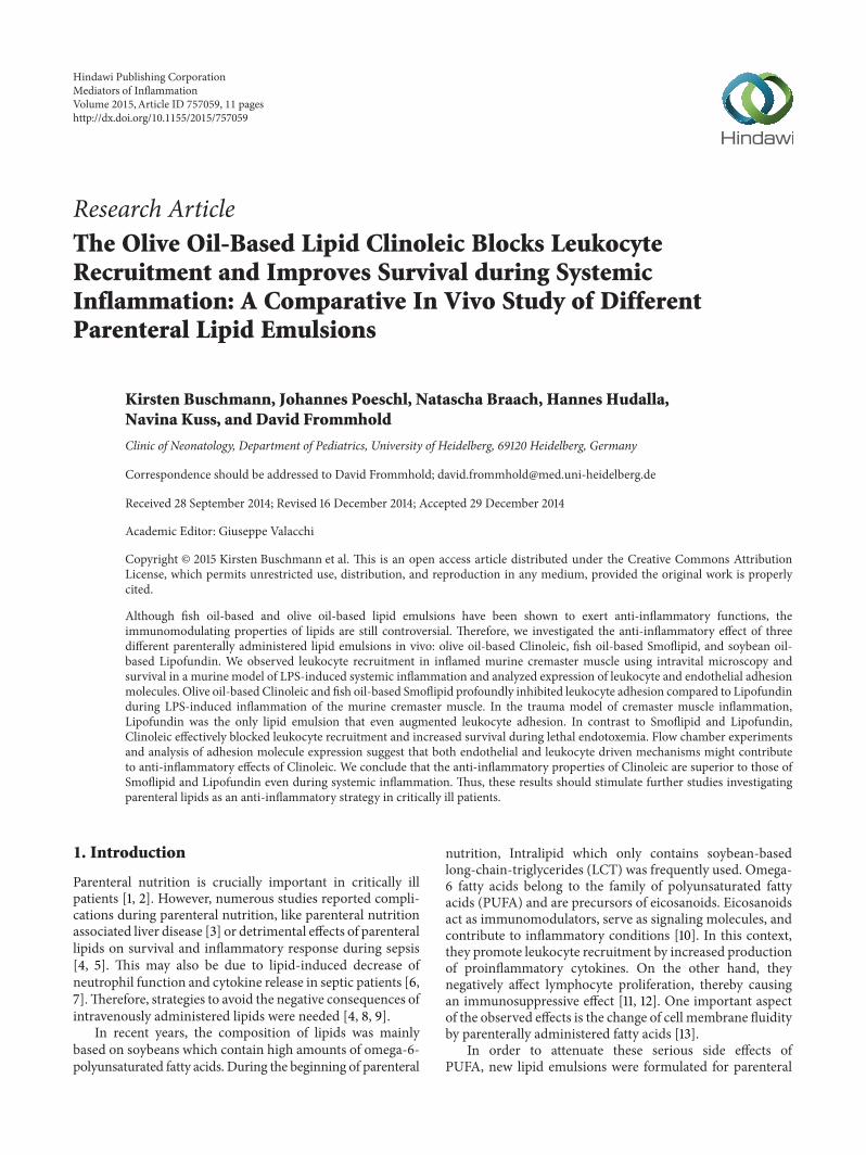
Figure 1: Effect of different lipid compositions on leukocyte recruitment in the traumamodel. Leukocyte adhesion (a) as number of adherentcells per mm2 of surface area and rolling (b) as rolling flux fraction in cremaster muscle venules of mice treated with Clinoleic, Lipofundin,Smoflipid (1 g/kg), or normal saline (control) during trauma-induced inflammation were investigated using intravital microscopy. All valuesare presented as mean ± SEM from three or more mice per group. Significant differences (𝑃 < 0.05) to control mice are indicated by theasterisk.
volume of normal saline was administered i.v. at 2 g/kg 30minutes, 8, and 24 hours after LPS challenge. In a first group,survival was observed for 14 days. In a second group, micewere perfused with saline and lungs were harvested 24 h afterLPS injection. After fixation in PFA (4%) they were preparedfor paraffin-embedded sections. Sections were performed at3 𝜇m thickness and finally stained with H&E (haematoxylinand eosin staining) for microscopic evaluation.
2.11. Statistics. Sigma Stat 3.5 (Systat Software, Erkrath, Ger-many) was used for statistical analysis. Leukocyte counts,vessel diameters, leukocyte adhesion, leukocyte rolling fluxfractions, wall shear rates, and in vitro leukocyte adhesionbetween groups and treatments were compared with one-way ANOVA followed by amultiple pairwise comparison test(Dunn’s test) or by Wilcoxon rank-sum test, as appropriate.To compare the survival during lethal endotoxemia, log-rank test of Kaplan-Meier survival distribution was used.Statistical significance was set at 𝑃 < 0.05.
3. Results and Discussion
3.1. Impact of Lipids on Leukocyte Recruitment duringTrauma-Induced Cremaster Muscle Inflammation. Surgicalpreparation of the cremaster muscle induces leukocyte adhe-sion mainly via the chemokine CXCL1-CXCR2 pathwayand 𝛽
2integrins LFA1 and MAC1 in the short-term model
of trauma-induced inflammation [33, 34]. In our presentexperiments, we analyzed the number of adherent and rollingleukocytes in postcapillary venules of the mouse cremastermuscle in response to intravenous injection of Clinoleic,Smoflipid, Lipofundin, or normal saline. To confirm thesystemic availability of the injected lipid, we first showed thatblood triglyceride levels significantly increased compared tocontrols after all three lipids, while blood levels of cholesteroland standard liver enzymes stayed unaltered (see Supple-mental Table 1 in Supplementary Material available online at
http://dx.doi.org/10.1155/2015/757059). Notably, the varyingcomposition of investigated lipid emulsions did not lead tosignificant differences in blood triglyceride levels. Next, weruled out that alterations of leukocyte recruitment might becaused by hemodynamic changes after fluid injection, sincetherewere nodifferences in hemodynamic andmicrovascularparameters between the different treatment groups (Supple-mental Table 2).
After injection of 1 g/kg Lipofundin, the number ofadherent leukocytes significantly increased when comparedto control conditions (Figure 1(a)). While the same amountof Smoflipid slightly increased leukocyte adhesion, Clinoleicinjection resulted in an insignificant decrease of adher-ent leukocytes. Since Lipofundin significantly reduced thenumber of rolling leukocytes (rolling flux fraction) whencompared to controls (Figure 1(b)), its proinflammatorystimulation triggers the transition from leukocyte rollingto adhesion and rolling leukocytes adhere more frequently.Neither Clinoleic nor Smoflipid treatment altered leukocyterolling compared to controls.
As reported previously during that mild and short-terminflammation of the traumamodel, anti-inflammatory effectsof candidate substances are less common than proinflam-matory effects [29, 30]. Therefore, we argue that potentiallyanti-inflammatory lipid effects are difficult to examine inthat model, although there was an obvious proinflammatorystatus in response to Lipofundin. Therefore, we continuedwith another established inflammation model of the mousecremaster muscle.
3.2. Impact of Lipids on Leukocyte Adhesion during LPS-Induced Cremaster Muscle Inflammation. As a potent proin-flammatory agent, we administered LPS in a dose of 50 ngintrascrotally 3 h prior to exteriorization of the cremastermuscle and observed the lipid-induced effects on leukocyteadhesion in murine cremaster muscle venules.
Microvascular and hemodynamic parameters were simi-lar between the investigated groups (Supplemental Table 3).

Mediators of Inflammation 5
0
200
400
600
800
1000
1200
Control Clinoleic Lipofundin Smoflipid
Adhe
rent
Leu
kocy
tes (
mm2)
∗
∗
(a)
0
2
4
6
8
Control Clinoleic Lipofundin Smoflipid
Rolli
ng fl
ux fr
actio
n (%
)
∗
(b)
Figure 2: Effect of different lipid compositions on leukocyte recruitment during LPS-induced inflammation. Leukocyte adhesion (a) asnumber of adherent cells per mm2 of surface area and rolling (b) as rolling flux fraction in cremaster muscle venules of mice treated withClinoleic, Lipofundin, Smoflipid (1 g/kg), or saline (control) were investigated by intravital microscopy 3 h after intrascrotal administrationof LPS (50 ng/mouse). Lipids were administered simultaneously with LPS. All values are presented as mean ± SEM from three or more miceper group. Significant differences (𝑃 < 0.05) to saline-treated mice are indicated by the asterisk.
In the model of LPS-induced inflammation, a proinflam-matory status is induced by TNF𝛼-mediated upregulationof chemokines and adhesion molecules on leukocytes andthe endothelium [34, 36, 37]. This strong inflammation afterintrascrotal injection of LPS is reflected by profound leuko-cyte adhesion in control mice (Figure 2(a)).The LPS-inducedleukocyte adhesion was robustly blocked by Clinoleic andless pronounced by Smoflipid. In contrast, administration ofLipofundin did not alter leukocyte adhesion in this modelwhen compared to control mice (Figure 2(a)). As depicted inFigure 2(b), leukocyte rolling was not affected by Lipofundinand Smoflipid, whereas rolling flux fraction was significantlyreduced by Clinoleic treatment when compared to controls.Therefore, we suggest that Clinoleic is able to interfere withboth leukocyte rolling and adhesion. We next analyzed thenumber of transmigrated leukocytes in cremaster musclewholemounts in the respective treatment groups, postulatingthat the lipid induced inhibition of leukocyte adhesionlargely translates into transmigration (Supplemental Figure1). Notably, Lipofundin seems to affect neutrophil transmi-gration more than leukocyte adhesion.
The protective effects of Smoflipid and Clinoleic onleukocyte recruitment are most likely explained by theirspecific olive oil and/or fish oil composition [17]. In line withour study, Glatzle et al. found a decrease of LPS- (5mg/kg i.p.)induced leukocyte recruitment after treatment with Clinoleicand less pronounced also with Smoflipid. However, lipidswere applied enterally in their study which is often notfeasible in patients in the intensive care unit [17].
Our observations seem to contrast the study of Buenes-tado et al. in ratmesenterium, which described a Lipofundin-induced inhibition of the whole leukocyte recruitment cas-cade after superfusionwith LPS but no such effect in responseto Clinoleic [16]. The conflicting results, however, might bedue to different experimental setups (LPS application, lipidadministration, and different investigated tissues and species)leading to different involved signaling pathways.
As a summary of our intravital microscopic experiments,we found that among all investigated lipids Clinoleic blocked
leukocyte recruitment most strongly, indicating a protectiverole of olive oil (omega-9 fatty acids) during inflammation.
3.3. Anti-Inflammatory Properties of Lipids during LethalEndotoxemia. Next, we aimed to investigate immunomod-ulatory effects of lipids in a clinically more relevant andwell-established mouse model of lethal endotoxemia. Inthis inflammatory model, an intraperitoneal injection ofEscherichia coli LPS (40mg/kg) is followed by treatmentwith 2 g/kg of the respective lipids (Clinoleic, Smoflipid,or Lipofundin) or control solution (equivalent volume ofnormal saline) after 0.5, 8, and 24 hours.
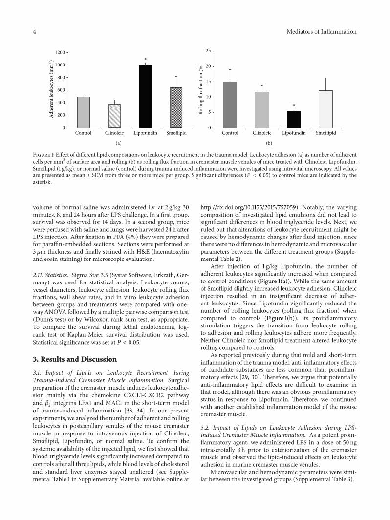
To quantify organ infiltration, some mice were used toinvestigate leukocyte infiltration into the lung 24 h after LPS-injection. We observed an increased leukocyte number afterapplication of LPS (Figure 3(a)) that was unchanged afterinjection of Lipofundin (Figure 3(c)) and hardly improvedafter injection of Smoflipid (Figure 3(d)). In line with ourresults in the above-mentioned inflammation models, wefound a marked reduction of infiltrated PMN after appli-cation of Clinoleic (Figure 3(b)). Garnacho-Montero et al.hypothesized that protective effects of Clinoleic during pul-monary inflammation may be caused by its antioxidativeproperties [38].
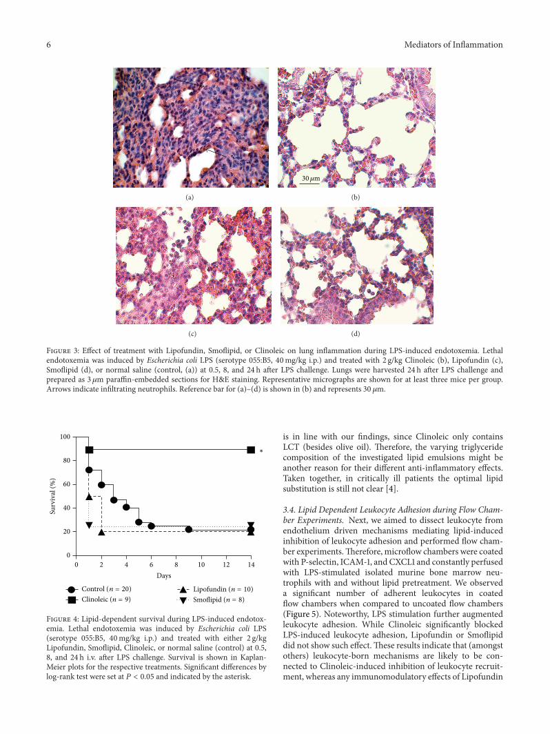
In a second group, survival was observed for 14 days.Consistent with previous findings, survival of control micewas about 20% [29] which stayed unaffected after injection ofLipofundin and Smoflipid (20% and 25%, resp., Figure 4). Incontrast, Clinoleic strongly improved survival during lethalendotoxemia when compared to controls (90% versus 20%,resp.).
These results are consistent with former studies thatobserved a protective effect of olive oil in septic mice [39] andcritically ill patients [17, 40]. However, they are in contrastto other studies describing an anti-inflammatory functionof Smoflipid during endotoxemia [41–43]. Moreover, Bois-rame-Helms et al. investigatedmembrane remodeling duringperitonitis-induced septic shock in rats and found proinflam-matory effects of MCT/LCT but not of LCT only [5]. This
6 Mediators of Inflammation
(a)
30𝜇m
(b)
(c) (d)
Figure 3: Effect of treatment with Lipofundin, Smoflipid, or Clinoleic on lung inflammation during LPS-induced endotoxemia. Lethalendotoxemia was induced by Escherichia coli LPS (serotype 055:B5, 40mg/kg i.p.) and treated with 2 g/kg Clinoleic (b), Lipofundin (c),Smoflipid (d), or normal saline (control, (a)) at 0.5, 8, and 24 h after LPS challenge. Lungs were harvested 24 h after LPS challenge andprepared as 3 𝜇m paraffin-embedded sections for H&E staining. Representative micrographs are shown for at least three mice per group.Arrows indicate infiltrating neutrophils. Reference bar for (a)–(d) is shown in (b) and represents 30 𝜇m.
100
80
60
40
20
0
Surv
ival
(%)
Lipofundin (n = 10)Clinoleic (n = 9)Control (n = 20)
Smoflipid (n = 8)
Days
∗
0 2 4 6 8 10 12 14
Figure 4: Lipid-dependent survival during LPS-induced endotox-emia. Lethal endotoxemia was induced by Escherichia coli LPS(serotype 055:B5, 40mg/kg i.p.) and treated with either 2 g/kgLipofundin, Smoflipid, Clinoleic, or normal saline (control) at 0.5,8, and 24 h i.v. after LPS challenge. Survival is shown in Kaplan-Meier plots for the respective treatments. Significant differences bylog-rank test were set at 𝑃 < 0.05 and indicated by the asterisk.
is in line with our findings, since Clinoleic only containsLCT (besides olive oil). Therefore, the varying triglyceridecomposition of the investigated lipid emulsions might beanother reason for their different anti-inflammatory effects.Taken together, in critically ill patients the optimal lipidsubstitution is still not clear [4].
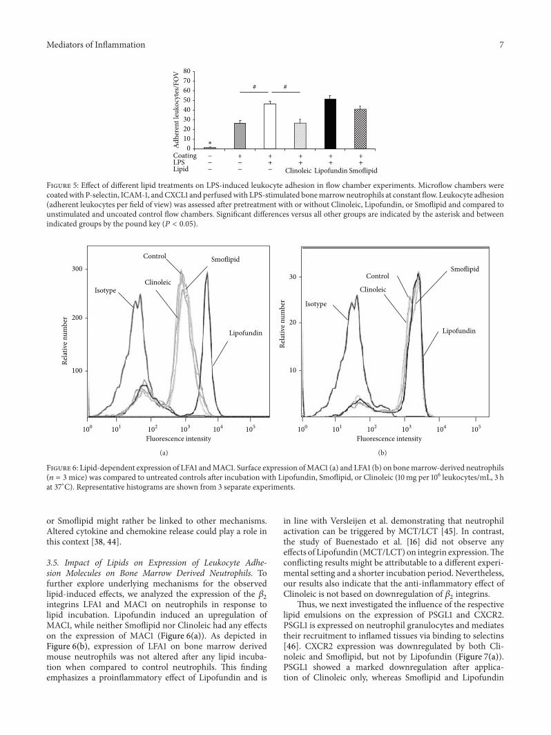
3.4. Lipid Dependent Leukocyte Adhesion during Flow Cham-ber Experiments. Next, we aimed to dissect leukocyte fromendothelium driven mechanisms mediating lipid-inducedinhibition of leukocyte adhesion and performed flow cham-ber experiments.Therefore, microflow chambers were coatedwith P-selectin, ICAM-1, and CXCL1 and constantly perfusedwith LPS-stimulated isolated murine bone marrow neu-trophils with and without lipid pretreatment. We observeda significant number of adherent leukocytes in coatedflow chambers when compared to uncoated flow chambers(Figure 5). Noteworthy, LPS stimulation further augmentedleukocyte adhesion. While Clinoleic significantly blockedLPS-induced leukocyte adhesion, Lipofundin or Smoflipiddid not show such effect.These results indicate that (amongstothers) leukocyte-born mechanisms are likely to be con-nected to Clinoleic-induced inhibition of leukocyte recruit-ment, whereas any immunomodulatory effects of Lipofundin
Mediators of Inflammation 7
CoatingLPSLipid Clinoleic SmoflipidLipofundin
Adhe
rent
leuk
ocyt
es/F
OV
# #
80
70
60
50
40
30
20
10
0−−−
−− −
+ ++
++
++
++
∗
Figure 5: Effect of different lipid treatments on LPS-induced leukocyte adhesion in flow chamber experiments. Microflow chambers werecoatedwith P-selectin, ICAM-1, andCXCL1 and perfusedwith LPS-stimulated bonemarrowneutrophils at constant flow. Leukocyte adhesion(adherent leukocytes per field of view) was assessed after pretreatment with or without Clinoleic, Lipofundin, or Smoflipid and compared tounstimulated and uncoated control flow chambers. Significant differences versus all other groups are indicated by the asterisk and betweenindicated groups by the pound key (𝑃 < 0.05).
Isotype
Smoflipid
Clinoleic
Lipofundin
Control
Fluorescence intensity
300
200
100
Rela
tive n
umbe
r
100 101 102 103 104 105
(a)
Lipofundin
Isotype
Smoflipid
Clinoleic
Control30
20
10
Rela
tive n
umbe
r
Fluorescence intensity100 101 102 103 104 105
(b)
Figure 6: Lipid-dependent expression of LFA1 andMAC1. Surface expression ofMAC1 (a) and LFA1 (b) on bonemarrow-derived neutrophils(𝑛 = 3mice) was compared to untreated controls after incubation with Lipofundin, Smoflipid, or Clinoleic (10mg per 106 leukocytes/mL, 3 hat 37∘C). Representative histograms are shown from 3 separate experiments.
or Smoflipid might rather be linked to other mechanisms.Altered cytokine and chemokine release could play a role inthis context [38, 44].
3.5. Impact of Lipids on Expression of Leukocyte Adhe-sion Molecules on Bone Marrow Derived Neutrophils. Tofurther explore underlying mechanisms for the observedlipid-induced effects, we analyzed the expression of the 𝛽
2
integrins LFA1 and MAC1 on neutrophils in response tolipid incubation. Lipofundin induced an upregulation ofMAC1, while neither Smoflipid nor Clinoleic had any effectson the expression of MAC1 (Figure 6(a)). As depicted inFigure 6(b), expression of LFA1 on bone marrow derivedmouse neutrophils was not altered after any lipid incuba-tion when compared to control neutrophils. This findingemphasizes a proinflammatory effect of Lipofundin and is
in line with Versleijen et al. demonstrating that neutrophilactivation can be triggered by MCT/LCT [45]. In contrast,the study of Buenestado et al. [16] did not observe anyeffects of Lipofundin (MCT/LCT) on integrin expression.Theconflicting results might be attributable to a different experi-mental setting and a shorter incubation period. Nevertheless,our results also indicate that the anti-inflammatory effect ofClinoleic is not based on downregulation of 𝛽
2integrins.
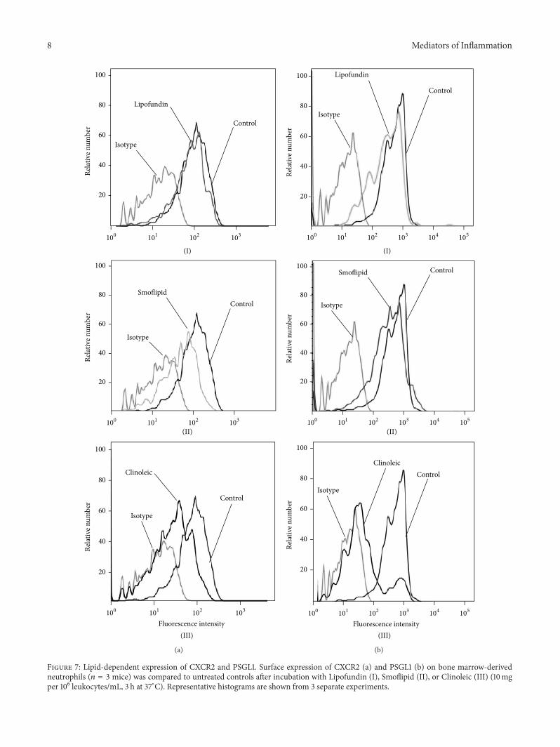
Thus, we next investigated the influence of the respectivelipid emulsions on the expression of PSGL1 and CXCR2.PSGL1 is expressed on neutrophil granulocytes and mediatestheir recruitment to inflamed tissues via binding to selectins[46]. CXCR2 expression was downregulated by both Cli-noleic and Smoflipid, but not by Lipofundin (Figure 7(a)).PSGL1 showed a marked downregulation after applica-tion of Clinoleic only, whereas Smoflipid and Lipofundin
8 Mediators of Inflammation
(I)
Isotype
Lipofundin
Control
100
80
60
40
20
Rela
tive n
umbe
r
100 101 102 103
(II)
Isotype
SmoflipidControl
100
80
60
40
20
Rela
tive n
umbe
r
100 101 102 103
(III)
Isotype
Clinoleic
Control
100
80
60
40
20
Rela
tive n
umbe
r
Fluorescence intensity100 101 102 103
(a)
100
80
60
40
20
Rela
tive n
umbe
r
Isotype
Lipofundin
Control
(I)
100 101 102 103 104 105
Isotype
Smoflipid Control
(II)
100
80
60
40
20
Rela
tive n
umbe
r
100 101 102 103 104 105
Fluorescence intensity
Isotype
ClinoleicControl
(III)
100
80
60
40
20
Rela
tive n
umbe
r
100 101 102 103 104 105
(b)
Figure 7: Lipid-dependent expression of CXCR2 and PSGL1. Surface expression of CXCR2 (a) and PSGL1 (b) on bone marrow-derivedneutrophils (𝑛 = 3 mice) was compared to untreated controls after incubation with Lipofundin (I), Smoflipid (II), or Clinoleic (III) (10mgper 106 leukocytes/mL, 3 h at 37∘C). Representative histograms are shown from 3 separate experiments.
Mediators of Inflammation 9
(III)Fluorescence intensity
100
80
60
40
20
LPS + Clinoleic
LPSIsotype
Control
Rela
tive n
umbe
r
100 101 102 103 104 105
(II)
300
200
100
LPS + Smoflipid
LPSIsotype
Control
Rela
tive n
umbe
r
100 101 102 103 104 105
(I)
100
80
60
40
20
LPS + Lipofundin
LPSIsotype
Control
Rela
tive n
umbe
r
100 101 102 103 104 105
(a)
100 101 102 103 104 105
(I)
Isotype
LPS + Lipofundin LPS
Control
100
80
60
40
20
Rela
tive n
umbe
r
(II)100 101 102 103 104 105
Isotype
LPS + SmoflipidLPS
Control
300
200
100
Rela
tive n
umbe
r
(III)Fluorescence intensity
100 101 102 103 104 105
Isotype
LPS + Clinoleic LPS
Control
100
80
60
40
20
Rela
tive n
umbe
r
(b)
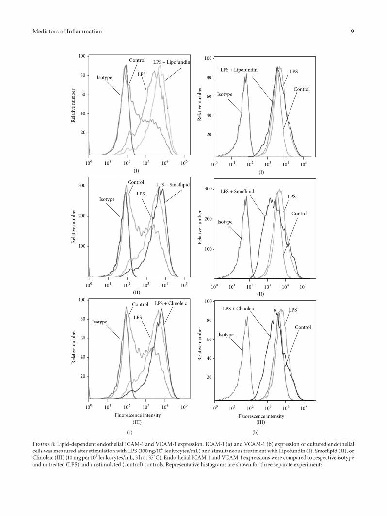
Figure 8: Lipid-dependent endothelial ICAM-1 and VCAM-1 expression. ICAM-1 (a) and VCAM-1 (b) expression of cultured endothelialcells was measured after stimulation with LPS (100 ng/106 leukocytes/mL) and simultaneous treatment with Lipofundin (I), Smoflipid (II), orClinoleic (III) (10mg per 106 leukocytes/mL, 3 h at 37∘C). Endothelial ICAM-1 and VCAM-1 expressions were compared to respective isotypeand untreated (LPS) and unstimulated (control) controls. Representative histograms are shown for three separate experiments.
10 Mediators of Inflammation
displayed no effect (Figure 7(b)). These results suggest thatanti-inflammatory properties of Clinoleic are mediated byPSGL1 and CXCR2 and those of Smoflipid by CXCR2, whichis in line with our observation of reduced LPS-induced leuko-cyte rolling after Clinoleic administration only. AlthoughVersleijen et al. demonstrated that neutrophil activation canbe triggered by MCT/LCT, this does not seem to involveleukocyte-expressed PSGL1 or CXCR2 [45].
3.6. Impact of Lipids on Endothelial Expression of Leuko-cyte Adhesion Molecules. We next addressed the questionwhether lipids alter the expression of endothelial leuko-cyte adhesion molecules like ICAM-1 and VCAM-1. There-fore, LPS-stimulated ICAM-1 and VCAM-1 expression wasassessed on MAECs by flow cytometry. In line with previousstudies, LPS induced an upregulation of ICAM-1 and—less pronounced—of VCAM-1. While none of the appliedlipids altered ICAM-1-expression (Figure 8(a)), LPS-inducedVCAM-1 expression was downregulated by Smoflipid andClinoleic (Figure 8(b)), indicating that an anti-inflammatoryeffect of omega 3 and omega 9 fatty acidsmight be attributableto endothelial VCAM-1 downregulation in our experimentalsetting. This finding is supported by the study of Tullet al. which investigated endothelial mechanisms of lipid-mediated immunomodulation [44].
4. Conclusion
Clinoleic exerted the strongest anti-inflammatory propertiesduring local and systemic inflammation in vivo when com-pared to the lipid composition Smoflipid or Lipofundin. Inturn, Clinoleic was the only investigated lipid emulsion thatimproved survival during lethal endotoxemia. Although oliveoil-based lipids seem to be a beneficial alternative to otherlipid emulsions, future studies are needed to confirm the anti-inflammatory potential of Clinoleic in critically ill patients.
Conflict of Interests
The authors declare that there is no conflict of interests forany of the authors.
Acknowledgments
The authors thank Melitta Weissinger and Claudia Felbingerfor their excellent technical assistance in performing intravi-tal microscopy and flow cytometry.The authors acknowledgethe financial support of the Deutsche Forschungsgemein-schaft and Ruprecht-Karls-Universitat Heidelberg within thefunding programme Open Access Publishing.
References
[1] D. K. Heyland, “Nutritional support in the critically ill patient.A critical review of the evidence,” Critical Care Clinics, vol. 14,no. 3, pp. 423–440, 1998.
[2] D. K. Heyland, S. MacDonald, L. Keefe, and J. W. Drover, “Totalparenteral nutrition in the critically III patient: ameta-analysis,”
Journal of the AmericanMedical Association, vol. 280, no. 23, pp.2013–2019, 1998.
[3] S. J. Carlson, P. Nandivada, M. I. Chang et al., “The additionof medium-chain triglycerides to a purified fish oil-based dietalters inflammatory profiles in mice,”Metabolism, vol. 64, no. 2,pp. 274–282, 2015.
[4] E. A.Miles andP. C. Calder, “Fatty acids, lipid emulsions and theimmune and inflammatory systems,”World Review of Nutritionand Dietetics, vol. 112, pp. 17–30, 2015.
[5] J. Boisrame-Helms, A. Said, M. Burban et al., “Medium-chaintriglycerides supplementation exacerbates peritonitis-inducedseptic shock in rats: role on cell membrane remodeling,” Shock,vol. 42, no. 6, pp. 548–553, 2014.
[6] K. Mayer, C. Fegbeutel, K. Hattar et al., “𝜔-3 vs. 𝜔-6 lipidemulsions exert differential influence on neutrophils in septicshock patients: impact on plasma fatty acids and lipid mediatorgeneration,” Intensive Care Medicine, vol. 29, no. 9, pp. 1472–1481, 2003.
[7] K. Mayer, S. Gokorsch, C. Fegbeutel et al., “Parenteral nutritionwith fish oil modulates cytokine response in patients withsepsis,” American Journal of Respiratory and Critical CareMedicine, vol. 167, no. 10, pp. 1321–1328, 2003.
[8] J. A. Meisel, H. D. Le, V. E. de Meijer et al., “Comparison of 5intravenous lipid emulsions and their effects on hepatic steatosisin a murine model,” Journal of Pediatric Surgery, vol. 46, no. 4,pp. 666–673, 2011.
[9] M. P. Casaer and G. van den Berghe, “Nutrition in the acutephase of critical illness,” The New England Journal of Medicine,vol. 370, no. 13, pp. 1227–1236, 2014.
[10] G. W. Fischer, K. W. Hunter, S. R. Wilson, and A. D. Mease,“Diminished bacterial defences with intralipid,”TheLancet, vol.2, no. 8199, pp. 819–820, 1980.
[11] K. Furukawa, H. Yamamori, K. Takagi et al., “Influences ofsoybean oil emulsion on stress response and cell-mediatedimmune function in moderately or severely stressed patients,”Nutrition, vol. 18, no. 3, pp. 235–240, 2002.
[12] P. C. Calder, E. J. Sherrington, J. Askanazi, andE.A.Newsholme,“Inhibition of lymphocyte proliferation in vitro by two lipidemulsions with different fatty acid compositions,” ClinicalNutrition, vol. 13, no. 2, pp. 69–74, 1994.
[13] P. C. Calder, P. Yaqoob, D. J. Harvey, A. Watts, and E. A.Newsholme, “Incorporation of fatty acids by concanavalin A-stimulated lymphocytes and the effect on fatty acid compositionandmembrane fluidity,” Biochemical Journal, vol. 300, no. 2, pp.509–518, 1994.
[14] E. Macia-Botejara, J. M. Moran-Penco, M. T. Espın-Jaime etal., “Brain lipid composition in rabbits after total parenteralnutrition with two different lipid emulsions,” Nutrition, vol. 29,no. 1, pp. 313–317, 2013.
[15] B. Juttner, J. Kroplin, S. M. Coldewey et al., “Unsaturatedlong-chain fatty acids induce the respiratory burst of humanneutrophils and monocytes in whole blood,” Nutrition andMetabolism, vol. 5, no. 1, article 19, 2008.
[16] A. Buenestado, J. Cortijo, M.-J. Sanz et al., “Olive oil-basedlipid emulsion’s neutral effects on neutrophil functions andleukocyte-endothelial cell interactions,” Journal of Parenteral &Enteral Nutrition, vol. 30, no. 4, pp. 286–296, 2006.
[17] J. Glatzle, S. Beckert, M. S. Kasparek et al., “Olive oil is morepotent than fish oil to reduce septic pulmonary dysfunctions inrats,” Langenbeck’s Archives of Surgery, vol. 392, no. 3, pp. 323–329, 2007.

Mediators of Inflammation 11
[18] G. Deshpande, K. Simmer, M. Deshmukh, T. A. Mori, K. D.Croft, and J. Kristensen, “Fish oil (SMOFlipid) and olive oillipid (clinoleic) in very preterm neonates,” Journal of PediatricGastroenterology and Nutrition, vol. 58, no. 2, pp. 177–182, 2014.
[19] H. Li, X. Z. Ruan, S. H. Powis et al., “EPA andDHA reduce LPS-induced inflammation responses in HK-2 cells: evidence for aPPAR-𝛾-dependent mechanism,” Kidney International, vol. 67,no. 3, pp. 867–874, 2005.
[20] P. C. Calder and P. Yaqoob, “Understanding omega-3 polyun-saturated fatty acids,” Postgraduate Medicine, vol. 121, no. 6, pp.148–157, 2009.
[21] T. A. Springer, “Traffic signals on endothelium for lymphocyterecirculation and leukocyte emigration,” Annual Review ofPhysiology, vol. 57, pp. 827–872, 1995.
[22] K. Ley, C. Laudanna, M. I. Cybulsky, and S. Nourshargh,“Getting to the site of inflammation: the leukocyte adhesioncascade updated,” Nature Reviews Immunology, vol. 7, no. 9, pp.678–689, 2007.
[23] M. Sperandio, A.Thatte, D. Foy, L. G. Ellies, J. D. Marth, and K.Ley, “Severe impairment of leukocyte rolling in venules of core2 glucosaminyltransferase-deficient mice,” Blood, vol. 97, no. 12,pp. 3812–3819, 2001.
[24] N. Braach, D. Frommhold, K. Buschmann et al., “RAGEcontrols activation and anti-inflammatory signalling of proteinC,” PLoS ONE, vol. 9, no. 2, Article ID e89422, 2014.
[25] H. Zeintl, F. U. Sack, M. Intaglietta, and K. Messmer, “Com-puter assisted leukocyte adhesion measurement in intravitalmicroscopy,” International Journal of Microcirculation, Clinicaland Experimental, vol. 8, no. 3, pp. 293–302, 1989.
[26] H. H. Lipowsky and B. W. Zweifach, “Application of the “two-slit” photometric technique to themeasurement of microvascu-lar volumetric flow rates,”Microvascular Research, vol. 15, no. 1,pp. 93–101, 1978.
[27] D. S. Long, M. L. Smith, A. R. Pries, K. Ley, and E. R.Damiano, “Microviscometry reveals reduced blood viscosityand altered shear rate and shear stress profiles in microvesselsafter hemodilution,” Proceedings of the National Academy ofSciences of the United States of America, vol. 101, no. 27, pp.10060–10065, 2004.
[28] M. L. Smith, D. S. Long, E. R. Damiano, and K. Ley, “Near-wall𝜇-PIV reveals a hydrodynamically relevant endothelial surfacelayer in venules in vivo,” Biophysical Journal, vol. 85, no. 1, pp.637–645, 2003.
[29] D. Frommhold, J. Tschada, N. Braach et al., “Protein C con-centrate controls leukocyte recruitment during inflammationand improves survival during endotoxemia after efficient in vivoactivation,” The American Journal of Pathology, vol. 179, no. 5,pp. 2637–2650, 2011.
[30] K. Buschmann, L. Koch, N. Braach et al., “CXCL1-triggeredinteraction of LFA1 and ICAM1 control glucose-induced leuko-cyte recruitment during inflammation in vivo,” Mediators ofInflammation, vol. 2012, Article ID 739176, 12 pages, 2012.
[31] M. Kobayashi, K. Inoue, E. Warabi, T. Minami, and T. Kodama,“A simple method of isolating mouse aortic endothelial cells,”Journal of Atherosclerosis andThrombosis, vol. 12, no. 3, pp. 138–142, 2005.
[32] J. Schymeinsky, A. Sindrilaru, D. Frommhold et al., “The Vavbinding site of the non-receptor tyrosine kinase Syk at Tyr348 is critical for𝛽2 integrin (CD11/CD18)-mediated neutrophilmigration,” Blood, vol. 108, no. 12, pp. 3919–3927, 2006.
[33] M. L. Smith, M. Sperandio, E. V. Galkina, and K. Ley, “Autop-erfused mouse flow chamber reveals synergistic neutrophil
accumulation through P-selectin and E-selectin,” Journal ofLeukocyte Biology, vol. 76, no. 5, pp. 985–993, 2004.
[34] D. Frommhold, A. Ludwig, M. G. Bixel et al., “SialyltransferaseST3Gal-IV controls CXCR2-mediated firm leukocyte arrestduring inflammation,” The Journal of Experimental Medicine,vol. 205, no. 6, pp. 1435–1446, 2008.
[35] N. Braach, K. Buschmann, J. Pflaum et al., “Anti-inflammatoryfunctions of protein c require RAGE and ICAM-1 in a stimulus-dependent manner,” Mediators of Inflammation, vol. 2014,Article ID 743678, 12 pages, 2014.
[36] D. Heumann and T. Roger, “Initial responses to endotoxins andGram-negative bacteria,” Clinica Chimica Acta, vol. 323, no. 1-2,pp. 59–72, 2002.
[37] D. Frommhold, A. Kamphues, I. Hepper et al., “RAGE andICAM-1 cooperate in mediating leukocyte recruitment duringacute inflammation in vivo,” Blood, vol. 116, no. 5, pp. 841–849,2010.
[38] J. Garnacho-Montero, C. Ortiz-Leyba, M. C. Garnacho-Montero et al., “Effects of three intravenous lipid emulsionson the survival and mononuclear phagocyte function of septicrats,” Nutrition, vol. 18, no. 9, pp. 751–754, 2002.
[39] M. S. Leite, P. Pacheco, R. N. Gomes et al., “Mechanisms ofincreased survival after lipopolysaccharide-induced endotoxicshock inmice consuming olive oil-enriched diet,” Shock, vol. 23,no. 2, pp. 173–178, 2005.
[40] A. Sala-Vila, V. M. Barbosa, and P. C. Calder, “Olive oil inparenteral nutrition,” Current Opinion in Clinical Nutrition andMetabolic Care, vol. 10, no. 2, pp. 165–174, 2007.
[41] P. C. Calder, “Omega-3 fatty acids and inflammatory processes,”Nutrients, vol. 2, no. 3, pp. 355–374, 2010.
[42] P. C. Calder, “Omega-3 polyunsaturated fatty acids and inflam-matory processes: nutrition or pharmacology?” British Journalof Clinical Pharmacology, vol. 75, no. 3, pp. 645–662, 2013.
[43] P. C. Calder, “Dietarymodification of inflammationwith lipids,”Proceedings of the Nutrition Society, vol. 61, no. 3, pp. 345–358,2002.
[44] S. P. Tull, C. M. Yates, B. H. Maskrey et al., “Omega-3 fattyacids and inflammation: novel interactions reveal a new step inneutrophil recruitment,” PLoS Biology, vol. 7, no. 8, Article IDe1000177, 2009.
[45] M. W. J. Versleijen, J. C. J. van Esterik, H. M. J. Roelofs,S. E. van Emst-de Vries, P. H. G. M. Willems, and G. J. A.Wanten, “Parenteral medium-chain triglyceride-induced neu-trophil activation is not mediated by a Pertussis Toxin sensitivereceptor,” Clinical Nutrition, vol. 28, no. 1, pp. 59–64, 2009.
[46] D. Asa, L. Raycroft, L. Ma et al., “The P-selectin glycoproteinligand functions as a common human leukocyte ligand for P-and E-selectins,” Journal of Biological Chemistry, vol. 270, no.19, pp. 11662–11670, 1995.

Submit your manuscripts athttp://www.hindawi.com
Stem CellsInternational
Hindawi Publishing Corporationhttp://www.hindawi.com Volume 2014
Hindawi Publishing Corporationhttp://www.hindawi.com Volume 2014
MEDIATORSINFLAMMATION
of
Hindawi Publishing Corporationhttp://www.hindawi.com Volume 2014
Behavioural Neurology
EndocrinologyInternational Journal of
Hindawi Publishing Corporationhttp://www.hindawi.com Volume 2014
Hindawi Publishing Corporationhttp://www.hindawi.com Volume 2014
Disease Markers
Hindawi Publishing Corporationhttp://www.hindawi.com Volume 2014
BioMed Research International
OncologyJournal of
Hindawi Publishing Corporationhttp://www.hindawi.com Volume 2014
Hindawi Publishing Corporationhttp://www.hindawi.com Volume 2014
Oxidative Medicine and Cellular Longevity
Hindawi Publishing Corporationhttp://www.hindawi.com Volume 2014
PPAR Research
The Scientific World JournalHindawi Publishing Corporation http://www.hindawi.com Volume 2014
Immunology ResearchHindawi Publishing Corporationhttp://www.hindawi.com Volume 2014
Journal of
ObesityJournal of
Hindawi Publishing Corporationhttp://www.hindawi.com Volume 2014
Hindawi Publishing Corporationhttp://www.hindawi.com Volume 2014
Computational and Mathematical Methods in Medicine
OphthalmologyJournal of
Hindawi Publishing Corporationhttp://www.hindawi.com Volume 2014
Diabetes ResearchJournal of
Hindawi Publishing Corporationhttp://www.hindawi.com Volume 2014
Hindawi Publishing Corporationhttp://www.hindawi.com Volume 2014
Research and TreatmentAIDS
Hindawi Publishing Corporationhttp://www.hindawi.com Volume 2014
Gastroenterology Research and Practice
Hindawi Publishing Corporationhttp://www.hindawi.com Volume 2014
Parkinson’s Disease
Evidence-Based Complementary and Alternative Medicine
Volume 2014Hindawi Publishing Corporationhttp://www.hindawi.com