research article the effect of primary cancer cell culture...
TRANSCRIPT

Research ArticleThe Effect of Primary Cancer Cell Culture Models on theResults of Drug Chemosensitivity Assays: The Application ofPerfusion Microbioreactor System as Cell Culture Vessel
Chia-Hsun Hsieh,1,2 Yi-Dao Chen,3 Shiang-Fu Huang,4
Hung-Ming Wang,5 and Min-Hsien Wu3
1Division of Hematology-Oncology, Department of Internal Medicine, Chang Gung Memorial Hospital at Linkou,Taoyuan 333, Taiwan2Department of Chemical and Materials Engineering, Chang Gung University, Taoyuan 333, Taiwan3Graduate Institute of Biochemical and Biomedical Engineering, Chang Gung University, Taoyuan 333, Taiwan4Department of Otolaryngology-Head and Neck Surgery, Chang Gung Memorial Hospital, Chang Gung University,Taoyuan 333, Taiwan5Division of Hematology-Oncology, Department of Internal Medicine, Chang Gung Memorial Hospital,Chang Gung University, Taoyuan 333, Taiwan
Correspondence should be addressed to Min-Hsien Wu; [email protected]
Received 7 August 2014; Revised 17 September 2014; Accepted 19 September 2014
Academic Editor: Jeroen Rouwkema
Copyright © 2015 Chia-Hsun Hsieh et al. This is an open access article distributed under the Creative Commons AttributionLicense, which permits unrestricted use, distribution, and reproduction in any medium, provided the original work is properlycited.
To precisely and faithfully perform cell-based drug chemosensitivity assays, a well-defined and biologically relevant culturecondition is required. For the former, a perfusion microbioreactor system capable of providing a stable culture condition wasadopted. For the latter, however, little is known about the impact of culture models on the physiology and chemosensitivityassay results of primary oral cavity cancer cells. To address the issues, experiments were performed. Results showed that minorenvironmental pH change could significantly affect the metabolic activity of cells, demonstrating the importance of stable culturecondition for such assays. Moreover, the culture models could also significantly influence the metabolic activity and proliferationof cells. Furthermore, the choice of culture models might lead to different outcomes of chemosensitivity assays. Compared with thesimilar test based on tumor-level assays, the spheroid model could overestimate the drug resistance of cells to cisplatin, whereas the2D and 3D culture models might overestimate the chemosensitivity of cells to such anticancer drug. In this study, the 3D culturemodels with same cell density as that in tumor samples showed comparable chemosensitivity assay results as the tumor-level assays.Overall, this study has provided some fundamental information for establishing a precise and faithful drug chemosensitivity assay.
1. Introduction
Chemotherapy is a kind of cancer treatments in whichchemical substances are utilized to kill cancer cells in humanbody. Currently, the decision of a chemotherapy regimen isstill based on the empirical information from clinical trials inpatients which ignores biological individuality of tumor [1].In fact, the therapeutic effects of anticancer drugs to cancercells exhibit high degree of variation [2] because individualpatient’s tumor is genotypically and phenotypically different[3]. For a more personalized chemotherapy, therefore, an in
vitro chemosensitivity assays is required to evaluate whichanticancer drugs the patient’s cancer cells will respond to.This can assist doctors to tailor a chemotherapy regimen forindividual patients. In vitro anticancer drug chemosensitivityassays mainly involve the basic procedures including (1)isolation of cancer cells from a tumor sample, (2) incubationof cancer cells with anticancer drugs, (3) evaluation of cancercell viability, and (4) interpretation of the results [1].
For most cell-based assays (e.g., drug chemosensitivityassays), static cell culture models [4, 5], where the culturemedium is virtually supplied in a manual and batch-wise
Hindawi Publishing CorporationBioMed Research InternationalVolume 2015, Article ID 470283, 10 pageshttp://dx.doi.org/10.1155/2015/470283

2 BioMed Research International
manner, were commonly adopted. Nevertheless, this couldlead to a fluctuating culture condition [6] that could in turnhamper the precise quantification of the link between thedrug conditions tested and cancer cells’ response. Moreover,most of the conventional cell culture models are relativelylarge in scale, which could therefore require larger number ofcells for a cell-based assay. In drug chemosensitivity assays,however, the clinical tumor samples harvested and thus thecancer cells isolated are normally limited. Therefore, theisolated primary cancer cells generally need to be expendedin number for the subsequent cell-based assays. Nevertheless,the expansion process of cell number (e.g., cell proliferationon a 2D surface) could possibly alter the cellular physi-ology [7] and in turn might affect the faithfulness of thefollowing chemosensitivity assays. In addition, the cell cultureconditions in a relatively large cell culture scale might notbe regarded as homogenous mainly due to the chemicalgradient phenomenon existing in the cell culture system.Such poorly defined culture conditions could restrict theprecise quantification of the link between cellular responsesand anticancer drug conditions. To tackle the above technicalissues, more recently, perfusion-based microscale bioreactorsystems were actively proposed for various cell-based assays[6, 8–10] by which a stable andwell-defined culture conditioncan be achieved due to the continuous medium perfusionformat and miniaturized cell culture scale [6, 8].
For the most drug chemosensitivity assays [11–13], more-over, two-dimensional (2D) monolayer cell cultures arecommonly used, where the cancer cells attach, spread, andgrow on a surface. Such a cell culture model has been widelyadopted in life science-related research for more than ahundred years. This is primarily because of its simplicityin terms of the cell culture preparation and the subsequentmicroscopic observation of cell culture. Nevertheless, 2Dculture conditions might not well simulate the in vivomicroenvironments surrounding biological cells since cellsinhabit environments with very 3D features [14]. It has beenrecognized that cancer cells in a 2D culture environmentdiffer physiologically from those in a 3D environment [15]. Inaddition to the conventional 2D cell culture model, spheroidculture models, in which cells self-aggregate to form sphere-like 3D cell clusters, are regarded as excellent models fortumor tissues [16]. Due to their 3Dnature, they are believed toprovide a more biologically relevant microenvironment than2Dmonolayer cultures [17]. Spheroid culturemodels are thuswidely utilized in various cancer cell researches [18, 19].
As aforementioned, cells inhabit environments with veryspecific 3D features in animal tissues. In their native 3Denvironment,mammalian cells are subject to not only variousbiological cues such as soluble signaling molecules, butalso cell-to-cell interactions and mechanics and dynamics ofthe surrounding extracellular matrix (ECM) [20]. All thesebiological signals may determine the fate of cells to undergoproliferation, differentiation, or apoptosis. Borrowing fromthe concept of tissue engineering, 3-dimensional (3D) culturemodels, where the biological cells are encapsulated in a 3Dpolymeric scaffold, are generally believed to provide a betterapproximation of the in vivo conditions than 2D culturemod-els.Therefore, they could provide amore biologically relevant
and thus physiologically meaningful culture condition forcell-based assays [21, 22]. Thus far, various 3D cell culturemodels have been proposed for cancer-related researches[23, 24].
In order to faithfully and precisely investigate the cancercells’ response to anticancer drugs, a stable, well-defined, andbiologically relevant cellular microenvironment is needed. Inthis study, a perfusion-based microscale cell culture systemcapable of providing a stable culture condition [6] wasused for the cell-based chemosensitivity assays. Before theapplication of drug chemosensitivity assays for guiding futurechemotherapy plans, however, there are some fundamentalbiological issues needed to be addressed. These include whatis the result difference of the chemosensitivity assays based onthe above-mentioned cell culture models (i.e., conventional2D, spheroid, and 3D culture models) and which resultsare closer to the chemosensitivity assay results based ontumor tissue-level assays, an assay model which is morerepresentative of the in vivo condition than the cell-basedassay counterpart. To more realistically answer the abovequestions, primary oral cavity cancer cells were used for theassays in this study compared with the cell line models inthe previous study [9, 15]. Results revealed that even minorenvironmental pH change could significantly influence themetabolic activity of the cultured primary cancer cells,demonstrating the importance of stable culture condition fora precise cell-based assay. Moreover, the choice of cell cultureformats (e.g., 2D, 3D, or spheroid culture models) might playan important role in the physiology (e.g., metabolic activityor cell proliferation) of the cultured cells. Also the use ofdifferent cell culture models could lead to different resultsof drug chemosensitivity assays. Compared with the tumortissue-level chemosensitivity assays, moreover, the 3D culturemodels with same cell density as that in tumor tissue samplesshowed comparable chemosensitivity assay results as thetumor tissue-level assays. As a whole, this study has providedsome fundamental information regarding the influence ofcell culture methods on the results of in vitro cell-basedassays. All these pieces of information are of great importancefor establishing a precise and faithful drug chemosensitivityassay.
2. Material and Methods
2.1. Fabrication and Experimental Setup of Perfusion Micro-bioreactor System. In this study, the perfusion microbioreac-tor system proposed in our previous study [6] was utilized tocarry out primary cancer cell-based chemosensitivity assays.Briefly, the perfusion microbioreactor system consists ofa microbioreactor chamber module, a plug module, anda bottom layer module (Figure 1(a)). The microbioreactorchamber module is composed of 9 cylindrical microbiore-actor chambers with each microbioreactor having the sameformat as the well of a standard 96-well microplate (D: 7mm;H: 7mm).The plugmodule (Figure 1(a)) contains 9 columns,which are able to plug up the 9 microbioreactor chambersaccordingly to form multiple closed systems for perfusioncell culture. Similar to the plug module, the bottom layer

BioMed Research International 3
Waste medium collector
ITO-microheater controller
Syringe pump Microbioreactor chamber modulePlug moduleBottom layer module
(a)
120mm
5mm 1.5mm 7mm5mm
2mm 0.2mm 0.2mm
23.5mm
Tumor and spheroidculture chamber
3D culture chamber 2D culture chamber
(b)
Medium inletMedium outletMedium perfusion layerCell culture layer
(c)
Figure 1: (a) The experimental setup of perfusion microbioreactor system, (b) the schematic illustration of bottom layer module (upperillustration: the topside view, lower illustration: the cross-section view), and (c) the cross-section view of microbioreactor.
module (Figure 1(b)) contains 9 columns to plug up the 9microbioreactor chambers at the bottom side accordingly.In this work, each column on the bottom layer modulenot only functions as a “stopper” to seal each cylindricalmicrobioreactor chamber in each column (Figure 1(b)) butalso acts as the compartment to accommodate the cancer cellsin 3 different formats for 2D monolayer, 3D, and spheroidcell culture. In addition, such compartment was also usedfor tumor tissue-based chemosensitivity assay. Figure 1(c)demonstrates the cross-section view of eachmicrobioreactor.In this study, all the modules in the microbioreactor systemwere constructed simply by casting of polydimethylsiloxane(PDMS) polymer (Sylgard 184, Dow Corning, USA) ona polymethylmethacrylate (PMMA) mold fabricated usingmicromachining technique as described previously [6]. Forperfusion cell culture purpose, each microbioreactor wasperfused with its own separate medium supply through
silicon tubing driven by a multichannel syringe pump (KDS220, KD Scientific Ltd., USA). In this work, the PDMS wallsof the microbioreactors were punched using a needle tosimply create holes for tubing insertion. In addition, theperfusion microbioreactor system was placed on the surfaceof a transparent indium tin oxide- (ITO-) based microheaterchip to provide a stable thermal condition of 37±0.2∘C for cellculture [6, 25]. The entire experimental setup was illustratedin Figure 1(a).
2.2. The Isolation of Primary Oral Cavity Cancer Cells.The study was approved by the Institutional Review Boardof the Chang Gung Memorial Hospital and the informedconsent was obtained from all patients (Approval ID: 102-3943B). The clinical tumor tissues were resected from theoral cavity cancer patients in a local medical center. Thesamples were harvested within 2-3 hrs of surgery. The tumor

4 BioMed Research International
(a) (b)
100𝜇m
(c)
100𝜇m
(d)
Figure 2: (a) Tumor tissue from an oral cavity cancer patient, (b) diced tumor tissue particles, (c) fluorescence microscopic observation ofthe isolated primary oral cavity cancer cells (green and red dots represent live and dead cells resp.), and (d) immunofluorescent microscopicimages of the isolated primary oral cavity cancer cells (Hoechst dye positive (nucleated cells): the blue dots; EGFR dye positive (cancer cells):the green dots).
tissue samples obtained (Figure 2(a)) were first rinsed usingphosphate buffered saline (PBS; Invitrogen, Taiwan) andthen diced to tiny cubic particles (approximate size: L:0.7mm; V : 0.343mm3) (Figure 2(b)) using a surgical scalpelblade. All process was carried out aseptically. The preparedtumor tissue particles were placed in a tissue culture flaskcontaining 10mL of RPMI-1640 medium (unless otherwisestated, all reagents were purchased from Sigma, Taiwan) sup-plemented with 2mgmL−1 collagenase-1. The tumor tissueparticles were enzymatically digested at 37∘C under shakingcondition for approximately 20 hrs. After incubation, thedigested suspension was filtered through a tea strainer toremove undigested tissue and subsequently through a 20 𝜇mpore sterile filter. The cancer cells in the filtrate were thenwashed three times with RPMI-1640 medium solution byrepeated centrifugation in a centrifuge (1,800 rpm for 5min)and resuspended in RPMI-1640 medium solution. The cellsuspension thus obtained was assessed microscopically forcell number and cell viability using a fluorescent dye kit(LIVE/DEAD Viability/Cytotoxicity Kit L-3224, MolecularProbes) [9, 15]. After cell staining, briefly, the images of live(green) and dead (red) cells were captured using a confocalmicroscope (LSM 510 META, Zeiss, Germany). This wasfollowed by an image analysis to evaluate cell viability [9].Only cell preparations with cell viability >95% were thenused. In addition, the purity of primary oral cavity cancer cells
isolated was also evaluated microscopically using an EGFRfluorescent dye kit [26] and the subsequent image analysis.
2.3. The Metabolic Activity of Primary Oral Cavity CancerCells Cultured under Different Medium pH Conditions in the2D, Spheroid, and 3DCell CultureModels. Conventional cell-based chemosensitivity assays are normally performed usinga static cell culturemodelwhichmight not be able to provide astable and thuswell-defined culture condition for such assays.In order to investigate the extent towhich the physiology (e.g.,metabolic activity) of primary cancer cells was influenced bysuch an unstable culture condition (e.g., culturemediumpH),we performed the following experiments. In this work, thefreshly isolated oral cavity cancer cells with equal total cellnumber (5.7× 10³) were cultured in 3 different culturemodels(i.e., 2D, 3D, and spheroid cell culture models) and underdifferent medium pH (pH 6.6, 6.8, 7.0, 7.2, and 7.4) conditionsusing the perfusion microbioreactor system (Figure 1(a)).Briefly, 5.7 × 10³ primary oral cavity cancer cells were seededon the bottom surface of themicrobioreactor chamber for 2Dculture (Figure 1). To achieve this, the bottom PDMS surfaceof chamber was treated with 0.01% fibronectin solution (1-hour immersion) to enhance cancer cell attachment. Afterloading cell suspension into the chamber, moreover, 4-hourtime was given to allow cell sedimentation and attachmentbased on our preliminary test. In addition, the equal amount

BioMed Research International 5
of cancer cells was encapsulated in collagen-alginate hydrogel(2.4% (w/v) Type I collagen and 0.2% (w/v) alginate) to forma3D cell culture construct (D: 1.5mm;H: 0.2mm;V : 0.353 𝜇L),having a cell density of 1.62 × 10⁷ cells mL−1, for the perfusion3D cell culture. For the spheroid cell culture, its preparationwas based on the conventional spheroid formation method,namely, the hanging drop technique [27]. In this study, thevolume of each hanging drop (cell density: 1.5 × 10⁵ cellsmL−1) was 38 𝜇L.
After the 3 cancer cell culture models were prepared,perfusion cell culture was performed using the microbiore-actor system (Figure 1(a)) for up to 2 days. In this work, theculture medium with different pH levels as aforementionedwas continuously supplied to the microbioreactors at theflow rate of 15 𝜇L hr−1. During the culture period, the wastemedium was collected for the measurement of lactate. Inthis study, the lactate produced and released into culturemedium was measured using a Lactate Reagent Kit (TrinityBiotech Plc., Ireland) [6]. The assay was carried out asdirected by manufacturer’s instructions. A lactate solutionat a concentration of 50∼500mg L−1 made from dissolvinglactate sodium salt in deionized water (DI) water was used asstandard. Moreover, the total number of cells after culturingwas also explored by quantifying the DNA content of the cells[6].
2.4. Drug Chemosensitivity of Primary Oral Cavity CancerCells to Cisplatin under Perfusion 2D, Spheroid, 3D, andTumor Tissue Culture Models. In this study, the anticancerdrug chemosensitivity of the primary oral cavity cancer cellscultured in the 3 different models as aforementioned wasevaluated. Briefly, the 3 cell culture models were preparedsimilarly to the descriptions in Section 2.3. In this work, thetotal cell numbers used in each cell culture models were 3 ×10⁴, in which the cell density of the 3D cell culture model was7.6 × 106 cells mL−1. For the 3D cell culture model, anothercase with higher cell density (8.5 × 10⁷ cells mL−1) used wasalso investigated. For comparison purpose, a tumor tissueculture model was also established, in which the preparedtumor tissue particles (Figure 2(b)) were directly culturedin the microbioreactor. In this work, the culture mediumsupplemented with the cisplatin at 3 different concentrations(0, 4, or 8𝜇gmL−1) was supplied to the microbioreactors atthe flow rate of 15 𝜇L hr−1. After 2-day culture, the culturedcells and tumors were assayed for cell viability using the CellCounting Kit 8 (CCK-8) [28]. Apart from the quantitativeevaluation, the viabilities of the oral cavity cancer cellscultured in the 3 different cell culturemodels and in the tumorculture model tested after treatment with different concen-trations of cisplatin were observed microscopically using thefluorescent dye kit (LIVE/DEAD Viability/Cytotoxicity KitL-3224, Molecular Probes) as aforementioned. For the 2D,3D, and spheroid culture models, the samples were directlytreated with fluorescent dye followed by fluorescence-basedmicroscopic observation. For the tissue model, the cancercells within the tissue sample cannot be easily treated withfluorescent dye and observed microscopically due to thedense tissue matrix. To tackle the issue, the drug treated
tissue sample was then digested based on the method forprimary cancer isolation (Section 2.2). The isolated cancercells were then treated with fluorescent dye and observedmicroscopically. To avoid excessive cell aggregation duringfluorescent dye treatment and microscopic observation, theobtained cell suspension was properly diluted.
2.5. Statistical Analysis. In this study, the data were presentedas the mean ± the standard deviation from three separateexperiments. For a given experiment, each condition wastested in triplicate. One-way ANOVA analysis with a statisti-cal significance level of 0.05 was used to examine the effect ofmedium pH condition on the metabolic activities of primarycancer cells and the effect of cell culture models on the cellproliferation as well as the outcomes of chemosensitivityassays. The Tukey honestly significant difference (HSD) posthoc test was used to compare the differences between twoconditions investigated when the null hypothesis of ANOVAanalysis was rejected.
3. Results and Discussion
3.1. Perfusion Microbioreactor System for Cancer Cell-BasedChemosensitivity Assays. Animal cell cultures are widelyused as in vitro cell-based models for various biologicalresearches. However, the most commonly used cell culturemodel (e.g., static 2D monolayer cell culture) at present hasseveral inherent limitations, mainly including the inabilityto precisely and faithfully probe real cellular responses totested conditions. These are mainly because of its inabilityto create physiologically relevant culture environments andto precisely control and define extracellular conditions [6,8]. For the latter, the manual culture medium replacementprocess in the conventional static cell culture practicesnormally leads to a fluctuating culture environment. Underthis circumstance, for example, the nutrient, waste, testeddrug, or pH level could vary with the periodic mediumchange process. Because the biological cells are fairly sensitiveto extracellular environments [29], such unstable cultureconditions might interfere the precise quantification of thecellular response to the specific culture condition (e.g., drugspecies or concentration) investigated.
To explore the extent to which the physiology of primarycancer cells was influenced by such an unstable culturecondition, experiments were carried out. In this work, theprimary cancer cells were isolated from oral tumor tissuesand were proved to have high cell viability (96 ± 2%)(Figure 2(c)) and purity (94± 4%) (Figure 2(d)). The effect ofmedium pH variations on the metabolic activity of primaryoral cancer cells was used as a demonstration case. In thisstudy, the variation range of medium pH (pH 6.6, 6.8, 7.0,7.2, and 7.4) studied is within the medium fluctuation rangenormally occurring in a static cell culture practice. Moreover,it is a well-known fact that most cancer cells predominantlyproduce energy by a high rate of glycolysis followed by lacticacid fermentation in the cytosol [30, 31].Therefore, the lactateproduced by the cancer cells was used as an indication ofmetabolic activity in this study. Results (Figure 3) revealed
6 BioMed Research International
2D culture model3D culture modelSpheroid culture model
6.6 6.8 7.0 7.2 7.40
2
4
6
8
Medium pH
Lact
ate y
ield
(𝜇g/𝜇
g D
NA
) ∗ ∗ ∗ ∗∗
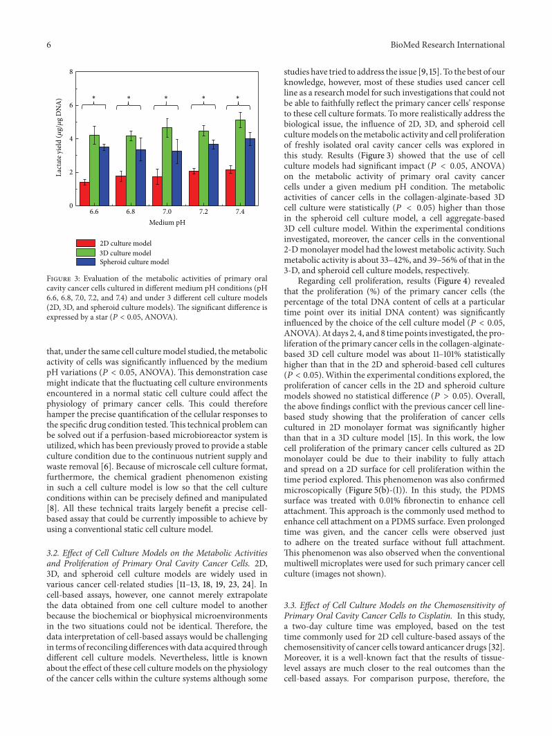
Figure 3: Evaluation of the metabolic activities of primary oralcavity cancer cells cultured in different medium pH conditions (pH6.6, 6.8, 7.0, 7.2, and 7.4) and under 3 different cell culture models(2D, 3D, and spheroid culture models). The significant difference isexpressed by a star (𝑃 < 0.05, ANOVA).
that, under the same cell culturemodel studied, themetabolicactivity of cells was significantly influenced by the mediumpH variations (𝑃 < 0.05, ANOVA). This demonstration casemight indicate that the fluctuating cell culture environmentsencountered in a normal static cell culture could affect thephysiology of primary cancer cells. This could thereforehamper the precise quantification of the cellular responses tothe specific drug condition tested.This technical problem canbe solved out if a perfusion-based microbioreactor system isutilized, which has been previously proved to provide a stableculture condition due to the continuous nutrient supply andwaste removal [6]. Because of microscale cell culture format,furthermore, the chemical gradient phenomenon existingin such a cell culture model is low so that the cell cultureconditions within can be precisely defined and manipulated[8]. All these technical traits largely benefit a precise cell-based assay that could be currently impossible to achieve byusing a conventional static cell culture model.
3.2. Effect of Cell Culture Models on the Metabolic Activitiesand Proliferation of Primary Oral Cavity Cancer Cells. 2D,3D, and spheroid cell culture models are widely used invarious cancer cell-related studies [11–13, 18, 19, 23, 24]. Incell-based assays, however, one cannot merely extrapolatethe data obtained from one cell culture model to anotherbecause the biochemical or biophysical microenvironmentsin the two situations could not be identical. Therefore, thedata interpretation of cell-based assays would be challengingin terms of reconciling differenceswith data acquired throughdifferent cell culture models. Nevertheless, little is knownabout the effect of these cell culturemodels on the physiologyof the cancer cells within the culture systems although some
studies have tried to address the issue [9, 15]. To the best of ourknowledge, however, most of these studies used cancer cellline as a researchmodel for such investigations that could notbe able to faithfully reflect the primary cancer cells’ responseto these cell culture formats. To more realistically address thebiological issue, the influence of 2D, 3D, and spheroid cellculturemodels on themetabolic activity and cell proliferationof freshly isolated oral cavity cancer cells was explored inthis study. Results (Figure 3) showed that the use of cellculture models had significant impact (𝑃 < 0.05, ANOVA)on the metabolic activity of primary oral cavity cancercells under a given medium pH condition. The metabolicactivities of cancer cells in the collagen-alginate-based 3Dcell culture were statistically (𝑃 < 0.05) higher than thosein the spheroid cell culture model, a cell aggregate-based3D cell culture model. Within the experimental conditionsinvestigated, moreover, the cancer cells in the conventional2-Dmonolayermodel had the lowest metabolic activity. Suchmetabolic activity is about 33–42%, and 39–56% of that in the3-D, and spheroid cell culture models, respectively.
Regarding cell proliferation, results (Figure 4) revealedthat the proliferation (%) of the primary cancer cells (thepercentage of the total DNA content of cells at a particulartime point over its initial DNA content) was significantlyinfluenced by the choice of the cell culture model (𝑃 < 0.05,ANOVA).At days 2, 4, and 8 timepoints investigated, the pro-liferation of the primary cancer cells in the collagen-alginate-based 3D cell culture model was about 11–101% statisticallyhigher than that in the 2D and spheroid-based cell cultures(𝑃 < 0.05). Within the experimental conditions explored, theproliferation of cancer cells in the 2D and spheroid culturemodels showed no statistical difference (𝑃 > 0.05). Overall,the above findings conflict with the previous cancer cell line-based study showing that the proliferation of cancer cellscultured in 2D monolayer format was significantly higherthan that in a 3D culture model [15]. In this work, the lowcell proliferation of the primary cancer cells cultured as 2Dmonolayer could be due to their inability to fully attachand spread on a 2D surface for cell proliferation within thetime period explored. This phenomenon was also confirmedmicroscopically (Figure 5(b)-(I)). In this study, the PDMSsurface was treated with 0.01% fibronectin to enhance cellattachment. This approach is the commonly used method toenhance cell attachment on a PDMS surface. Even prolongedtime was given, and the cancer cells were observed justto adhere on the treated surface without full attachment.This phenomenon was also observed when the conventionalmultiwell microplates were used for such primary cancer cellculture (images not shown).
3.3. Effect of Cell Culture Models on the Chemosensitivity ofPrimary Oral Cavity Cancer Cells to Cisplatin. In this study,a two-day culture time was employed, based on the testtime commonly used for 2D cell culture-based assays of thechemosensitivity of cancer cells toward anticancer drugs [32].Moreover, it is a well-known fact that the results of tissue-level assays are much closer to the real outcomes than thecell-based assays. For comparison purpose, therefore, the

BioMed Research International 7
0 2 4 6 8
100
150
200
Cel
l num
ber (
%)
Days
2D culture model3D culture modelSpheroid culture model
∗
∗
∗
Figure 4: Proliferation curves of primary oral cavity cancer cellscultured in 3 different cell culture models (2D, 3D, and spheroidculture models). The significant difference is expressed by a star(𝑃 < 0.05).
results of the chemosensitivity assays based on the cell culturemodels tested were compared with that based on the tumortissue-level chemosensitivity assays. For the 3D cell culturesstudied, furthermore, 2 kinds of culturemodels with differentcell density levels (7.6 × 10⁶ and 8.5 × 10⁷ cells mL−1, indicatedas “H” and “L,” respectively, in Figure 5) were prepared forthe chemosensitivity tests. For the higher cell density case,its cell density level (8.5 × 10⁷ cells mL−1) was same as thatin the tumor tissue samples tested, based on our preliminarystudies. It is not surprising that the results (Figure 5(a))revealed that the cell survival percentage of primary cancercells decreased upon increasing the dosage of cisplatin foreach cell culture model tested. At the drug concentrationof 8 𝜇gmL−1, the choice of the cell culture models had asignificant impact on the chemosensitivity assay results (𝑃 <0.05, ANOVA) (Figures 5(a) and 5(b)). For the cell culture-based assays explored, overall, the percentage of cell survivalin the chemosensitivity assays based on the spheroid cellculture models was significantly higher than that in the high-cell density 3D and 2D cell culture models. In addition, thecell survivals in these 3 models were statistically higher thanthat based on the low-cell density 3D cell culture model.Cisplatin is particularly effective at killing cancer cells duringtheir proliferation process [33]. The higher proliferationrates observed in the 3D cell culture model (Figure 4) areconsistent with their accordingly lower cell survival (%)in the low-density 3D cell culture-based chemosensitivityassays (Figure 5(a)). The above relationship between cellproliferation and cell survival, however, could not explain thechemosensitivity difference between the assays based on thespheroid and 2D cell culture models because the cell prolif-eration in these 2 models revealed no significant difference(Figure 4). Compared with the lower cell survival percentageobserved in the 2D culture-based chemosensitivity assays, the
higher cell survival occurring in the spheroid culture-basedchemosensitivity assays (Figure 5(a)) could be due to themass transfer barrier existing in the compact cell aggregatesystem, by which the anticancer drug might not be able toeffectively act on the cells within. This phenomenon wasalso observedmicroscopically (Figure 5(b)-(V)), inwhich thedead cancer cells (red dots) are mainly distributed aroundthe surface of cell aggregate particle, whereas the cancer cellslocated inside still kept viable (green dots).
Regarding the comparison of the chemosensitivity out-comes based on the cell-based and tumor tissue-level assays,the following descriptions and discussions were based onthe treatment of cisplatin at the concentration of 8𝜇gmL−1(Figures 5(a) and 5(b)). Results exhibited that the cell sur-vival percentage in the spheroid culture-based assays wassignificantly higher than that in the tumor tissue-level tests(𝑃 < 0.05). This result could imply that the spheroidculture-based chemosensitivity assays might overestimatethe resistance of primary cancer cells to the anticancerdrug tested. This phenomenon, again, could be reasonablyexplained by the aforementioned mass transfer barrier effectbecause the cancer cells formed a cell aggregate particle,which was more compact than the native tumor tissue.In addition, the drug chemosensitivity of primary cancercells in the tumor tissue-level and the high-cell density 3Dculture models showed no statistical difference, indicatingthat the drug chemosensitivity of cancer cells in such 3Dculture system was closer to that in the native tumor tissue.Moreover, the cell survivals in the 2D and low-cell density3D culture-based assays were significantly lower (𝑃 < 0.05)than those in the tissue-level assays, which might imply thatthe use of these 2 cell culture models could overestimate thechemosensitivity of primary cancer cells to cisplatin. Unlikethe 3D culture models with the same cell density (8.5 × 10⁷
cellsmL−1) as the tumor tissue sample tested (namely, thehigh cell density 3D model), the high chemosensitivity ofcancer cells to cisplatin occurred in the similar 3D culturemodel with lower cell density (7.6 × 10⁶ cellsmL−1) couldbe due to the cell proliferation effect. Primary cancer cellsin the low-cell density 3D environment could have morespace to proliferate whereas their proliferation was inhibitedunder a high cell density condition.Therefore, the higher cellproliferation phenomenon occurring in the low-cell density3D culturemodel could accordingly lead to a higher cytotoxiceffect of cisplatin to the cancer cells.This speculation was alsoconfirmed microscopically (Figures 5(b)-(II) and 5(b)-(III)).In order to find out the cellular response to the anticancerdrug tested under different cell culture models, overall, thecells had to be inevitably arranged in different cell cultureformats. In these situations, the influence of mass transportphenomenon cannot be perfectly ruled out. In this study,one of the technical advantages of using microscale cellculturemodel is its ability tominimize the chemical gradientsexisting in the cell culture system (e.g., cell culture construct).Further experiments (e.g., further miniaturized cell culturemodels using microfluidic technology) are required to possi-bly rule out the impact of mass transport phenomenon so as

8 BioMed Research International
0 4 860
70
80
90
100
Cel
l sur
viva
l (%
)
2D cell culture model3D cell culture model (L)3D cell culture model (H)
Tumor cultureSpheroid culture model
∗
∗
∗
Cisplatin concentration (𝜇g/mL)
(a)
(i)
(ii)
(I) (II) (III) (IV) (V)
100𝜇m
100𝜇m
100𝜇m
100𝜇m 100𝜇m
100𝜇m 100𝜇m 100𝜇m
100𝜇m100𝜇m
(b)
Figure 5: (a) Chemosensitivity evaluations of primary oral cavity cancer cells cultured in various cell culturemodels and treatedwith cisplatinat various concentrations; significant difference is expressed by a star (𝑃 < 0.05), (b) confocalmicroscopic observation of cell viability of cancercells cultured in various cell culture models ((I): 2D culture model; (II): 3D culture model (low cell density); (III): 3D culture model (high celldensity); (IV): tumor culture model; (V): spheroid culture model) and treated with cisplatin at the concentration of (i) 0 and (ii) 8 𝜇g/mL;green and red dots represent live and dead cells, respectively.
to more precisely investigate the real cellular response to thedrug condition tested.
4. Conclusions
Cell cultures are widely used as in vitro cell-based modelsfor various biological researches (e.g., drug chemosensitiv-ity assays). The most commonly used cell culture modelsare static 2D monolayer cell culture models. However, theinfluence of fluctuating culture conditions occurring in suchcell culture model on the physiology of the cultured cellsis generally ignored. In this study, experiments showed thateven minor environmental pH change could significantlyaffect the metabolic activity of cells, demonstrating theimportance of a stable culture condition for such assays. Totackle this issue, a perfusion-based microscale cell culturemodel capable of providing a stable culture condition wasadopted. In addition to the conventional 2D monolayer cell
culture models, several new types of cell culture methods(e.g., spheroid or 3D culture models) were actively proposed.However, little is known about the impact of these cell culturemodels on the physiology and the drug chemosensitivityassay results of primary oral cavity cancer cells. To addressthe fundamental biological issues, experiments were carriedout. Results revealed that the choice of cell culture formats(e.g., 2D, 3D, or spheroid culture models) might play animportant role in the physiology (e.g., metabolic activityor cell proliferation) of the cultured cells. Also the use ofdifferent cell culture models could lead to different resultsof drug chemosensitivity assays. Compared with the similartest based on tumor tissue-level assays, the use of spheroidculture model could overestimate the drug resistance ofcells to cisplatin, whereas the utilization of 2D and 3Dculture model might overestimate the chemosensitivity ofcells to such anticancer drug. In cell culture-based assays,thus, the extrapolation of experimental results from one cell

BioMed Research International 9
culture format to another might lead to biases because thebiochemical or biophysical microenvironments in the twosituations could not be identical. Compared with the tumortissue-level chemosensitivity assays, moreover, the 3D culturemodels with same cell density as that in tumor tissue samplesshowed comparable chemosensitivity assay results as thetumor tissue-level assays. As a whole, this study has providedsome fundamental information regarding the impact of cellculture methods on the results of in vitro cell-based assays.All these pieces of information are of great importance forestablishing a precise and faithful drug chemosensitivityassay.
Conflict of Interests
The authors declare that there is no conflict of interestsregarding the publication of this paper.
Authors’ Contribution
C.-H. Hsieh, Y.-D. Chen, and S.-F. Huang contributed equallyto the work.
Acknowledgments
The authors would like to thank financial support from theMinistry of Science and Technology in Taiwan (101-2221-E-182-001-MY3 and 103-2314-B-182A-064) and Chang GungMemorial Hospital (CMRPD2C0102, CMRPG3D0211, andCMRPG3B0973).
References
[1] J. Hatok, E. Babusikova, T. Matakova, D. Mistuna, D. Dobrota,and P. Racay, “In vitro assays for the evaluation of drugresistance in tumor cells,” Clinical and Experimental Medicine,vol. 9, no. 1, pp. 1–7, 2009.
[2] C.-J. Qi, Y.-L. Ning, Y.-L. Zhu,H.-Y.Min, H. Ye, andK.-Q. Qian,“In vitro chemosensitivity in breast cancer using ATP-tumorchemosensitivity assay,”Archives of Pharmacal Research, vol. 32,no. 12, pp. 1737–1742, 2009.
[3] N. P. Bown, M. M. Reid, A. J. Malcolm, E. V. Davison, A. W.Craft, and A. D. J. Pearson, “Cytogenetic abnormalities of smallround cell tumours,”Medical and Pediatric Oncology, vol. 23, no.2, pp. 124–129, 1994.
[4] S. Sarvi, A. C. Mackinnon, N. Avlonitis et al., “CD133+ cancerstem-like cells in small cell lung cancer are highly tumorigenicand chemoresistant but sensitive to a novel neuropeptideantagonist,” Cancer Research, vol. 74, no. 5, pp. 1554–1565, 2014.
[5] A. V. Heideman, B. Tholander, B. Grundmark et al.,“Chemotherapeutic drug sensitivity of primary culturesof epithelial ovarian cancer cells from patients in relationto tumour characteristics and therapeutic outcome,” ActaOncologica, vol. 53, no. 2, pp. 242–250, 2014.
[6] M.-H. Wu and C.-Y. Kuo, “Application of high throughputperfusion micro 3-D cell culture platform for the precise studyof cellular responses to extracellular conditions-effect of serumconcentrations on the physiology of articular chondrocytes,”Biomedical Microdevices, vol. 13, no. 1, pp. 131–141, 2011.
[7] B. T. Estes, B. O. Diekman, and F. Guilak, “Monolayer cellexpansion conditions affect the chondrogenic potential ofadipose-derived stem cells,” Biotechnology and Bioengineering,vol. 99, no. 4, pp. 986–995, 2008.
[8] M.-H. Wu, J. P. G. Urban, Z. Cui, and Z. F. Cui, “Developmentof PDMS microbioreactor with well-defined and homogenousculture environment for chondrocyte 3-D culture,” BiomedicalMicrodevices, vol. 8, no. 4, pp. 331–340, 2006.
[9] M. H. Wu, Y. H. Chang, Y. T. Liu et al., “Development ofhigh throughput microfluidic cell culture chip for perfusion 3-dimensional cell culture-based chemosensitivity assay,” Sensorsand Actuators, B: Chemical, vol. 155, no. 1, pp. 397–407, 2011.
[10] M.-H. Wu, H.-Y. Wang, C.-L. Tai et al., “Development ofperfusion-based microbioreactor platform capable of provid-ing tunable dynamic compressive loading to 3-D cell cultureconstruct: demonstration study of the effect of compressivestimulations on articular chondrocyte functions,” Sensors andActuators B: Chemical, vol. 176, pp. 86–96, 2013.
[11] Y. Ning, P. C. Manegold, Y. K. Hong et al., “Interleukin-8is associated with proliferation, migration, angiogenesis andchemosensitivity in vitro and in vivo in colon cancer cell linemodels,” International Journal of Cancer, vol. 128, no. 9, pp.2038–2049, 2011.
[12] Z.-Q. Ling, C.-J.Qi, X.-X. Lu et al., “Heterogeneity of chemosen-sitivity in esophageal cancer usingATP-tumor chemosensitivityassay,” Acta Pharmacologica Sinica, vol. 33, no. 3, pp. 401–406,2012.
[13] A. Forestier, F. Sarrazy, S. Caillat, Y. Vandenbrouck, and S.Sauvaigo, “Functional DNA repair signature of cancer celllines exposed to a set of cytotoxic anticancer drugs using amultiplexed enzymatic repair assay on biochip,” PLoS ONE, vol.7, no. 12, Article ID e51754, 2012.
[14] J. El-Ali, P. K. Sorger, and K. F. Jensen, “Cells on chips,” Nature,vol. 442, no. 7101, pp. 403–411, 2006.
[15] S.-B. Huang, S.-S. Wang, C.-H. Hsieh, Y. C. Lin, C.-S. Lai,and M.-H. Wu, “An integrated microfluidic cell culture systemfor high-throughput perfusion three-dimensional cell culture-based assays: effect of cell culture model on the results ofchemosensitivity assays,” Lab on a Chip, vol. 13, no. 6, pp. 1133–1143, 2013.
[16] G.Mehta, A. Y.Hsiao,M. Ingram,G.D. Luker, and S. Takayama,“Opportunities and challenges for use of tumor spheroids asmodels to test drug delivery and efficacy,” Journal of ControlledRelease, vol. 164, no. 2, pp. 192–204, 2012.
[17] L. G. Griffith and M. A. Swartz, “Capturing complex 3D tissuephysiology in vitro,”Nature Reviews Molecular Cell Biology, vol.7, no. 3, pp. 211–224, 2006.
[18] E. Burdett, F. K. Kasper, A. G. Mikos, and J. A. Ludwig,“Engineering tumors: a tissue engineering perspective in cancerbiology,” Tissue Engineering B: Reviews, vol. 16, no. 3, pp. 351–359, 2010.
[19] G. Oktem, O. Sercan, U. Guven et al., “Cancer stem celldifferentiation: TGF𝛽1 and versican may trigger molecules forthe organization of tumor spheroids,”Oncology Reports, vol. 32,no. 2, pp. 641–649, 2014.
[20] J. L. Tan, J. Tien, D. M. Pirone, D. S. Gray, K. Bhadriraju, and C.S. Chen, “Cells lying on a bed of microneedles: an approach toisolate mechanical force,” Proceedings of the National Academyof Sciences of the United States of America, vol. 100, no. 4, pp.1484–1489, 2003.

10 BioMed Research International
[21] M. S. Kim, J. H. Yeon, and J.-K. Park, “A microfluidic platformfor 3-dimensional cell culture and cell-based assays,”BiomedicalMicrodevices, vol. 9, no. 1, pp. 25–34, 2007.
[22] A. Abbott, “Cell culture: biology’s new dimension,” Nature, vol.424, no. 6951, pp. 870–872, 2003.
[23] D. Loessner, K. S. Stok, M. P. Lutolf, D. W. Hutmacher, J.A. Clements, and S. C. Rizzi, “Bioengineered 3D platform toexplore cell-ECM interactions and drug resistance of epithelialovarian cancer cells,” Biomaterials, vol. 31, no. 32, pp. 8494–8506, 2010.
[24] F. Pampaloni, E. G. Reynaud, and E. H. K. Stelzer, “The thirddimension bridges the gap between cell culture and live tissue,”Nature Reviews Molecular Cell Biology, vol. 8, no. 10, pp. 839–845, 2007.
[25] J. L. Lin, M. H. Wu, C. Y. Kuo, K. D. Lee, and Y. L. Shen,“Application of indium tin oxide (ITO)-based microheater chipwith uniform thermal distribution for perfusion cell cultureoutside a cell incubator,” Biomedical Microdevices, vol. 12, no.3, pp. 389–398, 2010.
[26] A. B. Riemer, H. Kurz, M. Klinger, O. Scheiner, C. C. Zielinski,andE. Jensen-Jarolim, “Vaccinationwith cetuximabmimotopesand biological properties of induced anti-epidermal growthfactor receptor antibodies,” Journal of the National CancerInstitute, vol. 97, no. 22, pp. 1663–1670, 2005.
[27] Y.-C. Tung, A. Y. Hsiao, S. G. Allen, Y.-S. Torisawa, M. Ho, andS. Takayama, “High-throughput 3D spheroid culture and drugtesting using a 384 hanging drop array,” Analyst, vol. 136, no. 3,pp. 473–478, 2011.
[28] N. Miyoshi, H. Ishii, K.-I. Nagai et al., “Defined factors inducereprogramming of gastrointestinal cancer cells,” Proceedings ofthe National Academy of Sciences of the United States of America,vol. 107, no. 1, pp. 40–45, 2010.
[29] M.-H. Wu, J. P. G. Urban, F. C. Zhan, Z. Cui, and X. Xu, “Effectof extracellular pH on matrix synthesis by chondrocytes in 3Dagarose gel,” Biotechnology Progress, vol. 23, no. 2, pp. 430–434,2007.
[30] R. A. Gatenby and R. J. Gillies, “Why do cancers have highaerobic glycolysis?” Nature Reviews Cancer, vol. 4, no. 11, pp.891–899, 2004.
[31] J.-W. Kim and C. V. Dang, “Cancer’s molecular sweet tooth andthe warburg effect,” Cancer Research, vol. 66, no. 18, pp. 8927–8930, 2006.
[32] H. Mese, Y. Ueyama, A. Suzuki et al., “Inhibition of telomeraseactivity as a measure of tumor cell killing by cisplatin insquamous cell carcinoma cell line,” Chemotherapy, vol. 47, no.2, pp. 136–142, 2001.
[33] M. A. Barry, C. A. Behnke, and A. Eastman, “Activation of pro-grammed cell death (apoptosis) by cisplatin, other anticancerdrugs, toxins and hyperthermia,” Biochemical Pharmacology,vol. 40, no. 10, pp. 2353–2362, 1990.

Submit your manuscripts athttp://www.hindawi.com
Stem CellsInternational
Hindawi Publishing Corporationhttp://www.hindawi.com Volume 2014
Hindawi Publishing Corporationhttp://www.hindawi.com Volume 2014
MEDIATORSINFLAMMATION
of
Hindawi Publishing Corporationhttp://www.hindawi.com Volume 2014
Behavioural Neurology
EndocrinologyInternational Journal of
Hindawi Publishing Corporationhttp://www.hindawi.com Volume 2014
Hindawi Publishing Corporationhttp://www.hindawi.com Volume 2014
Disease Markers
Hindawi Publishing Corporationhttp://www.hindawi.com Volume 2014
BioMed Research International
OncologyJournal of
Hindawi Publishing Corporationhttp://www.hindawi.com Volume 2014
Hindawi Publishing Corporationhttp://www.hindawi.com Volume 2014
Oxidative Medicine and Cellular Longevity
Hindawi Publishing Corporationhttp://www.hindawi.com Volume 2014
PPAR Research
The Scientific World JournalHindawi Publishing Corporation http://www.hindawi.com Volume 2014
Immunology ResearchHindawi Publishing Corporationhttp://www.hindawi.com Volume 2014
Journal of
ObesityJournal of
Hindawi Publishing Corporationhttp://www.hindawi.com Volume 2014
Hindawi Publishing Corporationhttp://www.hindawi.com Volume 2014
Computational and Mathematical Methods in Medicine
OphthalmologyJournal of
Hindawi Publishing Corporationhttp://www.hindawi.com Volume 2014
Diabetes ResearchJournal of
Hindawi Publishing Corporationhttp://www.hindawi.com Volume 2014
Hindawi Publishing Corporationhttp://www.hindawi.com Volume 2014
Research and TreatmentAIDS
Hindawi Publishing Corporationhttp://www.hindawi.com Volume 2014
Gastroenterology Research and Practice
Hindawi Publishing Corporationhttp://www.hindawi.com Volume 2014
Parkinson’s Disease
Evidence-Based Complementary and Alternative Medicine
Volume 2014Hindawi Publishing Corporationhttp://www.hindawi.com