research article structure, innervation and response ... · crocodylus, tomistoma and gavialias...
TRANSCRIPT

4217
INTRODUCTIONCrocodilians’ faces are covered in arrays of minute, pigmented skinelevations that are clearly visible around the upper and lower jaws.Early anatomical studies revealed differences in their distributionin the three families comprising Crocodilia (von Wettstein, 1937).In Alligatoridae, including the American alligator and caimanspecies, the protuberances are found only on the head near the mouthwhereas in Crocodilidae and Gavialidae, they are found on virtuallyevery scale of the body surface as well as on the head (Fig.1).Despite their prominence, few studies have investigated theirfunction. They have been hypothesized to play a role in secretingcleansing and waterproofing oils (Grigg and Gans, 1993), enablingosmoreception (Jackson and Brooks, 2007; Jackson et al., 1996),mediating mechanotransduction (Necker, 1974) and facilitatingcourtship behavior (Brazaitis and Watanabe, 2011). Othersuggestions include possibly acting as electroreceptors, as aconsequence of the aquatic habitat of crocodilians (Bullock, 1999),or acting as magnetoreceptors, an ability noted in alligators (Rodda,1984). More recently, Soares (Soares, 2002) discovered that thesestructures in juvenile Alligator mississippiensis mediate an orientingresponse to the center of a water surface disturbance. As a result,it was proposed that the main role of these structures is the detectionof surface waves generated by prey moving in water, and they weretermed ‘dome pressure receptors’ [these organs have been given avariety of names in different studies – we have chosen to followthe functionally neutral and commonly used term ‘integumentary
sensory organ’ (ISO) (Brazaitis, 1987)]. Although detection of waterdisturbances is clearly useful to crocodilians, a number ofobservations suggest that ISOs could facilitate a wider array ofmechanosensory abilities. For example, the ISOs of crocodylids arefound across their entire body and are thus poorly situated forreceiving surface waves. In alligators, the highest densities of ISOsare found around the teeth, inside the mouth,and at the rostralmargins of the mandibles and maxilla, suggesting a role indiscriminating food items or determining appropriate bite force(Erickson et al., 2012). Reflecting this uncertainty regarding theirfunctions, these receptors have also been identified as‘integumentary osmoreceptors’ in respect to the body receptorsfound in crocodylids (Jackson and Brooks, 2007).
The goal of this study was to provide further insight into thefunction of the ISOs by comparing them across two species ofcrocodilians: the Nile crocodile (Crocodylus niloticus) and theAmerican alligator (A. mississippiensis). Here we examine theanatomy of ISOs, the branching patterns of afferents providinginnervation and the physiological response properties of afferentsfrom the skin areas covered with ISOs, and document somebehaviors of alligators and crocodiles capturing prey under infraredillumination and with white noise to mask audition. Our resultssuggest that both the cranial and body ISOs of juvenile crocodiliansare employed as a high-resolution mechanosensory system thatallows for an otherwise armored skin surface to have a sensitivitygreater than primate fingertips.
SUMMARYIntegumentary sensory organs (ISOs) are densely distributed on the jaws of crocodilians and on body scales of members of thefamilies Crocodilidae and Gavialidae. We examined the distribution, anatomy, innervation and response properties of ISOs on theface and body of crocodilians and documented related behaviors for an alligatorid (Alligator mississippiensis) and a crocodylid(Crocodylus niloticus). Each of the ISOs (roughly 4000 in A. mississippiensis and 9000 in C. niloticus) was innervated by networksof afferents supplying multiple different mechanoreceptors. Electrophysiological recordings from the trigeminal ganglion andperipheral nerves were made to isolate single-unit receptive fields and to test possible osmoreceptive and electroreceptivefunctions. Multiple small (<0.1mm2) receptive fields, often from a single ISO, were recorded from the premaxilla, the rostraldentary, the gingivae and the distal digits. These responded to a median threshold of 0.08mN. The less densely innervated caudalmargins of the jaws had larger receptive fields (>100mm2) and higher thresholds (13.725mN). Rapidly adapting, slowly adaptingtype I and slowly adapting type II responses were identified based on neuronal responses. Several rapidly adapting unitsresponded maximally to vibrations at 20–35Hz, consistent with reports of the ISOsʼ role in detecting prey-generated water surfaceripples. Despite crocodiliansʼ armored bodies, the ISOs imparted a mechanical sensitivity exceeding that of primate fingertips. Weconclude that crocodilian ISOs have diverse functions, including detection of water movements, indicating when to bite based ondirect contact of pursued prey, and fine tactile discrimination of items held in the jaws.
Supplementary material available online at http://jeb.biologists.org/cgi/content/full/215/23/4217/DC1
Key words: brain, mechanosensory, trigeminal, behavior, touch, reptile, crocodile, alligator.
Received 28 June 2012; Accepted 23 August 2012
The Journal of Experimental Biology 215, 4217-4230© 2012. Published by The Company of Biologists Ltddoi:10.1242/jeb.076836
RESEARCH ARTICLE
Structure, innervation and response properties of integumentary sensory organs in crocodilians
Duncan B. Leitch1,2 and Kenneth C. Catania2,*1Neuroscience Graduate Program and 2Department of Biological Sciences, Vanderbilt University, Nashville, TN 37235, USA
*Author for correspondence ([email protected])
THE JOURNAL OF EXPERIMENTAL BIOLOGYTHE JOURNAL OF EXPERIMENTAL BIOLOGYTHE JOURNAL OF EXPERIMENTAL BIOLOGY

4218
MATERIALS AND METHODSAnimals
Eighteen American alligators (Alligator mississippiensis Daudin1801) and four Nile crocodiles (Crocodylus niloticus Laurenti1768) were studied. The alligators were provided by the LouisianaDepartment of Wildlife and Fisheries and were from theRockefeller Wildlife Refuge (Grand Chenier, LA, USA), and Nilecrocodiles were purchased from a commercial reptile breeder(Brooksville, FL, USA). They ranged in total body length from15 to 92cm and in mass from 30g to 3.2kg (from newly hatchedto ~3years old).
Scanning electron microscopyAnimals used to examine exernal skin structures were killed withsodium pentobarbital (120mgkg–1) and perfused with 4%paraformaldehyde (PFA). Tissues from the head and body surfacewere immersion fixed for 24 to 48h, rinsed with phosphate-bufferedsaline (PBS), and dehydrated in a graded series of ethanol. Followingdehydration, samples were critically point dried in an E3000 drier(Quorum Technologies, Guelph, ON, Canada) and coated with goldin a Cressington 108 sputter coater (Cressington ScientificInstruments, Watford, UK). Specimens were imaged using a TescanVega II SEM (Tescan, Cranberry Twp, PA, USA).
Sudan Black BSpecimens were fixed in 4% PFA for at least 1week, washed in tapwater for 12h and then cleared in 10% hydrogen peroxide for 2 to3days. Following washes in deionized water, maceration in trypsinsolution and washing in potassium hydroxide, the samples werestained in Sudan Black B solution (0.5g Sudan Black B, Sigma-Aldrich, St Louis, MO, USA). Specimens were destained in ethanoland preserved in glycerin.
Receptor density measurementsSurfaces of the heads from two alligators [snout–vent length(SVL)=46cm] were photographed by incrementally rotating thesamples. Individual photographs were aligned based ondistinguishing landmarks to create a complete montage of thescaled surfaces of the dorsal and ventral surfaces of the upper andlower jaws, and inside the oral cavity. A grid of 36 squares, each2�2mm, was superimposed on the completed montages. Thenumber of receptors within each box was counted, excluding thetop and left walls, and the distance between individual receptorswas measured using ImageJ (National Institutes of Health,Bethesda, MD, USA). Results from the four hemispheres wereaveraged.
DiI and confocal microscopyScale surface samples were removed post-mortem from PFA-fixedtissues. Small crystals of DiI (1,1�-dioctadecyl-3,3,3�,3�-tetramethylindocarbocyanine perchlorate; Molecular Probes,Invitrogen, Carlsbad, CA, USA) were applied with insect pins tothe exposed branches of the maxillary and mandibular nervesinnervating facial regions and to the intercostal nerves for the ventralbody surface. The scales were embedded in 2% agarose, immersedin 4% PFA and stored in darkness for ~1week. The specimens weresectioned sagittally on a Vibratome Series1000 (Technical ProductsInternational, St Louis, MO, USA) and imaged on an uprightLSM510 confocal microscope (Zeiss, Thornwood, NY, USA).
Trigeminal nerve light microscopySegments of the ophthalmic, maxillary and mandibular branches ofthe trigeminal nerve from three age-matched yearling alligators andtwo Nile crocodiles, ~2years old, were dissected followingperfusions with 4% PFA. Tissue was sampled 2 to 4mm from thebody of the trigeminal ganglion, and specimens were immersed inphosphate-buffered 2.5% glutaraldehyde solution for least 24h.Samples were post-fixed in osmium tetroxide, dehydrated in a gradedethanol series, transferred into propylene oxide and embedded inEMBed 812 (EM Sciences, Hatfield, PA, USA). Samples weresectioned transversely at ~0.5m thickness using a diamond knife(Diatome US, Hatfield, PA, USA) on a Reichert Ultracut Eultramicrotome (Leica Microsystems, Wetzlar, Germany). Tissuewas examined at 100� under light microscopy (Zeiss Axioskop,Zeiss, Jena, Germany), and digital images were captured (Axiovision4.5, Zeiss) and compiled in Adobe Photoshop CS5 (Adobe Systems,San Jose, CA, USA) into complete montages of the transversesection of the nerve of interest. Myelinated axons were manuallycounted.
Trigeminal and peripheral responsesEighteen alligators and two Nile crocodiles were anesthetized witha combination of urethane (0.4gkg–1), ketamine (100mgkg–1) andxylazine (20mgkg–1). Supplemental doses were given as needed.The trigeminal ganglion ipsilateral to the stimulated body surfacewas exposed. For recordings from the body integument, the radialand ulnar nerves were exposed in the proximal regions of theforelimb, and the median nerve was exposed in the hindlimb.Receptive fields were marked on photographs of the body. Multi-unit and single-unit electrode recordings were made ~400 to 800mfrom the ganglion’s surface using tungsten electrodes (1.0 to 1.5M at 1000Hz). Responses were collected using a Bak headstageand preamplifier (BAK Electronics, Mt Airy, MD, USA) and sent
The Journal of Experimental Biology 215 (23)
Fig.1. The phylogeny of extant crocodilians, as modified from Brochu (Brochu, 2003). The distribution of integumentary sensory organs (ISOs) is indicatedby the shaded regions. Within the order Alligatoridae, which includes all Caiman and Alligator species, ISOs are restricted to cranial regions. Within theCrocodylus, Tomistoma and Gavialias genera, ISOs are located on the cranium as well as along the rest of the post-cranial integument. Debate continueson the phylogenetic relationship of Tomistoma to Gavialis, depending on the genomic materials used in analysis (Piras et al., 2010).
THE JOURNAL OF EXPERIMENTAL BIOLOGYTHE JOURNAL OF EXPERIMENTAL BIOLOGYTHE JOURNAL OF EXPERIMENTAL BIOLOGY
4219Crocodilian receptors
to a Neurolog amplifier and filters (Digitimer, Welwyn Garden City,Herts, UK). Responses were monitored using speakers andwaveforms from single units were collected at 100,000sampless–1
using LabChart 7.0 software using a PowerLab 4/30 system(ADInstruments, Colorado Springs, CO, USA) attached to aMacBook laptop (Apple, Cupertino, CA, USA). The skin surfacewas kept moist during recordings.
Several combinations of somatosensory stimuli were used to elicitresponses from the skin surface. Scales were examined with smallwooden probes and von Frey filaments (Stoelting Company, WoodDale, IL, USA). Filaments just beyond threshold for eliciting aresponse were used in detailing the borders of receptive fields onphotographs of the skin surface. Using the Chubbuck stimulator(Chubbuck, 1966) and the digital sine and square wave generatorin LabChart 7.0, the frequency of the tactile stimuli was systemicallyaltered. The motion of the stimulator was recorded to observe thetiming of responses. Other stimuli included various positions of apair of 9V batteries and room-temperature hypertonic salt solutions(47p.p.t. or greater) using Instant Ocean sea salts (AquariumSystems, Mentor, OH, USA).
Following recordings, selected trigeminal ganglion electrodepenetrations were lesioned with a 10A current for 15s while theelectrode was withdrawn from the ganglion at 50ms–1; otherselected penetrations were marked with Toluidine Blue stain.Crocodilians were given an overdose of pentobarbital and perfusedwith PFA, as described above. Images of the intact ganglion werematched with photographs marked with locations of electrodepenetrations. The results from six alligators’ trigeminal ganglia were
used to reconstruct the somatotopy of the ganglion. Measurementsof receptive field size were made using ImageJ.
Single-unit recordings (N110) from four alligators’ trigeminalganglia were used to assess characteristics of receptor-covered skin.Data collected through LabChart 7.0 were analyzed using the SpikeHistogram module to measure the interspike interval betweenconsecutive action potentials. This was measured in the static phase(200 to 500ms) after the dynamic response to initial stimuluspresentation.
To assess differences between the von Frey force thresholds, thesurface area of receptive fields and the location of the field (fromeither crocodile or alligator and from the cranial or post-cranial bodyscales from both), a series of independently sampled t-tests was run.Pearson correlation coefficients and Spearman’s rank correlationcoefficients were calculated, as the force thresholds represented adiscontinuous data set whereas receptive field surface areas werecontinuous. All statistical analyses were two-tailed and set at the0.05 level of significance. These were completed using JMP Version9.0 (SAS Institute, Cary, NC, USA).
BehaviorNile crocodiles and American alligators were filmed with aMotionPro HS-3 camera with video recorded on a MacBook Procomputer running MotionProX software (Redlake, Integrated DesignTools, Tallahassee, FL, USA). Animals were placed in aquaria withroom-temperature water and permitted to move freely. Infrared (IR)lighting was provided on indicated trials with two IR-Flood Ultra-Covert 940nm illuminators (Night Vision Experts, Buffalo, NY,
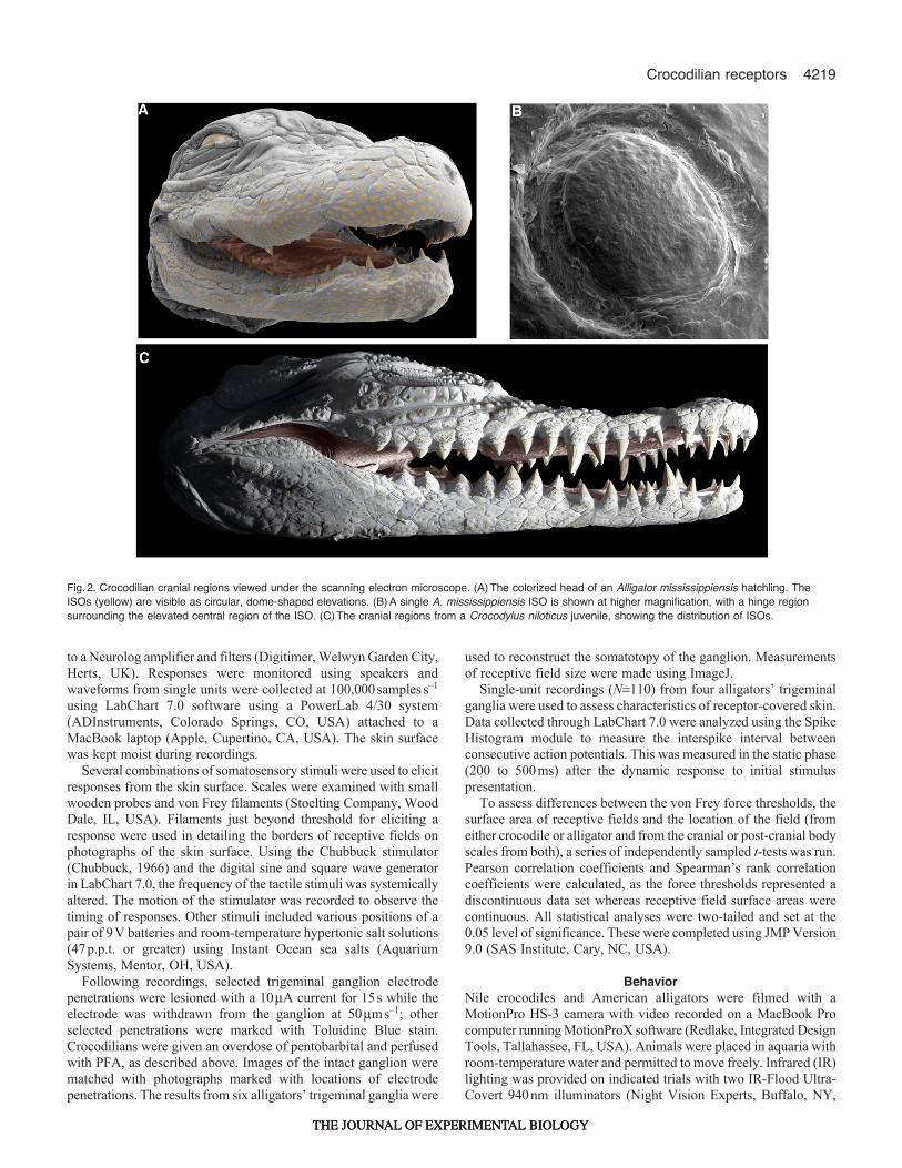
Fig.2. Crocodilian cranial regions viewed under the scanning electron microscope. (A)The colorized head of an Alligator mississippiensis hatchling. TheISOs (yellow) are visible as circular, dome-shaped elevations. (B)A single A. mississippiensis ISO is shown at higher magnification, with a hinge regionsurrounding the elevated central region of the ISO. (C)The cranial regions from a Crocodylus niloticus juvenile, showing the distribution of ISOs.
THE JOURNAL OF EXPERIMENTAL BIOLOGYTHE JOURNAL OF EXPERIMENTAL BIOLOGYTHE JOURNAL OF EXPERIMENTAL BIOLOGY

4220
USA). White noise was presented on indicated trials as generatedin Audacity (Carnegie Mellon Computer Music Group, Pittsburgh,PA, USA). Video was analyzed using iMovie (Apple). Allprocedures conformed to the National Institutes of Health standardsconcerning the use and welfare of experimental animals and wereapproved by the Vanderbilt University Animal Care and UseCommittee.
RESULTSIn examining the crocodilian ISOs, several levels of analysis wereadopted for both alligatorids and crocodylids. These data begin withdescribing the distribution of the organs, then their structure andinnervation, and next, the trigeminal and spinal afferentelectrophysiological responses recorded from the stimulation of skinand individual ISOs. Finally, some behavioral observations of theanimals orienting towards food pellets and live prey are noted inreference to supplementary material Movie1.
ISO distributionFig.2A shows the head of a juvenile American alligator. Skin onthe dorsal and ventral areas was covered in small, elevated sensoryorgans (Fig.2B). In each of the three juvenile alligators examined,which included a single 1-year-old alligator (head length7.2cm)and two ~3-year-old alligators (head length15.0cm), there were~4200±94 ISOs (mean ± s.d.) distributed across the cranial regions.In the same areas on two juvenile Nile crocodiles (Fig.2C), therewere 3001 and 2811 ISOs. These cranial ISOs varied in size acrossthe facial surface, with the smallest receptors found in appositionto and between the teeth and the largest receptors located on thedorsal surface of the maxilla, with mean diameters of 0.2±0.03 to1.2±0.04mm, respectively. Within the oral cavity, the ISOs weredistributed across the upper palate and the gingivae near the tongue.In alligators, the greatest concentration of ISOs(>2.00receptorsmm–2 on a 15cm head) was surrounding the teeth,and lower densities were found on the dorsal maxilla (Fig.3). Thedistance between ISOs ranged from 0.3±0.09mm in the areassurrounding the incisor teeth on the rostral dentary to 4.9±0.13mmon the dorsal surface of the maxilla. For the anterior–posterior axis,ISO density was greater near the most anterior point, particularlyon the lower jaw. Interestingly, disjunct areas of greater ISO densitywere found directly ventral to the eye and surrounding the nares.No evidence was found for other receptor organs (e.g. ampullaryorgans, ciliated receptors).
The post-cranial receptors of crocodylids were less denselydistributed but found across the entire integument, including on thearmored post-nuchal scales and osteoderms on the dorsal surface.Similar to the cranial receptors, they were visible as small, pigmentedprotuberances; however, there was typically only one ISO locatedcaudally on each scale (occasionally as many as two to three). Asa result, they were densest where the scales themselves were small,most notably near the cloaca.
ISO structure and innervationBelow the outer keratinized layers of epidermis of each ISO, adiversity of mechanoreceptors was positioned just beneath thestratum spinosum, supplied by a network of myelinated andunmyelinated axons. Transverse sections from the receptor revealeda number of anatomically distinct endings of the innervating axons(Fig.4). The connective tissue below each receptor contained manymelanocytes and provided the ISOs with their distinctivepigmentation. Just below the stratum basale, the melanocytesbranched extensively and were filled with darkly colored melanin
granules. These granules were interspersed with themechanoreceptors. Unmyelinated free nerve endings, ~0.5±0.09min diameter, passed through the connective tissue layers andterminated in the outer stratum spinosum. Branching from largerbundles of myelinated axons, free intraepidermal terminals werevisible as ubiquitous ‘discoid receptors’ and could be distinguishedbased on their rounded, expanded structure located just below thecells of the stratum lucidum and corneum. Discoid receptors wereclosely coupled to the tonofibrillar structures of the adjacent cellsof the spinosum and lucidum, and fluorescent, lipophilic dyeapplied to the proximal ends of the myelinated bundles often labeledthe keratinized cells of the stratum corneum. The extracellular spacebetween individual stratum spinosum cells was compressed at thepoint of receptor termination where the discoid receptors werelocated and surrounded by the tonofibrils of individual neighboringcells. Reflecting this compression, the keratinized layers of thestratified epithelium directly over the ISOs were ~60% thinner thanthat of adjacent scaled regions (N24, mean=61m, s.d.27m).The most superficial of the keratinized layers, the stratum corneum,appeared thinnest in the domed receptor region and at the hingedregion of epidermal folds between individual scales.
Within the connective tissue beneath each receptor, numerousaxon terminals were ensheathed in lamellations of Schwann cellprocesses (Fig.4C). They appeared similar to the Paciniformcorpuscles found in mammalian skin (Pease and Quilliam, 1957).There were also mechanoreceptor components that were notaffiliated with Schwann cell elaborations. These included free axonterminals running parallel to the collagen fibers and ending in thedermis, with the morphology of previously identified branchedlanceolate terminals (von Düring and Miller, 1979).
The most prominent sensory nerve endings of the ISOs wereassociated with the dermal Merkel cell column, located below thecenter of the dome and the surrounding stratum spinosum, wheremany of the axons traversing the longitudinal axis between domesconverged (Fig.4D). A similar configuration has been described inCaiman (von Düring, 1974). This structure was easily distinguishedas a mass of elongated, flattened Merkel cells with intercalated axonterminals and was distinct in its limited distribution to regions underthe ISOs.
The ISOs were supplied by fibers that originated below thesuperficial layers of the collagenous tissue from an elaboratenetwork of myelinated fibers that ran parallel to the skin surface.These branched most distally at the dome receptor regions intofascicles that typically contained 15 or more myelinated axons(Fig.4B, arrows). At more superficial levels, these branchescoursed together in circular patterns, ringing the inner
The Journal of Experimental Biology 215 (23)
ISO mm–2
>2.252.001.751.501.251.000.750.500.250
2 cm
Fig.3. Density of the ISOs across the cranium of a juvenile A.mississippiensis. Density was greatest directly adjacent to the teeth andnear the rostral-most points of the maxilla and dentary. Isolated patches ofgreater density were found surrounding the nares and below the eye.
THE JOURNAL OF EXPERIMENTAL BIOLOGYTHE JOURNAL OF EXPERIMENTAL BIOLOGYTHE JOURNAL OF EXPERIMENTAL BIOLOGY

4221Crocodilian receptors
circumference of the dome when viewed in horizontal sections,with nerve endings branching from larger groupings. At lowerlevels, the myelinated bundles were markedly larger in diameter(75±13m).
Cleared specimens stained with Sudan Black B revealed theorigin of the nerves in the trigeminal system (Fig.5). Thispreparation was useful for identifying the large rami of thetrigeminal nerve and for following the finer terminals to areas
covered in ISOs (Fig.5B). These data are shown in the schematiccreated from two hatchling alligators. A stained specimen froma juvenile Nile crocodile showed a similar pattern of innervation.The trigeminal nerve bifurcated into the mandibular andmaxillary/ophthalmic lobes ~1 to 2mm from the Gasserianganglion. The mandibular nerve then ran through the externalmandibular fenestra and extended both caudally to the back ofthe angular bone and rostrally to the teeth and anterior portions
Fig.4. The structure of the crocodilian ISO. (A)Schematic representation based on samples from A. mississippiensis and C. niloticus cranial and bodyreceptors. A diverse array of tactile components was localized to the epidermis and dermis of the ISO. Discoid receptors with enlarged terminals and freenerve endings ran through the keratinized epidermal layers that overlaid the prominent dermal Merkel complex and large branching network of myelinatedaxons. Cap, capillary; Discoid, discoid receptor; En LamC, encapsulated lamellated corpuscle; FBR, free branched receptor of the connective tissue; Ker, -keratinocyte; LamC, lamellated corpuscle; Mel, melanocyte; PNS, perineural sheath; RAx, branched receptor axons of the ISO connective tissue; StC,stratum corneum; StL, stratum lucidum; StS, stratum spinosum. (B)Confocal fluorescence of DiI-labeled free nerve endings (arrows) from a sectiontangential to the surface of the receptor, 20m below the apex. Scale bar, 10m. (C)Lamellated corpuscles (arrows) were visible in Toluidine-Blue-stainedsagittal sections from the dermis of the receptor. Scale bar, 50m. (D)A large dermal Merkel complex (arrow) and related branches of DiI-labeled nervefibers as seen under confocal microscopy in a cross-section. Scale bar, 200m.
THE JOURNAL OF EXPERIMENTAL BIOLOGYTHE JOURNAL OF EXPERIMENTAL BIOLOGYTHE JOURNAL OF EXPERIMENTAL BIOLOGY

4222
of the dentary. From the fenestra, the mandibular ramus branchedextensively into at least three smaller ramules running parallel tothe mandible and narrowed as it extended rostrally. The maxillaryramus ran from the jugal and quadratojugal and appeared equallydiverse in its arborization near the ISOs. Both the dentary andthe maxilla had many small foramina, and nerve fibers ran throughthe bone to project out of these openings in both directions onthe rostrocaudal axis. Typically, the afferents of a single cranialforamen innervated three ISOs. In addition to supplying fibers tothe external surface of the jaws, both the mandibular and maxillaryrami innervated the palate and gingivae extensively, both of whichwere covered in ISOs.
The ophthalmic ramus, which mainly innervated the largelyreceptor-free nasal and lacrimal bone areas as well as thedorsomedial surface of the cranium, was much smaller than themandibular and maxillary rami and did not branch extensively. Infour yearling alligators, the mandibular and maxillary rami contained~46,500±2700 and 48,300±3300 myelinated axons, respectively,whereas the ophthalmic ramus contained only 3600±200 myelinatedfibers. Similarly, in four Nile crocodiles matched in age and bodysize to the alligators, there were ~46,300±2800 myelinated axonsin the mandibular, 49,400±3000 in the maxillary and 3300±300 inthe ophthalmic rami.
Responses of neurons in the trigeminal ganglionThe trigeminal ganglion was found ventral to the ear, behind thejugal in anatomical dissections. To examine the responses of thecranial ISOs, we recorded extracellular activity from afferent cellbodies in the ganglion. We began by characterizing the location andsize of receptive fields corresponding to multi-unit activity elicitedby stimulating the skin with fine wooden probes and calibrated vonFrey hairs. Receptive fields were found for the majority of the skinsurfaces across the face of each crocodilian, including areas on theexternal surface of the mandible, the maxilla, the jugal bones ventralto the eye, and within the oral cavity, among other regions. Theextent of these multi-unit fields was documented and then singleunits were isolated to investigate individual afferents in greater detail.
The majority of receptive fields corresponded to skin areascovered in ISOs (Fig.6). Large receptive fields were found acrossthe jaws and often extended ventrally across the mandible or dorsallyto areas near the nares on the maxilla. Although some receptivefields were located on facial areas where ISOs were absent (i.e. theskin area dorsal to the suprangular and ventral to the quadratojugal),the majority of responses were elicited by stimulation on or nearISO-covered skin. Furthermore, receptive fields were organized inan overlapping manner, with the same area of ISOs often representedin two different locations in the ganglion. Ganglion cells responsive
The Journal of Experimental Biology 215 (23)
Fig.5. Innervation of the cranial ISOs by the trigeminal nerve. (A)Side view of the rami of the trigeminal nerve with hypertrophied mandibular and maxillarybranches comprising a network of finer fibers innervating regions where ISOs are present. Branching patterns were drawn from Sudan Black B preparations(see Materials and methods). The brain is shown to indicate the relative location of the trigeminal ganglion. Cb, cerebellum; gV, trigeminal ganglion; OB,olfactory bulb; OT, optic tectum; Sp, spinal cord; Tel, telencephalon. Scale bar, 1cm. (B)Example photograph of Sudan Black B preparation showing thedarkly stained processes of the maxillary nerve within the cleared whole-mounted specimen. Scale bar, 750m. (C)Transverse section of a mandibularnerve from C. niloticus. More than 46,000 myelinated axons (s.d.2700), as seen in the inset, were present within the nerve whereas fewer (3600±200)were present in the ophthalmic component. Scale bar, 50m.
THE JOURNAL OF EXPERIMENTAL BIOLOGYTHE JOURNAL OF EXPERIMENTAL BIOLOGYTHE JOURNAL OF EXPERIMENTAL BIOLOGY

4223Crocodilian receptors
to mechanical stimulation near the eye were located rostromediallynear the ophthalmic branch whereas cells responsive to mechanicalstimulation of the upper and lower jaws were found more caudallyin the ganglion. A large area of the ganglion between the maxillaryand mandibular branches contained cells that responded tostimulation of the teeth, upper palate and tongue (Fig.6C).
In total, 216 single units from the trigeminal ganglion wererecorded and analyzed in juvenile alligators, and 53 were examinedin Nile crocodiles. Their receptive fields were plotted on photographstaken of each individual crocodilian. In general, the tactile receptivefields corresponding to the most rostral regions of the animal’s facewere smallest and more numerous compared with thosecorresponding to caudal regions innervated by the mandibular andmaxillary nerves (Fig.6A,B). The smallest receptive fieldsencompassed single ISOs, with surface areas of less than 1mm2.These fields comprised of a single ISO were most often (92%) foundnear the rostral aspect of jaws, though a few were found on morecaudal regions of the face. Larger cranial receptive fields containedmore than 240 ISOs and were as much as 130mm2 in area (notillustrated).
After recording the area of each receptive field, mechanosensorythresholds were measured using calibrated von Frey hairs. We foundthat wet skin surfaces provided lower thresholds compared with dryskin, and thus all recorded data came from preparations withmoisture maintained. Among the 174 single unit receptive fieldsmeasured for indentation force, results ranged from 13.725 to0.078mN, corresponding to von Frey filaments numbered 4.17 to1.65, respectively. The lowest threshold could not be establishedfor the 28 receptive fields that were sensitive to the 1.65 filamentas this was the smallest calibrated force that could be applied. Themost sensitive areas were concentrated near the rostral premaxillaand mandible, as well as in apposition to the teeth. All of thereceptive fields that were restricted to a single ISO were responsiveto the 0.078mN (smallest) indentation force. Afferents with thehighest thresholds were generally found near the relatively sparselyinnervated regions on the dorsal surface of the maxillary, betweenthe nares and the eyes, and at the caudal margins the jaws. In general,afferents that were activated by the least pressure had smallreceptive fields whereas afferents responding to the stimulation ofmany ISOs (large receptive fields) required greater force (13.725mNor more).
To investigate the responses of afferents to precisely controlledstimuli, we employed a dedicated mechanosensory stimulator(Chubbuck, 1966). The Chubbuck stimulator was driven by a sinewave or square wave generator that controlled the linear movementof a small probe in a single dimension. The probe’s location wasprecisely tracked by a calibrated analog output of the stimulator(Fig.7).
Afferents that responded to the onset and offset of square-wavestimuli were characterized as rapidly adapting (RA) (Fig.7E,D,Fig.8) whereas afferents that responded throughout the duration ofthe stimulus were characterized as slowly adapting (SA)(Fig.7A,D,F). The SA responses could be further subdivided intoSA I and SA II, based on the coefficient of variation (CV) of theinterspike interval (ISI) during the static phase (200 to 500ms) ofthe maintained stimulus. This was calculated as the standarddeviation of the ISI divided by the mean ISI for the 2s train ofaction potentials (Chambers et al., 1972; Wellnitz et al., 2010). SAI units displayed irregularly timed discharges in response to themaintained stimulus whereas SA II units had regularly timeddischarges during the same period. Among 110 units in fouralligators, 51% of the responses were RA and 49% were SA
A
B
C
Alligator 11-4
Alligator 11-4
1 mm
Eye
OphthalmicTeeth/oral regions
MandibularMaxillary
Upper jaw – centerand caudal
Upper jaw – rostral
Lower jaw –center and caudal
Lower jaw –rostral
Motor branch
MedialRostral Caudal
Lateral
Fig.6. Representations in the trigeminal ganglion. (A)Ventral view of thelower jaw with representative receptive fields, which were often small andnear the rostral margins of the head. (B)Side view from the same case,showing the larger, overlapping fields that are characteristic of the morecaudal regions of the dentary and maxilla. (C)Composite figure from 10 A.mississippiensis and two C. niloticus trigeminal recording cases. Largeregions representing the teeth and mouth are present on the center of thebody of the trigeminal ganglion, while a smaller region located rostrallycontains neurons responding to the ISO-sparse areas near the eye.
THE JOURNAL OF EXPERIMENTAL BIOLOGYTHE JOURNAL OF EXPERIMENTAL BIOLOGYTHE JOURNAL OF EXPERIMENTAL BIOLOGY

4224
(Table1). Of the slowly adapting responses, 39% were SA I and37% were SA II. The remaining 24% of the SA responses had CVsthat were more than the 0.30 cut-off for SA II but less than the 0.50cut-off for SA I responses. The Chubbuck stimulator was not usedon a comparable number of Nile crocodile afferents, but based onclassification of 15 afferents from two Nile crocodiles usinghandheld probes, similar proportions of RA and SA units were found(55% SA and 45% RA responses).
Among a set of RA responses (N14), neurons were maximallyphase-locked with one-to-one correspondence of response perstimuli cycle to the lower vibrations (10 to 35Hz) and were lessattuned to 100Hz, 200Hz, 300Hz, and higher-frequency stimuli(Fig.8). Furthermore, smaller displacements of the probe wererequired to elicit responses for 20–30Hz vibrations compared withlower- (5 and 10Hz) or higher-frequency (50, 75, 100, 150 and200Hz) stimuli. RA units continued to respond to frequencies greaterthan 350Hz in four cases, and the median highest frequency for theSA responses was 250Hz in the SA II units.
In order to test for other possible sensory functions of the cranialISOs, we monitored activity in response to hyperosmotic solutionsand to electric fields (N40 afferents in four alligators and N15
afferents in two crocodiles). Single-unit neuronal responses wereisolated as described above, and cranial regions were exposed toroom-temperature deionized water and 31 to 47p.p.t. sea saltsolutions. These were applied via pipette or swab to the specificreceptive field and allowed to remain for at least 3min. No single-or multi-unit activity was detected apart from responses to the forceof the initial application of the solution. In other cases, the head ofthe crocodilian was lowered into a tank of room-temperature water,immersing the previously identified receptive field while theelectrode was held in place. A 9V battery was placed in the waterand moved in different configurations around the head, similar toparadigms used to elicit electrolocating behaviors in platypus(Scheich et al., 1986). No single- or multi-unit responses wereobserved.
Responses of the spinal nervesThe forelimb of the crocodile was supplied by the median, radialand ulnar nerves, and all three ran to the five digits as well as tothe skin of the dorsal surface of the limb (Fig.9A). The three nerveswere exposed near the proximal humerus. An electrode was insertedinto the nerve, and single unit responses were recorded. In total, 67
The Journal of Experimental Biology 215 (23)
Fig.7. Responses of trigeminal afferents from ISO-covered skin in crocodilians. (A)The small receptive field located on the juvenile A. mississippiensis.(B)Recording of the movement of the stimulator based on a calibrated analog output proportional to displacement. (C)Response of the afferent to thedisplacements shown in B showing the discharge pattern of a typical slowly adapting (SA) type II afferent. The interspike interval increased monotonicallywith increased displacement amplitude, maintaining the ʻregularʼ firing pattern indicative of SA type II fibers. In D–F, the output of the stimulator is recordedbelow the response of the afferent. (D)SA (in this case type I) and rapidly adapting (RA) responses were present for receptive fields covering individualISOs near the teeth in this A. mississippiensis case. (E)An RA unit from a juvenile C. niloticus is shown responding to block and sinusoidal (20Hz) stimuli.The photograph of the crocodile has been reversed to show the small receptive field more clearly. (F)Larger receptive fields, covering multiple ISOs, werefound at the caudal margins of the jaws as illustrated in an A. mississippiensis case.
THE JOURNAL OF EXPERIMENTAL BIOLOGYTHE JOURNAL OF EXPERIMENTAL BIOLOGYTHE JOURNAL OF EXPERIMENTAL BIOLOGY

4225Crocodilian receptors
single units from two alligators and 45 units from two crocodileswere examined from the medial, radial and ulnar nerves. For bothbody regions, receptive fields were drawn on the photographs ofthe animal.
The receptive fields found on the limbs of the alligators rangedfrom less than 1mm2 to more than 58mm2 (mean ± s.d.16.4±11.5),and the largest were found on the anterior surface of the hindlimb,above the tibia. On the forelimb, the smallest examples were isolated
to the distal regions of the digits of the forelimb. In particular, digitsIV and V had minute receptive fields near the ‘fingertip’ areas. Thesedigits are notable in that they lack the claws found on digits I toIII, were more slender, and appeared to be proportionally reducedin crocodilians compared with other reptile groups (Vargas et al.,2008). They have also been speculated to have a specialized tactilerole in detecting tactile stimuli from aquatic prey (Vliet and Groves,2010). Numerous low threshold receptive fields were founddistributed on the distal portion of these digits as well, withafferents responding to indentation forces of 0.392mN or less. Therewas an orderly progression of sensitivity as one moved moreproximally up the limb with the dorsal surface of most digitsresponding to the forces between 0.686 and 1.569mN, to regionscovering the carpals responding to 0.392 to 9.804mN, then to areascovering the radius and ulna responding to forces of 13.725mN(Fig.9B,C). Other areas of heightened sensitivity included thewebbing between digits I through III.
The hindlimb followed a similar pattern such that afferents fromdistal portions of the digits had small receptive fields and respondedat the lowest thresholds, whereas afferents innervating areas overthe tibia and fibula had larger receptive fields and responded athigher thresholds. By exposing the median and saphenous nervesnear the proximal end of the femur, recordings were obtained formuch of the hindlimb and its plantar surface (Fig.10). In recordingsfrom the forelimbs and hindlimbs in both species of crocodilians,both RA (N18 in Nile crocodiles; N29 in alligators) and SAafferents (N27 in Nile crocodiles; N39 in alligators) wereobserved.
In all of the electrophysiological recordings, the relationshipbetween receptive field surface area and the threshold forcenecessary to elicit activity was noted. In both alligators and Nilecrocodiles, smaller receptive fields were correlated with lowerdisplacement forces on the face (alligators: Spearman’s 0.651,N127, P<0.001; crocodiles: Spearman’s 0.5664, N16,P0.0222) and body (alligators: Spearman’s 0.618, N67,P<0.001; crocodiles: Spearman’s 0.6506; n45, P<0.001).Likewise, larger receptive fields were correlated with greaterdisplacement forces.
We also tested for responses to salinity changes or electric fields.The receptive field of interest on the limb was identified and
Table 1. Response properties of afferents of the ISO-covered scales from the trigeminal ganglia of four juvenile Alligator mississippiensis
Type Number % RF area (mm2)
Mean area (mm2)
Median min. displacement
(μm)
Median threshold
(mN)
Highest frequency
response (Hz) Associated structures
Discharge pattern
Coefficient of variation
RA 56 50.9 0.07–45.51 6.34±1.23 24.18 0.08 350 Lamellated corpuscles
At onset and offset only
–
SA I 21 19.1 0.18–15.46 6.48±0.24 52.58 0.08 250 Merkel discs and cells
Irregular discharges to maintained
stimulus
<0.30
SA II 20 18.2 0.03–20.12 4.81±0.30 41.53 0.3 200 Specialized end organs
Regular discharges to maintained
stimulus
>0.50
SA indeterminate 13 11.8 0.08–5.78 2.45±0.61 22.19 0.08 250 – Regular and irregular
discharges to maintained
stimulus
0.30–0.50
Total 110
ISO, integumentary sensory organ; RA, rapidly adapting response; SA I, slowly adapting type I response; SA II slowly adapting type II response.
Fig.8. Tuning curve for an A. mississippiensis cranial ISO. Upper panels:recordings of 1:1 entrained responses from one trigeminal A.mississippiensis RA unit to sinusoidal stimulation of increasing frequency.The movement of the stimulator is illustrated below each afferent recording.Scale bars, 100ms. Lower panel: the threshold displacement of the proberequired to produce 1:1 entrained responses for a single afferent from 10 to150Hz. Thresholds were lowest in the 20–30Hz range and were greaterwith both lower and higher frequencies. Tick marks on the x-axis indicate10Hz intervals.
THE JOURNAL OF EXPERIMENTAL BIOLOGYTHE JOURNAL OF EXPERIMENTAL BIOLOGYTHE JOURNAL OF EXPERIMENTAL BIOLOGY

4226
examined for threshold sensitivity and then the animal waspositioned to allow the body region to be submerged in a containerfilled with distilled water or 31 to 47p.p.t. sea salt solution (Fig.10).Immersing the limb in room-temperature water or hyperosmotic seasalt solution, as monitored for at least 3min, evoked no observablesingle- or multi-unit responses in either alligators or crocodiles.Similarly, there were no responses to the 9V battery in the water.
BehaviorJuvenile crocodilians, ranging in age from hatchling (SVL10.2cm)to 3years (SVL76.2cm) were observed and videotaped orientingtowards and capturing prey or ingesting food pellets dropped intothe water. With full-spectrum lighting, they generally turned rapidlytowards water ripples generated by dried food pellets dropped fromabove. Often, they closed both their lower, movable nictitatingmembrane and external eyelids as they snapped laterally towardsthe initial source of the disturbance. Although the jaws often securedthe food with the initial bite, subsequent bites re-orienting towardthe item appeared to rely on direct contact with the submerged pelletbecause the closed eyes were positioned well above the water surface(supplementary material Movie1, clip 1). These sideways snaps ofthe jaws were directed toward the food pellet within 50 to 70ms ofthe item’s contact with the skin.
Crocodilians were also monitored under 940nm IR illuminationand with white noise to block auditory cues. Both alligators andcrocodiles were capable of orienting towards the location of waterdisturbances when floating with their heads at the water surface.When positioned in this manner, the areas of greatest ISO density
near the rostral margins of the jaws and adjacent to the teeth wereoften below the air–water interface and just the eyes, ear flap andmore dorsal regions of the maxilla were exposed. Following theinitial directed movement towards the water disturbance(supplementary material Movie1, clip 2), both alligators andcrocodiles often swept their heads laterally when in the area of thesource of the ripples (Fig.11, supplementary material Movie1, clips3 and 4). The animal was obviously searching for the source of thedisturbance and often continued for 3–4s or until its jaws touchedan object. Within 200ms of contact with the object, the crocodilianusually bit the item and began to attempt ingestion. In the eventthat the animal had inadvertently secured a non-edible item (suchas floating aquarium fauna), the object was released after severalsnaps of the jaws whereas edible objects were quickly eaten.
Crocodilians were also observed orienting towards freelyswimming fish under IR illumination. Despite facing the oppositedirection and having their heads above water, crocodiles werecapable of rapidly turning and diving underwater towards thelocation of the fish (supplementary material Movie1, clip 5). Inanother predatory strategy, crocodiles would often remainsubmerged until prey came into contact with the skin surfaces(supplementary material Movie1, clip 6) or the open mouth(supplementary material Movie1, clip 7), at which point the animalimmediately attempted to capture the fish in its jaws.
DISCUSSIONAlligatorids have a dense array of sensory receptors (ISOs) extendingaround the mouth and cranial regions (4200±94 ISOs in A.
The Journal of Experimental Biology 215 (23)
Fig.9. Receptive fields of the forelimb in crocodilians. (A)In order to record from the forelimb, individual single units were isolated in the median, radial andulnar nerves near the proximal areas of the humerus. (B)Select receptive fields of single peripheral afferents from an A. mississippiensis case. The numbersrepresent the indentation threshold from the von Frey filament, measured in mN. Scale bar, 1cm. (C)Select receptive fields in a C. niloticus case. Individualbody ISOs are visible as small black dots on each scale (black dotted arrow). Same conventions as in B.
THE JOURNAL OF EXPERIMENTAL BIOLOGYTHE JOURNAL OF EXPERIMENTAL BIOLOGYTHE JOURNAL OF EXPERIMENTAL BIOLOGY

4227Crocodilian receptors
mississippiensis) whereas crocodylids have ISOs distributed acrossalmost every scale of the body surface (6200±389 ISOs) as well ason the head (2900±134 ISOs in C. niloticus). Since the earliestreports of ISOs (Maurer, 1895; von Wettstein, 1937) and their usein the dichotomous identification of crocodilian skins (King andBrazaitis, 1971), their function has remained a topic of speculation.Although detailed morphological studies undertaken in Caimanreceptors (von Düring, 1973; von Düring, 1974; von Düring andMiller, 1979) strongly suggested a mechanosensory role for ISOs,physiological characterization of their function has been limited tothe trigeminal receptors of a single species (A. mississippiensis)(Soares, 2002). Anatomical studies of crocodylid post-cranial ISOsfrom Crocodylus porosus focused on a potential role of the organsas osmoreceptors (Jackson and Brooks, 2007; Jackson et al., 1996).This hypothesis is based in part on models of how ISOs mechanicallyflatten under osmotic pressure in a saltwater environment and onexperiments measuring the mass of water consumed by the estuarinecrocodiles. This led to the hypothesis that ISOs are the firstidentified vertebrate integumentary osmoreceptors (Jackson andBrooks, 2007). Other investigators have proposed that ISOs couldfunction as magnetoreceptors (Rodda, 1984) or electroreceptors(Bullock, 1999).
Structure of ISOsThe ISOs appear to share many structural similarities with knownmechanoreceptors. These include the push-rod receptor organsdistributed across the snouts of monotremes (Andres and von Düring,1984; Andres et al., 1991) and the Eimer’s organs found on theglabrous skin on the rhinarium of moles (Catania, 1995). Numerous‘Tastflecken’ (‘touch spots’) found on the small warts of bufonidtoads and ranid species of frogs have also been identified (Lindblom,1963; Ogawa et al., 1981). Cutaneous cephalic corpuscles withprotruding centers appear in some colubrid snakes (Jackson, 1971;Jackson and Doetsch, 1977). Herbst and Grandry corpusclescomprise the tactile bill tip organs found in ducks (Berkhoudt, 1979;Gottschaldt and Lausmann, 1974). In all these cases, the receptorappears as a smooth, domed structure with an apex suitable fortransducing deflection to a series of specialized afferents.
In juvenile crocodilian ISOs, the external, keratinized dometypically had a diameter of 0.5mm or less for those distributed across
the jaws whereas larger ISOs (1.2mm) were found on crocodylidbody scales. Despite this size difference, both populations of ISOsappeared remarkably similar in internal composition. The stratumcorneum is thin over the organ (5m), presumably allowing a rangeof motions to compress the structure. This layer of three to five -keratin cells (Alibardi, 2011) functions both in structural integrityof the ISO and acts as scaffolding for the most apical of the finenerve terminals. In transverse sections, highly branched melanocytescan be seen throughout the keratinized layers and underlyingcollagenous layers and impart the distinctive pigmentation seen inmost of the ISO bodies. A number of mechanoreceptors are apparent
Fig.10. Electrophysiological recording preparation as used in bodyrecordings. In this case, the single-unit responses from the hindlimb wererecorded from the saphenous nerve. Once units were identifiedmechanically, the receptive field was submerged in hyperosmotic solutionsto monitor for activity. No activity related to immersion in the saltwatersolution was detected.
Fig.11. Crocodilian behavioral responses following water surfacedisturbance. (A)Individual images from a film sequence recorded underinfrared lighting and white noise presentation to block audition as a juvenileA. mississippiensis orients towards a surface wave generated by a smallfood pellet (white arrows). (B)Schematic of the orienting movementspresented in A. From the animalʼs initial location, a lateral, sweeping headmovement is repeated until the head makes tactile contact with the floatingpellet and it is rapidly captured. Scale bar, 10cm.
THE JOURNAL OF EXPERIMENTAL BIOLOGYTHE JOURNAL OF EXPERIMENTAL BIOLOGYTHE JOURNAL OF EXPERIMENTAL BIOLOGY

4228
in sectioned ISOs. These mechanoreceptors can be broadlycategorized based on their morphology and distribution, as describedby von Düring and Miller (von Düring and Miller, 1979). Thesedistinctions are as follows: (1) receptors of the epidermis, (2)receptors of the connective tissue with Schwann cell elaborationsor myelination, (3) receptors of the connective tissue lackingSchwann cells and (4) Merkel cell neurite complexes. Among tactilespecializations of the first group, crocodilians, as well as reptilesmore generally (Landmann and Villiger, 1975; von Düring, 1973),are notable for having expansions of the receptor terminals,compared with the finer, tapered free nerve terminals found in mostother vertebrates (Fig.4B). The dermal Merkel column, similar tothe ubiquitous epidermal Merkel neurite complex, traditionally hasbeen interpreted as slowly adapting in other species. These columnswere isolated to regions under each ISO whereas similar Merkelcells are found ubiquitously across the epidermal body surface infishes (Lane and Whitear, 1977), amphibians (Nafstad and Baker,1973), birds (Nafstad, 1971) and mammals (Halata, 1970; Munger,1965). Lamellated corpuscles, comparable to the paciniformstructures of mammals (Pease and Quilliam, 1957), have beencharacterized as rapidly adapting (Andres and von Düring, 1973;Iggo and Muir, 1969; von Düring and Miller, 1979). Indeed, bothrapidly adapting and slowly adapting afferents were observed inour physiological data.
The close association between the discoid terminals and thesupporting epidermal cells of the stratum corneum and lucidum hasbeen observed before in reptile scales (von Düring and Miller, 1979)and in mammalian glabrous skin (Munger and Ide, 1988), and thisrelationship also holds for the crocodilian ISOs both from thecephalic and body regions. Highlighting the intimate associationwith the free nerve terminals, fluorescent lipophilic label (DiI)applied to bundles of myelinated fibers of the maxillary nerve oftenlabeled the keratinized epidermal layers directly over the ISO whileremaining absent from adjacent scaled regions.
Several features of the trigeminal system of crocodilians stoodout when examining the innervation of the cranium. First, there wasan exceptional density of nerve fibers supplying the skin of the faceand a vast network of branching nerve bundles just below theepidermis. Throughout the dermis, ensheathed groups of myelinatedafferents projected across the rostro-caudal length and outwardstowards the epidermis, as seen in the cleared Sudan Black Bspecimens. The bundles emerged through small foramina of themaxilla and dentary. This organization is reminiscent ofmechanosensory end organs found in the foramina of anteriormargins of the beaks of water-foraging birds with bill tip organs(Cunningham et al., 2010) and highlights the shared archosaurianphylogeny between crocodilians and birds (Hedges and Poling,1999). It seems likely that by having the majority of the maxillaryand mandibular nerves shielded in bone, crocodilians are armoredagainst many potential injuries that might be encountered whenfeeding communally, while simultaneously maintaining an acutelysensitive skin surface via the fibers running through the foramina.
Trigeminal afferents and their organizationA large proportion of the neurons in the trigeminal ganglionresponded to stimulation of the areas most densely covered in ISOsnear rostral points of the pre-maxilla and mandible and surroundingthe teeth. In addition, many afferents responded to very light contactto the teeth, underscoring previous ultrastructural investigations ofsensory nerve endings within the dental ligament and attachmenttissues in Caiman crocodilus (Berkovitz and Sloan, 1979; Tadokoroet al., 1998). In general, the smallest receptive fields were found
rostrally on the upper and lower jaws and near the teeth. This overallpattern of small receptive field size and corresponding‘overrepresentation’ in the ganglion is reminiscent of corticalmagnification of behaviorally important skin surfaces observed inmammals (Krubitzer, 2007; Sur et al., 1980). For many species, themost important skin surfaces used for exploring objects are denselyinnervated by afferents with the smallest receptive fields, and theskin surfaces have correspondingly large representations in thecentral nervous system. Examples of functionally significant skinsurfaces with consequently large nervous system representationsinclude the forelimb of the raccoon (Welker and Seidenstein, 1959),the bill surface in the platypus (Pettigrew, 1999) and the lips andtongue of humans (Penfield and Boldrey, 1937). The overall patternfound for crocodilians, which have the highest density of ISOs andsmallest receptive fields around the teeth, provides an importantclue to ISO function. We suggest that ISOs play a key role not onlyin capturing prey based on water movements (Soares, 2002) andcontact, but also in discriminating objects that have been graspedin the jaws and guiding the manipulation of prey once it has beensecured. This interpretation is consistent with other recent findingsin vertebrates that have revealed very large cortical representationsof the dentition and oral structures that had been previouslyunappreciated (Jain et al., 2001; Kaas et al., 2006; Remple et al.,2003).
Within the ganglion, neurons that responded to the rostral headwere typically located ventrolaterally whereas neurons respondingto stimulation of the caudal regions of the jaws were positioneddorsomedially. As would be expected, responses to stimulation ofthe pre-maxilla, maxilla and quadratojugal of the upper jaw wererecorded from the anterior regions of the ganglion, in proximity tothe entrance of the maxillary nerve into the ganglion, and areasresponsive to stimulation of the dentary were recorded from theposterior regions, near the mandibular nerve’s division from theganglion. The electrophysiologically derived topography of thecrocodilian trigeminal ganglion was consistent with maxillaryrepresentations in the maxillo-mandibular lobe as documented inhorseradish peroxidase tracer studies from hatchling chicks (Noden,1980).
In both the Nile crocodiles and alligators, receptive fields, someas small as the area of a single ISO, were sensitive to indentationthresholds produced by the finest von Frey filaments correspondingto a force of 0.078mN. These measurements represent sensitivitiesmore acute than those of primate fingertips (Johansson et al., 1980)– skin surfaces that are widely appreciated for their sensitivity(Darrian-Smith, 1984; Kaas, 2004). Similarly, tactile responses wereelicited by mechanical displacements as small as 3.9m – anindentation threshold lower than found for the human hand(Johansson, 1978). These findings are evidence of the extreme andsurprising sensitivity of the crocodilian face and may represent arequirement for the detection of subtle water disturbances (Soares,2002) in addition to the discrimination of different objects and prey.
RA, SA I and SA II type responses were identified in recordingsfrom the surface of single trigeminal ISOs of alligators as well, inkeeping with the diverse array of mechanoreceptors and afferentend organs found in each receptor. A prior electrophysiological studyfrom the plantar nerve of alligators and caiman, which lack ISOson the body, have also found RA, SA I and SA II afferents on thehindlimbs (Kenton et al., 1971). However, in this preceding study,the finest indentation forces found for these cutaneous regionslacking ISOs (as is the case in alligatorid limbs) were more thansix times greater than the median indentation forces for responsesfrom the ISO covered areas (0.08mN) in this report, suggesting that
The Journal of Experimental Biology 215 (23)
THE JOURNAL OF EXPERIMENTAL BIOLOGYTHE JOURNAL OF EXPERIMENTAL BIOLOGYTHE JOURNAL OF EXPERIMENTAL BIOLOGY

4229Crocodilian receptors
the organs provide a considerable increase in sensitivity. ISOstherefore seem to be structures that impart great sensitivity to anotherwise armored and shielded body surface.
Analysis of 14 RA responses collected for stimulus frequenciesup to 350Hz indicated that the lowest indentation thresholds werefound at 20–30Hz within the 5–150Hz range. Larger displacementdistances were necessary to elicit 1:1 entrained responses of theafferent to frequencies both below and above the 20–30Hz window.The 20Hz vibration stimulus has been noted as one of the optimalfrequencies to induce orientation behaviors towards water surfacedisturbance in Notonecta glauca – a predatory aquatic insect thatlocalizes and orients towards prey-borne surface waves transmittedvia mechanoreceptive tarsal scolopoidal organs and abdominalsensory hairs (Lang, 1980; Weise, 1974). Thus the tuning of afferentsto this frequency in crocodilians is consistent with prior behavioralobservations of juvenile alligators orienting towards water surfaceripples (Soares, 2002). In addition, responses from SA (both typesI and II) and RA units often extended beyond 200 and 300Hz andwere elicited by 40–80m displacements, suggesting that relativelyhigher-frequency vibrations can also be readily transduced by ISOs.
Spinal nerve afferentsAs one of the goals of this project was to collect physiological dataregarding sensory function of post-cranial ISOs in crocodiles, it wasnecessary to record from spinal nerves innervating the integumentarysurface. Although still responding to forces of 13.725mN and finer,afferents from the body were not as sensitive as those distributedacross cephalic regions in either crocodiles or alligators. Discretesingle units across the limbs were typically large except for thosefound in distal regions of certain digits (IV and V on the forelimband IV on the hindlimb). There also appeared to be sensitive regions,responding to indentation forces of 0.392 and 0.686mN, on thewebbing present between digits III and IV on the forelimb. Theseresults are consistent with the concept of the ISOs being discretetactile receptor units as they are present on some of the smallestscales of the body; perhaps the increased receptor density per unitarea imparts a greater degree of acuity. This idea is supported bythe notion that the digits IV and V of the forelimb, which are notablymore slender and do not have the claws found on the other digits,might be adapted to detecting somatosensory cues when the animalis floating in the water (Vliet and Groves, 2010). When foragingfor fish, Caiman yacare partially open their mouths and fully extendtheir forelimbs, adopting a ‘cross posture’, and indeed, fish havebeen observed nipping at the caiman’s digits (Olmos and Sazima,1990), suggesting that tactile information from the digits couldmediate predatory behaviors.
Another motivation for physiological investigation of theintegument comes from Jackson’s intriguing series of experimentsinto the potential osmoreceptive capabilities of post-cranial ISOsof crocodiles (Jackson and Brooks, 2007; Jackson et al., 1996).However, in recording directly from the afferents innervating ISO-covered body surfaces in Nile crocodiles, no single- or multi-unitresponses attributable to exposure to hyperosmotic solutions wereobserved. The results were similar to those from alligators that wereused as an experimental control without body ISOs. Finally, noresponses were detected in response to electrical stimuli (Scheichet al., 1986), suggesting that ISOs play no role in electric fielddetection, and by extension, that crocodilians do not haveelectroreception.
We suggest that the crocodilian ISOs function as part of anelaborate mechanosensory system and are adaptive to a number ofaquatic behaviors. When filmed under 940nm IR illumination, both
crocodiles and alligators readily struck at and captured fish(supplementary material Movie1, clip 1) and occasionally orientedtowards minute water surface disturbances, similar to the resultsreported by Soares (Soares, 2002). Beyond providing positional cuesto the source of the stimuli, the ISOs are densely distributedthroughout the upper palate and areas adjacent to the teeth withinthe oral cavity – a location unlikely to receive and transduce thepressure from expanding surface waves. Disjunct regions of greaterreceptor density were observed near the eye and nares, similar tosupraorbital and rhinal microvibrissae areas found in mammalsrelying on trigeminally mediated tactile discrimination (Brecht,2007; Ling, 1966; Lyne, 1959). When actively foraging, crocodiliansopen their mouths and move so as to sweep the arrays of cranialISOs across the surface and underwater, rapidly capturing andsecuring objects that make contact with their heads, and releasingany non-edible matter, indicating that it is likely that they candiscriminate between multiple different materials using tactile cuesalone. As a testament to these discriminatory abilities, mothercrocodilians often manipulate their eggs as they begin hatching,gently cracking away the shell with their teeth (part of a feedingapparatus capable of inflicting crushing bites and dismemberinglarge prey) and allowing the hatchlings to seek protection in hermouth (Hunt, 1987; Pooley and Gans, 1976) – a situation in whichblunted tactile acuity would be maladaptive. Although the questionremains as to why ISO distribution differs between the alligatoridand crocodylid species, results from recording from the spinal nervessuggest that both species tested are sensitive to low thresholds offorce. Some have speculated that ISOs homologous to the post-cranial populations of crocodylids are present far deeper within theintegument in alligatorids (Richardson et al., 2002). Whilecrocodilians are certainly capable of accurately ambushing andcapturing prey by relying on their acute visual systems (Heric andKruger, 1966; Pritz, 1975) in lighted conditions, even on the darkestnights, prey still face a formidable mechanosensory system if theyunexpectedly come into contact with these reptiles.
ACKNOWLEDGEMENTSWe thank Dr Ruth Elsey and the staff of the Louisiana Department of Wildlife andFisheries at the Rockefeller Wildlife Refuge for providing alligator specimens, andthe assistance of Officer Walter Cook of the Tennessee Wildlife ResourcesAgency. We also thank Peter Brazaitis for anatomical advice and Eva Sawyer fortechnical assistance. These experiments would have been impossible without thecontributions of Danielle Gauthier, who assisted with crocodilian care, datacollection and analysis from this projectʼs inception.
FUNDINGThis research was supported by the National Science Foundation [grant 0844743to K.C.C.] and by a Vanderbilt University Discovery Grant.
REFERENCESAlibardi, L. (2011). Histology, ultrastructure, and pigmentation in the horny scales of
growing crocodilians. Acta Zool. 92, 187-200.Andres, K. H. and von Düring, M. (1973). The morphology of cutaneous receptors. In
Handbook of Sensory Physiology, Vol. 2 (ed. A. Iggo), pp. 1-28. Berlin: Springer-Verlag.
Andres, K. H. and von Düring, M. (1984). The platypus bill: a structural andfunctional model of a pattern-like arrangement of cutaneous sensory receptors. InSensory Receptor Mechanisms (ed. W. Hamann and A. Iggo), pp. 81-89. Singapore:World Scientific Publishing.
Andres, K. H., von Düring, M., Iggo, A. and Proske, U. (1991). The anatomy andfine structure of the echidna Tachyglossus aculeatus snout with respect to itsdifferent trigeminal sensory receptors including the electroreceptors. Anat. Embryol.184, 371-393.
Berkhoudt, H. (1979). The morphology and distribution of cutaneousmechanoreceptors (Herbst and Grandry corpuscles) in bill and tongue of the mallard(Anas platyrhynchos L.). Neth. J. Zool. 30, 1-34.
Berkovitz, B. K. B. and Sloan, P. (1979). Attachment tissues of the teeth in Caimansclerops (Crocodilia). J. Zool. 187, 179-194.
THE JOURNAL OF EXPERIMENTAL BIOLOGYTHE JOURNAL OF EXPERIMENTAL BIOLOGYTHE JOURNAL OF EXPERIMENTAL BIOLOGY

4230
Brazaitis, P. (1987). Identification of crocodilian skins and products. In WildlifeManagement: Crocodiles and Alligators (ed. G. J. Webb, S. C. Manolis and P. J.Whitehead), pp. 373-86. Chipping Norton, NSW: Surrey Beatty & Sons.
Brazaitis, P. and Watanabe, M. (2011). Crocodilian behaviour: a window to dinosaurbehavior? Hist. Biol. 23, 73-90.
Brecht, M. (2007). Barrel cortex and whisker-mediated behaviors. Curr. Opin.Neurobiol. 17, 408-416.
Brochu, C. A. (2003). Phylogenetic approaches toward crocodylian history. Annu. Rev.Earth Planet. Sci. 31, 357-397.
Bullock, T. H. (1999). The future of research on electroreception andelectrocommunication. J. Exp. Biol. 202, 1455-1458.
Catania, K. C. (1995). A comparison of the Eimerʼs organs of three North Americanmoles: the hairy-tailed mole (Parascalops breweri), the star-nosed mole (Condyluracristata), and the eastern mole (Scalopus aquaticus). J. Comp. Neurol. 354, 150-160.
Chambers, M. R., Andres, K. H., von Duering, M. and Iggo, A. (1972). The structureand function of the slowly adapting type II mechanoreceptor in hairy skin. Q. J. Exp.Physiol. Cogn. Med. Sci. 57, 417-445.
Chubbuck, J. G. (1966). Small motion biological stimulator. Appl. Phys. Lab. Tech.Digest 5, 18-23.
Cunningham, S. J., Castro, I., Jensen, T. and Potter, M. A. (2010). Remote touchprey-detection by Madagascar crested ibises Lophotibis cristata urschi. J. Avian Biol.41, 350-353.
Darrian-Smith, I. (1984). The sense of touch: performance and peripheral neuralprocesses. In Handbook of Physiology, The Nervous System III (ed. I. Darrian-Smith), pp. 739-788. Bethesda, MD: American Physiological Society.
Erickson, G. M., Gignac, P. M., Steppan, S. J., Lappin, A. K., Vliet, K. A.,Brueggen, J. D., Inouye, B. D., Kledzik, D. and Webb, G. J. (2012). Insights intothe ecology and evolutionary success of crocodilians revealed through bite-force andtooth-pressure experimentation. PLoS ONE 7, e31781.
Gottschaldt, K. M. and Lausmann, S. (1974). The peripheral morphological basis oftactile sensibility in the beak of geese. Cell Tissue Res. 153, 477-496.
Grigg, G. C. and Gans, C. (1993). Morphology and physiology of the Crocodilia. InFauna of Australia, Vol. 2A (ed. C. J. Glasby, G. J. B. Ross and P. L. Beesley), pp.326-336. Canberra, Australia: Australian Government Publishing Service.
Halata, Z. (1970). Nerve endings (Merkelʼs corpuscles) in hairless skin of the nose ofthe cat. Z. Zellforsch. Mikrosk. Anat. 106, 51-60.
Hedges, S. B. and Polling, L. L. (1999). A molecular phylogeny of reptiles. Science283, 998-1001.
Heric, T. M. and Kruger, L. (1966). The electrical response evoked in the reptilianoptic tectum by afferent stimulation. Brain Res. 1, 187-199.
Hunt, R. H. (1987). Nest excavation and neonate transport in wild Alligatormississippiensis. J. Herpetol. 21, 348-350.
Iggo, A. and Muir, A. R. (1969). The structure and function of a slowly adapting touchcorpuscle in hairy skin. J. Physiol. 200, 763-796.
Jackson, K. and Brooks, D. R. (2007). Do crocodiles co-opt their sense of “touch” to“taste”? A possible new type of vertebrate sensory organ. Amphib.-reptil. 28, 277-285.
Jackson, K., Butler, D. G. and Youson, J. H. (1996). Morphology and ultrastructureof possible integumentary sense organs in the estuarine crocodile Crocodylusporosus. J. Morphol. 229, 315-324.
Jackson, M. K. (1971). Cutaneous sense organs on the heads of some small groundsnakes in the genera Leptotyphylops, Tantilla, Sonora, and Virginia (Reptilia:Serpentes). Am. Zool. 11, 707-708.
Jackson, M. K. and Doetsch, G. S. (1977). Functional properties of nerve fibersinnervating cutaneous corpuscles within cephalic skin of the Texas rat snake. Exp.Neurol. 56, 63-77.
Jain, N., Qi, H. X., Catania, K. C. and Kaas, J. H. (2001). Anatomic correlates of theface and oral cavity representations in the somatosensory cortical area 3b ofmonkeys. J. Comp. Neurol. 429, 455-468.
Johansson, R. S. (1978). Tactile sensibility in the human hand: receptive fieldcharacteristics of mechanoreceptive units in the glabrous skin area. J. Physiol. 281,101-125.
Johansson, R. S., Vallbo, A. B. and Westling, G. (1980). Thresholds ofmechanosensitive afferents in the human hand as measured with von Frey hairs.Brain Res. 184, 343-351.
Kaas, J. H. (2004). Somatosensory system. In The Human Nervous System (ed. G.Paxinos and J. K. Mai), pp. 1059-1092. San Diego, CA: Elsevier Academic Press.
Kaas, J. H., Qi, H. X. and Iyengar, S. (2006). Cortical network for representing theteeth and tongue in primates. Anat. Rec. A 288, 182-190.
Kenton, B., Kruger, L. and Woo, M. (1971). Two classes of slowly adaptingmechanoreceptor fibres in reptile cutaneous nerve. J. Physiol. 212, 21-44.
King, W. F. and Brazaitis, P. (1971). Species Identification of Commercial CrocodilianSkins. New York: New York Zoological Society.
Krubitzer, L. (2007). The magnificent compromise: cortical field evolution in mammals.Neuron 56, 201-208.
Landmann, L. and Villiger, W. (1975). Glycogen in epidermal nerve terminals ofLacerta sicula (Squamata: Reptilia). Experientia 31, 967-968.
Lane, E. B. and Whitear, M. (1977). On the occurrence of Merkel cells in theepidermis of teleost fishes. Cell Tissue Res. 182, 235-246.
Lang, H. H. (1980). Surface wave sensitivity of the back swimmer Notonecta glauca.Naturwissenchaften 67, 204-205.
Lindblom, U. (1963). Phasic and static excitability of touch receptors in toad skin. ActaPhysiol. Scand. 59, 410-423.
Ling, J. K. (1966). The skin and hair of the southern elephant seal, Mirounga leonina(Linn.) I. The facial vibrissae. Aust. J. Zool. 14, 855-866.
Lyne, A. G. (1959). The systematic and adaptive significance of the vibrissae in themarsupalia. Proc. Zool. Soc. Lond. 133, 79-133.
Maurer, F. (1895). Die epidermis und ihre Abkommlinge. Leipzig: Wilhelm Engelmann.Munger, B. L. (1965). The intraepidermal innervation of the snout skin of the
opossum. A light and electron microscope study, with observations on the nature ofMerkelʼs Tastzellen. J. Cell Biol. 26, 79-97.
Munger, B. L. and Ide, C. (1988). The structure and function of cutaneous sensoryreceptors. Arch. Histol. Cytol. 51, 1-34.
Nafstad, P. H. (1971). Comparative ultrastructural study on Merkel cells and dermalbasal cells in poultry (Gallus domesticus). Z. Zellforsch. Mikrosk. Anat. 116, 342-348.
Nafstad, P. H. and Baker, R. E. (1973). Comparative ultrastructural study of normaland grafted skin in the frog, Rana pipiens, with special reference to neuroepithelialconnections. Z. Zellforsch. Mikrosk. Anat. 139, 451-462.
Necker, R. (1974). Dependence of mechanoreceptor activity on skin temperature insauropsids. J. Comp. Physiol. A 92, 65-73.
Noden, D. M. (1980). Somatotopic and functional organization of the avian trigeminalganglion: an HRP analysis in the hatchling chick. J. Comp. Neurol. 190, 405-428.
Ogawa, H., Morimoto, K. and Yamashita, Y. (1981). Physiological characteristics oflow threshold mechanoreceptor afferent units innervating frog skin. Q. J. Exp.Physiol. 66, 105-116.
Olmos, S. and Sazima, I. (1990). A fishing tactic in the floating Paraguayan caiman:the cross-posture. Copeia 1990, 875-877.
Pease, D. C. and Quilliam, T. A. (1957). Electron microscopy of the paciniancorpuscle. J. Biophys. Biochem. Cytol. 3, 331-342.
Penfield, W. and Boldrey, E. (1937). Somatic motor and sensory representation in thecerebral cortex of man as studied by electrical stimulation. Brain 60, 389-443.
Pettigrew, J. D. (1999). Electroreception in monotremes. J. Exp. Biol. 202, 1447-1454.Piras, P., Colangelo, P., Adams, D. C., Buscalioni, A., Cubo, J., Kotsakis, T.,
Meloro, C. and Raia, P. (2010). The Gavialis-Tomistoma debate: the contribution ofskull ontogenetic allometry and growth trajectories to the study of crocodylianrelationships. Evol. Dev. 12, 568-579.
Pooley, A. C. and Gans, C. (1976). The Nile crocodile. Sci. Am. 234, 114-119, 122-124.
Pritz, M. B. (1975). Anatomical identification of a telencephalic visual area incrocodiles: ascending connections of nucleus rotundus in Caiman crocodilus. J.Comp. Neurol. 164, 323-338.
Remple, M. S., Henry, E. C. and Catania, K. C. (2003). Organization ofsomatosensory cortex in the laboratory rat (Rattus norvegicus): evidence for twolateral areas joined at the representation of the teeth. J. Comp. Neurol. 467, 105-118.
Richardson, K. C., Webb, G. J. W. and Manolis, S. C. (2002). Crocodiles Inside Out:A Guide to the Crocodilians and Their Functional Morphology. Chipping Norton,NSW: Surrey Beatty & Sons.
Rodda, G. H. (1984). The orientation and navigation of juvenile alligators: evidence ofmagnetic sensitivity. J. Comp. Physiol. A 154, 649-658.
Scheich, H., Langner, G., Tidemann, C., Coles, R. B. and Guppy, A. (1986).Electroreception and electrolocation in platypus. Nature 319, 401-402.
Soares, D. (2002). Neurology: an ancient sensory organ in crocodilians. Nature 417,241-242.
Sur, M., Merzenich, M. M. and Kaas, J. H. (1980). Magnification, receptive-field area,and “hypercolumn” size in areas 3b and 1 of somatosensory cortex in owl monkeys.J. Neurophysiol. 44, 295-311.
Tadokoro, O., Mishima, H., Maeda, T. and Kozawa, Y. (1998). Innervation of theperiodontal ligament in the alligatorid Caiman crocodilius. Eur. J. Oral Sci. 106Suppl. 1, 519-523.
Vargas, A. O., Kohlsdorf, T., Fallon, J. F., Vandenbrooks, J. and Wagner, G. P.(2008). The evolution of HoxD-11 expression in the bird wing: insights from Alligatormississippiensis. PLoS ONE 3, e3325.
Vliet, K. A. and Groves, J. (2010). Crocodilian Biology and Captive ManagementMonograph. Silver Springs, MD: Association of Zoos and Aquariums.
von Düring, M. (1973). The ultrastructure of lamellated mechanoreceptors in the skinof reptiles. Z. Anat. Entwicklungsgesch. 143, 81-94.
von Düring, M. (1974). The ultrastructure of the cutaneous receptors in the skin ofCaiman crocodilus. Abhandlungen Rhein. Westfal. Akad. 53, 123-134.
von Düring, M. and Miller, M. R. (1979). Sensory nerve endings in the skin anddeeper structures. In Biology of the Reptilia, Vol. 9, Neurology A (ed. C. Gans, R. G.Northcutt and P. Ulinski), pp. 407-442. New York: Academic Press.
von Wettstein, O. (1937). Ordnuung der klasse Reptilia: Crocodilia. In Handbuch derZoologie eine Naturgeschichte der Stamme Destierreiches (ed. W. G. Kükenthal andT. Krumbach), pp. 236-248. Berlin: Walter de Gruyter & Co.
Weise, K. (1974). The mechanoreceptive system of prey localization in Notonecta. II.The principle of prey location. J. Comp. Physiol. A 92, 317-325.
Welker, W. I. and Seidenstein, S. (1959). Somatic sensory representation in thecerebral cortex of the racoon (Procyon lotor). J. Comp. Neurol. 111, 469-501.
Wellnitz, S. A., Lesniak, D. R., Gerling, G. J. and Lumpkin, E. A. (2010). Theregularity of sustained firing reveals two populations of slowly adapting touchreceptors in mouse hairy skin. J. Neurophysiol. 103, 3378-3388.
The Journal of Experimental Biology 215 (23)
THE JOURNAL OF EXPERIMENTAL BIOLOGYTHE JOURNAL OF EXPERIMENTAL BIOLOGYTHE JOURNAL OF EXPERIMENTAL BIOLOGY