research article pharmacological inhibition of nlrp3...
TRANSCRIPT

Research ArticlePharmacological Inhibition of NLRP3 InflammasomeAttenuates Myocardial Ischemia/Reperfusion Injury byActivation of RISK and Mitochondrial Pathways
Raffaella Mastrocola,1 Claudia Penna,1 Francesca Tullio,1
Saveria Femminò,1 Debora Nigro,1 Fausto Chiazza,2 Loredana Serpe,2
Debora Collotta,2 Giuseppe Alloatti,3 Mattia Cocco,2
Massimo Bertinaria,2 Pasquale Pagliaro,1 Manuela Aragno,1 and Massimo Collino2
1Department of Clinical and Biological Sciences, University of Turin, Torino, Italy2Department of Drug Science and Technology, University of Turin, Torino, Italy3Department of Life Sciences and Systems Biology, University of Turin, Torino, Italy
Correspondence should be addressed to Massimo Collino; [email protected]
Received 7 September 2016; Revised 12 October 2016; Accepted 23 October 2016
Academic Editor: Cecilia Zazueta
Copyright © 2016 Raffaella Mastrocola et al. This is an open access article distributed under the Creative Commons AttributionLicense, which permits unrestricted use, distribution, and reproduction in any medium, provided the original work is properlycited.
Although the nucleotide-binding oligomerization domain- (NOD-) like receptor pyrin domain containing 3 (NLRP3) inflamma-some has been recently detected in the heart, its role in cardiac ischemia/reperfusion (IR) is still controversial. Here, we investigatewhether a pharmacological modulation of NLRP3 inflammasome exerted protective effects in an ex vivo model of IR injury.Isolated hearts from male Wistar rats (5-6 months old) underwent ischemia (30min) followed by reperfusion (20 or 60min) withand without pretreatment with the recently synthetized NLRP3 inflammasome inhibitor INF4E (50𝜇M, 20min before ischemia).INF4E exerted protection against myocardial IR, shown by a significant reduction in infarct size and lactate dehydrogenase releaseand improvement in postischemic left ventricular pressure. The formation of the NLRP3 inflammasome complex was inducedby myocardial IR and attenuated by INF4E in a time-dependent way. Interestingly, the hearts of the INF4E-pretreated animalsdisplayed amarked improvement of the protective RISK pathway and this effect was associated increase in expression of markers ofmitochondrial oxidative phosphorylation. Our results demonstrate for the first time that INF4E protected against the IR-inducedmyocardial injury anddysfunction, by amechanism that involves inhibition of theNLRP3 inflammasome, resulting in the activationof the prosurvival RISK pathway and improvement in mitochondrial function.
1. Introduction
Ischemic heart disease is one of the main culprits of illnessand death [1, 2]. The main outcome of a transient cardiacischemia is the progressive decline of the left ventriclecontractile function, frequently paralleled by impairmentof the mitochondrial energy metabolism [3, 4]. Duringthe reperfusion phase the sudden mitochondrial oxygenoverload induces oxidative stress and further worsens themetabolic derangement [4], thus paradoxically exacerbatingmyocardial injury and inducing pyroptosis [2, 5]. Pyroptosis
is a caspase-1-dependent process leading to cell lysis, whichhas been demonstrated to be strongly regulated by the mul-tiprotein platform complex nucleotide-binding oligomeriza-tion domain- (NOD-) like receptor pyrin domain contain-ing 3 (NLRP3) inflammasome. The NLRP3 inflammasomecomprises (a) NLRP3, (b) an apoptosis-associated speck-likeprotein containing a caspase activation recruitment domain(ASC), and (c) procaspase-1. In response to a wide rangeof danger signals, including oxygen-free radicals, K+ efflux,or mitochondrial stress [6–8], NLRP3 recruits the adaptorprotein ASC which in turn interacts with procaspase-1.
Hindawi Publishing CorporationOxidative Medicine and Cellular LongevityVolume 2016, Article ID 5271251, 11 pageshttp://dx.doi.org/10.1155/2016/5271251

2 Oxidative Medicine and Cellular Longevity
Inflammasome oligomerization promotes the autocatalyticactivation of procaspase-1 and the processing of prointer-leukin- (IL-) 1𝛽 [2]. More recently, a new protein has beenidentified as member of the NLRP3 inflammasome com-plex, the Gasdermin D (GSDMD), which is recruited withkinetics similar to those required for caspase-1 activation.The proteolytic cleavage of GSDMD by caspase-1 detachesits N-terminal fragment, which contributes to mediate IL-1𝛽 secretion and pyroptosis [9]. Since NLRP3 is detectable inmany cardiac cell types, including cardiofibroblasts (themostimportant cell type in the heart in terms of number of cells)and cardiomyocytes (the most important cell type in terms ofcell volumes), it is likely that it may play a pivotal role in acutemyocardial infarction [10, 11]. Indeed, we and others haveshown that NLRP3 is upregulated by ischemia/reperfusion(IR) injury and its myocardial activation is exacerbated bymetabolic derangements [12, 13]. Interestingly, genetic mod-ulation of NLRP3 has been reported to reduce myocardialinfarct sizes upon IR [13]. However, a very recent study failedto find any role of NLRP3 in determining myocardial IRinjury [14] and another investigation supported cardiopro-tective effects due to NLRP3 inflammasome activation, thushighlighting that the interpretation of NLRP3 inflammasomerole in myocardial IR injury is far from clear. Nevertheless,a cross-talk between NLRP3 and mitochondria, the mainplayer of IR injury, has been described, with NLRP3 beingable to sense the presence of reactive oxygen species (ROS)produced by normal or dysfunctional mitochondria [15].Thus, the present study aimed to investigate the effects of anewly synthesized NLRP3 inflammasome inhibitor, namedINF4E [16], in an ex vivo model of myocardial IR injury.We deepened our investigation evaluating its ability, inthe rat heart, (i) to interfere with the IR-induced NLRP3inflammasome activation and pyroptotic cascade and (ii) toimprove the mitochondrial metabolic response to IR insult.
2. Materials and Methods
2.1. INF4E Preparation. INF4E was dissolved at 200mMconcentration in DMSO. Stock solution was then diluted ata final concentration of 50𝜇M in the perfusion buffer (seebelow). The description of the synthesis and the specificityof the inhibitor is included in the Supplemental Material(available online at http://dx.doi.org/10.1155/2016/5271251),according to previous publications [16, 17].
2.2. Animals Protocol and Ex Vivo Ischemia/Reperfusion (IR)Injury. Male Wistar rats (Harlan Laboratories, Udine, Italy)5-6 months old, reaching a body weight of 450–550 g, werecared in compliance with the EuropeanDirective 2010/63/EUon the protection of animals used for scientific purposes.Theanimal protocols followed in this study were approved by thelocal “Animal Use and Care Committee.” After one week ofquarantine, with drink and food ad libitum, rats were anes-thetized and killed. The hearts were rapidly perfused. A con-stant flow was maintained to obtain a typical coronary perfu-sion pressure of about 80mmHg by the Langendorff tech-nique with Krebs-Henseleit bicarbonate buffer containing
(mM) NaCl 118, NaHCO3 25, KCl 4.7, KH2PO4 1.2, MgSO41.2, CaCl2 1.25, and Glucose 11. The buffer was gassed with95% O2 : 5% CO2. The temperature of the perfusion systemwas maintained at 37∘C. The hearts underwent 30min stabi-lization and then were exposed to 30min of global no-flow,normothermic ischemia followed by a period of 20 or 60minof reperfusion. Hearts from a subgroup of rats (IR+INF4E)were pretreatedwith 50𝜇MINF4E in the perfusate for 20minbefore ischemia (after the first 10min of stabilization). Heartsfrom sham animals were exposed to 60min perfusion onlyand served as reference group in Western blot analysis.
The hearts were electrically paced at 280–300 bpm andkept in a temperature-controlled chamber (37∘C). Pacingwas stopped at the beginning of the ischemia and restartedafter the third min of reperfusion. Left ventricular pressure(LVP) and coronary perfusion pressure (CPP) were recordedand monitored with two electromanometers placed withinthe left ventricle and along the perfusion line, respectively.Coronary flow, CPP, and LVP were used as indices ofpreparation conditions. Moreover, end diastolic LVP wasrecorded as index of contracture development during I/R anddeveloped ventricular pressure as index of contractile activitythroughout the experiment using PowerLab data acquisitionsystem and analyzed using Chart software (ADInstruments,Oxford, UK).
The perfusate flowing out of the heart was collected formeasurement of lactate dehydrogenase (LDH) release 5minimmediately before ischemia and for the entire reperfusionperiod. To assess the conditions of experimental preparationthe coronary flow rate was determined by the amount ofperfusate measured in a specific time period. The heart wasthen cut in two parts by a coronal section (perpendicular tothe long axis). The apical part (less than 1/3 of ventricularmass) was used for molecular analysis and, thus, frozenrapidly in liquid nitrogen and stored at −80∘C. Infarctsize assessment was performed by using the basal part ofventricle.
2.3. Measurement of the Infarct Size. Infarct mass was eval-uated at the end of the reperfusion with the nitro-blue-tetrazolium (NBT) technique by using a gravimetric method[18]. The basal part of the ventricles was dissected by trans-verse sections into two/ three slices, which were incubatedfor 20min with a solution of NBT (0.1%) in phosphate buffer.Two independent and blind observers carefully separated andthen weighted both stained and unstained tissues. Total massof necrosis was then calculated and expressed as percentageof ventricular mass. Since the ischemia was global and sincewe analyzed only the basal part of the ventricles the necroticmass was expressed as a percentage of the analyzed ischemictissue.
2.4. LDH Assay. Spectrophotometric analysis at 340 nm wasperformed on the collected perfusion effluent to measureLDH released from the heart.
2.5. Preparation of Tissue Extracts. Total proteins extractswere obtained from 10% (w/v) apex homogenates in RIPA
Oxidative Medicine and Cellular Longevity 3
buffer (0.5% Nonidet P-40, 0.5% sodium deoxycholate, 0.1%SDS, 10mmol/l EDTA, and protease inhibitors) as previouslydescribed [19]. Protein concentrations were measured byBradford assay (BioRad, Hercules, CA, USA) and sampleswere then stored at −80∘C for subsequent analysis.
2.6. Determination of IL-1𝛽 in Hearts Homogenates. Com-mercially available ELISA kit (R&D Systems, Abingdon,UK) was used to measure concentrations of IL-1𝛽 in tissuehomogenates, according to the manufacturer’s instructions.
2.7. Western Blot Analysis. Total proteins extracts were sep-arated by SDS-PAGE and blotted to nitrocellulose mem-brane (GE-Healthcare Europe, Milano, Italy). Membraneswere incubated with rabbit anti-NLRP3 (Abcam, Cam-bridge, UK), rabbit anti-caspase-1 (Santa Cruz Biotech-nology, Dallas, TX, USA), mouse anti-GSDMDC1 (SantaCruz Biotechnology), rabbit anti-IL-1𝛽 (Santa Cruz Biotech-nology), rabbit anti-caspase-1 (Santa Cruz Biotechnology,Dallas, TX, USA), mouse anti-Tyr204 ERK1/2 (Cell Sig-naling Technology), rabbit anti-total ERK1/2 (Cell Signal-ing Technology), mouse anti-Ser473 Akt (Cell SignalingTechnology), rabbit anti-total Akt (Cell Signaling Technol-ogy), rabbit anti-Ser9 GSK-3𝛽 (Abcam, Cambridge, UK),anti-total GSK-3𝛽 (Cell Signaling Technology), rabbit anti-mitochondrial transcription factor A (mtTFA) (Novus Bio-logicals, Cambridge, UK), mouse anti-nuclear respiratoryfactor-1 (NRF-1) (Santa Cruz Biotechnology), and mouseanti-sarcomeric mitochondrial creatine kinase (sMtCK)(Santa Cruz Biotechnology) and then probed with properHRP-conjugated secondary antibodies (BioRad). ClarityWestern ECL substrate (BioRad) was used for protein detec-tion and quantification was performed by densitometricanalysis (Quantity-One, Bio-Rad software). Data were nor-malized according to the related antitubulin densitometricvalues.
2.8. Real-Time PCR. Total RNA was extracted from heartsamples using the AllPrep� DNA/RNA/protein kit (Qiagen,Hilden, Germany), according to the manufacture instruc-tions.The total RNA concentration (𝜇g/mL) was determinedby the fluorometer Qubit and the Quant-iT� RNA AssayKit (Invitrogen, Milano, Italy). A total of 500 ng of RNA wasreverse-transcribed using QuantiTect Reverse TranscriptionKit (Qiagen). The synthesized cDNA was used for real-time polymerase chain reaction (RT-PCR). The cDNA wasamplified by real-time PCR using SsoFast� EvaGreen (Bio-Rad,) and primers specific for cytokine IL-1𝛽 (Mm Il1b 2 SG,cat. number QT01048355, Qiagen). The PCR reaction wasperformed at 95∘C for 30 s followed by 40 cycles of 95∘Cfor 5 s, 55∘C for 10 s. All samples were run in duplicate.At least two nontemplate controls were included in allPCR. The transcript of the reference gene ribosomal RNA18S (Mm Rn18s 3 SG, cat. number QT02448075, Qiagen)was used to normalize mRNA data, and the quantificationdata analyses were performed by using the Bio-Rad CFXManager Software, version 1.6 (Bio-Rad) according to themanufacturer’s instructions.
2.9. Materials. Compounds here used were obtained fromthe Sigma-Aldrich Company Ltd., unless otherwise stated.
2.10. Statistical Analysis. Data described in the text andfigures are presented as means ± standard error of the mean(s.e.m.) of 𝑛 observations, where 𝑛 represents the numberof animals studied. Statistical analysis was performed usingANOVA test followed by Bonferroni's posttest. A 𝑃 value ofless than 0.05 was considered to be statistically significant.
3. Results
3.1. INF4E Pretreatment Limits Infarct Size and ImprovesContractility Recovery. Rat hearts exposed to a 30min globalischemia and 60min reperfusion developed a 60 ± 3%infarct size in the basal portion of ventricle evaluated byNBT staining. When hearts were pretreated (20min priorto ischemia) with INF4E in the perfusate, the infarct sizewas significantly reduced compared to untreated hearts (IR)(Figure 1(a)). Importantly, LDH release in the perfusate wasalmost halved by drug treatment (Figure 1(b)). In addition,the INF4E pretreated hearts showed a twofold increase incontractile recovery after IR, as assessed by left ventricularpressure monitoring (Figure 1(c)).
Of note, IR caused an impairment of the mechanicalperformance, as evidenced by the dramatic reduction ofdeveloped LVP (DLVP) immediately after ischemia and theincomplete recovery during reperfusion. In fact, developedLVP fell from about 80mmHg in the preischemic conditionto about 25mmHg after 10min of reperfusion and thenrecovered to only 50mmHg at 60min reperfusion, thusreaching only less than 65% of the preischemic value (Figures1(c) and 1(d)). The pretreatment with the NLRP3 inhibitordid not modify the recovery of DLVP at early reperfusiontime (10 and 20min), whereas an improvement starts to bedetectable after 40min, reaching values up to 85mmHg at60min, corresponding to over 100% of the preischemic valueat the end of reperfusion.The end diastolic LVP (EDLVP)wasabout 5mmHg during stabilization; then, the 30min globalischemia and the subsequent reperfusion caused a sustainedincrease of this parameter in both the IR and IR+INF4Egroups (Figure 1(e)). Actually, an increase of EDLVP wasappreciated immediately after the end of ischemia and con-tinued during the first 20min of reperfusion. Then EDLVPfurther increased in the IR group reaching values over30mmHg, while in the group pretreated with the NLRP3inhibitor it decreased progressively to about 20mmHg after 1hour of reperfusion.The increase in EDLVP was significantly(𝑃 < 0.05) attenuated by the NLRP3 inhibitor only at 60minreperfusion.
3.2. INF4E Pretreatment Prevents NLRP3 Inflammasome Acti-vation and Downstream Signaling. To confirm the abilityof INF4E to interfere with NLRP3 inflammasome complexformation and activation in our experimental model, theexpression level and the activation of the downstream sig-naling of NLRP3 inflammasome were assessed by Westernblotting analysis in protein extracts obtained from the apical

4 Oxidative Medicine and Cellular Longevity
IR IR+INF4E
§
0
10
20
30
40
50
60
70
Infa
rct s
ize (
% o
f left
ven
tric
le)
60 min reperfusion
(a)
IR IR+INF4E
§§
0
50
100
150
200
250
300
LDH
rele
ase (
U/m
g of
tiss
ue)
60 min reperfusion
(b)
IR IR+INF4E
§
0
25
50
75
100
125
150
Con
trac
tility
(% re
cove
ry)
60 min reperfusion
(c)
§
DLV
P (m
mH
g)
(60 min)
0
25
50
75
100
Ischemia 20 40 6010Reperfusion (min)
IRIR+INF4E
(d)
§
EDLV
P (m
mH
g)
0
10
20
30
40
50
(60 min)Ischemia 20 40 6010
Reperfusion (min)
IRIR+INF4E
(e)
Figure 1: Infarct area, LDH release, and contractility recovery in hearts from rats exposed to 30min of ischemia plus 60min of reperfusion,pretreated or not with 50𝜇M INF4E in the perfusate 20min before ischemia. (a): infarct size after IR exposition is expressed as a percentageof ischemic tissue (% IS/IT). (b): LDH release in the perfusion effluent during the IR was expressed as units per mg of wet tissue weight.(c)–(e): monitoring of left ventricular pressure (LVP) was used to assess the contractility response to ischemia reperfusion injury. (c) showsthe percentage of contractility recovered at the end of 60min reperfusion. (d) and (e) show the entire time-course of developed LVP (DLVP)and end diastolic LVP (EDLVP), respectively. Data are means of 6 rats ± SEM. §𝑃 < 0.05 versus IR. §§𝑃 < 0.01 versus IR.
portion of hearts pretreated or not with INF4E and exposedto IR (Figure 2). Besides, rat hearts were exposed to twodifferent periods of reperfusion, short and long (20minand 60min, resp.) to better elucidate the kinetics of thepharmacological modulation of NLRP3 inflammasome andrelated pathways. As shown in Figure 2, the protein level ofNLRP3 was increased in a time-dependent manner, reachingstatistical significance only after the 60min reperfusion.Notably, the INF4E pretreatment effectively reduced itsupregulation. Western blotting analysis with an antibodyrecognizing the C-terminal region of caspase-1 allowed the
identification of two bands corresponding to the procaspase-1 and the cleaved active p10 subunit of caspase-1. After 20minof reperfusion the active form of caspase-1 was markedlyincreased, without significant changes in the expression ofprocaspase-1, although the INF4E-pretreated hearts showeda slight nonsignificant trend towards reduced procaspasecleavage. After 60min of reperfusion, both precursor andactive forms of caspase-1 were dramatically increased inthe untreated IR group, whereas the INF4E-pretreatmentsignificantly prevented procaspase cleavage. As shown inFigure 2(c), caspase-1 activation is associated with cleavage

Oxidative Medicine and Cellular Longevity 5
Sham IR+INF4EIR IR+INF4EIR0.0
0.5
1.0
1.5
2.0
2.5
3.0
3.5
NLR
P3fo
ld to
Sha
m (O
D)
§§§
60 min reperfusion20 min reperfusion
IRSham
NLRP3
Tubulin
IR+INF4E IR IR+INF4E
60 min reperfusion20 min reperfusion
∗∗∗
(a)
Procaspase-1Ca
spas
e-1
Caspase-1
Sham
§§
60 min reperfusion20 min reperfusionIRSham
Procaspase-1
Caspase-1 (p10)
Tubulin
IR+INF4E IR IR+INF4E
60 min reperfusion20 min reperfusionIR+INF4EIR IR+INF4EIR
∗∗∗∗∗∗
∗∗∗
∗ ∗
0
1
2
3
fold
s to
Sham
(OD
)
(b)
Sham
GSDMDC1 precursorCleaved GSDMDC1
GSD
MD
C1
§§§
60 min reperfusion20 min reperfusionIRSham
Tubulin
IR+INF4E IR IR+INF4E
GSDMDC1
Cleaved
precursor
GSDMDC1
60 min reperfusion20 min reperfusionIR IR+INF4E IR IR+INF4E
∗∗∗∗ ∗∗∗∗
0
5
10
15
(den
sitom
etric
val
ue)
(c)
Sham IR IR+INF4E IR IR+INF4E
§§§
60 min reperfusion20 min reperfusion
IRSham
Tubulin
IR+INF4E IR IR+INF4E
Pro-IL-1𝛽
Pro-IL-1𝛽Cleaved IL-1𝛽
IL-1𝛽Cleaved
IL-1𝛽
60 min reperfusion20 min reperfusion
∗∗∗
0.0
0.5
1.0
1.5
2.0
2.5
fold
to sh
am (O
D)
(d)
Figure 2: Inflammasome expression and activation in the rat heart exposed to 30min ischemia plus 20 or 60min reperfusion, pretreated ornot with INF4E. Representative Western blotting showing cardiac levels of NLRP3 and of downstream activation of caspase-1, GSDMDC1,and IL-1𝛽 cleavage assessed on heart extracts. Histograms report densitometric analysis normalized for the corresponding tubulin content.Data are means of 6 rats ± SEM. ∗𝑃 < 0.05, ∗∗𝑃 < 0.01, and ∗∗∗𝑃 < 0.001 versus Sham; §§𝑃 < 0.01, §§§𝑃 < 0.001 versus IR.

6 Oxidative Medicine and Cellular Longevity
§§§
∗
∗∗∗
∗∗∗IL
-1𝛽
(pg/
mg
prot
ein)
0
10
20
30
40
50
60 min reperfusion20 min reperfusionSham IR IR+INF4EIR IR+INF4E
(a)
60 min reperfusionSham IR IR+INF4E
0.0
0.5
1.0
1.5
2.0
2.5
fold
incr
ease
IL-1𝛽
mRN
A
(b)
Figure 3: IL-1𝛽 concentrations evaluated by ELISA (a) and mRNA levels of IL-1𝛽measured by RT-PCR (b) in extracts of rat hearts exposedto 30min ischemia plus 20 or 60min reperfusion, pretreated or not with INF4E. Data are means of 6 rats ± SEM. ∗𝑃 < 0.05, ∗∗∗𝑃 < 0.001versus Sham; §§§𝑃 < 0.001 versus IR.
of theGSDMDC1 component of the inflammasome platform,detectable only after the longest reperfusion time. Interest-ingly, INF4E pretreatment significantly reduced GSDMCD1cleavage. Accordingly, the protein levels of the active form ofIL-1𝛽 showed a robust increase after the 60min reperfusion,as demonstrated by Western blot analysis (Figure 2) andconfirmed by ELISA (Figure 3(a)). The INF4E pretreatmentprevented the slight increase in IL-1𝛽 production due to20min of reperfusion and strongly reduced the massiveIL-1𝛽 release recorded after 60min of reperfusion (Figures2 and 3(a)). Interestingly, neither the IR injury nor thedrug treatment affected the mRNA levels of IL-1𝛽 (Fig-ure 3(b)), thus confirming a selective effect of INF4E onNLRP3 inflammasome-dependent IL-1𝛽 cleavage rather thanexpression.
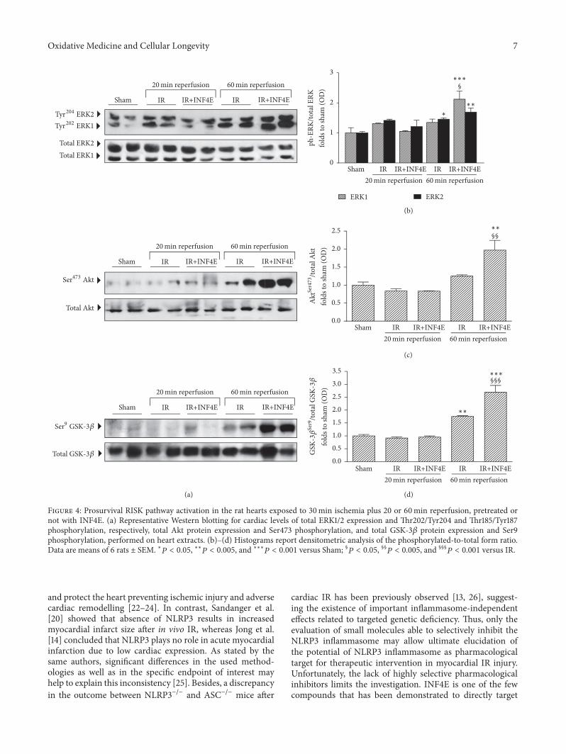
3.3. INF4E Pretreatment Enhances RISK Pathway ProtectiveActivity. Very recently, a role for NLRP3 inflammasomeactivation in the modulation of the Reperfusion InjurySalvage Kinase (RISK) pathway has been suggested [20].Thus, we quantified expression and activity (in terms ofphosphorylation) of the key members of this pathway. After20min of reperfusion no modulation of the activity and/orexpression level of members of the RISK pathway wasrecorded (Figure 4). On the contrary, the longer IR challeng-ing induced increased phosphorylation rate of both ERK1/2(Figures 4(a) and 4(b)) and GSK-3𝛽 (Figures 4(a) and 4(d)),while phosphorylation of Akt tended to increase, withoutreaching statistical significance, in untreated hearts exposedto either short or long IR protocol (Figures 4(a) and 4(c)).Interestingly, the INF4E pretreatment further increased thephosphorylation rate of ERK1/2, Akt, and GSK-3𝛽 inducedby the 60min reperfusion in untreated hearts (Figures 4(b)and 4(c)), suggesting an enhancement of the activation of thisprotective pathway by the pharmacological intervention.
3.4. INF4E Pretreatment Improves Mitochondrial Biogenesisand Energy Metabolism. Since mitochondrial metabolism
is highly involved in the myocytes response to ischemicinsult, and a cross-talk betweenmitochondria andNLRP3hasbeen described [21], we analyzed markers of mitochondrialbiogenesis after 30min ischemia and 60min reperfusion.The MtTFA and the NRF-1 were markedly downregulatedby IR. Conversely, mitogenesis was significantly preservedby INF4E pretreatment (Figures 5(a)–5(c)). A crucial phys-iological reaction of cardiac myocytes to oxygen deprivationis the enhancement of mitochondrial energy production, assuggested in ourmodel by the increased expression of sMtCKafter IR (Figures 5(a) and 5(d)). Intriguingly, the INF4Epretreatment further stimulated the expression of sMtCK by25% with respect to untreated rat hearts (Figures 5(a) and5(d)).
4. Discussion
The present study improves our understanding on the effectsof the NLRP3 inflammasome targeting in acute myocardialinfarction. Here we confirmed that myocardial IR inducestranscription of all the inflammasome components in a time-dependent way, with slight effect detectable when hearts wereexposed to 20min reperfusion and robust overexpressionand activation at the longest reperfusion time (60min).These data are in agreement with previous papers showingincreased expression levels of NLRP3 and procaspase-1 in theinfarcted and noninfarcted areas in both cardiomyocytes andnonmyocyte cell, associated with augmentation of caspase-1 activity [13, 22]. We also previously demonstrated that anenhanced susceptibility to a myocardial ischemic insult, dueto metabolic derangements, is paralleled by greater NLRP3inflammasome activation in the heart [12]. However, thepotential role of the innate immune NLRP3 protein complexas therapeutic target for cardiac infarction is ill defined.This ismainly due to the contrasting results so far obtained with tar-geted deletion of the inflammasome components. Few studiesdemonstrated that the depletion of even one of the inflam-masome complex components, either the sensor (NLRP3)or the effector enzyme (caspase-1), can prevent its activation
Oxidative Medicine and Cellular Longevity 7
(a)
(b)
(c)
(d)
Sham IR IR+INF4E IR IR+INF4E
Tyr204 ERK2
Tyr202 ERK1
Total ERK2
Total ERK1
60 min reperfusion20 min reperfusion
Sham IR IR+INF4E IR IR+INF4E
Ser473 Akt
Total Akt
60 min reperfusion20 min reperfusion
Sham IR IR+INF4E IR IR+INF4E
Ser9 GSK-3𝛽
Total GSK-3𝛽
60 min reperfusion20 min reperfusion
0
1
2
3
ERK1
ph-E
RK/to
tal E
RKfo
lds t
o sh
am (O
D)
ERK2
§
∗
∗∗∗
∗∗
Sham60 min reperfusion20 min reperfusion
IR+INF4EIR IR+INF4EIR
0.0
0.5
1.0
1.5
2.0
2.5
fold
s to
sham
(OD
)
§§∗∗
Sham60 min reperfusion20 min reperfusion
IR+INF4EIR IR+INF4EIR
Akt
Ser4
73/to
tal A
kt
0.0
0.5
1.0
1.5
2.0
2.5
3.0
3.5
fold
s to
sham
(OD
)
§§§∗∗∗
∗∗
Sham60 min reperfusion20 min reperfusion
IR+INF4EIR IR+INF4EIR
GSK
-3𝛽
Ser9
/tota
l GSK
-3𝛽
Figure 4: Prosurvival RISK pathway activation in the rat hearts exposed to 30min ischemia plus 20 or 60min reperfusion, pretreated ornot with INF4E. (a) Representative Western blotting for cardiac levels of total ERK1/2 expression and Thr202/Tyr204 and Thr185/Tyr187phosphorylation, respectively, total Akt protein expression and Ser473 phosphorylation, and total GSK-3𝛽 protein expression and Ser9phosphorylation, performed on heart extracts. (b)–(d) Histograms report densitometric analysis of the phosphorylated-to-total form ratio.Data are means of 6 rats ± SEM. ∗𝑃 < 0.05, ∗∗𝑃 < 0.005, and ∗∗∗𝑃 < 0.001 versus Sham; §𝑃 < 0.05, §§𝑃 < 0.005, and §§§
𝑃 < 0.001 versus IR.
and protect the heart preventing ischemic injury and adversecardiac remodelling [22–24]. In contrast, Sandanger et al.[20] showed that absence of NLRP3 results in increasedmyocardial infarct size after in vivo IR, whereas Jong et al.[14] concluded that NLRP3 plays no role in acute myocardialinfarction due to low cardiac expression. As stated by thesame authors, significant differences in the used method-ologies as well as in the specific endpoint of interest mayhelp to explain this inconsistency [25]. Besides, a discrepancyin the outcome between NLRP3−/− and ASC−/− mice after
cardiac IR has been previously observed [13, 26], suggest-ing the existence of important inflammasome-independenteffects related to targeted genetic deficiency. Thus, only theevaluation of small molecules able to selectively inhibit theNLRP3 inflammasome may allow ultimate elucidation ofthe potential of NLRP3 inflammasome as pharmacologicaltarget for therapeutic intervention in myocardial IR injury.Unfortunately, the lack of highly selective pharmacologicalinhibitors limits the investigation. INF4E is one of the fewcompounds that has been demonstrated to directly target

8 Oxidative Medicine and Cellular Longevity
mtTFA
Sham IR IR+INF4E
60 min reperfusion
NRF-1
Sham IR IR+INF4E
60 min reperfusion
Tubulin
sMtCK
Sham IR IR+INF4E
60 min reperfusion
0.0
0.5
1.0
1.5
mtT
FAfo
lds t
o sh
am (O
D)
§§
∗∗∗
∗∗∗
Sham60 min reperfusion
IR+INF4EIR
0.00
0.25
0.50
0.75
1.00
1.25
NRF
-1fo
lds t
o sh
am (O
D)
§∗∗∗
∗∗∗
Sham60 min reperfusion
IR+INF4EIR
0.0
0.5
1.0
1.5
2.0
2.5
sMtC
Kfo
lds t
o sh
am (O
D)
§§§∗∗∗
∗∗∗
Sham60 min reperfusion
IR+INF4EIR
(b)
(c)
(d)(a)
Figure 5: Mitochondrial biogenesis and energy metabolism evaluated in the rat hearts exposed to 30min ischemia plus 20 or 60minreperfusion, pretreated or not with INF4E. (a) Representative Western blotting for cardiac levels of MtTFA, NRF-1, and sMtCK performedon heart extracts. (b)–(d) Histograms report densitometric analysis normalized for the corresponding tubulin content. Data are means of 6rats ± SEM. ∗∗∗𝑃 < 0.001 versus Sham; §𝑃 < 0.05, §§𝑃 < 0.005, and §§§
𝑃 < 0.001 versus IR.
the NLRP3 inflammasome and inhibit the ATPase activity ofNLRP3 required for its activation. Although there are clearindications that this drug exerts a specific effect on NLRP3inflammasome independently of the activating stimulus, theexact mechanism of action has still to be clarified [16].Here we demonstrate, for the first time, that administrationof the NLRP3 inflammasome inhibitor INF4E in a singledose significantly reduces infarct size, the main endpoint to
target in cardioprotective studies.Moreover, the pretreatmentwith INF4E preserves systolic function, an index of reducedmyocardial stunning. To the best of our knowledge, so far,only another small molecule acting as NLRP3 inflammasomeinhibitor has been tested in models of acute myocardialinjury, showing protective effects similar to those recordedin our ex vivo model of myocardial IR injury [27, 28].Interestingly, this compound did not reduce infarct size at

Oxidative Medicine and Cellular Longevity 9
3 h of reperfusion, while it significantly reduced infarct size at24 h, if administered at the beginning of reperfusion, but notafter 3 h reperfusion [29], thus confirming the importance ofthe first period of reperfusion for IR injury development andthe efficacy of pharmacological strategies [5]. Here we docu-mented significant reduction of infarct size even after 60minof reperfusion, with INF4E administered as pretreatment. Ithas to be noted that our ex vivo model of IR injury mayallow excluding the involvement of infiltrating inflammatorycells that usually may affect the reperfusion-related injuryas documented in the previous studies investigating theeffects of NLRP3 inhibitors when given during reperfusion[28, 29]. Thus, our data further extend the previous findingssuggesting that the continuous pharmacological inhibition ofNLRP3 inflammasome pathway already during the ischemicperiod may significantly contribute to the beneficial effectsrecorded at the end of reperfusion. The kinetics of thereperfusion injurymay also affect the entity of the drug targetexpression and, thus, protection. In fact, when comparingthe 20min reperfusion and the 60min reperfusion modelswe observed a progressive expression and activity of NLRP3inflammasome complex, thus confirming previous findingson the timing of NLRP3 inflammasome formation in theheart during ischemia reperfusion [29].
Very recently, a role for NLRP3 inflammasome activa-tion in the cardioprotective RISK pathway has been faintlysuggested, but not convincingly demonstrated [20]. Herewe measured the entire RISK pathway, which includesAkt and ERK1/2 activation and GSK3𝛽 inhibition throughphosphorylation [30]. Our data demonstrate that the acti-vation of ERK/Akt/GSK-3𝛽 signaling is further enhancedby pharmacological inhibition of the NLRP3 inflammasomecomplex and this effect may significantly contribute to itscardioprotective effects. These data confirm our previousfindings, showing that pharmacological inhibition of NLRP3inflammasome significantly potentiates the activity of theprosurvival Akt pathway [31]. Besides, the cross-talk betweenNLRP3 inflammasome and RISK pathways is further con-firmed by the comparative analysis of the expression/activityof members of both pathways, which show robust increaseonly in the 60min reperfusion model in both cases.
Although we demonstrated a direct effect of the testedcompound on NLRP3 inflammasome activation, we cannotrule out the potential interactions of INF-4E with other sig-naling pathways, including those involved in the regulationof the expression of NLRP3 and/or activation of the RISKpathway. Thus, a further rigorous evaluation of effects of thetested compound on other signaling pathways affected by IRis needed to better elucidate its pharmacodynamics profile.
The effects of the pharmacological inhibition of NLRP3inflammasome on the RISK pathway are closely relatedto those recorded on mitochondrial metabolic response.Indeed, the mitochondrial metabolic dysfunction is crucialfor cardiac damage development, as both the block of mito-chondrial ATP-sensitive potassium channel (mitoKATP) andthe opening of the mitochondrial permeability transitionpore (mPTP) are common end effectors of the IR injury.Accordingly, pharmacological strategieswhich aimed to openthe mitoKATP and inhibit mPTP opening effectively reduce
myocardial IR injury and improve cardiomyocytes energyhomeostasis via modulation of the activity of members ofthe RISK pathway [32–35].We and others have demonstratedthat the Akt-mediated inactivation of GSK-3𝛽 is criticalfor the prevention of myocardial IR injury, being a keyevent in the regulation of apoptosis and the enhancementof mitochondrial biogenesis [36–38]. Besides, the use ofselective GSK-3𝛽 inhibitors evokes protection against IRinjury and promotes cell survival by limiting mPTP opening[39, 40]. In agreement with the above-mentioned observa-tions, the INF4E-induced amplification of the RISK pathwayactivation here described positively impacted on mitochon-drial metabolism. In fact, our data clearly demonstrate thatmarkers of mitochondrial biogenesis, suppressed by IR, weresignificantly upregulated by INF4E administration. Besides,the administered compound enhancedmitochondrial energymetabolism, shown in terms of reinforced expression of thesMtCK, which is a marker of increased mitochondrial ATPproduction.
Unfortunately, our study does not allow identifying thespecific cell types involved in NLRP3-mediated responses.Cardiomyocytes are the most prominent cell type in theheart and loss of contractile tissue is the most importantconsequence of a myocardial infarction. However, in car-diomyocytes, the activation onNLRP3 inflammasome evokescaspase-1 activation and pyroptosis, but not relevant releaseof mature IL-1𝛽 [22, 41]. In contrast, NLRP3 inflammasomeactivation inmyocardial fibroblasts induces the production oflarge amounts of mature IL-1𝛽, causing a rapid amplificationof the inflammatory response [13, 23, 42]. As cardiomy-ocytes are crucial target for the IL-1𝛽 produced by residentfibroblasts, which impairs contractile function and inducesapoptotic cell death, we may speculate that the beneficialeffects of INF4E are due, at least in part, to an indirectimprovement in cardiomyocytes functionality.
5. Conclusion
Taken together, our results strengthen the crucial role ofNLRP3 inflammasome activation even in the early step ofmyocardial injury caused by IR and show, for the first time,that its pharmacological inhibition by INF4E reduced theorgan injury/dysfunction. Preservation of cardiac function byINF4E is, at least in part, attributable to a cardioprotectiveeffect mediated by the activation of the RISK survival path-ways and the improvement inmitochondrial function, which,in turn, may improve cardiac function and postischemicoutcome. It has to be stressed, however, that the lack oflong-term evaluation as well as data on drug effects whenadministered during reperfusion limit the interpretation ofthe clinical transferability of our findings. Thus, further rig-orous evaluations are needed to gain a better understandingof both efficacy and the mechanism of action of INF4E in thesettings of acute myocardial infarction.
Competing Interests
The authors declare that they have no competing interests.
10 Oxidative Medicine and Cellular Longevity
Acknowledgments
This work was supported and funded by the University ofTurin (Ricerca Locale 2015 Linea A and Linea B).
References
[1] A. S. Go,D.Mozaffarian, V. L. Roger et al., “Executive summary:heart disease and stroke statistics—2013 update: a report fromthe American Heart Association,” Circulation, vol. 127, no. 1, pp.143–152, 2013.
[2] M. Takahashi, “NLRP3 inflammasome as a novel player inmyocardial infarction,” International Heart Journal, vol. 55, no.2, pp. 101–105, 2014.
[3] M. G. Perrelli, P. Pagliaro, and C. Penna, “Ischemia/reperfusioninjury and cardioprotective mechanisms: role of mitochondriaand reactive oxygen species,” World Journal of Cardiology, vol.3, no. 6, pp. 186–200, 2011.
[4] C. Penna, M.-G. Perrelli, and P. Pagliaro, “Mitochondrialpathways, permeability transition pore, and redox signalingin cardioprotection: therapeutic implications,” Antioxidants &Redox Signaling, vol. 18, no. 5, pp. 556–599, 2013.
[5] D. M. Yellon and D. J. Hausenloy, “Myocardial reperfusioninjury,” The New England Journal of Medicine, vol. 357, no. 11,pp. 1121–1135, 2007.
[6] R. Zhou, A. Tardivel, B. Thorens, I. Choi, and J. Tschopp,“Thioredoxin-interacting protein links oxidative stress toinflammasome activation,” Nature Immunology, vol. 11, no. 2,pp. 136–140, 2010.
[7] R. Zhou, A. S. Yazdi, P. Menu, and J. Tschopp, “A role formitochondria inNLRP3 inflammasome activation,”Nature, vol.469, no. 7329, pp. 221–225, 2011.
[8] J. R. Yaron, S. Gangaraju, M. Y. Rao et al., “K+ regulates Ca2+to drive inflammasome signaling: dynamic visualization of ionflux in live cells,” Cell Death and Disease, vol. 6, no. 10, ArticleID e1954, 2015.
[9] X. Liu, Z. Zhang, J. Ruan et al., “Inflammasome-activatedgasdermin D causes pyroptosis by forming membrane pores,”Nature, vol. 535, no. 7610, pp. 153–158, 2016.
[10] J. A. Kummer, R. Broekhuizen, H. Everett et al., “InflammasomecomponentsNALP 1 and 3 showdistinct but separate expressionprofiles in human tissues suggesting a site-specific role inthe inflammatory response,” Journal of Histochemistry andCytochemistry, vol. 55, no. 5, pp. 443–452, 2007.
[11] N. A. Bracey, P. L. Beck, D. A. Muruve et al., “The Nlrp3inflammasome promotes myocardial dysfunction in structuralcardiomyopathy through interleukin-1𝛽,” Experimental Physiol-ogy, vol. 98, no. 2, pp. 462–472, 2013.
[12] R. Mastrocola, M. Collino, C. Penna et al., “Maladaptive mod-ulations of nlrp3 inflammasome and cardioprotective path-ways are involved in diet-induced exacerbation of myocardialischemia/reperfusion injury in mice,” Oxidative Medicine andCellular Longevity, vol. 2016, Article ID 3480637, 12 pages, 2016.
[13] Ø. Sandanger, T. Ranheim, L. E. Vinge et al., “The NLRP3inflammasome is up-regulated in cardiac fibroblasts and medi-ates myocardial ischaemia-reperfusion injury,” CardiovascularResearch, vol. 99, no. 1, pp. 164–174, 2013.
[14] W.M. Jong andC. J. Zuurbier, “A role forNLRP3 inflammasomein acute myocardial ischaemia-reperfusion injury?” Cardiovas-cular Research, vol. 99, no. 1, p. 226, 2013.
[15] J. Zhou and W.-J. Chng, “Roles of thioredoxin binding protein(TXNIP) in oxidative stress, apoptosis and cancer,” Mitochon-drion, vol. 13, no. 3, pp. 163–169, 2013.
[16] M. Cocco, D. Garella, A. Di Stilo et al., “Electrophilic warhead-based design of compounds preventingNLRP3 inflammasome-dependent pyroptosis,” Journal of Medicinal Chemistry, vol. 57,no. 24, pp. 10366–10382, 2014.
[17] M. Cocco, G. Miglio, M. Giorgis et al., “Design, synthesis, andevaluation of acrylamide derivatives as direct NLRP3 inflam-masome inhibitors,” ChemMedChem, vol. 11, no. 16, pp. 1790–1803, 2016.
[18] C. Penna, M. Brancaccio, F. Tullio et al., “Overexpression ofthe muscle-specific protein, melusin, protects from cardiacischemia/reperfusion injury,” Basic Research in Cardiology, vol.109, no. 4, article 418, 2014.
[19] M. Aragno, R. Mastrocola, C. Ghe et al., “Obestatin inducedrecovery of myocardial dysfunction in type 1 diabetic rats:underlying mechanisms,” Cardiovascular Diabetology, vol. 11,article 129, 2012.
[20] Ø. Sandanger, E. Gao, T. Ranheim et al., “NLRP3 inflammasomeactivation during myocardial ischemia reperfusion is cardio-protective,” Biochemical and Biophysical Research Communica-tions, vol. 469, no. 4, pp. 1012–1020, 2016.
[21] P. Gurung, J. R. Lukens, and T. D. Kanneganti, “Mitochondria:diversity in the regulation of theNLRP3 inflammasome,”Trendsin Molecular Medicine, vol. 21, no. 3, pp. 193–201, 2015.
[22] E. Mezzaroma, S. Toldo, D. Farkas et al., “The inflamma-some promotes adverse cardiac remodeling following acutemyocardial infarction in themouse,” Proceedings of the NationalAcademy of Sciences of the United States of America, vol. 108, no.49, pp. 19725–19730, 2011.
[23] M. Kawaguchi, M. Takahashi, T. Hata et al., “Inflammasomeactivation of cardiac fibroblasts is essential for myocardialischemia/reperfusion injury,” Circulation, vol. 123, no. 6, pp.594–604, 2011.
[24] Y. Liu, K. Lian, L. Zhang et al., “TXNIP mediates NLRP3inflammasome activation in cardiac microvascular endothelialcells as a novel mechanism in myocardial ischemia/reperfusioninjury,” Basic Research in Cardiology, vol. 109, no. 5, article 415,2014.
[25] S. Toldo, C. Marchetti, and A. Abbate, “nLRP3 inflammasomeactivation during myocardial ischemia reperfusion is cardio-protective,” Biochemical and Biophysical Research Communica-tions, vol. 470, no. 4, pp. 811–812, 2016.
[26] C. J. Zuurbier, W. M. C. Jong, O. Eerbeek et al., “Deletion ofthe innate immune NLRP3 receptor abolishes cardiac ischemicpreconditioning and is associated with decreased IL-6/STAT3signaling,” PLoS ONE, vol. 7, no. 7, Article ID e40643, 2012.
[27] C. Marchetti, J. Chojnacki, S. Toldo et al., “A novel pharmaco-logic inhibitor of the NLRP3 inflammasome limits myocardialinjury after ischemia-reperfusion in the mouse,” Journal ofCardiovascular Pharmacology, vol. 63, no. 4, pp. 316–322, 2014.
[28] C. Marchetti, S. Toldo, J. Chojnacki et al., “Pharmacologic inhi-bition of the NLRP3 inflammasome preserves cardiac functionafter ischemic and nonischemic injury in the mouse,” Journal ofCardiovascular Pharmacology, vol. 66, no. 1, pp. 1–8, 2015.
[29] S. Toldo, C. Marchetti, A. G. Mauro et al., “Inhibition of theNLRP3 inflammasome limits the inflammatory injury follow-ing myocardial ischemia-reperfusion in the mouse,” Interna-tional Journal of Cardiology, vol. 209, pp. 215–220, 2016.
[30] D. J.Hausenloy, S. Lecour, andD.M.Yellon, “Reperfusion injurysalvage kinase and survivor activating factor enhancement

Oxidative Medicine and Cellular Longevity 11
prosurvival signaling pathways in ischemic postconditioning:two sides of the same coin,” Antioxidants and Redox Signaling,vol. 14, no. 5, pp. 893–907, 2011.
[31] F. Chiazza, A. Couturier-Maillard, E. Benetti et al., “Targetingthe NLRP3 inflammasome to reduce diet-induced metabolicabnormalities in mice,” Molecular Medicine, vol. 21, no. 1, pp.1025–1037, 2015.
[32] Y. Chen, J. Zhao, J. Du, G. Xu, C. Tang, and B. Geng, “Hydrogensulfide regulates cardiac sarcoplasmic reticulumCa2+ uptake viaKATP channel and PI3K/Akt pathway,” Life Sciences, vol. 91, no.7-8, pp. 271–278, 2012.
[33] M. Najafi, S. Farajnia, M. Mohammadi et al., “Inhibition ofmitochondrial permeability transition pore restores the cardio-protection by postconditioning in diabetic hearts,” Journal ofDiabetes & Metabolic Disorders, vol. 13, no. 1, article 106, 2014.
[34] Y. Hu, L. I. Li, W. Yin, L. Shen, B. You, and H. Gao, “Protectiveeffect of proanthocyanidins on anoxia-reoxygenation injury ofmyocardial cells mediated by the PI3K/Akt/GSK-3𝛽 pathwayand mitochondrial ATP-sensitive potassium channel,” Molecu-lar Medicine Reports, vol. 10, no. 4, pp. 2051–2058, 2014.
[35] G. Ikeda, T. Matoba, Y. Nakano et al., “Nanoparticle-mediatedtargeting of cyclosporine a enhances cardioprotection againstischemia-reperfusion injury through inhibition of mitochon-drial permeability transition pore opening,” Scientific Reports,vol. 6, Article ID 20467, 2016.
[36] A. Kapoor, M. Collino, S. Castiglia, R. Fantozzi, and C.Thiemermann, “Activation of peroxisome proliferator-activatedreceptor-𝛽/𝛿 attenuates myocardial ischemia/reperfusioninjury in the rat,” Shock, vol. 34, no. 2, pp. 117–124, 2010.
[37] M. Zhang, C. Wang, J. Hu et al., “Notch3/Akt signal-ing contributes to OSM-induced protection against cardiacischemia/reperfusion injury,” Apoptosis, vol. 20, no. 9, pp. 1150–1163, 2015.
[38] D. Sun, S. Li, H. Wu et al., “Oncostatin M (OSM) protectsagainst cardiac ischaemia/reperfusion injury in diabetic miceby regulating apoptosis, mitochondrial biogenesis and insulinsensitivity,” Journal of Cellular and Molecular Medicine, vol. 19,no. 6, pp. 1296–1307, 2015.
[39] M. Collino, M. Aragno, S. Castiglia et al., “Insulin reducescerebral ischemia/reperfusion injury in the hippocampus ofdiabetic rats: a role for glycogen synthase kinase-3beta,” Dia-betes, vol. 58, no. 1, pp. 235–242, 2009.
[40] E. R. Gross, A. K. Hsu, and G. J. Gross, “Delayed cardioprotec-tion afforded by the glycogen synthase kinase 3 inhibitor SB-216763 occurs via a KATP- and MPTP-dependent mechanismat reperfusion,” American Journal of Physiology—Heart andCirculatory Physiology, vol. 294, no. 3, pp. H1497–H1500, 2008.
[41] S. Toldo, A. Das, E. Mezzaroma et al., “Induction of microRNA-21 with exogenous hydrogen sulfide attenuates myocardialischemic and inflammatory injury in mice,” Circulation: Car-diovascular Genetics, vol. 7, no. 3, pp. 311–320, 2014.
[42] A. Saxena, W. Chen, Y. Su et al., “IL-1 induces proinflammatoryleukocyte infiltration and regulates fibroblast phenotype in theinfarctedmyocardium,”The Journal of Immunology, vol. 191, no.9, pp. 4838–4848, 2013.

Submit your manuscripts athttp://www.hindawi.com
Stem CellsInternational
Hindawi Publishing Corporationhttp://www.hindawi.com Volume 2014
Hindawi Publishing Corporationhttp://www.hindawi.com Volume 2014
MEDIATORSINFLAMMATION
of
Hindawi Publishing Corporationhttp://www.hindawi.com Volume 2014
Behavioural Neurology
EndocrinologyInternational Journal of
Hindawi Publishing Corporationhttp://www.hindawi.com Volume 2014
Hindawi Publishing Corporationhttp://www.hindawi.com Volume 2014
Disease Markers
Hindawi Publishing Corporationhttp://www.hindawi.com Volume 2014
BioMed Research International
OncologyJournal of
Hindawi Publishing Corporationhttp://www.hindawi.com Volume 2014
Hindawi Publishing Corporationhttp://www.hindawi.com Volume 2014
Oxidative Medicine and Cellular Longevity
Hindawi Publishing Corporationhttp://www.hindawi.com Volume 2014
PPAR Research
The Scientific World JournalHindawi Publishing Corporation http://www.hindawi.com Volume 2014
Immunology ResearchHindawi Publishing Corporationhttp://www.hindawi.com Volume 2014
Journal of
ObesityJournal of
Hindawi Publishing Corporationhttp://www.hindawi.com Volume 2014
Hindawi Publishing Corporationhttp://www.hindawi.com Volume 2014
Computational and Mathematical Methods in Medicine
OphthalmologyJournal of
Hindawi Publishing Corporationhttp://www.hindawi.com Volume 2014
Diabetes ResearchJournal of
Hindawi Publishing Corporationhttp://www.hindawi.com Volume 2014
Hindawi Publishing Corporationhttp://www.hindawi.com Volume 2014
Research and TreatmentAIDS
Hindawi Publishing Corporationhttp://www.hindawi.com Volume 2014
Gastroenterology Research and Practice
Hindawi Publishing Corporationhttp://www.hindawi.com Volume 2014
Parkinson’s Disease
Evidence-Based Complementary and Alternative Medicine
Volume 2014Hindawi Publishing Corporationhttp://www.hindawi.com