research article open access tissue engineering rib …
TRANSCRIPT

Tang et al. Journal of Cardiothoracic Surgery 2013, 8:133http://www.cardiothoracicsurgery.org/content/8/1/133
RESEARCH ARTICLE Open Access
Tissue engineering rib with the incorporation ofbiodegradable polymer cage and BMSCs/decalcified bone: an experimental study in acanine modelHua Tang1†, Bin Wu1†, Xiong Qin1, Lu Zhang2, Jim Kretlow3 and Zhifei Xu1*
Abstract
Background: The reconstruction of large bone defects, including rib defects, remains a challenge for surgeons. Inthis study, we used biodegradable polydioxanone (PDO) cages to tissue engineer ribs for the reconstruction of4cm-long costal defects.
Methods: PDO sutures were used to weave 6cm long and 1cm diameter cages. Demineralized bone matrix (DBM)which is a xenograft was molded into cuboids and seeded with second passage bone marrow mesenchymal stemcells (BMSCs) that had been osteogenically induced. Two DBM cuboids seeded with BMSCs were put into the PDOcage and used to reconstruct the costal defects. Radiographic examination including 3D reconstruction, histologicexamination and mechanical test was performed after 24 postoperative weeks.
Results: All the experimental subjects survived. In all groups, the PDO cage had completely degraded after 24weeks and been replaced by fibrous tissue. Better shape and radian were achieved in PDO cages filled with DBMand BMSCs than in the other two groups (cages alone, or cages filled with acellular DBM cuboids). When therepaired ribs were subjected to an outer force, the ribs in the PDO cage/DBMs/BMSCs group kept their originalshape while ribs in the other two groups deformed. In the PDO cage/DBMs/BMSCs groups, we also observed bonyunion at all the construct interfaces while there was no bony union observed in the other two groups. This resultwas also confirmed by radiographic and histologic examination.
Conclusions: This study demonstrates that biodegradable PDO cage in combination with two short BMSCs/DBMcuboids can repair large rib defects. The satisfactory repair rate suggests that this might be a feasible approach forlarge bone repair.
Keywords: Tissue engineering, Rib reconstruction, PDO, Long defect of bone
BackgroundRib defects are seen in many medical situations such aspost excision of chest wall tumours [1,2], infection, ne-crosis [3], trauma and when part of a rib is used as thedonor material to reconstruct other bone defects [4,5].In the past, little attention was paid to rib defect recon-struction as it was always thought that to have little
* Correspondence: [email protected]†Equal contributors1Department of Thoracic and Cardiovascular Surgery, Shanghai ChangzhengHospital, The Second Military Medical University, No.415 Fengyang Road,Shanghai 200003, ChinaFull list of author information is available at the end of the article
© 2013 Tang et al.; licensee BioMed Central LtCommons Attribution License (http://creativecreproduction in any medium, provided the or
impact on respiratory function. With the developmentof improved surgical techniques and the increase ofpatient aesthetic concerns, rib reconstruction has grad-ually gained more attention. As rib defects are alwayslarge, to now there are few experimental reports on ribreconstruction.Tissue engineering has been demonstrated to be a vi-
able technique for regenerating large segments of bone[6,7]; however, few attempts have been made to tissueengineer ribs where a complete segmental defect exists.When tissue engineering bone, two important factors
must be considered chiefly among many others—seed
d. This is an Open Access article distributed under the terms of the Creativeommons.org/licenses/by/2.0), which permits unrestricted use, distribution, andiginal work is properly cited.

Tang et al. Journal of Cardiothoracic Surgery 2013, 8:133 Page 2 of 9http://www.cardiothoracicsurgery.org/content/8/1/133
cell and scaffold. Bone marrow mesenchymal stem cells(BMSCs) have repeatedly been demonstrated to be a suit-able seed cell for bone tissue engineering [8-10]. As forthe scaffold, significant research has been performed toidentify the best material for bone tissue engineering. Au-togenous bone is often considered to be the best scaffoldfor bone tissue engineering [8,11,12], but concerns overthe limited ability and donor site morbidity limit its use inthe treatment of large defects, so allograft and xenograftbone often become the first choice in clinical applications.Polydioxanone (PDO), a synthetic resorbable polymer is
now widely used as a suture material due to its strengthand rate of degradation, but there are few reports aboutits use for other applications. Our previous work has in-cluded successful reconstruction of a chest wall defectspanning multiple ribs using a single PDO mesh [13].For this study, we hypothesized that two 2-cm long
DBM cuboids seeded with autogenous BMSCs could beplaced with a 6-cm long PDO cage woven from PDOsutures and used to repair a 4-cm long single rib defect inthe canine, proving the potential of reconstructing a singlerib defect using multiple scaffolds seeded with BMSCs.We hypothesized that the PDO cage alone, or a PDO cagefilled with two acellular cuboids would not equal the re-generative capability of the cell-seeded scaffolds.
MethodsAnimalsTwelve mongrel dogs aged 1 to 2 years, weighing 12 to15 kg, were used in this study. The 4th and 7th ribs ofeach dog were made defect. All the 7th ribs were re-ceived PDO cage/DBMS/BMSCs, and six of all the 4th
ribs received PDO cages/DBM or PDO cages (Table 1).The experimental protocol was approved by the AnimalCare and Experiment Committee of The Second MilitaryMedical University.
Preparation of DBMs/BMSCsThe demineralization process: The swine cancellous boneof the tibial plateau was prepared and washed with 50°Cwater repeatedly until it was clean. Then it would be driedup and degreased with Chloroform methanol under the50°C temperature for 24 hours. The scaffold would besoaked in the H2O2 (volume fraction 300 ml/L) for an-other 24 hours. The degreasing process and soaking
Table 1 The grouping of the experimental dogs
Number ofthe animals
The reconstructionof 4th rib defect
The reconstructionof 7th rib defect
group 1 6 no material PDO cage/DBM/BMSCs
group 2 6 PDO cage/ DBM PDO cage/DBM/BMSCs
process would be repeated twice. Then the scafooldswould be dialyzed with double-distilled water for 12 hours.The last step is to deminerlized with 0.3 mol/L hydro-chloric acid for 5 minutes and washed with clean water.The residual calcium was ranged from 12% to 20%. Thepore size ranged from 150 um to 400 um.Swine demineralized bone matrix (Shanghai GuoRui
Life Technology, Ltd., China) was molded into cuboids(20 × 10 × 5 mm) mimicking the normal structure of therib (Figure 1A) with knife. Scanning electron micros-copy (SEM, Figure 1B) and micro-CT (Figure 1C) wereperformed to characterize the DBM and showed that allpores of the cuboids were interconnected. Osteogenicallyinduced BMSCs at passage 2 were harvested from the rightor left iliac crest, collected, and resuspended in the osteo-genic media (OM) consisting of Modified Eagle Medium(DMEM, Gibco, Grand Island, NY) supplemented with10% fetal bovine serum (FBS, Gibco), 10-8 mol/L dexa-methasone, 10 mmol/L β-phosphoglycerol and 50 mmol/LL-2-ascorbic acid (all from Sigma) at a density of 5 × 10⁷
cells/ml. When the cells reached 80–90% confluence, theywere detached with 0.25% trypsin/EDTA (Gibco) and thensubcultured at a density of 1 × 10⁵ cells/cm2 in 100-mmdishes. Cells at second passage were used in this study.Cells in suspension were slowly injected into the cuboidsusing a syringe (1 mL/cuboid). The DBMs/BMSCs weresubsequently cultured for 7 days in vitro before implant-ation. In a parallel experiment, the same cuboids were pre-pared and seeded with BMSCs at an identical cell density.After 7 days incubation, the constructs were fixed in 2%formalin and examined with SEM (Figure 1D).
Preparation of biodegradable polymer cageThe biodegradable PDO thread with diameter 0.23 cmwas prepared to construct the cage which is 6 cm inlength and 1.2 cm in diameter by Donghua University.The cage can contain two 2 cm-long DBM cuboids andthe remaining unfilled ends of the cage can overlap withthe cut end of the partially resected rib that is to berepaired. The cage was also examined by scanning elec-tron microscopy (Figure 2).
Surgical proceduresThe dogs were operated on under general anesthesia withendotracheal intubation. Anesthesia was performed withintravenous sodium pentobarbital (30 mg/kg) throughoutthe procedure. The chest wall was randomly chosen asright or left. After anesthesia, the hair over the chest wallwas shaved, and endotracheal intubation was performedbefore the dog was put in lateral position. The skin wassterilized with povidone iodine solution and wrappedusing sterile technique. An incision was made parallel tothe rib in the anterolateral region. A 4cm-long segmentaldefect of the seventh rib was created in the midportion
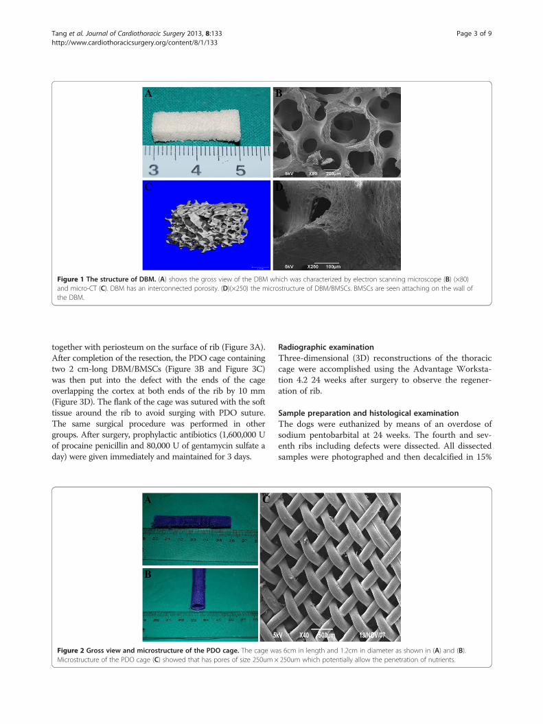
Figure 1 The structure of DBM. (A) shows the gross view of the DBM which was characterized by electron scanning microscope (B) (×80)and micro-CT (C). DBM has an interconnected porosity. (D)(×250) the microstructure of DBM/BMSCs. BMSCs are seen attaching on the wall ofthe DBM.
Tang et al. Journal of Cardiothoracic Surgery 2013, 8:133 Page 3 of 9http://www.cardiothoracicsurgery.org/content/8/1/133
together with periosteum on the surface of rib (Figure 3A).After completion of the resection, the PDO cage containingtwo 2 cm-long DBM/BMSCs (Figure 3B and Figure 3C)was then put into the defect with the ends of the cageoverlapping the cortex at both ends of the rib by 10 mm(Figure 3D). The flank of the cage was sutured with the softtissue around the rib to avoid surging with PDO suture.The same surgical procedure was performed in othergroups. After surgery, prophylactic antibiotics (1,600,000 Uof procaine penicillin and 80,000 U of gentamycin sulfate aday) were given immediately and maintained for 3 days.
Figure 2 Gross view and microstructure of the PDO cage. The cage waMicrostructure of the PDO cage (C) showed that has pores of size 250um×
Radiographic examinationThree-dimensional (3D) reconstructions of the thoraciccage were accomplished using the Advantage Worksta-tion 4.2 24 weeks after surgery to observe the regener-ation of rib.
Sample preparation and histological examinationThe dogs were euthanized by means of an overdose ofsodium pentobarbital at 24 weeks. The fourth and sev-enth ribs including defects were dissected. All dissectedsamples were photographed and then decalcified in 15%
s 6cm in length and 1.2cm in diameter as shown in (A) and (B).250um which potentially allow the penetration of nutrients.

Figure 3 The surgical procedure. (A) the 4 cm long rib together with periosteum defect was created. (B) the preparation of DBMs/BMSCs andPDO cage. (C) The DBMs/BMSCs were put into the PDO cage. (D) The 6cm-long PDO cage containing two 2cm-long BMSCs/DBMs was then fitinto the defect with the ends of the cage overlapping the cortex at both ends of the rib by 10 mm.
Tang et al. Journal of Cardiothoracic Surgery 2013, 8:133 Page 4 of 9http://www.cardiothoracicsurgery.org/content/8/1/133
formic acid in formalin for 2–6 weeks. Tissue sections ofsamples were obtained for H&E staining. The costal tis-sue was also stained by H&E. The contralateral ribs wereremoved as normal controls.
Mechanical testThe length of the samples was uniformly processed as6 cm, including the reconstructed part. Each sample wastested using a three bending point test. The parametersof the test were as follows: L (test span) = 60 mm, loadrate = 0.5N/mm, primary load = 1N. The bending stresswas calculated using the equation: σ = 3PL/2bh2, whereσ, P, L, b, and h represent the bending stress, the bend-ing strength load, test span, the width and thickness ofthe specimen, respectively.
Statistical analysisMechanical test data were analyzed by one-way ANOVA.The differences between the PDO cage/DBMs/BMSCsgroup (n = 12), PDO cage/DBMs group (n = 6), PDO cagegroup (n = 6) and normal rib group (n = 12) were assessedby Student–Newman–Keuls-q. The level of statistical sig-nificance was defined as p = 0.05.
ResultsGross observationAll experimental dogs survived without any difficultyafter surgery. No complications such as wound infectionor paradoxical movement occurred after surgery. Bettershape was achieved in the PDO cage/DBMs/BMSCsgroup than in the other two groups. When the repaired
rib was subjected to an outer force, the ribs in the PDOcage/DBMs/BMSCs group kept their original shape whileribs in other two groups were easily deformed Additionalfile 1. In the PDO cage/DBMs/BMSCs group, we also ob-served bony union at all junctions. In the PDO cage/DBMs group, no bony union was observed not only inthe junction of primary rib and scaffold but also in thejunction of scaffold and scaffold; in the flank group, thedefects between two cut ends of rib were filled with fi-brous tissue (Figure 4; Additional file 2). In all groups, thePDO cage degraded completely.
Radiographic examinationTo observe new bone formation, CT-images were taken24 weeks after surgery. Bony union was observed in thePDO cage/DBMs/BMSCs group while there was nounion observed in the other two groups. Additionally,the reconstructed rib had an appropriate anatomic shapewhich correlated well to the primary radian (Figure 5).
Histological examinationH&E staining was performed to verify the regenerationof new bone. In the PDO cage/DBMs/BMSCs group,there was bony union both in the junction of two scaf-folds and in the junction of the primary rib and scaffold(Figure 6). Marrow was also observed in the scaffold. Inthe PDO cage/DBMs group, there was fibrous tissue atthe junction of scaffolds and the center of the scaffold(Figure 7). In the flank group, no new bone was found.After 24 weeks, the PDO cages were almost com-
pletely degraded. Only some pieces of PDO were found

Figure 4 The result of the experiment. (A) and (B) show the situation of the wound 24 weeks after surgery; the wound healed well. (C) showsthe gross view of reconstructed rib. (I) the flank group whose rib defect receives no material; (II) the control group whose rib defect receivedPDO cage/DBM; (III) the experimental group whose rib defect received PDO cage/DBM/BMSCs; (IV) the normal rib. The arrow shows the junctionof the normal rib and scaffold.
Tang et al. Journal of Cardiothoracic Surgery 2013, 8:133 Page 5 of 9http://www.cardiothoracicsurgery.org/content/8/1/133
in the tissue when examined by histology. Also, smallvessels were observed in the tissue, which may help thepenetration of nutrients (Figure 8).
Mechanical testTo evaluate the mechanical properties of the reconstructedrib, three point bending tests were performed at 24 weeksafter surgery. As there was no bone-union in the PDOcage group and the PDO cage/DBMs group, the threepoint bending test was not applied. In PDO cage/DBMs/BMSCs group (n = 12), the bending stress was 44.27 ± 2.31Mpa, equivalent to 94.8% of the contralateral normal rib(46.67 ± 4.62 Mpa, n = 12). As there were only two groups,the assessment of the data was performed using a pairedsamples t-test. No significant difference were found be-tween the two groups (P > 0.05).
Figure 5 3-D reconstruction of the thoracic cage. We could see that ththe red arrow. In the PDO cage/DBMs group there was no new bone regejunction between the rib and scaffold, which is shown as the green arrow.primary rib. In the PDO cage/DBMS/BMSCs, there was bone union in both
DiscussionThe 4cm-long rib defect in this study meets the standardcriteria of a critical size defect which is defined as a defectwith length at least 2.5 times the diameter of the bone[14]. Also, when the defect was not repaired during sur-gery, after 24 postoperative weeks there was no evidenceof bone regeneration in the defect. However, when thisdefect was reconstructed with the experimental construct,a biodegradable PDO cage and two BMSCs/decalcifiedbones, there was bone regeneration into the defect.In this study, we tried to address two problems— fix-
ation of scaffolds and repair of large bone defects. As forthe first problem, two aspects including material andform have to be considered. As for the material for fix-ation, typically nondegradable materials, such as stain-less steel or titanium [15,16], are deemed to be ideal as
ere was no bone regeneration in the flank group, which is shown byneration not only in the junction of two scaffolds but also in theThe radian of the reconstructed rib, however, was similar to thethe junctions which are shown with the blue arrow.

Figure 6 H&E staining of the junction of primary rib (red triangular arrow) and scaffold (green triangular arrow). (A) (×10) The primaryrib and scaffold of the PDO/DBM/BMSCS group. No clear borderline was observed in the junction of the primary rib and scaffold. (B) (×40) At thejunction of primary rib and scaffold of PDO/DBM/BMSCS group, marrow (black arrow) and new bone were observed; (C) (×10) In the primary riband scaffold of the PDO/DBM group, a clear borderline was observed at the junction. (D) (×40) In the junction of the primary rib and scaffold ofPDO/DBM group, fibrous tissue (blue arrow) was found both in the junction and in the scaffold.
Tang et al. Journal of Cardiothoracic Surgery 2013, 8:133 Page 6 of 9http://www.cardiothoracicsurgery.org/content/8/1/133
they have good mechanical properties which can main-tain the stability of scaffolds. Such materials are nowwidely used in orthopedics for the fixation of variousfractures. However, several problems still remain such ashigh cost, infection, difficulty molding the materials andthe lack of degradation, which may result in many com-plications. Often, such materials have to be removed inan additional operation, adding to the patient’s pain andthe cost associated with the initial operation. Further-more, nondegradable materials are almost completely
Figure 7 H&E staining of the junction of scaffold and scaffold. (A) (×1was bone connection in the junction, and marrow was observed as is showthe PDO/DBM group. No bone connection was observed, and fibrous tissufigure (D) (×100).
radiopaque, which obscures observation of the tissuearound the material. Some imaging modalities, such asMRI, cannot be used when needed, which may delay thediagnosis of other diseases. Thus there is an increasingtrend towards using degradable materials. In this study,we used PDO as the material of fixation. PDO has nowbeen widely used for suture as it has excellent mechan-ical strength and degradability. It is reported that PDOsutures (PDS) can maintain their strength for abouttwenty weeks and will be completely resorbed after 180
0) the junction of two scaffolds of the PDO/DBM/BMSCS group. Theren in Figure B (×100); (C) (×10) the junction of the two scaffolds ofe (blue arrow) was seen in the junction and scaffold as is shown in

Figure 8 H&E staining of the tissue around the reconstructedrib (×100). The PDO degraded almost completely. Some pieces ofPDO can be observed in the tissue and are shown by the blackarrow. New blood vessels can be also seen in the tissue and areshown by the yellow arrow.
Tang et al. Journal of Cardiothoracic Surgery 2013, 8:133 Page 7 of 9http://www.cardiothoracicsurgery.org/content/8/1/133
days, which matches well to the speed of tissue regen-eration [17,18]. In our study, we also found that thePDO cage was completely replaced by fibrous tissuewhich accords well with the result of other researchers’experiments. As for degradation, hydrolysis into H2Oand CO2 is the main method of degradation and shouldhave little effect on the growth of BMSCs .For the form of fixation, different methods such as
chambers, cages and threads were used [19]. Porous cagesare ideal for the fixation of scaffolds. First, the cage allowscontact between the scaffold and primary rib, which canform an osseous connection in those junctions, adding tothe stability of the rib. Second, the porous cage wall alsoallows for nutrient exchange with the material within thecage. The form of cage was first reported by Cobos et al.[20]. They repaired segmental long bone defects with cy-lindrical titanium cages. Over the next several years, thismodel was widely used. It is suitable for the reconstruc-tion of load bearing bone as it has appropriate mechanicalproperties. The titanium cage, however, is nondegradableand radiopaque, which affects the observation of newbone and the tissue behind it. In this study, we utilized aPDO cage instead of a metal cage as the container for thescaffold. The PDO cage had appropriate degradability andis radiolucent, and thus did not affect the observation oforgans in the thoracic cage. The PDO cage, however, hadpoor mechanical properties and can only be used in non-load bearing bones such as the rib, upper limb, etc. SEMcharacterization showed that the PDO cage also had a
pores whose size is about 250 um× 250 um, sufficient toallow the penetration and exchange of nutrients andwaste. Additionally, this cage is flexible and can matchthe radian of the chest wall.The second problem is the repair of large bone de-
fects, which was the critical part of our study. In the pastseveral years, significant research directed towards ad-dressing this problem, but little progress has been made.Tissue engineering is now an acknowledged techniquefor the reconstruction of bone defects, but, to date largebone defect repair remains a challenging problem. In thepast, a single scaffold was widely used as a prosthetic forbone reconstruction or regeneration [21-23]. Some spe-cial considerations must be made when designing a scaf-fold to repair a rib defect. First, the rib has a variableradian and thus a single scaffold may not be suitablefor rib reconstruction as the scaffold must be easilymolded. In this study, we used multiple pieces of scaf-fold to reconstruct one bone defect. This method wasfirst reported by Masatoshi et al. [24]. They successfullyreconstructed a long rib defect with sixteen small, por-ous TCP scaffolds connected with titanum wire and cov-ered with periosteum.There still, however, remain several problems such as
the mechanical integrity of the scaffolds. Although rib is abone that suffers very little outer force, because of the ef-fect of the chest muscle, the scaffold still is exposed todeforming forces, especially when the animal vocalizes. Inthe present study, we used DBM as the scaffold due to thesuitable mechanical properties and porosity, which seemedto make it a more ideal scaffold than tricalcium phosphate.First, DBM has good osteoconductivity, osteoinductivityand osteogenic potential and has been widely used inorthopedic applications. Second, the rib is not a load bear-ing bone but may move with respiration. Thus we need afirm fixation of the scaffold. Third, the cortex of bone isthe main component of the rib and yields very few seedcells for native tissue regeneration. Thus, a tissue engin-eering approach which can supply seed cells is the mostsuitable method for rib reconstruction.Although we have achieved a good result with respect
to the fixation of the scaffold and rib reconstruction,some problems should be studied further. First, the PDOcage as the material for fixation can only be used in nonload bearing bone. Other materials or a method to aug-ment the mechanical strength of the PDO should be in-vestigated. Second, attempts should be made to optimizethe bone regeneration. Nutrients are important to theseed cells, but the scaffolds used do not allow enough nu-trients to reach cells seeded at the center of the scaffold.Thus it will be necessary to optimize the scaffold size andconfiguration with regard to the resultant mechanicalproperties so that a stable scaffold that allows maxi-mal nutrient transfer is used. Third, xenograft may have

Tang et al. Journal of Cardiothoracic Surgery 2013, 8:133 Page 8 of 9http://www.cardiothoracicsurgery.org/content/8/1/133
rejection reaction if it is not properly managed and thus itmay have worse effect of bone regeneration compared toautograft. Fourth, much research is needed into the seedcell source and any necessary manipulations that may beperformed prior to implantation such as cell expansion ortransformation. A number of options are currently beinginvestigated, and the ideal seed cell for bone tissue engin-eering has not been identified.
ConclusionIn our study, we successfully reconstructed large rib de-fects with biodegradable PDO cages in combination withtwo short DBM/BMSCs constructs. New bone regener-ation was verified not only between the two scaffoldsbut also in the junction of scaffold and primary rib. Wethink that such a technique might be a feasible approachfor large bone repair but further research should bedone.
Additional files
Additional file 1: The normal rib.
Additional file 2: The rib defect was reconstructed with PDOcage/DBM.
AbbreviationsPDO: Polydioxanone; DBM: Demineralized bone matrix; BMSCs: Bone marrowmesenchymal stem cells; 3D: Three-dimensional; SEM: Scanning ElectronMicroscope.
Competing interestsThe authors declare that they have no competing interests.
Authors’ contributionsHT designed the study, carried out animal study, followed up the animalsand drafted the manuscript. BW performed the operation of the animal. XQparticipated in the design of the study and performed the statistical analysis.LZ conceived of the study, and participated in its design. KJD participated inthe design and helped to draft the manuscript. ZX designed the study,carried out animal study and rectified the manuscript. All authors read andapproved the final manuscript.
AcknowledgmentThis study was funded by NSFC (30901467) and Shanghai Science andTechnology Development Foundation (08ZR1404900).We thank Dr Yaochang Sun,jianqiu Li, ,Lei Zhong,Lei Xue,Hao Pen,GuangyuanSun for their techinal assistance to this study.We also appreciate Lili, Wangsu,Weilinyun, Dingxinyu, Zhanglu, Xuliang, Guangpeng Liu,Guangdong Zhou,JunZhou, Chenkai Gao, Jinchun Zhuo for their help .
Author details1Department of Thoracic and Cardiovascular Surgery, Shanghai ChangzhengHospital, The Second Military Medical University, No.415 Fengyang Road,Shanghai 200003, China. 2Shanghai Key Laboratory of Tissue Engineering,Shanghai 9th People’s Hospital, Shanghai Jiao Tong University School ofMedicine, Shanghai, China. 3Institute of Biosciences and Bioengineering,Rice University, Rice, USA.
Received: 3 December 2012 Accepted: 13 May 2013Published: 20 May 2013
References1. Gupta SS, Singh O, Soni M, Raikwar RS, Mathur RK: Extra-osseous Ewing's
sarcoma of chest wall. ANZ J Surg 2009, 79(10):752–753.2. Schwartz GS, Rios L, Zivin-Tutela T, Bhora FY, Connery CP: Uncommon
etiology of an anterior chest wall mass. Ann Thorac Surg 2009, 88(5):e58–e59.3. Hidalgo DA, Saldana EF, Rusch VW: Free flap chest wall reconstruction for
recurrent breast cancer and radiation ulcers. Ann Plast Surg 1993, 30(4):375–380.
4. Kridel RW, Ashoori F, Liu ES, Hart CG: Long-term use and follow-up ofirradiated homologous costal cartilage grafts in the nose. Arch FacialPlast Surg 2009, 11(6):378–394.
5. Bapat MR, Chaudhary K, Garg H, Laheri V: Reconstruction of large iliac crestdefects after graft harvest using autogenous rib graft: a prospectivecontrolled study. Spine 2008, 33(23):2570–2575.
6. Jie Y, Lei C, Wen Jie Z, Yilin C: Repair of canine mandibular bone defectswith bone marrow stromal cells and porous β-tricalcium phosphate.Biomaterials 2007, 28(6):1005–1013.
7. Garreta E, Gasset D, Semino C: Fabrication of a three-dimensionalnanostructured biomaterial for tissue engineering of bone. Biomol Eng2007, 24(1):75–80.
8. Syed-Picard FN, Larkin LM, Shaw CM, Arruda EM: Three-dimensionalengineered bone from bone marrow stromal cells and their autogenousextracellular matrix. Tissue Eng Part A 2009, 15(1):187–195.
9. Zhou Y, Chen F, Ho ST, Woodruff MA, Lim TM, Hutmacher DW: Combinedmarrow stromal cell-sheet techniques and high-strength biodegradablecomposite scaffolds for engineered functional bone grafts. Biomaterials2007, 28(5):814–824.
10. Shao X, Goh JC, Hutmacher DW, Lee EH, Zigang G: Repair of large articularosteochondral defects using hybrid scaffolds and bone marrow-derivedmesenchymal stem cells in a rabbit model. Tissue Eng 2006, 12(6):1539–1551.
11. Barone A, Covani U: Maxillary alveolar ridge reconstruction withnonvascularized autogenous block bone: clinical results. J Oral MaxillofacSurg 2007, 65(10):2039–2046.
12. Da Silva RV, Bertran CA, Kawachi EY, Camilli JA: Repair of cranial bonedefects with calcium phosphate ceramic implant or autogenous bonegraft. J Craniofac Surg 2007, 18(2):281–286.
13. Qin X, Tang H, Zhifei X, Zhao X, Sun Y, Gong Z: Chest wall reconstructionwith two types of biodegradable polymer prostheses in dogs. Eur JCardiothorac Surg 2008, 34(4):870–874.
14. Lindsey RW, Probe R, Miclau T, Alexander JW, Perren SM: The effects ofantibiotic-impregnated autogenic cancellous bone graft on bonehealing. Clin Orthop Relat Res 1993, 291:303–312.
15. Segal N, Hell J, Berzins DW: Influence of stress and phase on corrosion ofa superelastic nickel-titanium orthodontic wire. Am J Orthod DentofacialOrthop 2009, 135(6):764–770.
16. Hamad AM, Marulli G, Bulf R, Rea F: Titanium plates support for chest wallreconstruction with Gore-Tex dual mesh after sternochondral resection.Eur J Cardiothorac Surg 2009, 36(4):779–780.
17. Mäkelä P, Pohjonen T, Törmälä P, Waris T, Ashammakhi N: Strength retentionproperties of self-reinforced poly L-lactide (SR-PLLA) sutures comparedwith polyglyconate (Maxon) and polydioxanone (PDS) sutures: an in vitrostudy. Biomaterials 2002, 23(12):2587–2592.
18. Kontio R, Ruuttila P, Lindroos L, Suuronen R, Salo A, Lindqvist C: Biodegradablepolydioxanone and poly(l/d)lactide implants: an experimental study onperi-implant tissue response. Int J Oral Maxillofac Surg 2005, 34(7):766–776.
19. Lindsey RW, Gugala Z, Milne E, Sun M, Gannon FH, Latta LL: The efficacy ofcylindrical titanium mesh cage for the reconstruction of a critical-sizecanine segmental femoral diaphyseal defect. J Orthop Res 2006, 24(7):1438–1453.
20. Cobos J, Lindsey RW, Gugala Z: The cylindrical titanium mesh cage forthe treatment of a long segmental bone defect: description of a newtechnique and report of two cases. J Orthop Trauma 2000, 14(1):54–59.
21. Dallari D, Fini M, Stagni C, Torricelli P, Nicoli Aldini N, Giavaresi G: In vivostudy on the healing of bone defects treated with bone marrow stromalcells, platelet-rich plasma, and freeze-dried bone allografts, alone and incombination. J Orthop Res 2006, 24(5):877–888.
22. Shao XX, Hutmacher DW, Ho ST, Goh JC, Lee EH: Evaluation of a hybridscaffold/cell construct in repair of high-load-bearing osteochondraldefects in rabbits. Biomaterials 2006, 27(7):1071–1080.
23. Chistolini P, Ruspantini I, Bianco P, Corsi A, Cancedda R, Quarto R:Biomechanical evaluation of cell-loaded and cell-free hydroxyapatite

Tang et al. Journal of Cardiothoracic Surgery 2013, 8:133 Page 9 of 9http://www.cardiothoracicsurgery.org/content/8/1/133
implants for the reconstruction of segmental bone defects. J Mater SciMater Med 1999, 10(12):739–742.
24. Hoshino M, Egi T, Terai H, Namikawa T, Takaoka K: Regenerative repair oflong intercalated rib defects using porous cylinders of β-Tricalciumphosphate: an experimental study in a canine model. Plast Reconstr Surg2007, 119(5):1431–1439.
doi:10.1186/1749-8090-8-133Cite this article as: Tang et al.: Tissue engineering rib with theincorporation of biodegradable polymer cage and BMSCs/decalcifiedbone: an experimental study in a canine model. Journal of CardiothoracicSurgery 2013 8:133.
Submit your next manuscript to BioMed Centraland take full advantage of:
• Convenient online submission
• Thorough peer review
• No space constraints or color figure charges
• Immediate publication on acceptance
• Inclusion in PubMed, CAS, Scopus and Google Scholar
• Research which is freely available for redistribution
Submit your manuscript at www.biomedcentral.com/submit