research article open access batch correction of ... · and 2) at the clinic of orthopedics,...
TRANSCRIPT
Kupfer et al. BMC Medical Genomics 2012, 5:23http://www.biomedcentral.com/1755-8794/5/23
RESEARCH ARTICLE Open Access
Batch correction of microarray data substantiallyimproves the identification of genesdifferentially expressed in Rheumatoid Arthritisand OsteoarthritisPeter Kupfer1, Reinhard Guthke1, Dirk Pohlers2,3, Rene Huber2,4, Dirk Koczan5 and Raimund W Kinne2*
Abstract
Background: Batch effects due to sample preparation or array variation (type, charge, and/or platform) mayinfluence the results of microarray experiments and thus mask and/or confound true biological differences. Of thepublished approaches for batch correction, the algorithm “Combating Batch Effects When Combining Batches ofGene Expression Microarray Data” (ComBat) appears to be most suitable for small sample sizes and multiplebatches.
Methods: Synovial fibroblasts (SFB; purity> 98%) were obtained from rheumatoid arthritis (RA) and osteoarthritis(OA) patients (n = 6 each) and stimulated with TNF-α or TGF-β1 for 0, 1, 2, 4, or 12 hours. Gene expression wasanalyzed using Affymetrix Human Genome U133 Plus 2.0 chips, an alternative chip definition file, and normalizationby Robust Multi-Array Analysis (RMA). Data were batch-corrected for different acquiry dates using ComBat and theefficacy of the correction was validated using hierarchical clustering.
Results: In contrast to the hierarchical clustering dendrogram before batch correction, in which RA and OApatients clustered randomly, batch correction led to a clear separation of RA and OA. Strikingly, this applied notonly to the 0 hour time point (i.e., before stimulation with TNF-α/TGF-β1), but also to all time points followingstimulation except for the late 12 hour time point. Batch-corrected data then allowed the identification ofdifferentially expressed genes discriminating between RA and OA. Batch correction only marginally modified theoriginal data, as demonstrated by preservation of the main Gene Ontology (GO) categories of interest, and byminimally changed mean expression levels (maximal change 4.087%) or variances for all genes of interest. Eightgenes from the GO category “extracellular matrix structural constituent” (5 different collagens, biglycan, andtubulointerstitial nephritis antigen-like 1) were differentially expressed between RA and OA (RA>OA), bothconstitutively at time point 0, and at all time points following stimulation with either TNF-α or TGF-β1.Conclusion: Batch correction appears to be an extremely valuable tool to eliminate non-biological batch effects,and allows the identification of genes discriminating between different joint diseases. RA-SFB show an upregulatedexpression of extracellular matrix components, both constitutively following isolation from the synovial membraneand upon stimulation with disease-relevant cytokines or growth factors, suggesting an “imprinted” alteration oftheir phenotype.
Keywords: Microarray analysis, Batch correction, Rheumatoid arthritis, Osteoarthritis, Collagen, Extracellular matrix
* Correspondence: [email protected] Rheumatology Unit, Department of Orthopedics, UniversityHospital Jena, Friedrich Schiller University Jena, Waldkrankenhaus “RudolfElle“, Klosterlausnitzer Str. 81, D-07607, Eisenberg, GermanyFull list of author information is available at the end of the article
© 2012 Kupfer et al.; This is an Open Access a(http://creativecommons.org/licenses/by/2.0),provided the original work is properly cited.
rticle distributed under the terms of the Creative Commons Attribution Licensewhich permits unrestricted use, distribution, and reproduction in any medium,

Kupfer et al. BMC Medical Genomics 2012, 5:23 Page 2 of 12http://www.biomedcentral.com/1755-8794/5/23
BackgroundGene expression microarray technology measures the ex-pression of thousands of genes in one single array by usingmultiple probes to assay each transcript. It is a widely-used tool for identifying genes whose expression changesin response to specific treatments. There are several con-cerns with regard to the reliability of DNA microarraytechnology in the study of diseases.Microarray experiments are costly and time-consuming.
Many studies use multiple arrays, with experiments per-formed at different times, on different array charges oreven with different microarray platforms. The resultinggene expression data could thus be affected by non-biological variables. These systematic differences betweenthe measurements are commonly referred to as “batcheffects”. There are several causes for batch effects, as out-lined below:
� Ambient conditions during the sample preparationand handling, such as room temperature and ozonelevels
� Storage and shipment conditions of the biologicalsamples and/or the arrays
� cRNA/cDNA synthesis� Amplification, labeling, and hybridization protocol:
Use of reagents from different batches� Sites/laboratories: Different laboratories have
different operating procedures� Chip type/charge/platform: The array quality varies
from batch to batch� Washing conditions: Temperature, ionic strength� Scanner: Type, settings, and the calibration drift [1].
Although some of these batch effects can be minimizedor even prevented with appropriate precautions and ex-perimental design, certain batch effects are unavoidable.Some studies require large sample sizes and have to becarried out over many months or sometimes years. Inclinical experiments, experiments are often driven by theavailability of the samples, which cannot be specificallycontrolled for in the original study design. Combiningdata from different batches without removing batch effectscan give rise to misleading results, since the bias intro-duced by the non-biological nature of the batch effectscan be strong enough to mask and/or confound true bio-logical differences. Thus, there is a need to identify andremove these masking effects before further processing.Microarray signal intensity normalization has been
widely used to adjust for experimental artifacts betweenthe samples. The effect of the normalization is to in-crease the precision of multi-array measurementsthrough calibration of the signal intensity distributions.There are several methods for normalization includingMAS5, Robust Multi-Array Analysis (RMA), and dChip
for Affymetrix chips, median scaling for GE-CodeLinkmicroarrays, and LOWLESS-based methods for cDNAtwo-color microarrays. Common to all normalizationmethods is that they are not specifically designed to re-move batch effects reflected by systematic differencesbetween two or more groups of samples. Consequently,batch effects may often remain after normalization.However, of thousands of papers dealing with DNAmicroarrays published in the last 5 years (>32,000), onlyfew address the potential existence of batch effects and/or their correction. Of the 219 papers using microarraydata published from January 1 to July 1, 2010, not eventen percent took this issue into account (NCBI GEOdatabase, studies with more than 30 samples) [2].There are several published approaches to identify and
remove batch effects [1,3]. An Empirical Bayes methodcalled “Combating Batch Effects When CombiningBatches of Gene Expression Microarray Data” (ComBat)estimates parameters for location and scale adjustmentof each batch for each gene independently [4-6]. In thepresent study, this method was applied to a data subsetof 24 arrays (of a total of 120) in order to remove batcheffects due to different acquiry dates of the microarrayanalyses. In contrast to the hierarchical clustering den-drogram before batch correction, in which rheumatoidarthritis (RA) and osteoarthritis (OA) patients clusteredrandomly, batch correction led to a clear separation ofthe RA and OA groups. Batch-corrected data thenallowed an unequivocal identification of differentiallyexpressed genes discriminating between RA and OApatients.
Materials and methodsPatientsSynovial membrane samples were obtained following tis-sue excision upon joint replacement/synovectomy fromRA and OA patients (n = 6 each; all Caucasian; Tables 1and 2) at the Clinic of Orthopedics, Waldkrankenhaus“Rudolf Elle” (Eisenberg, Germany). Informed patientconsent was obtained and the study was approved by theethics committee of the University Hospital Jena. RApatients were classified according to the American Col-lege of Rheumatology (ACR) criteria valid in the sampleassessment period [7], OA patients according to the re-spective criteria for osteoarthritis [8]. Negative purifica-tion of primary synovial fibroblasts (SFB) from RA andOA patients (purity: > 98%) was performed as previ-ously described [9].
Cell stimulation and isolation of total RNAAt the end of the fourth passage, the SFB were washedin serum-free DMEM and then stimulated by 10 ng/mlTNF-α or TGF-β1 (PeproTech, Hamburg, Germany) inserum-free DMEM for 0, 1, 2, 4, or 12 hours. At each

Table 1 Clinical data of patients
Samplename
Gender/Age
Diseaseduration
RF ESR(mm/h)
CRP(mg/ml)
ARAcriteria
Concurrentmedication
RheumatoidArthritis
EB87 F/65 12 + 50,00 106,7 5 NSAIDs
EB88 F/62 10 + 90,00 169,5 6 NSAIDs
EB213 F/69 15 + 94,00 99,1 4 NSAIDs,Leflunomide,Prednisolone
EB220 F/57 20 + 23,00 2,3 4 NSAIDs, MTX,Prednisolone.
EB221 F/66 12 + 7,00 5,4 4 NSAIDs, MTX
EB227 F/49 25 + 12,00 2,4 5 NSAIDs, MTX,Prednisolone.
Osteoarthritis
EB168 F/68 20 - 18,00 20,3 0 NSAIDs
EB173 F/81 2,5 - 38,00 32,0 2 NSAIDs,Prednisolone.
EB190 M/56 5 - 6,00 0,5 1 NSAIDs
EB194 F/79 8 - 28,00 0,3 1 NSAIDs
EB202 M/65 5 - 7,00 3,0 0 NSAIDs
EB205 F/55 3 - 47,00 36,0 0 NSAIDs
ARA, American Rheumatism Association (now American College ofRheumatology); CRP, C-reactive protein (normal range <5 mg/l); ESR,erythrocyte sedimentation rate; F, female; M, male; MTX, methotrexate;NSAIDs, non steroidal anti-inflammatory drugs; RF, rheumatoid factor;-,negative; +, positive.
Table 2 Sample features (incl. acquiry date) and clinicaldiagnosis of RA and OA patients
Batch Acquiry date Sample ID Treatment Diagnosis
1 2006-04-07 EB87.TGF.0 h TGF-β1 RA
EB87.TNF.0 h TNF-α
2 2006-11-30 EB190.TGF.0 h TGF-β1 OA
EB190.TNF.0 h TNF-α
3 2006-12-07 EB202.TGF.0 h TGF-β1
EB202.TNF.0 h TNF-α
EB205.TGF.0 h TGF-β1
EB205.TNF.0 h TNF-α
4 2006-12-12 EB220.TGF.0 h TGF-β1 RA
EB220.TNF.0 h TNF-α
EB221.TGF.0 h TGF-β1
EB221.TNF.0 h TNF-α
5 2009-03-04 EB173.TGF.0 h TGF-β1 OA
EB173.TNF.0 h TNF-α
EB227.TGF.0 h TGF-β1 RA
EB227.TNF.0 h TNF-α
6 2009-03-14 EB194.TGF.0 h TGF-β1 OA
EB194.TNF.0 h TNF-α
EB213.TGF.0 h TGF-β1 RA
EB213.TNF.0 h TNF-α
7 2009-03-26 EB88.TGF.0 h TGF-β1
EB88.TNF.0 h TNF-α
EB168.TGF.0 h TGF-β1 OA
EB168.TNF.0 h TNF-α
All samples (total 120 arrays) were derived from 5 different time points of therespective stimulation experiment. Batch = Integer label for the BatchCorrection; Acquiry date = date of hybridization of the microarray; SampleID = unique array identification; Stimulation = Stimulation of the samples;Diagnosis =Disease of the patient.
Kupfer et al. BMC Medical Genomics 2012, 5:23 Page 3 of 12http://www.biomedcentral.com/1755-8794/5/23
time point, the medium was removed and the cells wereharvested after treatment with trypsin (0.25% in versene;Invitrogen, Karlsruhe, Germany). After washing withphosphate-buffered saline, they were lysed with RLT buf-fer (Qiagen, Hilden, Germany) and frozen at −70°C.Total RNA was isolated using the RNeasy Kit (Qiagen)according to the supplier's recommendation.
Microarray analysisAnalysis of gene expression was performed using U133Plus 2.0 RNA microarrays (Affymetrix, Santa Clara, CA,USA; total of 120 microarrays). Labeling of RNA probes,hybridization, and washing were carried out accordingto the supplier's instructions. Microarrays were analyzedby laser scanning (Hewlett-Packard Gene Scanner).Original data from microarray analysis are accessiblethrough Gene Expression Omnibus series accessionnumber GSE13837 [10].For annotating the genes, the alternative Chip Defin-
ition File (CDF) of Ferrari et al. was used to resolve theproblem of choosing reliable and non-contradictory pro-besets for each transcript [11]. Several publicationsdemonstrated the advantage of such alternative CDFsfor the removal of cross-hybridization and other system-based biases.
The microarray data were preprocessed using RMA inthe default configuration for background adjustment andnormalization.
ComBatFor Batch correction of the patient data (Table 2), theEmpirical Bayes' (EB) method ComBat was used (non-parametric prior method) [5]. EB methods are veryappealing in microarray analyses because of their abilityto robustly handle high-dimensional data derived fromsmall sample sizes. EB methods are primarily designedto “borrow information” from a certain number of genesand/or experimental conditions in order to obtain betterestimates or more stable inferences for the expression ofall genes. In several papers, EB methods were designedto stabilize the expression values/ratios for genes withextreme values or else the variance of genes or genegroups by shrinking variances across all other genes,
Kupfer et al. BMC Medical Genomics 2012, 5:23 Page 4 of 12http://www.biomedcentral.com/1755-8794/5/23
possibly diminishing the effects of artifacts in the data[6,12-19]. Johnson et al. extended the EB methods to theproblem of adjusting for batch effects in microarraydata, which are not addressed by the use of one or sev-eral normalization procedures [5]. Johnson et al. pub-lished a location and scale (L/S) adjustment methodfor batch correction, which is available as R-packageComBat at the developer's homepage [20]. In contrast toother L/S methods, this method is the only procedurecurrently known to robustly adjust batches with smallsample sizes.As other L/S adjustments, ComBat assumes that the
batch effects can be modeled by standardizing meansand variances across batches. It uses a straightforwardL/S adjustment to independently center the mean andstandardize the variance for each gene in each batch.This method incorporates systematic batch biases com-mon across several genes to make adjustments on theassumption that phenomena resulting in batch effectsoften affect many genes in a similar way (i.e., increasedexpression, higher variability, etc.). To determine thedata parameters which describe the particular L/S model[5], ComBat estimates the L/S model parameters thatbest represent the batch effects by “pooling information”across some or all genes in each batch in order to“shrink” the parameter estimates and thereby reduce theinfluence of batch effects.In the present study, a modified method of ComBat
was used to correct for batch effects among arrays gen-erated at different dates. The algorithm was modified inorder to allow processing of RMA-normalized data in-stead of dChip-normalized data. The Sample Informa-tion File was created as described in the ComBatmanual. The creation date was tagged as “batch effect”and the parameters time point (total of 5), disease group(RA and OA), and stimulation (TNF-α and TGF-β1)were marked as covariates for every array.
LimmaTo identify differentially expressed genes, the softwareLimma was used. Limma is an R-package for differentialexpression analysis of data arising from microarrayexperiments [21]. The package is designed to analyzecomplex experiments involving simultaneous compari-sons between many RNA targets. The basic concept isto fit a linear model to the expression data for each gene.The expression data from experiments are representedas a data matrix with rows corresponding to probe setsand columns to arrays. For the analysis with linear mod-els, the approach requires two matrices. First, the designmatrix was created, which provides a representation ofthe different RNA targets. Secondly, the contrast matrixwas generated, which allows to assign the coefficientsdefined by the design matrix to contrasts of interests
(in our case time point 0 hours, RA versus OA patients,Table 2). Afterwards, the linear model was fitted usingthe lmFit function, which fully models the systematicpart of the data. The actual analysis was analogous tothe classic t-test except that the eBayes-function wasused which employs the shrunken empirical Bayes esti-mates [21]. Differentially expressed genes (DEG) wereobtained by using the toptable function and user-specificthresholds (i.e., 2-fold changes in gene expression and aq-value with a threshold of 0.05; [22]). The q-value (FDRadjusted p-value) was calculated using the Benjaminiand Hochberg’s method [21].
GOstatsThe Bioconductor package GOstats shows substantialimprovements for testing the association between GeneOntology (GO) terms and a given gene list [23]. Falconet al. implemented a conditional hypergeometric (hg)test that uses the relationships among GO terms to ad-dress the hierarchical structure of GO. To use this hgtest, the gene universe (in this case containing all geneson the microarray) and a list of selected genes from thatuniverse was defined (DEG). After setting a cutoff forthe adjusted p-value of the hg test, GOstats returns aGOHyperGResult with a summary of the test performedand the number of significant GO terms. Furthermore,the GOHyperGResult contains the expected gene countand actual gene count for each GO term.
GPowerThe Tool GPower 3.1.3 was used for post-hoc calcula-tion of the power (1-ß error probability) of the t-testwith the error probability alpha = 0.05, where the samplesizes of the two groups (RA, OA) were 12 each and therespective mean values and SD of the two groups werederived from Additional file 1: Table S1 [24,25].
ResultsClustering of RA and OA patients before and after batchcorrectionStarting with RMA-normalized data of the 24 arrays fortime point 0, standard hierarchical clustering dendro-grams (generated using the R function hclust with eu-clidean distances) were employed to monitor the effectof the batch correction by ComBat. These dendrogramsmeasure inter-cluster distances between the arrays. Theresulting distances can be interpreted as the dissimilarityof the arraysBefore batch correction, the normalized data formed
clear clusters for the different batches (7 differentacquiry dates for groups of 2 – 4 individual Affymetrixchips; Figure 1a). Except for 2 patients (extreme left andextreme right), two main clusters were formed, separat-ing the microarrays created in 2006 (red shades) from

Figure 1 Hierarchical clustering of uncorrected and batch-corrected data from time point 0 hours: a) The uncorrected data formclusters reflecting the 7 different acquiry dates (red shades for arrays generated in 2006; blue shades for those generated in 2009; forprecise definition of the individual acquiry dates see Table 2). In contrast, RA and OA are not grouped. b) The ComBat-corrected data(7 batches) form clusters reflecting the diseases (RA and OA) instead of the acquiry dates.
Figure 2 Venn Plot for genes differentially expressed betweenRA and OA at time point 0 hours. BC results in a doubling ofdifferentially expressed genes (87 ! 181). A total of 57 genes(65.51%) were represented in the intersection of the two gene lists(DEG_over).
Kupfer et al. BMC Medical Genomics 2012, 5:23 Page 5 of 12http://www.biomedcentral.com/1755-8794/5/23
those generated in 2009 (blue shades). Indeed, the com-pany providing the hybridization agents for the microar-rays confirmed a change in the chemical componentsused in the production process in the year 2007. As aconsequence of the batch effect, several unique clusterswere observed for the RA and OA patients (right andleft branches of the dendrogram), but other clusterswere mixed and contained members of both groups. Inaddition, patient EB87 (extreme right cluster) showedthe highest dissimilarity with all arrays and was inter-preted as an outlier, as also confirmed by Principal Com-ponent Analysis (PCA; not shown).Following batch correction for the 7 different acquiry
dates, the arrays instead formed two distinct clusters forRA and OA in the hierarchical clustering dendrogram(Figure 1b). The effects of the different creation dateswere completely removed and the outlier EB87 was inte-grated, as also confirmed by PCA (not shown). This un-equivocal clustering of RA and OA patients was onlyachieved when correcting for 7 batches and not for 2
yearly batches only (2006 and 2009; Additional file 2:Figure S1).Strikingly, distinct clustering for RA and OA following
batch correction for the 7 different acquiry dates wasnot only observed at the 0 hour time point (i.e., before

Table 3 GO categories overrepresented in the gene set DEG_wBC (time point 0 hours) top-ranked with b) or withoutBC a)
TermID Members (DEG/all) P-value Category
a)
GO:0005201 3/74 5.87e-03 extracellular matrix structural constituent
GO:0050840 2/26 6.62e-03 extracellular matrix binding
GO:0005100 2/28 7.65e-02 Rho GTPase activator activity
GO:0008307 2/40 1.52e-02 structural constituent of muscle
GO:0005178 2/57 2.96e-02 integrin binding
GO:0001871 3/154 3.61e-02 pattern binding
GO:0030246 3/156 3.70e-02 carbohydrate binding
b)
GO:0005201 8/74 7.60E-07 extracellular matrix structural constituent
GO:0048407 4/11 3.29E-06 platelet-derived growth factor binding
GO:0005540 3/19 9.04E-04 hyaluronic acid binding
GO:0015355 2/5 1.02E-03 secondary active monocarboxylatetransmembrane transporter activity
GO:0004035 2/6 1.52E-03 alkaline phosphatase activity
GO:0005178 4/57 2.74E-03 integrin binding
GO:0004000 2/9 3.58E-03 adenosine deaminase activity
GO:0050840 2/26 7.46E-03 extracellular matrix binding
GO:0008307 3/40 7.88E-03 structural constituent of muscle
GO:0046332 3/42 9.02E-03 SMAD binding
GO:0030674 3/44 1.02E-02 protein binding
GO:0016853 4/83 1.02E-02 isomerase activity
GO:0000287 10/441 1.53E-02 magnesium ion binding
GO:0005507 3/62 2.56E-02 copper ion binding
GO:0042802 7/289 3.17E-02 identical protein binding
GO:0005100 2/28 3.31E-02 Rho GTPase activator activity
GO:0005518 2/31 3.94E-02 collagen binding
GO:0016903 2/33 4.47E-02 oxidoreductase activity
GO categories overrepresented in both data sets are highlighted in bold. Please note that both the number of the category members and the p-values fordifferential expression considerably improve by BC.
Kupfer et al. BMC Medical Genomics 2012, 5:23 Page 6 of 12http://www.biomedcentral.com/1755-8794/5/23
stimulation with TNF-α/TGF-β1), but also at all timepoints after stimulation except for the TGF-β1 late 12hour time point (Additional file 3: Figure S2).
Comparison of differentially expressed genes before andafter batch correctionLimma was used to obtain differentially expressed genes(DEG; ≥ 2-fold change; p-value of ≤ 0.05) for the keyquestion concerning a generic difference between RAand OA patients. For the uncorrected data, 87 geneswere extracted (DEG_woBC). The batch-corrected datacontained 181 genes (DEG_wBC), with a total overlap of65.51% (in total 57 genes; DEG_over) between the 2gene sets. This was illustrated using a Venn plot(Figure 2).
For both data sets (DEG_woBC and DEG_wBC), agene enrichment analysis was done with GOstats(p-value ≤ 0.05). In both data sets, the highest rankedGO term was “extracellular matrix structural constitu-ent” with a p-value of 5.87E-03 and 3/74 genes for theuncorrected and a p-value of 7.60E-07 and 8/74 genesfor the corrected data set (Table 3). In addition, 5 of 7GO categories identified before batch correction werealso overrepresented in the data set after batch correc-tion, indicating that no major information was lost uponbatch correction (Table 3). Furthermore, the p-value ofthe 3 genes from the top-ranked GO term contained inboth data sets largely improved following batch correc-tion (by up to 20 orders of magnitude), underlining thevalidity of the differential expression (Table 4).

Table 4 Genes of the top-ranked GO category (time point 0 hours) identified with b) or without BC a)
GC ID P-value SYMBOL ENTREZ ID Gene name
Without BC
GC01M103055_at 3.90E-05 COL11A1 1301 collagen, type XI, alpha 1
GC09P115957_at 3.73E-03 COL27A1 85301 collagen, type XXVII, alpha 1
GC0XP152413_at 3.59E-02 BGN 633 biglycan
With BC
GC01M103055_at 5.31E-25 COL11A1 1301 collagen, type XI, alpha 1
GC0XP152413_at 5.07E-12 BGN 633 biglycan
GC02P189547_at 1.39E-08 COL3A1 1281 collagen,type III, alpha 1
GC09P115957_at 2.98E-06 COL27A1 85301 collagen, type XXVII, alpha 1
GC01P031814_at 6.96E-05 TINAGL1 64129 tubulointerstitial nephritis antigen-like 1
GC07P093861_at 4.37E-04 COL1A2 1278 collagen, type I, alpha 2
GC02M189604_at 1.13E-03 COL5A2 1290 collagen, type V, alpha 2
GC17M045617_at 1.28E-02 COL1A1 1277 collagen, type I, alpha 1
Genes of the GO category (GO:0005201 extracellular matrix structural constituent) top-ranked with or without BC (see Table 3), including their p-value fordifferential expression between RA and OA. Genes represented in both data sets are highlighted in bold. Please note that both the number of genes and thep-value for differential expression considerably improve.
Kupfer et al. BMC Medical Genomics 2012, 5:23 Page 7 of 12http://www.biomedcentral.com/1755-8794/5/23
To analyze the magnitude of changes in either meanor standardized variance induced by the batch correc-tion, variance/mean plots were generated to illustratethe “shrinkage” of the expression values. For this pur-pose, the variances were standardized (range between 0and 1) to prevent negative values. Analyzing the 181genes from the DEG_wBC data set differentiallyexpressed between RA and OA, only marginal changesof the means, but moderate to substantial reductions ofthe variances were observed for all genes (Figure 3).The same was true when the genes lost for analysis
following batch correction (DEG_lost; Figure 4 a,b), thegenes of interest from the GO term “extracellular matrixstructural constituent” (DEG_interest; Figure 4 c,d) orall genes identified without batch correction (DEG_woBC; Additional file 4: Figure S3) were displayed in thevariance/mean plot.To further analyze the magnitude of changes in the
mean induced by the batch correction, the mean valuesin both data sets were compared and the changesexpressed as percentages of the initial value; in both RAand OA, very limited changes were observed (maximalchange 4.087%), either at the time point 0 hours (Table 5)or at any time point following stimulation with TNF-αor TGF-β1 (Additional file 1: Table S1). Despite thelimited changes of the means induced by BC, BCresulted in a substantial improvement of the power forthe differentiation of RA and OA for all genes of interest(Additional file 5: Table S2).There was no indication for any type-1 error concern-
ing differential expression of the genes of interest, asdemonstrated by permuting the disease status (RA and
OA) 20 times with subsequent BC; applying the thresh-olds 2-FC and p< 0.05, none of the 8 genes of interestwere contained in the list of differentially expressedgenes for any of the permutations (Additional file 6:Table S3). Also, a complete lack of clustering for RA andOA was observed and, strikingly, the pairs of one patientfor the time point 0 were separated in some cases(Additional file 7: Figure S4)In addition to the differential expression between RA
and OA after BC for the 8 genes of interest from thetop-ranked GO term at time point 0 hours (Table 4b),these genes also remained differentially expressed be-tween RA and OA at all time points following stimula-tion with either TNF-α or TGF-β1 (Figure 5, Additionalfile 8: Table S4).
DiscussionIn the present study, the ComBat method was highlyeffective in removing batch effects due to differentacquiry dates of the microarrays; this was demonstratedby i) unequivocal clustering of the RA and OA patientsin the batch-corrected data for almost all time pointsinvestigated (shown by hierarchical clustering dendro-grams and PCA); ii) integration of the outlier EB87,resulting in reduced standardized variances for a numberof genes; iii) identification of a large number of genesdifferentially expressed between RA and OA with highlysignificant p-values (up to 5.31E-25); iv) identification ofnumerous overrepresented GO terms (with an increasednumber of members and strongly improved p-values).The 7 different batches (aquiry dates) were clearly
grouped into the two main clusters representing microarray

Figure 3 Means and variances of 181 differentially expressed genes from the DEG_wBC set (time point 0 hours) in RA (a; n = 12; i.e. 6patients with two replicates each) and OA (b; n = 12; i.e. 6 patients with two replicates each) with (red dots) or without BC (blue dots).There are generally only marginal changes of the means, but moderate to substantial reductions of the variances, as indicated by an exclusivelyhorizontal shift.
Kupfer et al. BMC Medical Genomics 2012, 5:23 Page 8 of 12http://www.biomedcentral.com/1755-8794/5/23
analyses from the years 2006 and 2009. These 2 main clus-ters are a clear example of inevitable batch effects since thesupplier of the hybridization agents changed the chemicalcomponents used in the production process in the year2007 (personal communication; Affymetrix). The only pos-sibility to avoid such batch effects would have been to col-lect all the samples for simultaneous analysis, an approachtechnically impossible for a total of 120 samples at the timepoint of analysis (and possibly even today). In addition,
unequivocal clustering of RA and OA patients was onlyachieved when correcting for 7 batches and not only for 2batches (2006 and 2009), indicating that both the technicalchange of the supplier and a basic variance among theacquiry dates contributed to the dissimilarity of the arrays.This should be taken into consideration for future micro-array analyses.The batch correction had no major influence on the
underlying data, as demonstrated by i) almost unaffected
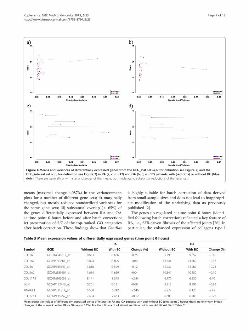
Figure 4 Means and variances of differentially expressed genes from the DEG_lost set (a,b; for definition see Figure 2) and theDEG_interest set (c,d; for definition see Figure 2) in RA (a, c; n = 12) and OA (b, d; n = 12) patients with (red dots) or without BC (bluedots). There are generally only marginal changes of the means, but moderate to substantial reductions of the variances.
Kupfer et al. BMC Medical Genomics 2012, 5:23 Page 9 of 12http://www.biomedcentral.com/1755-8794/5/23
means (maximal change 4.087%) in the variance/meanplots for a number of different gene sets; ii) marginallychanged, but mostly reduced standardized variances forthe same gene sets; iii) substantial overlap (> 65%) ofthe genes differentially expressed between RA and OAat time point 0 hours before and after batch correction;iv) preservation of 5/7 of the top-ranked GO categoriesafter batch correction. These findings show that ComBat
Table 5 Mean expression values of differentially expressed ge
RA
Symbol GCID Without BC With BC
COL1A1 GC17M045617_at 10.663 10.636
COL1A2 GC07P093861_at 13.894 13.895
COL3A1 GC02P189547_at 13.616 13.599
COL5A2 GC02M189604_at 11.664 11.659
COL11A1 GC01M103055_at 8.141 8.373
BGN GC0XP152413_at 10.201 10.131
TINAGL1 GC01P031814_at 6.580 6.742
COL27A1 GC09P115957_at 7.454 7.463
Mean expression values of differentially expressed genes of interest in RA and OA pchanges of the means in either RA or OA (up to 3.7%). For the full data of all stimu
is highly suitable for batch correction of data derivedfrom small sample sizes and does not lead to inappropri-ate modification of the underlying data as previouslypublished [2].The genes up-regulated at time point 0 hours (identi-
fied following batch correction) reflected a key feature ofRA, i.e., SFB-driven fibrosis of the affected joints [26]. Inparticular, the enhanced expression of collagens type I
nes (time point 0 hours)
OA
Change (%) Without BC With BC Change (%)
-0.25 9.793 9.852 +0.60
+0.01 13.544 13.562 +0.13
-0.12 12.937 12.967 +0.23
-0.04 10.841 10.852 +0.10
+2.84 6.470 6.230 -3.70
-0.68 8.912 8.995 +0.93
+2.46 6.277 6.125 -2.42
+0.12 6.688 6.703 +0.23
atients with and without BC (time point 0 hours); there are only very limitedli and time points see Additional file 1: Table S1.

Figure 5 Time courses of genes of interest (DEG_interest; see Table 4b) in synovial fibroblasts from RA patients (blue and purple) orOA (red and green) stimulated with TNF-α (red and blue) or TGF-β1 (green and purple). There were only marginal differences for the geneexpression values with or without BC (see also Additional file 1: Table S1). As expected, there was a clearly different regulation of the expression of6 of 8 genes (BGN, COL1A1, COL27A1, COL5A2, COL1A2 and COL3A1; for definition of the abbreviations see Table 4) following stimulation of SFBwith either TNF-α or TGF-β1; this differential regulation was common for SFB from RA and OA patients (see Additional file 1: Table S1). However, 2of 8 genes (COL11A1 and TINAGL) were regulated in a similar fashion by TNF-α and TGF-β1 in both RA and OA patients. Strikingly, significantdifferences between RA and OA patients were observed for all genes of interest already at the time point 0 hours (see Additional file 8: Table S4a).These differences were unaffected by stimulation with either TNF-α or TGF-β1.
Kupfer et al. BMC Medical Genomics 2012, 5:23 Page 10 of 12http://www.biomedcentral.com/1755-8794/5/23

Kupfer et al. BMC Medical Genomics 2012, 5:23 Page 11 of 12http://www.biomedcentral.com/1755-8794/5/23
and III (COL1A1, COL1A2, COL3A1), as well as bigly-can (BGN) by RA-SFB has been previously published[27-29]. In the context of RA, collagens types V and XIare less well known as mediators of fibrosis, but are tar-gets of matrix metalloproteinase-mediated proteolyticactivity [30,31]. However, together with collagens type I,II, and III, these collagens are defined as (fibrillar) inter-stitial collagens [32] and represent major components ofthe extracellular matrix [33], thus suggesting a potentialrole in fibrosis also for these proteins. The fibrillar typeXXVII collagen (COL27A1) and lipocalin-7 (also knownas tubulointerstitial nephritis antigen-like 1; TINAGL1),which were identified as up-regulated molecules inRA-SFB in this study for the first time, are not intrinsic
extracellular matrix molecules, but both exhibit differen-tiation potential. Type XXVII collagen, for example, ispredominantly expressed in cartilaginous tissues andgenerally involved in skeletogenesis [34], whereas matri-cellular lipocalin-7 appears to be a (positive) regulator ofangiogenesis [35], potentially influencing enhancedangiogenesis in the synovial membrane [36]. Taken to-gether, the enhanced formation of these matrix compo-nents may contribute to joint fibrosis in an attempt tocounteract the progressive destruction of cartilage andbone.Strikingly, differential expression of the 8 genes of
interest from the top-ranked GO term “extracellularmatrix structural constituent” (DEG_interest) was notonly observed at the time point 0 hours, but also at alltime points following stimulation with either TNF-α orTGF-β1. This suggests that RA-SFB show a constitu-tively altered “rheumatic phenotype”, which is preservedupon stimulation with TNF-α and TGF-β1 (“spacer ef-fect”). Possible reasons for such an “imprinted” RAphenotype may include both genomic changes, e.g.,mutations or chromosome aberrations, or epigeneticmodifications, e.g. methylation or histone acetylationstatus [37-39].
ConclusionThe present study clearly underscores the necessity of cor-rection and removal of batch effects in the analysis ofmicroarray data. The application of batch correctionallowed the unequivocal identification of genes differentiallyexpressed between RA and OA and returned the top-ranked GO category “extracellular matrix structural con-stituent” (8/74 genes; p-value decreased by 4 orders of mag-nitude). Batch correction strongly reduced the variance, butonly marginally influenced the mean expression levels, i.e.,led to reliable results without falsification of the underlyingdata.RA-SFB show a constitutively altered “rheumatic pheno-
type”, which is preserved upon stimulation with TNF-α andTGF-β1, suggesting an “imprinted” RA phenotype.
Additional files
Additional file 1: Table S1. Mean expression values of differentiallyexpressed genes of interest in RA and OA patients with and without BCfor all time points.
Additional file 2: Figure S1. Hierarchical clustering of uncorrected andbatch-corrected data from time point 0: a) The uncorrected data formclusters reflecting the 2 different years of acquiry (red shades for arraysgenerated in 2006; blue shades for those generated in 2009). In contrast,RA and OA are not grouped. b) The ComBat-corrected data (2 batches)still fail to form clusters reflecting the diseases (RA and OA).
Additional file 3: Figure S2. Cluster plots for the time points 1, 2, 4,and 12. a) Time point 1: TNF-α. b) Time point 1: TGF-β1. c) Time point 2:TNF-α. d) Time point 2: TGF-β1. e) Time point 4: TNF-α. f) Time point 4:TGF-β1. g) Time point 12: TNF-α. h) Time point 12: TGF-β1.
Additional file 4: Figure S3. Means and variances of differentiallyexpressed genes from the uncorrected data set (DEG_woBC) in RA (a)and OA (b) patients with (red dots) or without BC (blue dots); there aregenerally only marginal changes of the means, but moderate tosubstantial reductions of the variances.
Additional file 5: Table S2. Comparison of the power values for thedifferentiation of RA and OA before and after BC for the differentiallyexpressed genes [applied values: means ± standard deviations (SD)]calculated using GPower [25].
Additional file 6: Table S3. Differentially expressed genes calculatedwith LIMMA for 20 permutations.
Additional file 7: Figure S4. Cluster plots for time point 0 on the basisof 20 permutations of the disease status (RA and OA).
Additional file 8: Table S4. Mann–Whitney U tests for the comparisonbetween RA and OA in a), or for the comparison between TNF-α andTGF-β1 in b); +, significant difference; -, lack of significant difference; *,significance test is not applicable (technical replicates).
AbbreviationsACR: American College of Rheumatology; CDF: Chip Definition File;ComBat: Combating Batch Effects When Combining Batches of GeneExpression Microarray Data; DEG: Differentially expressed genes; DMEM,Dulbecco's modified Eagle's medium; EB: Empirical Bayes; GO: GeneOntology; hg: Hypergeometric; L/S: Location and scale; OA: Osteoarthritis;PCA: Principal Component Analysis; RA: Rheumatoid arthritis; RMA: RobustMulti-Array Analysis; SFB: Synovial fibroblasts.
Competing interestsThe authors declare that they have no competing interests.
Authors' contributionsPK performed the bioinformatic analysis, contributed to the design of thestudy, and participated in the layout, writing, and finalization of themanuscript. RG and RWK contributed to the design of the study andparticipated in the layout, writing, and the finalization of the manuscript. DP,RH, and DK performed the experiments with synovial fibroblasts and therespective data analysis and participated in writing the manuscript. Allauthors read and approved the final manuscript.
AcknowledgementsThis work was supported by grants from the German Federal Ministry ofEducation and Research (BMBF FKZ 0315719A and FKZ 0315719B; ERASysBioPLUS; LINCONET).
Author details1Research Group Systems Biology/Bioinformatics, Leibniz Institute for NaturalProduct Research and Infection Biology – Hans Knöll Institute Jena, Germany.2Experimental Rheumatology Unit, Department of Orthopedics, UniversityHospital Jena, Friedrich Schiller University Jena, Waldkrankenhaus “RudolfElle“, Klosterlausnitzer Str. 81, D-07607, Eisenberg, Germany. 3Present address:Center of diagnostics GmbH, Chemnitz Hospital Chemnitz, Germany.4Present address: Institute of Clinical Chemistry, Hannover Medical School

Kupfer et al. BMC Medical Genomics 2012, 5:23 Page 12 of 12http://www.biomedcentral.com/1755-8794/5/23
Hannover, Germany. 5Institute of Immunology, University of Rostock Rostock,Germany.
Received: 19 December 2011 Accepted: 21 May 2012Published: 8 June 2012
References1. Luo J, Schumacher M, Scherer A, Sanoudou D, Megherbi D, Davison T,
Shi T, Tong W, Shi L, Hong H, Zhao C, Elloumi F, Shi W, Thomas R, Lin S,Tillinghast G, Liu G, Zhou Y, Herman D, Li Y, Deng Y, Fang H, Bushel P, Woods M,Zhang J: A comparison of batch effect removal methods for enhancement ofprediction performance using MAQC-II microarray gene expression data.Pharmacogenomics J 2010, 10:278–291.
2. Chen C, Grennan K, Badner J, Zhang D, Gershon E, Jin L, Liu C: Removing batcheffects in analysis of expression microarray data: an evaluation of six batchadjustment methods. PLoS One 2011, 6:e17238.
3. Scherer A: Batch Effects and Noise in Microarray Experiments: Sources and Solutions,Wiley Series Probability Statistics.; 2009.
4. Benito M, Parker J, Du Q, Wu J, Xiang D, Perou CM, Marron JS:Adjustment of systematic microarray data biases. Bioinformatics 2004,20:105–114.
5. Johnson WE, Li C, Rabinovic A: Adjusting batch effects in microarrayexpression data using empirical Bayes methods. Biostatistics 2007,8:118–127.
6. Kendziorski C, Newton M, Lan H, Gould M: On parametric empirical Bayesmethods for comparing multiple groups using replicated gene expressionprofiles. Stat Med 2003, 22:3899–3914.
7. Arnett FC, Edworthy SM, Bloch DA, McShane DJ, Fries JF, Cooper NS,Healey LA, Kaplan SR, Liang MH, Luthra HS: The American RheumatismAssociation 1987 revised criteria for the classification of rheumatoid arthritis.Arthritis Rheum 1988, 31:315–324.
8. Altman R, Asch E, Bloch D, Bole G, Borenstein D, Brandt K, Christy W,Cooke TD, Greenwald R, Hochberg M: Development of criteria for theclassification and reporting of osteoarthritis. Classification of osteoarthritis ofthe knee. Diagnostic and Therapeutic Criteria Committee of the AmericanRheumatism Association. Arthritis Rheum 1986, 29:1039–1049.
9. Zimmermann T, Kunisch E, Pfeiffer R, Hirth A, Stahl HD, Sack U, Laube A, LiesausE, Roth A, Palombo-Kinne E, Emmrich F, Kinne RW: Isolationand characterization of rheumatoid arthritis synovial fibroblastsfrom primary culture–primary culture cells markedly differ fromfourth-passage cells. Arthritis Res 2001, 3:72–76.
10. Gene Expression Omnibus [http://www.ncbi.nlm.nih.gov/geo/]11. Ferrari F, Bortoluzzi S, Coppe A, Sirota A, Safran M, Shmoish M, Ferrari S,
Lancet D, Danieli GA, Bicciato S: Novel definition files for humanGeneChips based on GeneAnnot. BMC Bioinforma 2007, 8:446.
12. Chen Y, Dougherty ER, Bittner ML: Ratio-based decisions and the quantitativeanalysis of cDNA microarray images. J Biomed Opt 1997, 2:364–374.
13. Efron B, Tibshirani R, Storey JD, Tusher V: Empirical Bayes Analysis of aMicroarray Experiment. J Am Stat Assoc 2001, 96:1151–1160.
14. Newton MA, Kendziorski CM, Richmond CS, Blattner FR, Tsui KW: Ondifferential variability of expression ratios: improving statisticalinference about gene expression changes from microarray data.J Comput Biol 2001, 8:37–52.
15. Tusher VG, Tibshirani R, Chu G: Significance analysis of microarraysapplied to the ionizing radiation response. Proc Natl Acad Sci 2001,98:5116–5121.
16. Smyth G: Linear models and empirical bayes methods for assessingdifferential expression in microarray experiments. Stat Appl Genet Mol Biol2004, 3: ISSN (Online) 1544-6115 (Article 3), doi:10.2202/1544-6115.1027.February 2004.
17. Lönnstedt I, Rimini R, Nilsson P: Empirical bayes microarray ANOVA andgrouping cell lines by equal expression levels. Stat Appl Genet Mol Biol2005, 4. Article7: ISSN (Online) 1544-6115 (Article 7), doi: 10.2202/1544-6115.1125. April 2005.
18. Pan W: Incorporating biological information as a prior in an empiricalbayes approach to analyzing microarray data. Stat Appl Genet Mol Biol2005, 4: ISSN (Online) 1544-6115 (Article 12), doi:10.2202/1544-6115.1124.May 2005.
19. Gottardo R, Raftery AE, Yeung KY, Bumgarner RE: Bayesian robust inference fordifferential gene expression in microarrays with multiple samples. Biometrics2006, 62:10–18.
20. Combat [http://jlab.byu.edu/ComBat/Abstract.html]
21. Smyth GK: Limma: linear models for microarray data. In Bioinformatics andComputational Biology Solutions using R and Bioconductor. Edited by GentlemanR, Carey V, Dudoit S, Irizarry R, Huber W. New York: Springer; 2005:397–420.
22. Shi L, Jones WD, Jensen RV, Harris SC, Perkins RG, Goodsaid FM, Guo L, Croner LJ,Boysen C, Fang H, Qian F, Amur S, Bao W, Barbacioru CC, Bertholet V, Cao XM,Chu T-M, Collins PJ, Fan X, Frueh FW, Fuscoe JC,Guo X, Han J, Herman D, Hong H, Kawasaki ES, Li Q-Z, Luo Y, Ma Y, Mei N,Peterson RL, Puri RK, Shippy R, Su Z, Sun YA, Sun H, Thorn B, Turpaz Y, Wang C,Wang SJ, Warrington JA, Willey JC, Wu J, Xie Q, Zhang L, Zhang L, Zhong S,Wolfinger RD, Tong W: The balance of reproducibility, sensitivity, andspecificity of lists of differentially expressed genes in microarray studies. BMCBioinforma 2008, 9:S10.
23. Falcon S, Gentleman R: Using GOstats to test gene lists for GO termassociation. Bioinformatics 2007, 23:257–8.
24. GPower Tutorial [http://www.psycho.uni-duesseldorf.de/aap/projects/gpower/tutorial_01.html]
25. Faul F, Erdfelder E, Lang A-G, Buchner A: G*Power 3: a flexible statistical poweranalysis program for the social, behavioral, and biomedical sciences. BehavRes Meth 2007, 39:175–191.
26. Szekanecz Z, Koch AE: Update on synovitis. Curr Rheumatol Rep 2001,3:53–63.
27. Postlethwaite AE, Holness MA, Katai H, Raghow R: Human fibroblasts synthesizeelevated levels of extracellular matrix proteins in responseto interleukin 4. J Clin Invest 1992, 90:1479–1485.
28. Kinne RW, Boehm S, Iftner T, Aigner T, Vornehm S, Weseloh G, Bravo R, EmmrichF, Kroczek RA: Synovial fibroblast-like cells strongly expressjun-B and C-fos proto-oncogenes in rheumatoid- and osteoarthritis. Scand JRheumatol Suppl 1995, 101:121–125.
29. Seki T, Selby J, Häupl T, Winchester R: Use of differential subtraction method toidentify genes that characterize the phenotype ofcultured rheumatoid arthritis synoviocytes. Arthritis Rheum 1998,41:1356–1364.
30. Okada Y, Morodomi T, Enghild JJ, Suzuki K, Yasui A, Nakanishi I,Salvesen G, Nagase H: Matrix metalloproteinase 2 from humanrheumatoid synovial fibroblasts. Purification and activation ofthe precursor and enzymic properties. Eur J Biochem 1990,194:721–730.
31. Gadher SJ, Eyre DR, Duance VC, Wotton SF, Heck LW, Schmid TM, Woolley DE:Susceptibility of cartilage collagens type II, IX, X, and XI to human synovialcollagenase and neutrophil elastase. Eur J Biochem 1988, 175:1–7.
32. Miller EJ, Gay S: The collagens: an overview and update. Meth Enzymol 1987,144:3–41.
33. Ricard-Blum S: The collagen family. Cold Spring Harb Perspect Biol 2011,3:a004978.
34. Hjorten R, Hansen U, Underwood RA, Telfer HE, Fernandes RJ, Krakow D, SebaldE, Wachsmann-Hogiu S, Bruckner P, Jacquet R, Landis WJ, Byers PH, Pace JM:Type XXVII collagen at the transition of cartilage to bone duringskeletogenesis. Bone 2007, 41:535–542.
35. Brown LJ, Alawoki M, Crawford ME, Reida T, Sears A, Torma T, Albig AR:Lipocalin-7 is a matricellular regulator of angiogenesis. PLoS One 2010,5:e13905.
36. Zvaifler NJ, Firestein GS: Pannus and pannocytes. Alternative modelsof joint destruction in rheumatoid arthritis. Arthritis Rheum 1994,37:783–789.
37. Karouzakis E, Gay RE, Gay S, Neidhart M: Epigenetic deregulationin rheumatoid arthritis. Adv Exp Med Biol 2011, 711:137–149.
38. Kinne RW, Liehr T, Beensen V, Kunisch E, Zimmermann T, Holland H, Pfeiffer R,Stahl HD, Lungershausen W, Hein G, Roth A, Emmrich F, Claussen U, Froster UG:Mosaic chromosomal aberrations in synovial fibroblasts of patients withrheumatoid arthritis, osteoarthritis, and other inflammatory joint diseases.Arthritis Res 2001, 3:319–330.
39. Dunger S, Neumann S, Zell R, Birch-Hirschfeld E, Stelzner A, Paschke R, KinneRW, Sickinger S: Mutation detection in mosaic situations: RNA mismatchassay and denaturing gradient gel electrophoresis are more sensitivethan conventional cycle sequencing. Anal Biochem 2001, 294:89–93.
doi:10.1186/1755-8794-5-23Cite this article as: Kupfer et al.: Batch correction of microarray datasubstantially improves the identification of genes differentiallyexpressed in Rheumatoid Arthritis and Osteoarthritis. BMC MedicalGenomics 2012 5:23.