research article identification and preservation of...
TRANSCRIPT

Research ArticleIdentification and Preservation ofIntestinal Parasites Using Methylene Blue-Glycerol Mount:A New Approach to Stool Microscopy
Vinay Khanna, Kriti Tilak, Shihnin Rasheed, and Chiranjay Mukhopadhyay
Department of Microbiology, Kasturba Medical College, Manipal University, Karnataka 576104, India
Correspondence should be addressed to Vinay Khanna; [email protected]
Received 18 February 2014; Revised 19 March 2014; Accepted 27 March 2014; Published 10 April 2014
Academic Editor: Bernard Marchand
Copyright © 2014 Vinay Khanna et al. This is an open access article distributed under the Creative Commons Attribution License,which permits unrestricted use, distribution, and reproduction in any medium, provided the original work is properly cited.
We have tried a new approach to routine stool microscopy by using a combination of methylene blue and glycerol in wet mountpreparation of fresh faecal samples for the demonstration of medically important intestinal parasites. This combination wasevaluated for finding differences in the details and clarity of morphology and internal structures of parasites under low- and high-power microscopy as compared to iodine and saline mount. It was further evaluated to estimate the time taken by methyleneblue-glycerol mount to dry up as compared to iodine and saline wet mount.
1. Introduction
Parasitic infections, caused by intestinal helminths and pro-tozoans, account for significant burden of human diseaseload in the developing countries. It is estimated that around3.5 billion people harbour parasites and 450 million areill as a result of these infections [1]. Intestinal helminths,however, are rarely a cause of mortality. Parasites of diar-rhoeal aetiology are widespread in water supplies, infecting asignificant proportion of the human population in the third-world countries [2], especially across the Asian subcontinent.With an exploding population leading to overcrowding andunhygienic practices, these parasites pose a serious threat thatis worsened by limited resources [3]. Over the last decade,new approaches to the diagnosis, treatment, and preven-tion of intestinal protozoan parasites have been developed.However, a considerable improvement in the diagnosis andtreatment of intestinal helminthic infections are yet to beachieved.
Faeces are the most frequent specimens collected andexamined for demonstration of parasites of the gastrointesti-nal tract. In a parasitology laboratory, routinely, two prepa-rations of each specimen are usually made on each slide: oneunstained preparation and another temporarily stained prep-aration. The saline wet mount is an unstained preparation
made by using physiological saline. The advantage of salinepreparation is that it helps to demonstrate the motilityof trophozoites. However, it does not contribute much tothe definitive diagnosis of cysts or trophozoites, becauseinternal structures are often poorly visible. To overcomethis disadvantage, several stain solutions have been usedfor preparation of temporarily stained wet mounts of faecalspecimens. The advantages of direct wet mount include thefollowing: (i) it is a fast, simple procedure and provides aquick answer when positive [4]; (ii) it provides an approxi-mation of the parasitic burden [4]; (iii) it can be used withunpreserved specimens to detect the characteristic motilityof trophozoites [4]; (iv) it can be used as a safeguard, as someprotozoa may at times not concentrate properly because ofunknown factors [5]; (v) it can detect the motile trophozoitesstage of the protozoan species [6].
The classical technique described for the microscopicexamination of parasites is the iodine mount method, whichaids in the differentiation and identification of parasites bycharacteristic morphological features and details of internalstructures. The method is simple to perform, quick, andinexpensive, facilitating direct visualization of parasitic ovaand cyst morphology. The disadvantage of this technique isthat the preparation dries within a few minutes, rendering itunreadable and unreliable to visualize live nematodal larvae
Hindawi Publishing CorporationJournal of Parasitology ResearchVolume 2014, Article ID 672018, 4 pageshttp://dx.doi.org/10.1155/2014/672018

2 Journal of Parasitology Research
[7]. Each time a fresh preparation is required to view slides forlater consultation or demonstration, which consumes consid-erable resources and technician’s valuable time. These short-comings can be overcome by using methylene blue dye andglycerol combination.Thiswetmount is a simple, economicalsubstitute to the iodine mounts and saline mounts as it lastslonger and provides better visualization and differentiationfrom vegetable matters and other debris in the stool, therebyfacilitating any subsequent expert confirmation. Glycerol, byits hygroscopic nature, absorbs water molecules from theenvironment and prevents drying out of wet mounts.
2. Aims and Objectives
Objective 1. This work aims to evaluate differences in themorphological details and clarity of parasites from surround-ing debris and artefacts in stool under low- and high-powermicroscopy.Objective 2. It also aims to evaluate the time taken bymethylene blue dye with different dilutions of glycerol mountto dry out at ambient tropical laboratory temperature (25 ±2∘C) compared to the time taken by control (conventionalmounts without glycerol).
3. Material and Methods
3.1. Specimens. One hundred and fifty fresh unpreservedfaecal specimens were collected from symptomatic patientsattending tertiary care hospital from south India with historyof various intestinal symptoms such as diarrhoea, abdominalpain, vomiting, and perirectal itching, over a study period of1 year. All faecal specimens were examined using methyleneblue-glycerol wet mount and compared with iodine andsaline wet mounts without glycerol.
3.2. Preparation of Wet Mounts of Faeces
3.2.1. Methylene Blue-Glycerol Wet Mount. A thick smear offaecal sample was prepared by adding a greater volume offaeces to a drop of methylene blue-glycerol solution on amicroscope glass slide and placing a coverslip over the smear.Methylene blue-glycerol solution was prepared by mixingmethylene blue dye separately with increasing quantities ofglycerol in concentrations of 10%, 12.5%, 16.6%, 25%, and50%.
3.2.2. Saline Wet Mount and Iodine Wet Mount. Saline wetmounts and iodine wet mount were prepared by separatelymixing a small volume of stool sample with a drop ofphysiological saline, methylene blue dye, and Lugol’s iodine(diluted in 1 : 5 distilled water), respectively, on a glass slideand placing a coverslip over the smear.
3.3. Faecal Microscopy. Saline wet mount and iodine wetmount of specimen were first examined followed by themethylene blue-glycerol wet mounts which were mountedusing solutions containing varying concentrations of glycerol.
Table 1: Total numbers of various intestinal parasites observed bydifferent wet mounts techniques of faecal specimen.
Parasites Structureobserved
Various parasitesamong total
positive samples(𝑁 = 50)
Entamoeba histolytica Cysts andtrophozoites 8
Giardia lamblia Cysts 7Ascaris lumbricoides Ova 2Hookworm Ova 8Trichuris trichiura Ova 6Hymenolepis nana Ova 1Isospora belli Ova 6Cyclospora spp. Ova 3Strongyloides stercoralis Larvae 8Paramecium spp. Organism 1
The entire wetmounts were examined initially by using a low-power (10x) objective and then again by using a high-power(40x) objective of the compound microscope.
4. Results and Discussion
A total of one hundred and fifty samples were observed usingmethylene blue-glycerol, saline, and Lugol’s iodine, out ofwhich fifty were found to be positive for various parasites.Majority of the parasites were found to be Entamoeba his-tolytica and Strongyloides stercoralis and one sample, con-taminated with Paramecium spp., was taken to observe themorphological details of the organism (Table 1).
After observing various parasites by different mount-ing techniques, it was found that methylene blue-glycerolcombination provided an excellent contrast between theparasitic structures and the artefacts as compared to salineand iodine mount. Internal structures of trophozoites, cysts,and ova were stained well by methylene blue dye, whichfacilitated their recognition and identification in specimens.Refractive index of glycerol is greater than that of water,which helps to demarcate the characteristic morphologicalfeatures and details of internal structures, thus assisting indifferentiation and identification of parasites. A similar typeof study was done by Parija and Prabhakar [8] to evaluatethe use of lactophenol cotton blue wet mount preparation asan alternative to routine mounts and found similar resultswith different parasites. We found that the preparation oflactophenol cotton blue requires more numbers of reagents,is expensive, and is more cumbersome as compared tomethylene-glycerol mount.
Vignesh et al. have evaluated 0.25% iodine-glycerol wetmount preparation for the detection of intestinal parasites[9]. One of the drawbacks of iodine wet mounts is that thecysts are stained poorly with the iodine and are less retractilethan unstained cysts in the saline preparation; therefore,they are difficult to visualize and can easily be missed. Incontrast, cysts in methylene blue-glycerol wet mounts were
Journal of Parasitology Research 3
(a) (b) (c) (d)
(e) (f) (g) (h)
(i) (j)
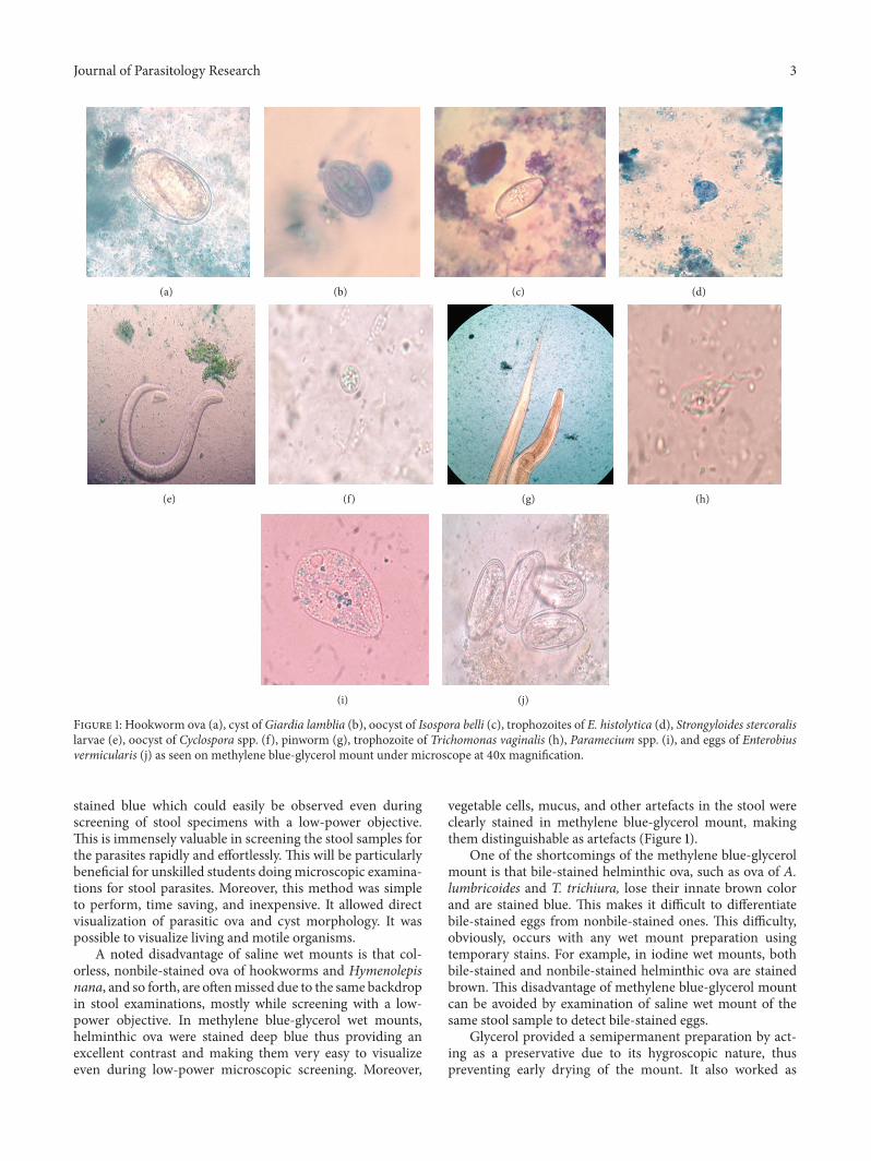
Figure 1: Hookworm ova (a), cyst ofGiardia lamblia (b), oocyst of Isospora belli (c), trophozoites of E. histolytica (d), Strongyloides stercoralislarvae (e), oocyst of Cyclospora spp. (f), pinworm (g), trophozoite of Trichomonas vaginalis (h), Paramecium spp. (i), and eggs of Enterobiusvermicularis (j) as seen on methylene blue-glycerol mount under microscope at 40x magnification.
stained blue which could easily be observed even duringscreening of stool specimens with a low-power objective.This is immensely valuable in screening the stool samples forthe parasites rapidly and effortlessly. This will be particularlybeneficial for unskilled students doing microscopic examina-tions for stool parasites. Moreover, this method was simpleto perform, time saving, and inexpensive. It allowed directvisualization of parasitic ova and cyst morphology. It waspossible to visualize living and motile organisms.
A noted disadvantage of saline wet mounts is that col-orless, nonbile-stained ova of hookworms and Hymenolepisnana, and so forth, are oftenmissed due to the same backdropin stool examinations, mostly while screening with a low-power objective. In methylene blue-glycerol wet mounts,helminthic ova were stained deep blue thus providing anexcellent contrast and making them very easy to visualizeeven during low-power microscopic screening. Moreover,
vegetable cells, mucus, and other artefacts in the stool wereclearly stained in methylene blue-glycerol mount, makingthem distinguishable as artefacts (Figure 1).
One of the shortcomings of the methylene blue-glycerolmount is that bile-stained helminthic ova, such as ova of A.lumbricoides and T. trichiura, lose their innate brown colorand are stained blue. This makes it difficult to differentiatebile-stained eggs from nonbile-stained ones. This difficulty,obviously, occurs with any wet mount preparation usingtemporary stains. For example, in iodine wet mounts, bothbile-stained and nonbile-stained helminthic ova are stainedbrown. This disadvantage of methylene blue-glycerol mountcan be avoided by examination of saline wet mount of thesame stool sample to detect bile-stained eggs.
Glycerol provided a semipermanent preparation by act-ing as a preservative due to its hygroscopic nature, thuspreventing early drying of the mount. It also worked as

4 Journal of Parasitology Research
Table 2:Association between the amount and time taken by glycerolmount to dry up.
Percentage of glycerolused in the solution
Time taken bythe mount to dry up
10% 6 hrs12.5% 8 hrs16.6% 12 hrs25% 36–48 hrs50% 5 days
a fixative. Therefore, repeated mounting was not required.Iodine or saline mount, which was taken as a control, driedin less than 10 minutes, whereas methylene blue-glycerolmount lasted much longer without drying. It started dryingonly after 6 hours and could be retained for up to 5 daysunder different dilutions. The time taken by the methyleneblue-glycerol mount to dry up varied with the amount ofglycerol in the mount. Table 2 clearly shows the associationbetween the amount of glycerol used in the mount and thetime taken by the mount to dry up. Optimal dilution ofglycerol preventedmorphological alteration of parasitic cysts,ova, and trophozoites and conserved their internal structures,which would have been distorted on using pure glycerol dueto its water extracting property.
Mount in which glycerol was used at 50% concentrationcould be preserved for the longest duration and also providedthe best contrast when viewed under the microscope. Similarresults were obtained byVignesh et al. [9] using 0.25% iodine-glycerol combination.
5. Conclusion
Methylene blue-glycerol wet mount is easy to perform andstays longer; the reagents are economical and easily prepared.The procedure also aids the better detection and recognitionof parasites in the stool in a peripheral laboratory, side clinicof doctors, and other laboratories in the developing countrieswhere permanent stained smear of stool does not form acomponent of routine stool examination.We advocate the useof methylene blue-glycerol wet mount along with saline wetmount in a routine parasitology laboratory.
Conflict of Interests
The authors declare that there is no conflict of interestsregarding the publication of this paper.
References
[1] M. Endris, Z. Tekeste, W. Lemma, and A. Kassu, “Comparisonof the Kato-Katz, Wet Mount, and Formol-Ether concentrationdiagnostic techniques for intestinal helminth infections inEthiopia,” ISRN Parasitology, vol. 2013, Article ID 180439, 5pages, 2013.
[2] R. Vignesh, P. Balakrishnan, E. M. Shankar et al., “Short report:high proportion of isosporiasis among hiv-infected patients
with diarrhea in Southern India,” American Journal of TropicalMedicine and Hygiene, vol. 77, no. 5, pp. 823–824, 2007.
[3] H. Guyatt, “Do intestinal nematodes affect productivity inadulthood?” Parasitology Today, vol. 16, no. 4, pp. 153–158, 2000.
[4] H. Afroz, R. Alam, M. Hossain et al., “Evaluation of iodine-glycerol for wet mount preparation of faeces,” InternationalJournal of Biological & Medical Research, vol. 4, no. 4, pp. 3615–3618, 2013.
[5] D. M. Melvin and J. W. Smith, “Intestinal parasitic infections.Part 1. Problems in laboratory diagnosis,” Laboratory Medicine,vol. 10, pp. 207–210, 1979.
[6] R. Neimeister, A. L. Logan, J. H. Egleton, and B. Kleger,“Evaluation of direct wet mount parasitological examination ofpreserved fecal specimens,” Journal of ClinicalMicrobiology, vol.28, no. 5, pp. 1082–1084, 1990.
[7] E. G. Estevez and J. A. Levine, “Examination of preserved stoolspecimens for parasites: lack of value of the direct wet mount,”Journal of ClinicalMicrobiology, vol. 22, no. 4, pp. 666–667, 1985.
[8] S. C. Parija and P. K. Prabhakar, “Evaluation of lacto-phenolcotton blue for wet mount preparation of feces,” Journal ofClinical Microbiology, vol. 33, no. 4, pp. 1019–1021, 1995.
[9] R. Vignesh, R. Sekar, E. M. Shankar et al., “Wet mountingusing iodine-glycerol provides a semi-permanent preparationfor microscopic observation of faecal parasites,” Journal ofMedical Microbiology, vol. 57, no. 5, pp. 679–680, 2008.

Submit your manuscripts athttp://www.hindawi.com
Hindawi Publishing Corporationhttp://www.hindawi.com Volume 2014
Anatomy Research International
PeptidesInternational Journal of
Hindawi Publishing Corporationhttp://www.hindawi.com Volume 2014
Hindawi Publishing Corporation http://www.hindawi.com
International Journal of
Volume 2014
Zoology
Hindawi Publishing Corporationhttp://www.hindawi.com Volume 2014
Molecular Biology International
GenomicsInternational Journal of
Hindawi Publishing Corporationhttp://www.hindawi.com Volume 2014
The Scientific World JournalHindawi Publishing Corporation http://www.hindawi.com Volume 2014
Hindawi Publishing Corporationhttp://www.hindawi.com Volume 2014
BioinformaticsAdvances in
Marine BiologyJournal of
Hindawi Publishing Corporationhttp://www.hindawi.com Volume 2014
Hindawi Publishing Corporationhttp://www.hindawi.com Volume 2014
Signal TransductionJournal of
Hindawi Publishing Corporationhttp://www.hindawi.com Volume 2014
BioMed Research International
Evolutionary BiologyInternational Journal of
Hindawi Publishing Corporationhttp://www.hindawi.com Volume 2014
Hindawi Publishing Corporationhttp://www.hindawi.com Volume 2014
Biochemistry Research International
ArchaeaHindawi Publishing Corporationhttp://www.hindawi.com Volume 2014
Hindawi Publishing Corporationhttp://www.hindawi.com Volume 2014
Genetics Research International
Hindawi Publishing Corporationhttp://www.hindawi.com Volume 2014
Advances in
Virolog y
Hindawi Publishing Corporationhttp://www.hindawi.com
Nucleic AcidsJournal of
Volume 2014
Stem CellsInternational
Hindawi Publishing Corporationhttp://www.hindawi.com Volume 2014
Hindawi Publishing Corporationhttp://www.hindawi.com Volume 2014
Enzyme Research
Hindawi Publishing Corporationhttp://www.hindawi.com Volume 2014
International Journal of
Microbiology