research article - global research onlineglobalresearchonline.net/journalcontents/v42-2/23.pdf ·...
TRANSCRIPT
Int. J. Pharm. Sci. Rev. Res., 42(2), January - February 2017; Article No. 23, Pages: 119-124 ISSN 0976 – 044X
International Journal of Pharmaceutical Sciences Review and Research International Journal of Pharmaceutical Sciences Review and Research Available online at www.globalresearchonline.net
© Copyright protected. Unauthorised republication, reproduction, distribution, dissemination and copying of this document in whole or in part is strictly prohibited. Available online at www.globalresearchonline.net
119
Sinha S*., Choubey S, Ajay Kumar A, Bhosale P Department of Microbiology, Dr. D. Y. Patil ACS College, Pimpri, Pune, India.
*Corresponding author’s E-mail: [email protected]
Received: 30-11-2016; Revised: 15-01-2017; Accepted: 03-02-2017.
ABSTRACT
The aim of the present study was to isolate the microorganisms from different geographical locations, capable of producing pigments with anti-microbial activity. Soil and water samples were collected from different areas under different climatic conditions. A total of five pigmented colonies were isolated that produced intracellular pigments and isolated organism were characterized by the Bergey’s Manual of Determinative Bacteriology. The pigments were extracted from the isolates and their antimicrobial activity was identified. The extracted pigments had inhibitory effect on both Gram positive and Gram negative bacteria as well on fungus. So it was concluded that soil and water has diverse organisms which showed antibacterial and antifungal activity.
Keywords: Serratia sp., Micrococcus sp., Erythrobacter sp., Azorhizobium sp., Pigment.
INTRODUCTION
igments produced from natural sources are of worldwide interest and is gaining significance. Natural pigments are obtained from ores, insects,
plants and microbes1. Natural pigments possess anticancer activity, contain pro-vitamin A and have some desirable properties like stability to light, heat and pH2. Natural colorants or dyes derived from flora and fauna are believed to be safe because of non-toxic, non-carcinogenic and biodegradable in nature. The advantages of pigment production from microorganisms comprise easy and fast growth in the cheap culture medium, independence from weather conditions and colors of different shades.
Pigments of various colors are synthesized to protect the cells of microorganisms from injurious effect of light rays of visible and near ultraviolet range. These pigments are synthesized by various types of microorganisms as secondary metabolites and not often found in all types of organisms. Pigments come in a wide variety of colors, some of which are water soluble
3.Micro-organisms which
have the ability to produce pigments in high yields include species of Monascus, Paecilomyces, Serratia, Cordyceps, Streptomyces and yellow-red and blue compounds produced by Penicilliumherquei and Penicillium atrovenetum, Rhodotorula, Sarcina, Cryptococcus, Monascus purpureus, Phaffia rhodozyma, Bacillus sp., Achromobacter, Yarrowia and Phaffia also produce a large number of pigment2. Microorganisms produce various pigments like carotenoids, melanins, quinones, flavins, prodigiosins and more specifically monascins, violacein or indigo4-7.
Pigments are compounds with uniqueness of importance to many industries. The utilization of natural pigments in foodstuff, dyestuff, cosmetic and pharmaceutical manufacturing processes has been mounting in recent
years3. In the food industry they are used as additives,
antioxidants, color intensifiers, etc. Microbial colorants play a significant role as food coloring agent, because of its production and easy downstream processing8, 9.
Some pigments produced by the bacteria shows antimicrobial activity against pathogens. These antimicrobial agents or substance produced by the bacteria are successfully used for preventing and treating microbial diseases. The pigments like carotenoids, melanin, flavin, violacein, prodigiosin showed distinct antimicrobial effect against many pathogenic bacteria10-15. Presently research is going on these pigment producing bacteria because the antimicrobial substance produced by these bacteria have been successfully used for clinical therapy. Inspired by these facts, the aim of this research was to isolate the pigment producing microorganisms from natural habitat and evaluate the antimicrobial activity of pigments against human pathogenic bacteria.
MATERIAL AND METHODS
Sample Collection
Different soil samples (Moshi dumping ground and forest area near Katraj tunnel, Pune) and water samples {Muthariver (in front of Pune Municipal Corporation) and Indrayani River, Alandi, Pune} were collected. From these samples, pigment producing bacteria were isolated and the bacterial cultures were used for the present study. Microbial cultures were obtained from NCL (National Chemical Laboratory) Escherichia coli ATCC 8739, Salmonella typhi ATCC 23564, Pseudomonas, Staphylococcus aureus ATCC 25923, Candida ATCC 18804. Media and chemicals used in the present study were from HIMEDIA Biosciences.
Identification, Characterization of Pigment Producing Bacteria from Soil and Water and Testing of Antimicrobial Activity of Bacterial Pigments
P
Research Article
Int. J. Pharm. Sci. Rev. Res., 42(2), January - February 2017; Article No. 23, Pages: 119-124 ISSN 0976 – 044X
International Journal of Pharmaceutical Sciences Review and Research International Journal of Pharmaceutical Sciences Review and Research Available online at www.globalresearchonline.net
© Copyright protected. Unauthorised republication, reproduction, distribution, dissemination and copying of this document in whole or in part is strictly prohibited. Available online at www.globalresearchonline.net
120
Isolation of pigment producing organism
Soil samples
Soil samples were collected from two places Moshi garbage dumping ground and forest area near Katraj tunnel. Collected soil samples were used for serial dilution up to 10-9. 10-4 to 10-9 dilutions were plated on sterile Nutrient agar (NA) petri plates and kept for incubation at 37
0C for 48hrs. The NA plates were
observed for growth after 48hrs of incubation. The pigment producing organism was observed on 10-4
dilution of Katraj soil sample. The isolated pigment producing organism from 10
-4 dilution of Katraj soil
sample was cream in color which was further streaked on sterile NA plate to obtain pure culture. The cream color pigment producing bacteria was further characterized for identification.
Water samples
Water samples were collected from two places Muthariver and Indrayani River in Alandi. Water samples were serially diluted up to 10-9. 10-3 to 10-9 dilutions were placed on sterile Nutrient agar (NA) plates and kept for incubation at 370C.Pigment producing organisms were isolated after 2- 3 days of incubation period and the pigment producing organisms were yellow, dark orange, light orange. The pigment producing organism like red was isolated after 2–3 weeks. The color of pigment producing organism of Muthariver was yellow and dark orange and Indrayani River was red and light orange. The isolated colonies of pigment producing organisms were streaked on another sterile NA plates to isolate pure cultures.
Characterization and Identification of pure cultures of pigment producing bacteria
Colony characterization of pigment producing bacteria from NA plate (24 hour incubation)was done based on its size, shape, color, margin, opacity, consistency, elevation, Gram staining and motility. The biochemical tests performed were Indole test, Methyl Red (MR), Voges Proskauer (VP), Simmon’s Citrate test, Oxidase test, Catalase tests and sugar fermentation test. Identification of isolates obtained in pure cultures were characterized by morphology, growth characteristics on selective and differential media, microscopically by Gram staining, and various biochemical tests recommended in the Bergey’s Manual of Determinative Bacteriology
16, 17.
Screening of pigment producing bacteria
Isolated colonies of identified cultures were suspended in 2% glycerol containing Nutrient broth in a flask and the flasks were incubated on rotary shaker for 48hrs. Extensive growth of pigment producing bacteria was seen in the flasks after 48hrs.
Extraction of pigments from pigment producing bacteria
The pigment producing bacteria was harvested by centrifuging at 6000rpm for 20min. Then supernatants
were discarded and the pellets were re-suspended in ethanol. Then the mixture was vortexed and the suspension was centrifuged at 6000rpm for10 min and supernatant was collected. Centrifugation was repeated till the pellet changes to colorless. After centrifugation, supernatants containing diffused pigments were filtered through Millipore membrane filter (pore diameter 0.22μm). The absorbance of filtrates was measured on UV-visible spectrophotometer in the range 350-750nm. Then the filtrates were kept in water bath for ethanol evaporation. After evaporation of ethanol, dry pigment residues were re-suspended in DMF (dimethylformamide) solvent. The solvent containing pigment was then used to evaluate its antimicrobial activity against human pathogens along with its control.
Optical density (O.D.)
Visible absorption spectrum of the separated pigments in nutrient broth was analyzed with UV-visible Spectrophotometer between the wavelengths of 350-750 nm.
Antimicrobial Activity
Antimicrobial activity of the pigments was tested by well diffusion method. Five human pathogens (Escherichia coli, Salmonella typhi, Pseudomonas, Staphylococcus aureus, Candida) and one unknown sample were used against extracted pigment to evaluate its antimicrobial activity. Unknown pus sample was collected from facial part of 32yrs old female and it was collected with the help of sterile cotton swab. The sample collected was plated on sterile NA plate by spread plate technique and incubated at 370C for 24hrs. Growth was observed after 24hrs, isolated colonies were further characterized for identification.
These human pathogens and unknown female pus sample were inoculated in nutrient broth and incubated at 370C for 24hrs.After incubation, they were used for antimicrobial activity and NA plates were used for well diffusion method. The plate was seeded with 24hrsgrown pathogen culture and wells were bored in the plates. Then the wells were filled with appropriate amount of pigment (20μl) and it was kept in refrigerator for half an hour. After that it was incubated at 37
0C for 24hrs and
the result was observed by measuring zone of inhibition.
Zone of inhibition
The anti-microbial activity of pigments against different human pathogens and unknown pus sample was observed after 24 hrs of incubation at 37
0C.
RESULTS AND DISCUSSION
Identification and characterization of the pigment producing bacteria
The soil and water samples collected from different parts of Pune were used for isolation of pigment producing bacteria. Five pigment producing bacteria were identified and characterized which were red, yellow, cream, light
Int. J. Pharm. Sci. Rev. Res., 42(2), January - February 2017; Article No. 23, Pages: 119-124 ISSN 0976 – 044X
International Journal of Pharmaceutical Sciences Review and Research International Journal of Pharmaceutical Sciences Review and Research Available online at www.globalresearchonline.net
© Copyright protected. Unauthorised republication, reproduction, distribution, dissemination and copying of this document in whole or in part is strictly prohibited. Available online at www.globalresearchonline.net
121
orange and dark orange in color (Fig. 1).These bacteria were then identified and characterized with the help of morphological characteristics and biochemical tests (Table 1). Their identification at genus level was done
with the help of Bergey’s Manual of Determinative Bacteriology, which is presented in Table 2.
Table 1: Morphophological, Biochemical and Physiological Characterization of the Pigment Producing Microorganism
Colony Morphology
Color Size Margin Shape Opacity Consistency Elevation Gram characters Motility
Red 2mm entire circular opaque Smooth Convex Gram Negative Rod Non Motile
Yellow 1mm Irregular circular opaque Smooth Convex Gram Positive Rod Motile
Cream 2mm entire circular opaque Smooth Flat Gram Negative Rod Motile
Dark Orange 1mm entire circular opaque Sticky Flat Gram Negative Rod Motile
Light Orange 1mm entire circular opaque Smooth Convex Gram Negative Rod Motile
Unknown sample 1mm entire circular opaque sticky convex Gram Negative cocci motile
Biochemical test for sugar
Pigment Glucose Mannitol Raffinose Galactose Fructose Sucrose Maltose Arabinose Cellobiose Lactose Xylose
Red - + - - - - + - + - +
Yellow + - + + + + - - - - -
Dark orange
- + + - - + - - + - -
Light orange
- - + - + - - + - - +
Cream - - - - - - - - - - +
Un known
sample
+ - + - - + + - - + -
IMVIC test, nitrate test, gelatinase test, catalase and oxidase test
Pigment Indole Methyl
Red
Citrate
Test
Voges
Proskauer Gelatinase
Nitrate
Test Oxidase Catalase
Red - - + + + - - +
Yellow - - + - + + - +
Dark orange - - - _ + - + +
Light orange - - + + + - - +
Cream - + + _ _ - + +
Unknown sample - - - + + - - +
+ Sign stands for positive and - stands for negative result.
Table 2: Genus level identification
Color of pigment Microorganisms
Red Serratia
Yellow Micrococcus
Dark orange Erythrobacter
Light orange Serratia
Cream Azorhizobium
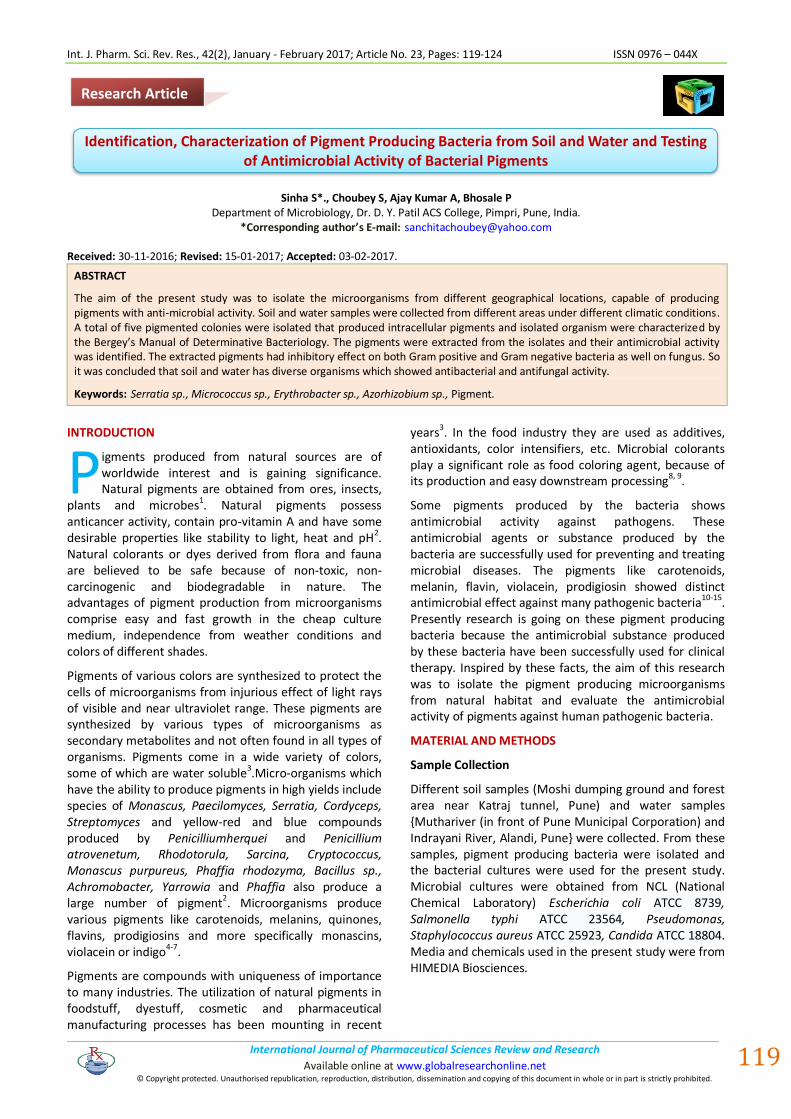
In the present study, the five pigment producing bacteria that produced intracellular pigments (Fig. 1)
were characterized. The identified genera were Serratia, Micrococcus, Erythrobacter and Azorhizobium.
Int. J. Pharm. Sci. Rev. Res., 42(2), January - February 2017; Article No. 23, Pages: 119-124 ISSN 0976 – 044X
International Journal of Pharmaceutical Sciences Review and Research International Journal of Pharmaceutical Sciences Review and Research Available online at www.globalresearchonline.net
© Copyright protected. Unauthorised republication, reproduction, distribution, dissemination and copying of this document in whole or in part is strictly prohibited. Available online at www.globalresearchonline.net
122
Serratia (Red) Serratia (Light orange)
Micrococcus (Yellow) Erythrobacter (Dark orange)
Azorhizobium (Cream) Staphylococcus (Female pus
Sample)
Figure 1: Nutrient agar showing pigment producing organism isolated from soil and water samples
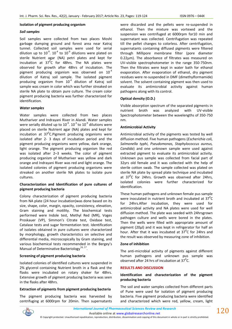
Extraction of pigments from pigment producing bacteria
For the extraction of pigment producing bacteria, various methods were used like centrifugation, filtration, and addition of ethanol so that cell gets lysed and intracellular pigment can be extracted. The pigments extracted were red, yellow, cream, light orange and dark orange in color (Fig.2). The optical density of the pigment were measured and further processed for antimicrobial activity.
Dark orange pigment Red pigment
Yellow pigment Cream pigment
Light orange pigment
Figure 2: Visual observation of culture of isolated pigment producing organism grown for pigment
extraction
Salmonella Typhi Staphylococcus aureus
Yellow pigment Dark orange pigment
Yellow pigment
Candida
Cream pigment Yellow pigment
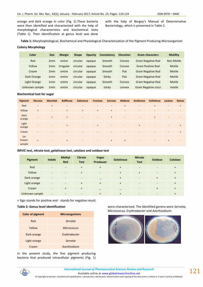
Dark orange pigment Figure 3: Measurement of antimicrobial activity of isolated pigmented organism as zone of inhibition
Int. J. Pharm. Sci. Rev. Res., 42(2), January - February 2017; Article No. 23, Pages: 119-124 ISSN 0976 – 044X
International Journal of Pharmaceutical Sciences Review and Research International Journal of Pharmaceutical Sciences Review and Research Available online at www.globalresearchonline.net
© Copyright protected. Unauthorised republication, reproduction, distribution, dissemination and copying of this document in whole or in part is strictly prohibited. Available online at www.globalresearchonline.net
123
The pigments produced in this study have shown different optical density. The peak optical density was at 450 nm and the pigment which showed maximum optical density was yellow pigment.
Antimicrobial activity against bacterial and fungal pathogens
The extracted pigments were dissolved with solvent DMF (dimethylformamide) to evaluate the antimicrobial activity against human pathogen by well diffusion method. The bacterial pathogens were E.coli, Pseudomonas, Staphylococcus aureus and Salmonella typhi and the fungal pathogen used was Candida.The zone of inhibition was measured to evaluate antimicrobial activity. All the pigments isolated showed
antibacterial activity against the test pathogens. Of all the different pigments tested, yellow pigment has shown the antimicrobial activity with maximum zone of inhibition 23 mm against S. aureus (Table 3). This study suggests that all the pigments showed better antibacterial activity against gram positive pathogens than gram negative pathogens. Mode of anti-bacterial action of most pigments was bacteriostatic. Anti-bacterial activity of the extracted pigments against the test pathogens are shown in Table3.
Antimicrobial activity against unknown sample
The unknown sample was the facial pus sample from female which was identified by morphological characteristic and biochemical tests.
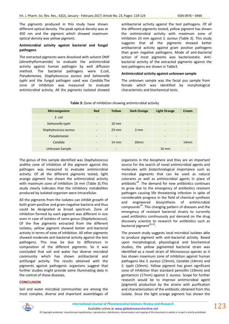
Table 3: Zone of inhibition showing antimicrobial activity
Microorganism Red Yellow Dark Orange Light Orange Cream
E. coli - - - - -
Salmonella typhi - 10 mm - - -
Staphylococcus aureus - 23 mm 3 mm - -
Pseudomonas - - - -
Candida - 14 mm 10mm - 14mm
Unknown Sample - - - 16 mm -
The genus of this sample identified was Staphylococcus andthe zone of inhibition of the pigment against this pathogen was measured to evaluate antimicrobial activity. Of all the different pigments tested, light orange pigment has shown the antimicrobial activity with maximum zone of inhibition 16 mm (Table 3).This study clearly indicates that the inhibitory metabolites produced by isolated organism were intracellular.
All the pigments from the isolates can inhibit growth of both gram-positive and gram-negative bacteria and thus could be designated as broad spectrum. Zone of inhibition formed by each pigment was different in size even in case of isolates of same genus (Staphylococcus). Of the five pigments extracted from the different isolates, yellow pigment showed better anti-bacterial activity in terms of zone of inhibition. All other pigments showed moderate anti-bacterial activity against the test pathogens. This may be due to differences in composition of the different pigments. So it was concluded that soil and water has diverse microbial community which has shown antibacterial and antifungal activity. The results obtained with the pigments against pathogenic organisms suggest that further studies might provide some illuminating data in the control of these diseases.
CONCLUSION
Soil and water microbial communities are among the most complex, diverse and important assemblages of
organisms in the biosphere and they are an important source for the search of novel antimicrobial agents and molecules with biotechnological importance such as microbial pigments that can be used as natural colorants as well as antimicrobial agents in place of antibiotic18. The demand for new antibiotics continues to grow due to the emergency of antibiotics resistant pathogen causing life threatening infection in spite of considerable progress in the field of chemical synthesis and engineered biosynthesis of antimicrobial compounds19. This changing pattern of diseases and the emergency of resistant bacterial strains to currently used antibiotics continuously put demand on the drug discovery scientist to research for antibiotics such as bacterial pigment20-22.
The present study suggests local microbial isolates able to produce pigment with anti-bacterial activity. Based upon morphological, physiological and biochemical studies, the yellow pigmented bacterial strain was identified as a novel strain of Micrococcus species as it has shown maximum zone of inhibition against human pathogens like S. aureus (23mm), Candida (14mm) and S. typhi (10mm). Yellow pigment has given significant zone of inhibition than standard penicillin (19mm) and gentamicin (17mm) against S. aureus. Scope for further research would be to improve antimicrobial agent (pigment) production by the strains with purification and characterization of the antibiotic obtained from this isolate. Since the light orange pigment has shown the
Int. J. Pharm. Sci. Rev. Res., 42(2), January - February 2017; Article No. 23, Pages: 119-124 ISSN 0976 – 044X
International Journal of Pharmaceutical Sciences Review and Research International Journal of Pharmaceutical Sciences Review and Research Available online at www.globalresearchonline.net
© Copyright protected. Unauthorised republication, reproduction, distribution, dissemination and copying of this document in whole or in part is strictly prohibited. Available online at www.globalresearchonline.net
124
antimicrobial activity against facial pus sample, so it can be used as a potential source for pharmaceutical and other cosmetic industries. Hence it was concluded that soil and water has diverse organisms which showed antibacterial and antifungal activity. Further, optimization of various parameters needs to be carried out for the maximal production of the pigment so that the above pigment could be exploited in future for various applications like pharmaceuticals and cosmetics. Further the antimicrobial ability of the pigment can be looked for so that it could of great use to the mankind.
REFERENCES
1. Pattnaik P, Roy U, Jain P. Biocolours: New Generation Additives for Food. IndianFood Ind. 16(5), 1997, 21-32.
2. Joshi, V.K., Attri, D., Bala, A. and Bhushan, S. Microbial Pigments. Indian J. Biotech., 2, 2003, 362-369.
3. Tibor, C. Liquid Chromatography of Natural pigments and synthetic dyes. J. Chromatography Library, 71, 2007 11-19.
4. Keneni, A. and Gupta, V. K. Characterization of a Red bacterium Strain isolated From Root Nodule of a Faba Bean (Viciafaba. L.) for Growth and Pigment production, J. Advanced Laboratory Research in Biology. 2(3), 2011, 138-146.
5. Sasidharan, P., R. Raja, C. Karthik, R. Sharma and Indra Arulselvi P. Isolation and Characterization of yellow pigment producing Exiguo bacterium sp., J Biochem Tech. (4), 2013, 632-635.
6. Tarangini, K., and Mishra, S.Production, Characterization and Analysis of Melanin from Isolated Marine Pseudomonas sp. using Vegetable Wastes. Research J. of Engineering Sci. 2(5), 2013, 40-46.
7. Moss, M. O., Bacterial pigments, Microbiologist. 2002, 10-12.
8. Malik K., Tokkas J., and Goyal S. Microbial Pigments: a review. International Journal of Microbial Resource Technology 1(4), 2012, 361-365.
9. C. K. Venil, et al., "Bacterial pigments and their applications," Process Biochemistry, vol. 48, 2013, 1065-1079.
10. A. Mortensen, "Carotenoids and other pigments as natural colorants," Pure and Applied Chemistry, 78, 2006, 1477-1491.
11. V. Vasantha bharathi, et al., "Melanin production from marine Streptomyces," African Journal of Biotechnology, 10, 2013, 11224-11234.
12. F. Pantanella, et al., "Violacein and biofilm production in Janthino bacterium lividum," Journal of Applied Microbiology, 102, 2007, 992-999.
13. Venil CK, Lakshmana perumalsamy P. An Insightful Overview on Microbial Pigment, Prodigiosin. Elect. J. Biol. 5(3), 2009, 49-61.
14. Cang S, Sanada M, Johdo O, Ohta S, Yoshimoto A. High production of prodigiosin by Serratiamarcescensgrowth in ethanol. Biotechnol.Lett. 22(22), 2000;1716-1765.
15. Kim D, Lee JS, Park YK, Kim JF, Jeong H, Oh TK, Kim BS, Lee CH. Biosynthesis of Antibiotic prodiginines in the marine bacterium Hahella chejuensis KCTC 2396. J. Appl. Microbiol. 102, 2007;937–944
16. Holt JG, Krieg NR, Sneath HA, Staley JT, Williams ST.Bergey’s Manual of Determinative bacteriology. 9th (ed) Williams and Wilkins. U.S.A, 1994, 787.
17. Ewing WH. Edwards and Ewing's identification of Entero bacteriaceae. Elsevier Science Publishing Co., Inc., New York, N.Y. 1986;169-181.
18. Selvameenal L, Radha krishnan M, Balagurunathan R. Antibiotic pigment from desert soil actinomycetes; biological activity, purification and chemical screening. Ind. J. Pharma. Sci. 71, 2009;499-504
19. Saadoun I, Gharaibeh R. The Streptomyces flora of Badia region of Jordan and its potential as a source of antibiotics active against antibiotic-resistant bacteria. J. Arid Environ. 53, 2003, 365-371.
20. Isaka M, Jaturapat A, Kramyu J, Tanticharoen M, Thebtaranonth Y. Potent In vitroantimalarial activity of meta cycloprodigiosin isolated from Streptomyces spectabilisBCC 4785.Antimic.Agents Chemo. 46(4), 2002, 1112–1113.
21. Motta AS, Cladera-Olivera F, Brandelli A. Screening for antimicrobial activity amongbacteria isolated from the Amazon Basin. Braz. J. Microbiol. 35, 2004, 307-310.
22. Singh S, Kumar P, Gopalan N, Shrivastava B, Kuhad RC, Chaudhary HS. Isolation and partial characterization of actinomycetes with antimicrobial activity against multidrug resistant bacteria. Asian Pacific J. Trop. Biomed. 2012, S1147-S1150.
Source of Support: Nil, Conflict of Interest: None.