research article evaluation of the esthetic properties of...
TRANSCRIPT
Research ArticleEvaluation of the Esthetic Properties of Developmental Defectsof Enamel: A Spectrophotometric Clinical Study
Fabrizio Guerra, Marta Mazur, Denise Corridore, Debora Pasqualotto,Gianna Maria Nardi, and Livia Ottolenghi
Department of Oral and Maxillo-Facial Sciences, “Sapienza” University of Rome, Via Caserta 6, 00161 Rome, Italy
Correspondence should be addressed to Marta Mazur; [email protected]
Received 22 July 2014; Accepted 27 October 2014
Academic Editor: Toni Zeinoun
Copyright © 2015 Fabrizio Guerra et al.This is an open access article distributed under the Creative CommonsAttribution License,which permits unrestricted use, distribution, and reproduction in any medium, provided the original work is properly cited.
Objectives. Detailed clinical quantification of optical properties of developmental defect of enamel is possible with spectropho-tometric evaluation. Developmental defects of enamel (DDE) are daily encountered in clinical practice. DDE are an alteration inquality and quantity of the enamel, caused by disruption and/or damage to the enamel organ during amelogenesis.Methods. Severalclinical indices have been developed to categorize enamel defects based on their nature, appearance, microscopic features, or cause.A sample of 39 permanent teeth presenting DDE on labial surface was examined using the DDEModified Index and SpectroShadeevaluation. The spectrophotometric approach quantifies L∗ (luminosity), a∗ (quantity of green-red), and b∗ (quantity of blue-yellow) of different DDE. Conclusions. SpectroShade evaluation of the optical properties of the enamel defect enhances clinicalunderstanding of severity and extent of the defect and characterizes the enamel alteration in terms of color discrepancy and surfacecharacterization.
1. Introduction
Developmental defects of enamel (DDE) are daily encoun-tered in clinical practice. DDE are alteration in qualityand quantity of the enamel, caused by disruption and/ordamage to the enamel organ during the amelogenesis process.The clinical aspect of the defect depends on the stage ofdevelopment during which the insult occurs as well asthe extent and duration of the insult. Enamel hypoplasia(HE) is a quantitative defect and presents a scarce enamelthickness, while enamel hypomineralization (EO) is a qual-itative enamel deficiency presenting alterations in enameltranslucency and opacity which may be diffuse (DIO) ordemarcated (DEO) with white, yellow, or brown colour [1,2]. DDE can have a significant impact on oral health andesthetics, tooth sensitivity, and altered occlusal functions[3, 4]. Enamel defects are also risk conditions for dentalcaries and erosion in children [5, 6]. Epidemiologic data(DDE prevalence in permanent dentition ranges from 10%to 49%) reflect an increasing trend of this condition, whichshould be considered as a public health problem and achallenge for dental practitioners. Several clinical indices
have been developed to categorize enamel defects and theycan be divided into (a) specific fluorosis indices (Dean/WHO,Thylstrup and Fejerskov, and TSIF indices), which identifyand categorize only dental fluorosis, and (b) descriptiveindices (the Al-Alousi and the Developmental Defects ofEnamel Index, the Modified DDE Index), with no etiologicalassumption [2].
The Modified DDE Index [7] is a descriptive indexderived from theDevelopmentalDefects of Enamel Index [8].It is more practical and comparable index for epidemiologicalstudies and it allows efficient recording of prevalence andseverity of enamel defects. The criteria for classificationare related with histopathological changes [9]. The DDEModified Index divides defects into three types: demarcated(Figures 1, 2, and 3), diffuse (Figure 5), and hypoplastic(Figure 4). The diffuse opacity category probably containsmost of the fluoride-related opacities. However, this groupcontains some nonfluoride opacities as well, and no attemptis made to differentiate between these types.The extent of thedefect should be recorded in thirds of the tooth surface areaand a limit in size of greater than 1mm in diameter should
Hindawi Publishing Corporatione Scientific World JournalVolume 2015, Article ID 878235, 9 pageshttp://dx.doi.org/10.1155/2015/878235

2 The Scientific World Journal
(a) (b) (c)
(d) (e) (f)
(g)
Figure 1: Example of 𝐿∗𝑎∗𝑏∗ measurements of demarcated opacity: (a) polarized image; (b) gloss mode allowing the identification of “pureenamel zones”; (c) contrast image; (d) evaluation of the three equal zones along the median axis: gingival, central, and incisal; (e) colourdistribution and detailed mapping of the defect surface; (f) overall detailed mapping; (g) translucency.
Table 1
Basic type of DDE Subtype of DDE
Demarcated opacities (DEO)Demarcated opacities(white/cream)Demarcated opacities(yellow/brown)
Diffuse opacities (DIO)Diffuse opacities lines/patchyDiffuse opacities confluentConfluent/patchy stain gloss ofenamel
Hypoplasia (HE) Hypoplasia pitsHypoplasia missing enamel
Discolouration
be used to distinguish between normal and abnormal enameldefects (Table 1).
The aetiology of DDE is not completely clear. Geneticand hereditary factors such as in amelogenesis imperfecta areinvolved, along with acquired, systemic, and environmentalfactors such as fluoride intake and medications, nutritionaldeficiencies, prenatal infections, or chicken pox or other earlychildhood diseases [3, 10–12].
Clinical detecting ofDDEoccurs in children, adolescents,and young adults. DDE on vestibular surface of upper andlower arch may cause the patient distress, while parentsare concerned about esthetic problem and ask for solution.Clinical evaluation of defect severity and extent is part ofthe esthetic management of these lesions and may influencethe treatment option choice. The aim of this paper is todevelop a clinical approach based on spectrophotometricmeasurements. Subjective human perception of colour issusceptible to bias. It is possible to exclude this bias byusing spectrophotometer that allows an objective, quanti-tative colorimetric method and can be used under routineclinical conditions [13, 14]. Spectrophotometricmeasurement
The Scientific World Journal 3
(a) (b) (c)
(d) (e) (f)
(g)
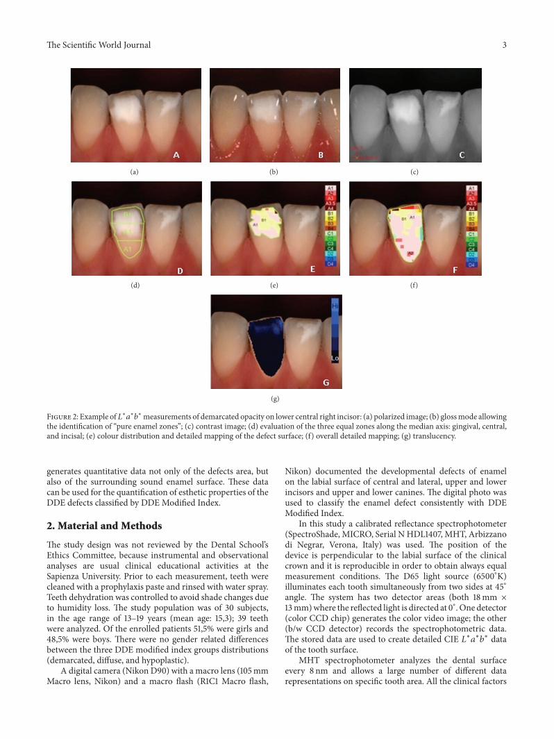
Figure 2: Example of𝐿∗𝑎∗𝑏∗measurements of demarcated opacity on lower central right incisor: (a) polarized image; (b) glossmode allowingthe identification of “pure enamel zones”; (c) contrast image; (d) evaluation of the three equal zones along the median axis: gingival, central,and incisal; (e) colour distribution and detailed mapping of the defect surface; (f) overall detailed mapping; (g) translucency.
generates quantitative data not only of the defects area, butalso of the surrounding sound enamel surface. These datacan be used for the quantification of esthetic properties of theDDE defects classified by DDE Modified Index.
2. Material and Methods
The study design was not reviewed by the Dental School’sEthics Committee, because instrumental and observationalanalyses are usual clinical educational activities at theSapienza University. Prior to each measurement, teeth werecleaned with a prophylaxis paste and rinsed with water spray.Teeth dehydration was controlled to avoid shade changes dueto humidity loss. The study population was of 30 subjects,in the age range of 13–19 years (mean age: 15,3); 39 teethwere analyzed. Of the enrolled patients 51,5% were girls and48,5% were boys. There were no gender related differencesbetween the three DDE modified index groups distributions(demarcated, diffuse, and hypoplastic).
A digital camera (Nikon D90) with a macro lens (105mmMacro lens, Nikon) and a macro flash (R1C1 Macro flash,
Nikon) documented the developmental defects of enamelon the labial surface of central and lateral, upper and lowerincisors and upper and lower canines. The digital photo wasused to classify the enamel defect consistently with DDEModified Index.
In this study a calibrated reflectance spectrophotometer(SpectroShade, MICRO, Serial N HDL1407, MHT, Arbizzanodi Negrar, Verona, Italy) was used. The position of thedevice is perpendicular to the labial surface of the clinicalcrown and it is reproducible in order to obtain always equalmeasurement conditions. The D65 light source (6500∘K)illuminates each tooth simultaneously from two sides at 45∘angle. The system has two detector areas (both 18mm ×13mm)where the reflected light is directed at 0∘. One detector(color CCD chip) generates the color video image; the other(b/w CCD detector) records the spectrophotometric data.The stored data are used to create detailed CIE 𝐿∗𝑎∗𝑏∗ dataof the tooth surface.
MHT spectrophotometer analyzes the dental surfaceevery 8 nm and allows a large number of different datarepresentations on specific tooth area. All the clinical factors
4 The Scientific World Journal
(a) (b) (c)
(d) (e) (f)
(g)
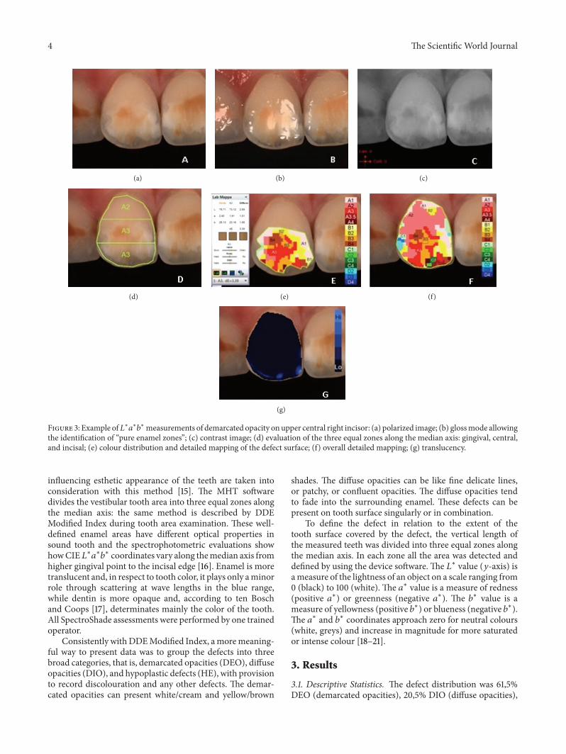
Figure 3: Example of𝐿∗𝑎∗𝑏∗measurements of demarcated opacity onupper central right incisor: (a) polarized image; (b) glossmode allowingthe identification of “pure enamel zones”; (c) contrast image; (d) evaluation of the three equal zones along the median axis: gingival, central,and incisal; (e) colour distribution and detailed mapping of the defect surface; (f) overall detailed mapping; (g) translucency.
influencing esthetic appearance of the teeth are taken intoconsideration with this method [15]. The MHT softwaredivides the vestibular tooth area into three equal zones alongthe median axis: the same method is described by DDEModified Index during tooth area examination. These well-defined enamel areas have different optical properties insound tooth and the spectrophotometric evaluations showhowCIE𝐿∗𝑎∗𝑏∗ coordinates vary along themedian axis fromhigher gingival point to the incisal edge [16]. Enamel is moretranslucent and, in respect to tooth color, it plays only aminorrole through scattering at wave lengths in the blue range,while dentin is more opaque and, according to ten Boschand Coops [17], determinates mainly the color of the tooth.All SpectroShade assessments were performed by one trainedoperator.
Consistently withDDEModified Index, amoremeaning-ful way to present data was to group the defects into threebroad categories, that is, demarcated opacities (DEO), diffuseopacities (DIO), and hypoplastic defects (HE), with provisionto record discolouration and any other defects. The demar-cated opacities can present white/cream and yellow/brown
shades. The diffuse opacities can be like fine delicate lines,or patchy, or confluent opacities. The diffuse opacities tendto fade into the surrounding enamel. These defects can bepresent on tooth surface singularly or in combination.
To define the defect in relation to the extent of thetooth surface covered by the defect, the vertical length ofthe measured teeth was divided into three equal zones alongthe median axis. In each zone all the area was detected anddefined by using the device software. The 𝐿∗ value (𝑦-axis) isameasure of the lightness of an object on a scale ranging from0 (black) to 100 (white). The 𝑎∗ value is a measure of redness(positive 𝑎∗) or greenness (negative 𝑎∗). The 𝑏∗ value is ameasure of yellowness (positive 𝑏∗) or blueness (negative 𝑏∗).The 𝑎∗ and 𝑏∗ coordinates approach zero for neutral colours(white, greys) and increase in magnitude for more saturatedor intense colour [18–21].
3. Results
3.1. Descriptive Statistics. The defect distribution was 61,5%DEO (demarcated opacities), 20,5% DIO (diffuse opacities),
The Scientific World Journal 5
(a) (b) (c)
(d) (e) (f)
(g)
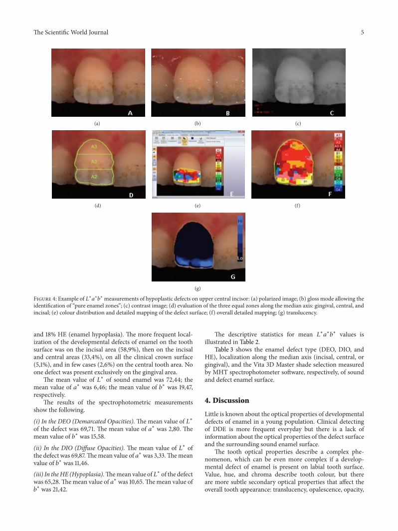
Figure 4: Example of 𝐿∗𝑎∗𝑏∗ measurements of hypoplastic defects on upper central incisor: (a) polarized image; (b) gloss mode allowing theidentification of “pure enamel zones”; (c) contrast image; (d) evaluation of the three equal zones along the median axis: gingival, central, andincisal; (e) colour distribution and detailed mapping of the defect surface; (f) overall detailed mapping; (g) translucency.
and 18% HE (enamel hypoplasia). The more frequent local-ization of the developmental defects of enamel on the toothsurface was on the incisal area (58,9%), then on the incisaland central areas (33,4%), on all the clinical crown surface(5,1%), and in few cases (2,6%) on the central tooth area. Noone defect was present exclusively on the gingival area.
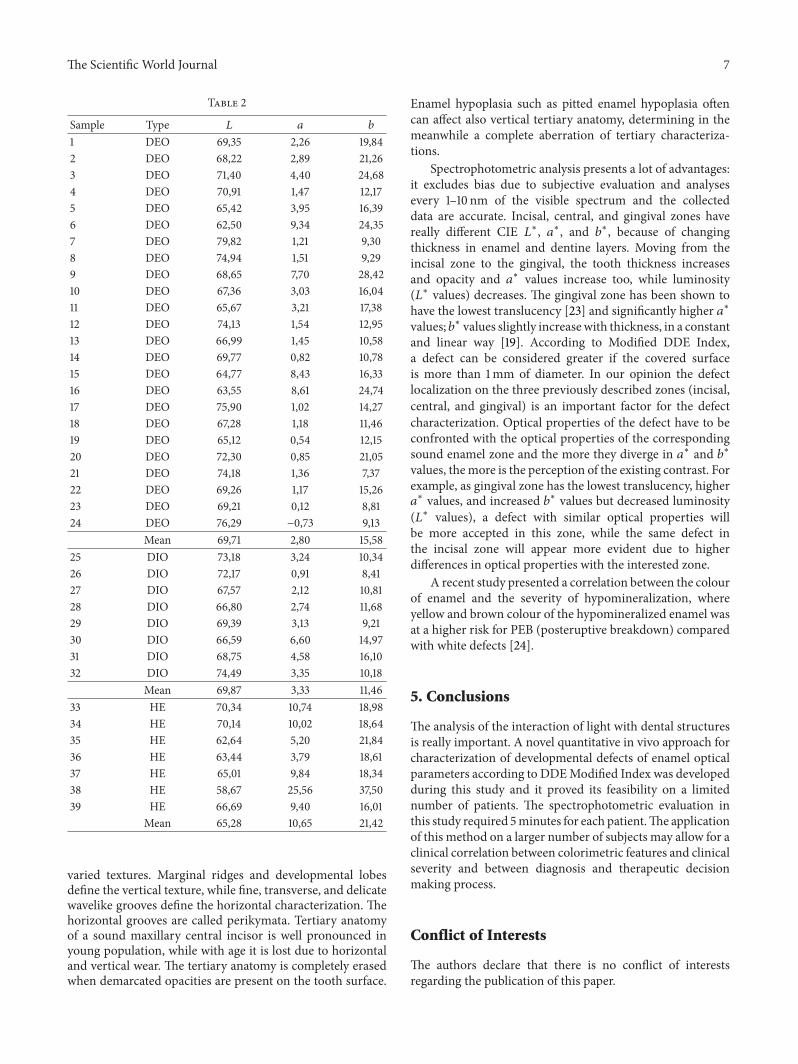
The mean value of 𝐿∗ of sound enamel was 72,44; themean value of 𝑎∗ was 6,46; the mean value of 𝑏∗ was 19,47,respectively.
The results of the spectrophotometric measurementsshow the following.
(i) In the DEO (Demarcated Opacities).Themean value of 𝐿∗of the defect was 69,71. The mean value of 𝑎∗ was 2,80. Themean value of 𝑏∗ was 15,58.
(ii) In the DIO (Diffuse Opacities). The mean value of 𝐿∗ ofthe defect was 69,87.Themean value of 𝑎∗ was 3,33.Themeanvalue of 𝑏∗ was 11,46.
(iii) In theHE (Hypoplasia).Themean value of𝐿∗ of the defectwas 65,28.Themean value of 𝑎∗ was 10,65.Themean value of𝑏∗ was 21,42.
The descriptive statistics for mean 𝐿∗𝑎∗𝑏∗ values isillustrated in Table 2.
Table 3 shows the enamel defect type (DEO, DIO, andHE), localization along the median axis (incisal, central, orgingival), and the Vita 3D Master shade selection measuredby MHT spectrophotometer software, respectively, of soundand defect enamel surface.
4. Discussion
Little is known about the optical properties of developmentaldefects of enamel in a young population. Clinical detectingof DDE is more frequent everyday but there is a lack ofinformation about the optical properties of the defect surfaceand the surrounding sound enamel surface.
The tooth optical properties describe a complex phe-nomenon, which can be even more complex if a develop-mental defect of enamel is present on labial tooth surface.Value, hue, and chroma describe tooth colour, but thereare more subtle secondary optical properties that affect theoverall tooth appearance: translucency, opalescence, opacity,
6 The Scientific World Journal
(a) (b) (c)
(d) (e) (f)
(g)
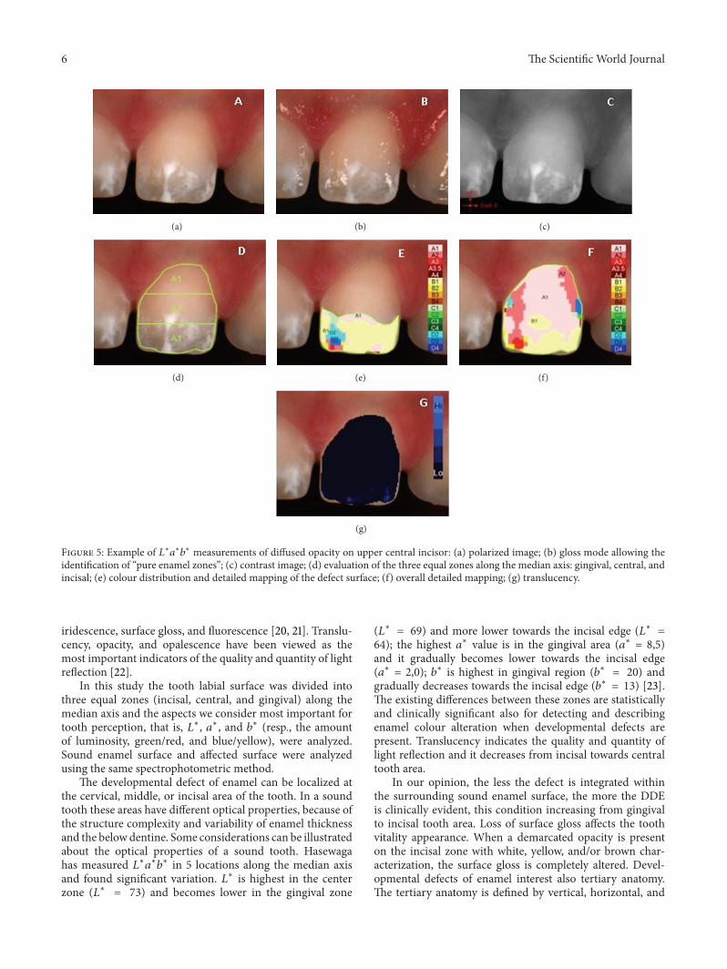
Figure 5: Example of 𝐿∗𝑎∗𝑏∗ measurements of diffused opacity on upper central incisor: (a) polarized image; (b) gloss mode allowing theidentification of “pure enamel zones”; (c) contrast image; (d) evaluation of the three equal zones along the median axis: gingival, central, andincisal; (e) colour distribution and detailed mapping of the defect surface; (f) overall detailed mapping; (g) translucency.
iridescence, surface gloss, and fluorescence [20, 21]. Translu-cency, opacity, and opalescence have been viewed as themost important indicators of the quality and quantity of lightreflection [22].
In this study the tooth labial surface was divided intothree equal zones (incisal, central, and gingival) along themedian axis and the aspects we consider most important fortooth perception, that is, 𝐿∗, 𝑎∗, and 𝑏∗ (resp., the amountof luminosity, green/red, and blue/yellow), were analyzed.Sound enamel surface and affected surface were analyzedusing the same spectrophotometric method.
The developmental defect of enamel can be localized atthe cervical, middle, or incisal area of the tooth. In a soundtooth these areas have different optical properties, because ofthe structure complexity and variability of enamel thicknessand the belowdentine. Some considerations can be illustratedabout the optical properties of a sound tooth. Hasewagahas measured 𝐿∗𝑎∗𝑏∗ in 5 locations along the median axisand found significant variation. 𝐿∗ is highest in the centerzone (𝐿∗ = 73) and becomes lower in the gingival zone
(𝐿∗
= 69) and more lower towards the incisal edge (𝐿∗ =64); the highest 𝑎∗ value is in the gingival area (𝑎∗ = 8,5)and it gradually becomes lower towards the incisal edge(𝑎∗ = 2,0); 𝑏∗ is highest in gingival region (𝑏∗ = 20) andgradually decreases towards the incisal edge (𝑏∗ = 13) [23].The existing differences between these zones are statisticallyand clinically significant also for detecting and describingenamel colour alteration when developmental defects arepresent. Translucency indicates the quality and quantity oflight reflection and it decreases from incisal towards centraltooth area.
In our opinion, the less the defect is integrated withinthe surrounding sound enamel surface, the more the DDEis clinically evident, this condition increasing from gingivalto incisal tooth area. Loss of surface gloss affects the toothvitality appearance. When a demarcated opacity is presenton the incisal zone with white, yellow, and/or brown char-acterization, the surface gloss is completely altered. Devel-opmental defects of enamel interest also tertiary anatomy.The tertiary anatomy is defined by vertical, horizontal, and
The Scientific World Journal 7
Table 2
Sample Type 𝐿 𝑎 𝑏
1 DEO 69,35 2,26 19,842 DEO 68,22 2,89 21,263 DEO 71,40 4,40 24,684 DEO 70,91 1,47 12,175 DEO 65,42 3,95 16,396 DEO 62,50 9,34 24,357 DEO 79,82 1,21 9,308 DEO 74,94 1,51 9,299 DEO 68,65 7,70 28,4210 DEO 67,36 3,03 16,0411 DEO 65,67 3,21 17,3812 DEO 74,13 1,54 12,9513 DEO 66,99 1,45 10,5814 DEO 69,77 0,82 10,7815 DEO 64,77 8,43 16,3316 DEO 63,55 8,61 24,7417 DEO 75,90 1,02 14,2718 DEO 67,28 1,18 11,4619 DEO 65,12 0,54 12,1520 DEO 72,30 0,85 21,0521 DEO 74,18 1,36 7,3722 DEO 69,26 1,17 15,2623 DEO 69,21 0,12 8,8124 DEO 76,29 −0,73 9,13
Mean 69,71 2,80 15,5825 DIO 73,18 3,24 10,3426 DIO 72,17 0,91 8,4127 DIO 67,57 2,12 10,8128 DIO 66,80 2,74 11,6829 DIO 69,39 3,13 9,2130 DIO 66,59 6,60 14,9731 DIO 68,75 4,58 16,1032 DIO 74,49 3,35 10,18
Mean 69,87 3,33 11,4633 HE 70,34 10,74 18,9834 HE 70,14 10,02 18,6435 HE 62,64 5,20 21,8436 HE 63,44 3,79 18,6137 HE 65,01 9,84 18,3438 HE 58,67 25,56 37,5039 HE 66,69 9,40 16,01
Mean 65,28 10,65 21,42
varied textures. Marginal ridges and developmental lobesdefine the vertical texture, while fine, transverse, and delicatewavelike grooves define the horizontal characterization. Thehorizontal grooves are called perikymata. Tertiary anatomyof a sound maxillary central incisor is well pronounced inyoung population, while with age it is lost due to horizontaland vertical wear. The tertiary anatomy is completely erasedwhen demarcated opacities are present on the tooth surface.
Enamel hypoplasia such as pitted enamel hypoplasia oftencan affect also vertical tertiary anatomy, determining in themeanwhile a complete aberration of tertiary characteriza-tions.
Spectrophotometric analysis presents a lot of advantages:it excludes bias due to subjective evaluation and analysesevery 1–10 nm of the visible spectrum and the collecteddata are accurate. Incisal, central, and gingival zones havereally different CIE 𝐿∗, 𝑎∗, and 𝑏∗, because of changingthickness in enamel and dentine layers. Moving from theincisal zone to the gingival, the tooth thickness increasesand opacity and 𝑎∗ values increase too, while luminosity(𝐿∗ values) decreases. The gingival zone has been shown tohave the lowest translucency [23] and significantly higher 𝑎∗values; 𝑏∗ values slightly increasewith thickness, in a constantand linear way [19]. According to Modified DDE Index,a defect can be considered greater if the covered surfaceis more than 1mm of diameter. In our opinion the defectlocalization on the three previously described zones (incisal,central, and gingival) is an important factor for the defectcharacterization. Optical properties of the defect have to beconfronted with the optical properties of the correspondingsound enamel zone and the more they diverge in 𝑎∗ and 𝑏∗values, themore is the perception of the existing contrast. Forexample, as gingival zone has the lowest translucency, higher𝑎∗ values, and increased 𝑏∗ values but decreased luminosity
(𝐿∗ values), a defect with similar optical properties willbe more accepted in this zone, while the same defect inthe incisal zone will appear more evident due to higherdifferences in optical properties with the interested zone.
A recent study presented a correlation between the colourof enamel and the severity of hypomineralization, whereyellow and brown colour of the hypomineralized enamel wasat a higher risk for PEB (posteruptive breakdown) comparedwith white defects [24].
5. Conclusions
The analysis of the interaction of light with dental structuresis really important. A novel quantitative in vivo approach forcharacterization of developmental defects of enamel opticalparameters according to DDEModified Index was developedduring this study and it proved its feasibility on a limitednumber of patients. The spectrophotometric evaluation inthis study required 5minutes for each patient.The applicationof this method on a larger number of subjects may allow for aclinical correlation between colorimetric features and clinicalseverity and between diagnosis and therapeutic decisionmaking process.
Conflict of Interests
The authors declare that there is no conflict of interestsregarding the publication of this paper.

8 The Scientific World Journal
Table 3
Sample Enamel defect Shade (Vita 3D Master)Type Localization Defect Sound enamel
1 DEO Incisal 1M1 3M12 DEO Incisal/central 2R2,5 1M13 DEO Incisal/central 3M3 2M14 DEO Incisal 1M1 1M25 DEO Incisal 5M1 3M26 DEO Incisal/central 5M3 3M17 DEO Incisal/central 0M1 1M28 DEO Incisal 0M3 1M29 DEO Incisal/central 3M3 3M310 DEO Incisal 1M1 3R1,511 DEO Incisal 2R1,5 3L1,512 DEO Incisal 2M1 4M113 DEO Incisal 1M1 2M114 DEO Incisal 1M1 3M115 DEO Incisal/central/gingival 3R1,5 4M116 DEO Incisal/central 5M2 4M117 DEO Incisal 2M1 3M118 DEO Incisal 1M1 3M119 DEO Incisal 2M1 3M120 DEO Incisal 2R2,5 2M121 DEO Incisal 1M1 2M122 DEO Incisal 1M2 2M223 DEO Incisal 2M1 2M124 DEO Incisal 0M1 3M125 DIO Incisal/central 1M1 2M126 DIO Incisal 5M1 2R1,527 DIO Incisal 0M1 3M128 DIO Incisal 2M1 4M129 DIO Incisal 2M1 4M130 DIO Incisal/gingival 3M1 3M131 DIO Incisal/central 3M1 3R1,532 DIO Incisal/central 1M1 2M133 HE Incisal/central 3M2 3M134 HE Incisal/central 5M1 3M135 HE Incisal 5M2 4M136 HE Incisal 4M1 4R2,537 HE Incisal/central 5M1 4M138 HE Central 5M3 3M239 HE Incisal/central 3R1,5 3M1
Acknowledgments
Fabrizio Guerra designed the study. Marta Mazur and Deb-ora Pasqualotto collaborated in the collection of data andpreparation of the paper. Gianna Maria Nardi and DeniseCorridore collaborated in writing and preparation of thepaper. LiviaOttolenghi coordinated the study and revised andprovided final approval of the paper.
References
[1] G. W. Suckling, “Developmental defects of enamel—historicaland present-day perspectives of their pathogenesis,” Advancesin Dental Research, vol. 3, no. 2, pp. 87–94, 1989.
[2] “A review of the developmental defects of enamel index (DDEIndex). Commission on Oral Health, Research and Epidemi-ology. Report of an FDI Working Group,” International DentalJournal, vol. 42, no. 6, pp. 411–426, 1992.
The Scientific World Journal 9
[3] W.K. Seow,D. Ford, S. Kazoullis, B. Newman, andT.Holcombe,“Comparison of enamel defects in the primary and permanentdentitions of children from a low-fluoride district in Australia,”Pediatric Dentistry, vol. 33, no. 3, pp. 207–212, 2011.
[4] B. Jalevik and G. A. Klingberg, “Dental treatment, dental fearand behaviour management problems in children with severeenamel hypomineralization of their permanent first molars,”International Journal of Paediatric Dentistry, vol. 12, no. 1, pp.24–32, 2002.
[5] S. Kazoullis,W. K. Seow, T.Holcombe, B. Newman, andD. Ford,“Common dental conditions associated with dental erosion inschoolchildren in Australia,” Pediatric Dentistry, vol. 29, no. 1,pp. 33–39, 2007.
[6] L. Hong, S. M. Levy, J. J. Warren, and B. Broffitt, “Associationbetween enamel hypoplasia and dental caries in primary secondmolars: a cohort study,” Caries Research, vol. 43, no. 5, pp. 345–353, 2009.
[7] J. Clarkson and D. O’Mullane, “A modified DDE Index for usein epidemiological studies of enamel defects,” Journal of DentalResearch, vol. 68, no. 3, pp. 445–450, 1989.
[8] Federation Dentaire Internationale (FDI), “An epidemiologicalindex of developmental defects of enamel,” Tech. Rep. 5, WorldDental Federation Publications, Ferney-Voltaire, France, 1982.
[9] G.W. Suckling, D. G. Nelson, andM. J. Patel, “Macroscopic andscanning electron microscopic appearance and hardness valuesof developmental defects in human permanent tooth enamel,”Advances in Dental Research, vol. 3, no. 2, pp. 219–233, 1989.
[10] D. Ford,W.K. Seow, S. Kazoullis, T. Holcombe, and B.Newman,“A controlled study of risk factors for enamel hypoplasia in thepermanent dentition,” Pediatric Dentistry, vol. 31, no. 5, pp. 382–388, 2009.
[11] M. Bossu, A. Bartoli, G. Orsini, E. Luppino, and A. Polimeni,“Enamel hypoplasia in coeliac children: a potential clinicalmarker of early diagnosis,” European Journal of PaediatricDentistry, vol. 8, no. 1, pp. 31–37, 2007.
[12] P. Arrow, “Risk factors in the occurrence of enamel defects ofthe first permanent molars among schoolchildren in WesternAustralia,”Community Dentistry and Oral Epidemiology, vol. 37,no. 5, pp. 405–415, 2009.
[13] S. Paul, A. Peter, N. Pietrobon, and C. H. F. Hammerle, “Visualand spectrophotometric shade analysis of human teeth,” Journalof Dental Research, vol. 81, no. 8, pp. 578–582, 2002.
[14] A. Dozic, C. J. Kleverlaan, I. H. A. Aartman, and A. J. Feilzer,“Relation in color of three regions of vital human incisors,”Dental Materials, vol. 20, no. 9, pp. 832–838, 2004.
[15] S. J. Paul, A. Peter, L. Rodoni, and N. Pietrobon, “Conventionalvisual vs spectrophotometric shade taking for porcelain-fused-to-metal crowns: a clinical comparison,” International Journalof Periodontics and Restorative Dentistry, vol. 24, no. 3, pp. 222–231, 2004.
[16] W. J. O’Brien, H. Hemmendinger, K. M. Boenke, J. B. Linger,and C. L. Groh, “Color distribution of three regions of extractedhuman teeth,” Dental Materials, vol. 13, no. 3, pp. 179–185, 1997.
[17] J. J. ten Bosch and J. C. Coops, “Tooth color and reflectanceas related to light scattering and enamel hardness.,” Journal ofDental Research, vol. 74, no. 1, pp. 374–380, 1995.
[18] Y.-K. Lee, H. Lu, and J. M. Powers, “Changes in opalescence andfluorescence properties of resin composites after acceleratedaging,” Dental Materials, vol. 22, no. 7, pp. 653–660, 2006.
[19] S. Ardu, A. J. Feilzer, A. Devigus, and I. Krejci, “Quantitativeclinical evaluation of esthetic properties of incisors,” DentalMaterials, vol. 24, no. 3, pp. 333–340, 2008.
[20] M. D. Russell, M. Gulfraz, and B. W. Moss, “In vivo mea-surement of colour changes in natural teeth,” Journal of OralRehabilitation, vol. 27, no. 9, pp. 786–792, 2000.
[21] D. A. Terry, W. Geller, O. Tric, M. J. Anderson, M. Tourville,and A. Kobashigawa, “Anatomical form defines color: function,form, and aesthetics,” Practical Procedures and Aesthetic Den-tistry, vol. 14, no. 1, pp. 59–68, 2002.
[22] R.Winter, “Visualizing the natural dentition,” Journal of EstheticDentistry, vol. 5, no. 3, pp. 102–117, 1993.
[23] A. Hasegawa, I. Ikeda, and S. Kawaguchi, “Color and translu-cency of in vivo natural central incisors,” Journal of ProstheticDentistry, vol. 83, no. 3, pp. 418–423, 2000.
[24] C. M. da Costa-Silva, G. M. B. Ambrosano, F. Jeremias, J. F.de Souza, and F. L. Mialhe, “Increase in severity of molar-incisor hypomineralization and its relationship with the colourof enamel opacity: a prospective cohort study,” InternationalJournal of Paediatric Dentistry, vol. 21, no. 5, pp. 333–341, 2011.

Submit your manuscripts athttp://www.hindawi.com
Hindawi Publishing Corporationhttp://www.hindawi.com Volume 2014
Oral OncologyJournal of
DentistryInternational Journal of
Hindawi Publishing Corporationhttp://www.hindawi.com Volume 2014
Hindawi Publishing Corporationhttp://www.hindawi.com Volume 2014
International Journal of
Biomaterials
Hindawi Publishing Corporationhttp://www.hindawi.com Volume 2014
BioMed Research International
Hindawi Publishing Corporationhttp://www.hindawi.com Volume 2014
Case Reports in Dentistry
Hindawi Publishing Corporationhttp://www.hindawi.com Volume 2014
Oral ImplantsJournal of
Hindawi Publishing Corporationhttp://www.hindawi.com Volume 2014
Anesthesiology Research and Practice
Hindawi Publishing Corporationhttp://www.hindawi.com Volume 2014
Radiology Research and Practice
Environmental and Public Health
Journal of
Hindawi Publishing Corporationhttp://www.hindawi.com Volume 2014
The Scientific World JournalHindawi Publishing Corporation http://www.hindawi.com Volume 2014
Hindawi Publishing Corporationhttp://www.hindawi.com Volume 2014
Dental SurgeryJournal of
Drug DeliveryJournal of
Hindawi Publishing Corporationhttp://www.hindawi.com Volume 2014
Hindawi Publishing Corporationhttp://www.hindawi.com Volume 2014
Oral DiseasesJournal of
Hindawi Publishing Corporationhttp://www.hindawi.com Volume 2014
Computational and Mathematical Methods in Medicine
ScientificaHindawi Publishing Corporationhttp://www.hindawi.com Volume 2014
PainResearch and TreatmentHindawi Publishing Corporationhttp://www.hindawi.com Volume 2014
Preventive MedicineAdvances in
Hindawi Publishing Corporationhttp://www.hindawi.com Volume 2014
EndocrinologyInternational Journal of
Hindawi Publishing Corporationhttp://www.hindawi.com Volume 2014
Hindawi Publishing Corporationhttp://www.hindawi.com Volume 2014
OrthopedicsAdvances in