research article effects of 5-amyno-4-(1,3-benzothyazol-2
TRANSCRIPT

Research ArticleEffects of 5-Amyno-4-(1,3-benzothyazol-2-yn)-1-(3-methoxyphenyl)-1,2-dihydro-3H-pyrrol-3-one Intake onDigestive System in a Rat Model of Colon Cancer
Halyna M. Kuznietsova, Valentyna K. Luzhenetska,Iryna P. Kotlyar, and Volodymyr K. Rybalchenko
Institute of Biology, Taras Shevchenko National University, 64/13 Volodymyrska Street, Kyiv 01601, Ukraine
Correspondence should be addressed to Halyna M. Kuznietsova; [email protected]
Received 29 April 2015; Revised 2 September 2015; Accepted 9 September 2015
Academic Editor: Peter Krajcsi
Copyright © 2015 Halyna M. Kuznietsova et al. This is an open access article distributed under the Creative Commons AttributionLicense, which permits unrestricted use, distribution, and reproduction in any medium, provided the original work is properlycited.
Introduction. Pyrrol derivate 5-amyno-4-(1,3-benzothyazol-2-yn)-1-(3-methoxyphenyl)-1,2-dihydro-3H-pyrrol-3-one (D1) hasshown antiproliferative activities in vitro, so investigation of the impact of D1 intake on gut organs in rats that experienced coloncancer seems to be necessary.Materials and Methods. D1 at the dose of 2.3mg/kg was administered per os daily for 27 (from the 1stday of experiment) or 7 (from the 21st week of experiment) weeks to rats that experienced 1,2-dimethylhydrazine (DMH)-inducedcolon cancer for 20 weeks. 5-Fluorouracil (5FU) was chosen as reference drug and was administered intraperitoneally weekly for7 weeks (from the 21st week of experiment) at the dose of 45mg/kg. Results. Antitumor activity of D1 comparable with the 5FUone against DMH-induced colon cancer in rats was observed (decrease of tumor number and tumor total area up to 46%). D1attenuated the inflammation of colon, gastric and jejunal mucosa, and the liver, caused by DMH, unlike 5FU, aggravating the latter.In addition, D1 partially normalizedmucosamorphometric parameters suggesting its functional restore.Conclusions. D1 possesses,comparable with 5-fluorouracil antitumor efficacy, less damaging effects on the tissues beyond cancerous areas and contributes topartial morphological and functional gut organs recovery.
1. Introduction
Cancer is one of the major public health problems inthe world, becoming the leading cause for deaths amonggeneral population [1, 2]. Chemotherapy is themost commonmethod for neoplasm treatment. However, it has significantdisadvantages such as high frequency and severity of sideeffects. This is the one of the main factors for choosing theproper chemotherapy and for treatment outcomes [3, 4].
Protein kinases inhibitors recently attract the attention asinhibitors of the proliferative activity having specific effectson malignant cells and low toxicity towards normal cellsin the organism. However information about their systemicexposure is controversial [5], which requires detailed studieson each case. Particularly it concerns the small molecularinhibitors of protein kinases, which are promising therapeu-tics because of their moderate specificity (have a wide range
of malignant “targets”), ease of use (oral administration), andlower cost compared with monoclonal antibodies [6].
An important criterion of anticancer drug development,in addition to its efficacy, is no negative impact on thesystemic condition after prolonged use. Digestive organsare first exposed to exogenous substances including drugs,especially if administered per os. Exactly the digestive systemoccursmost side effects of anticancer therapy. Gut epitheliumis hypersensitive to the cytostatic action because of highproliferative activity [7], whereas the liver detoxicates thexenobiotics and often is damaged by their metabolites [8].
Pyrrol derivates have been synthesized at the Depart-ment of Chemistry of Taras Shevchenko National Univer-sity of Kyiv as inhibitors of protein kinases such as Yes,Src(h), ZAP70, Syk(h), PDK1, EGFR, IGF-1R, and VEGFR[9, 10]. One of them, 5-amyno-4-(1,3-benzothyazol-2-yn)-1-(3-methoxyphenyl)-1,2-dihydro-3H-pyrrol-3-one, called D1
Hindawi Publishing Corporatione Scientific World JournalVolume 2015, Article ID 376576, 13 pageshttp://dx.doi.org/10.1155/2015/376576

2 The Scientific World Journal
NS
N
O
OCH3
12
34
5
H2N
Figure 1: 5-amyno-4-(1,3-benzothyazol-2-yl)-1-(3-methoxyphen-yl)-1,2-dihydro-3H-pyrrol-3-one (D1) structural formula.
(Figure 1), has shown high cytostatic activity in vitro againstHT29, HCT-15, and COLO-205 (colorectal cancer) cell lines,which suggests its potential anticancer activity. As D1 showedthe most promising results on colorectal cancer cell lines,rat 1,2-dimethylhydrazine-induced colon cancer model waschosen for current in vivo investigations.
1,2-dimethylhydrazine/azoxymethane- (DMH/AOM-)induced colon carcinogenesis is a multistep process withmorphological and histological features similar to thoseseen in human sporadic colon carcinogenesis [11, 12]. Themain cellular and molecular defects found in human coloncarcinogenesis have also been observed in DMH/AOM ratcolon carcinogenesis. These alterations have been found tobe involved in such pathways, as the Wnt pathway, K-raspathway, TGF𝛽 signaling pathway, and inflammatory relatedprocess [13].
The Wnt pathway has been implicated as a crucial stepin the initiation and development of colonic tumorigenesis.In the absence of the extracellular Wnt signal, free 𝛽-catenin is bound to the APC-axin-conductin-GSK3𝛽 com-plex. Phosphorylation of 𝛽-catenin by this complex marksit for ubiquitination and subsequent proteolytic degradationby the proteasome. When APC or 𝛽-catenin is mutated, 𝛽-catenin cannot be degraded but accumulates in the cytoplasmand translocates into the nucleus, where it binds to T-cellfactor (TCF) and activates the Wnt target genes. Well-knowndownstream targets of APC/𝛽-catenin/Tcf-4 mediated tran-scriptional activation in colorectal neoplasia include genes,such as cyclin D1, c-myc, which have important roles inproliferation, apoptosis, and cell cycle progression and areresponsible for tumor formation.
Mutations in the K-ras gene are responsible for activationof the K-ras pathway. In human tumors, as well as intumors of AOM-treated rats, increased expression of Akt,a downstream target of the K-ras pathway, has been found.PI3K/Akt pathway is implicated in glucose metabolism, cellproliferation, apoptosis, transcription, and cell migration.PI3K/Akt also promotes cyclin D1. In addition, activation ofthe MEK/MAPK/ERK pathway, another downstream K-rassignaling, has been found in tumors without K-ras mutation.Activation of this pathway has been associated with overex-pression of c-erbB-2 receptors and decreased expression of
GTPase activating protein, resulting in constitutive activationof normal K-ras protein.There are several bodies of evidencethat Wnt/Apc/𝛽-catenin/Tcf pathway is involved in COX-2expression [14].
COX-2 expression in colorectal cancer is significantlyhigher than in normal colorectal tissues. COX-2 can (1)increase the production of prostaglandins and inhibit thebody’s immune response, (2) inhibit tumor cell apoptosis andpromote cell proliferation, (3) regulate cell cycle progression,(4) promote tumor angiogenesis, (5) increase the expressionof matrix metalloproteinases in tumor cells, and (6) induceactivation of precursors of carcinogenic substances. AOMaugments the expression of cyclooxygenase- (COX-) 2 andconsequently prostaglandin E2 (PGE2) levels within adeno-carcinomas and upregulates a number of proinflammatorycytokines, including tumor necrosis factor- (TNF-) a andinterleukin-1a/b. Elevated COX-2 expression has been shownto occur throughout the carcinogenic process, beginning inrats within normal-appearing colonic mucosa as early as 1week followingAOMexposure [15]. COX-2 activates intrinsictyrosine kinase of EGFR. EGF can form a complex with 𝛽-catenin, possibly through receptor tyrosine kinase-PI3K/Aktpathway, and further activate Wnt signaling pathway. Coss-communication betweenWnt- andEGFR-signaling pathwaysallows the integration of the diversity of stimuli in colono-cytes and promotes tumor progression [16]. TGF𝛽 in normalcells stops the cell cycle at theG1 stage, induces differentiationor promotes apoptosis, and thus exerts tumor suppressiveeffects. In cancer cells, it also modulates processes such ascell invasion, immune regulation, and microenvironmentmodification that cancer cells may exploit to their advantage.It has been found that TGF𝛽 and COX-2 were concurrentlyoverexpressed in the same colonic neoplastic lesions inAOM-treated rats, suggesting that COX-2 expression in AOM-induced colonic tumor could, in part, be due to the overex-pression of TGF𝛽 [13].
Colon is the tissue where carcinogenesis processes pass,so we suggested, that conditions in the colon differ from thatones in other parts of digestive system.Therefore the outcomeof the latter under colon cancer conditions has to be studied.So investigation of the impact of pyrrol derivate D1 intakeon the colon, gastric and jejunal mucosa, and the liver inrats that experienced colon cancer was aimed. 5-Fluorouracilcommonly used for colorectal cancer treatment [7, 17] waschosen as reference drug.
2. Materials and Methods
2.1. Animals. All experimental procedures executed withanimals were in compliance with international principles ofthe European Convention for the protection of vertebrateanimals used for experimental and other scientific purposes(European convention, Strasburg, 1986), article 26 of the Lawof Ukraine “on protection of animals from cruelty” (number3447-IV, 21.02.2006), and all norms of bioethics and biosafety.
80 Male Wistar rats weighing 120–130 g (4 weeksold) were obtained from Central Animal House of TarasShevchenko National University (Kyiv, Ukraine), and 5 ani-mals were housed per plastic cage on softwood chip bedding.

The Scientific World Journal 3
These animals were maintained under constant conditions(12 hr light/dark cycle, 60% humidity at 20–22∘C) and fed onstandard diet and tap water ad libitum.
2.2. Chemicals. D1 was dissolved in vegetable oil contain-ing 15% dimethylsulfoxide. Animals were treated by D1at the dose of 2.3mg/kg of body weight per os daily.The common anticancer therapeutic 5-fluorouracil (5FU,Ebewe Pharma, Austria) was used for referencing. Undi-luted 5FU was injected intraperitoneally weekly at the doseof 45mg/kg of body weight [18]. 1,2-Dimethylhydrazine(DMH, “Acros Organics”, USA), a highly specific colorectalcarcinogen in rodents, was dissolved immediately beforeuse in saline adjusted to pH 6.5 with sodium hydrox-ide. To induce tumor development, animals were subcuta-neously injected with 20mg/kg DMH weekly for 20 weeks[19].
2.3. Experimental Design. Colorectal tumors were inducedas described [19]: animals were administered with DMHsubcutaneously during the first 20 weeks of experiment;then tumor development followed for 7 weeks. D1 applica-tions were started simultaneously with the administrationof DMH and followed for 27 weeks or at the 21st week ofexperiment and followed for 7 weeks. 27-week intake wascalled as preventive regimen and started from the 1st dayof experiment, and 7-week intake was called as therapeuticregimen and started from the 21st week of experiment, whenthe first tumor nodes appear. 5FU injections were started atthe 21st week of experiment and followed for 7weeks. Controlanimals received the appropriate vehicles: saline or vegetableoil that contained 15% dimethylsulfoxide.
The rats were divided into 8 groups (10 rats each): (1)vehicle-treated control, (2) D17 weeks, where the animals weretreated with D1 for 7 weeks starting at the 21st week ofexperiment, (3) D127 weeks, where the animals were treatedwith D1 for 27 weeks, (4) 5FU, where the animals were treatedwith 5FU for 7 weeks starting at the 21st week of experiment,(5) DMH, where the animals were treated with DMH for 20weeks and then tumor development followed for 7 weeks, (6)DMH+D17 weeks, where the animals were treated with DMHfor 20 weeks and with D1 for 7 weeks starting at the 21st week,(7) DMH + D127 weeks, where the animals were treated withDMH for 20 weeks and with D1 for 27 weeks starting at the1st day of experiment, and (8)DMH+5FU,where the animalswere treated with DMH for 20 weeks and with 5FU for 7weeks starting at the 21st week of experiment.
2.4. Tissue Preparation. One day after the last treatmentthe rats were sacrificed by carbon dioxide asphyxia, theabdomen was opened, and the entire gastrointestinal tractand liver were removed. The bowel internal side was exam-ined, the tumor number per animal and the areas of tumorswere measured, and the tumor total area per animal wascalculated.
The colon segments with no tumors, the jejunum ones,and the fundus parts of the stomach were fixed for 14days in neutral saline containing 10% formalin; the liver
samples were fixed for the same time in Bouin’s fixative.Then, they were embedded into paraffin and sliced into 5𝜇msections, which were stained with hematoxylin-eosin-orange[20] and examined under the light microscope (OlympusBX-41, Olympus Europe GmbH, Japan). Systemic conditionsof colon, gastric and jejunal mucosa, and the liver wereassessed. Gastric mucosa thickness, cross-sectional areas ofchief and parietal cells and their nuclei, colon and jeju-nal mucosa thickness, colonocytes/enterocytes height andtheir nuclei cross-sectional area, as well as goblet cells one,and cross-sectional areas of hepatocytes and their nucleifrom centrolobular and periportal hepatic zones separatelyand hepatic sinusoid’s diameter were measured from themicrophotographs with magnification 400x, using WCIFImageJ software.Themitotic index (MI) was calculated as thesum of epithelial cells in any phase of mitosis divided by thetotal sum of epithelial cells. No less than 300 colon or jejunalcrypt epithelial cells per animal were assessed.
2.5. Liver Function Tests. Immediately after animals sacrificethe blood from inguinal vein was collected, kept for 20minto coagulate, and then centrifuged for 5min at 1000 g. Bloodserum alanine aminotransferase (ALT), aspartate amino-transferase (AST), alkaline phosphatase (ALP), and lactatedehydrohenase (LDH) were determined by standard kits(“Reagent”, Ukraine). De Ritis coefficient was defined asAST/ALT ratio.
2.6. Urinary 8-oxoG Extraction Test. Urinary 7,8-dihydro-8-oxoguanine (8-oxoG) is one of the main products of DNAoxidative damage and an indicator of tumor availability andprogression as well as drugs genotoxicity [21, 22]. Urinary 8-oxoG was determined as described [21]: animals were keptfor 1 day in the urine collection chamber (on a standarddiet with free access to water), collected urine was filtered,and daily volume was measured; 8-oxoG was extracted fromurine aliquots (1mL) by its filtration through a cartridge filledwith a DSC-18 (Cayman Chemical Company, USA) followedby washing with methanol; 8-oxoG amount was determinedspectrophotometrically; daily urine 8-oxoG extraction per1 kg of body weight was calculated.
2.7. Statistical Analysis. Statistical analysis was carried outusing SPSS 17.0 software for Windows. One-way ANOVAfollowed by the post hoc Bonferroni’s test was employed todetermine statistical significance. 𝑝 < 0.05 was consideredstatistically significant.
3. Results
3.1. D1 Influence on the Digestive Organs of Healthy Rats
3.1.1. Colon. Submucosa and lamina propria’s slight inflam-mation manifested by lymphatic and histiocytic cells infil-tration loci, as well as no epithelium damage, caused byD1 acting for 7 and 27 weeks, were observed. D1 actingfor 7 weeks increased mucosa thickness, colonocyte height,and their nuclei area by 11–19%, whereas D1 acting for 27

4 The Scientific World Journal
weeks decreased mucosa thickness by 12% and MI by 27%,suggesting the depletion of adaptive reserves and inhibitionof cell proliferation. On the contrary, moderate lymphaticand histiocytic cells infiltration of the upper mucosa layerand sometimes blood capillary dilationwere observed in 5FUgroup, suggesting the inflammatory process. 5FU increasedcolonocyte height (by 11%) as D17 weeks did but also inhib-ited cell proliferation, manifested by MI decrease by 37%(Table 2).
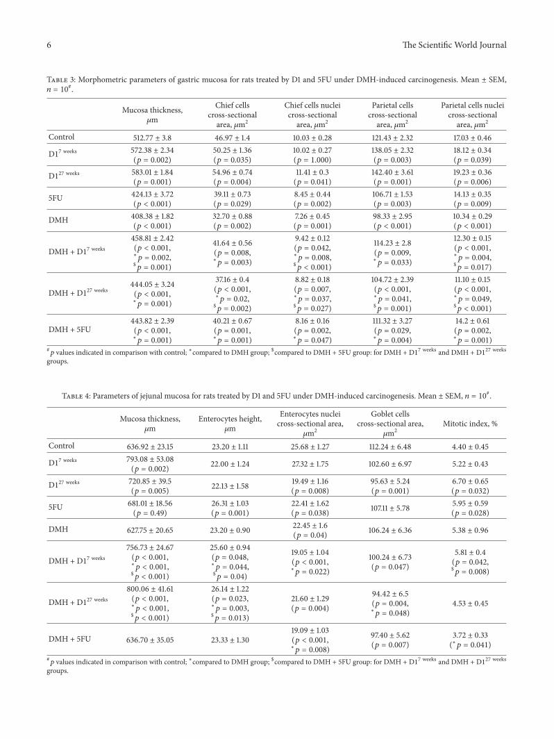
3.1.2. Stomach. Moderate submucosal and lamina propria’sedema, isolated inflammation features manifested by lym-phatic and histiocytic cells infiltration loci, caused by D1acting for 7 and 27 weeks, were observed. Some areas ofsurface epithelium desquamation also appeared. On thecontrary, 5FU caused severe mucosa damage, manifested bysurface epitheliumdesquamation, gastric glands’ destruction,sometimes colonocytes dystrophy, and necrosis foci. D1increased the values almost of all measured parameters intime-dependent manner by 6–13% acting for 7 weeks and by11–22% acting for 27 weeks (Table 3), which could indicatemucosa compensatory processes by intensification of syn-thetic processes in colonocytes [23]. However decrease of allmeasured parameters by 11–17% together withmorphologicalchanges, caused by 5FU, could indicate chronic atrophicgastritis development in 5FU group.
3.1.3. Jejunum. There were slight edema, hyperaemia, andsometimes stroma lymphatic and histiocytic cells infiltrationin jejunal villi. In D17 weeks group no changes were observedcompared to control one, whereas in D127 weeks group gobletcells’ and enterocytes nuclei areas were reduced by 15% and24%, respectively, supposingmucosa functional activity inhi-bition (Table 4). On the contrary, 5FU caused edema of villiapexes, epithelium desquamation, inflammation, enterocytesnuclei area reduction by 12.7% together with their heightincrease by 13.4%, and intensification of progenitor cellsproliferation by 35.4%, which evidenced chronic enteritisdevelopment.
3.1.4. Liver. D1 acting for 7 weeks sometimes caused sinu-soids inflammatory dilation, in some cases granular dys-trophy. Hepatocytes area increase by 12% and 13%, respec-tively, has been shown in both hepatic zones (Table 3),indicating the activation of detoxificating processes [24]. InD127 weeks group no inflammation and dystrophy featureswere observed, although centrolobular and periportal hep-atocytes areas were increased by 10% and 9%, respectively,indicating partial liver adaptation and its structure recoverywhile continuing detoxificating processes. Cell cytoplasminhomogeneity especially in periportal zone, hyperchromicnuclei, and lymph infiltration of perivascular areas causedby 5FU could evidence the drug hepatotoxicity. Hepatocytesarea increase by 12% has been shown in periportal zonethat indicated the liver detoxificating activity (Table 5). D1decreased serum ALT by 20–22%, when administered forboth terms, confirming the morphological data about liverfunctional load associated with the process of xenobiotics
detoxification [25]. Statistically significant changes of serumsALP, AST, and LDH were not observed, confirming no D1pronounced hepatotoxicity. On the contrary, 5FU decreasedserum AST by 23% and did not affect other measuredenzymes (Table 6). Furthermore, in contrast to classicalanticancer drugs [26] having strong genotoxicity, D1 didnot induce significant DNA oxidative damage, evidencedby a moderate (by 33–46%) urinary 8-oxoG increase. 5FUincreased urinary 8-oxoG up to 5 times towards 0.49 ±0.07 nmol/kg body weight per day in control.
3.2. Antitumor Action of D1. Visual inspection of the bowelsof rats that experienced colon cancer detected tumors mainlyin the distal bowel part and predominantly of exophytictype, which is consistent with published data [19]. All testedcompounds, D17 weeks, D127 weeks, and 5FU, reduced thetumor total area at a similar manner (by 40–46%) (Table 1).Statistically significant decrease of tumor number was foundin DMH + D17 weeks and DMH + 5FU groups (by 41 and50%, resp.). Moreover, predominantly exophytic tumors wereaffected by tested compounds. Thus, antitumor effects of D1and 5FU were similar. Notably, antitumor effect of D1 actingfor 7 weeks was stronger comparing to 27-week treatment,which may be explained by adaptation to D1 under itsprolonged influence.
3.3. D1 Influence on the Digestive Organs of Rats ThatExperienced Colon Cancer
3.3.1. Colon. Inflammatory features, manifested by lymphaticand histiocytic cells infiltration and blood capillary dilation(Figure 2(e)), increasedmucosa thickness, colonocyte height,and their nuclei area (by 13–25%); sometimes enlarged andoverextended crypts in DMHgroup could be interpreted [27]as precursor lesions. Local lymphatic and histiocytic cellsaggregations in DMH + D127 weeks group (Figure 2(m)) andno changes in DMH + D17 weeks group (Figure 2(i)) wereobserved comparing to control. On the contrary, inflamma-tion was enhanced in DMH + 5FU group (Figure 2(q)): thelymphatic and histiocytic cells infiltrationwasmore prevalentand the vessel dilations were more frequent. Damage ofsurface epithelium was also observed in this group. D17 weeksincreased colonocyte height and their nuclei area (by 14%and 6%, resp.), suggesting the cells functional activity gain,and didn’t affect the cell proliferation. Otherwise, D127 weeksand 5FU increased all mucosa parameters by 10–24%, exceptfor MI, which was diminished by 29% and 32%, respectively(Table 2).
3.3.2. Stomach. Severe epithelium desquamation and lym-phatic and histiocytic cells submucosal infiltration wereobserved inDMH-treated rats. Reduce the number of parietalcells due to relatively deep mucosal defects (Figure 2(f)) anddecrease the values of all parameters by 19–39% (Table 3)were also detected. These findings suppose [23] chronicatrophic gastritis and therefore mucosa functional activitysuppression.

The Scientific World Journal 5
Table 1: Tumor parameters for rats treated by D1 and 5FU under DMH-induced carcinogenesis. Mean ± SEM, 𝑛 = 10#.
DMH DMH + D17 weeks DMH + D127 weeks DMH + 5FUTotal tumors
Tumor number 9.56 ± 1.74 5.60 ± 1.27 (𝑝 = 0.015) 7.17 ± 1.70 4.78 ± 1.44 (𝑝 = 0.003)Tumor total area, cm2 1.58 ± 0.65 0.94 ± 0.40 (𝑝 = 0.025) 0.86 ± 0.47 (𝑝 = 0.019) 0.91 ± 0.68 (𝑝 = 0.038)
Exophytic tumorsTumor number 9.00 ± 1.53 4.90 ± 1.38 (𝑝 = 0.004) 6.50 ± 2.23 4.44 ± 1.42 (𝑝 = 0.001)Tumor total area, cm2 1.03 ± 0.28 0.49 ± 0.16 (𝑝 = 0.016) 0.56 ± 0.37 (𝑝 = 0.023) 0.46 ± 0.21 (𝑝 = 0.013)
Endophytic tumorsTumor number 0.67 ± 0.47 0.70 ± 0.60 0.67 ± 0.67 0.33 ± 0.31Tumor total area, cm2 0.55 ± 0.53 0.45 ± 0.42 0.3 ± 0.27 0.45 ± 0.42#𝑝 values indicated in comparison with DMH group.
Table 2: Morphometric parameters of colon mucosa, adjacent to colonic tumors for rats treated by D1 and 5FU under DMH-inducedcarcinogenesis. Mean ± SEM, 𝑛 = 10#.
Mucosa thickness,𝜇m
Colonocytesheight, 𝜇m
Colonocytes nucleicross-sectional
area, 𝜇m2
Goblet cellscross-sectional
area, 𝜇m2Mitotic index, %
Control 210.64 ± 9.62 16.75 ± 0.87 14.93 ± 1.22 107.08 ± 11.3 4.25 ± 0.32
D17 weeks 233.12 ± 10.39(𝑝 = 0.01)
18.69 ± 0.70(𝑝 = 0.02)
17.83 ± 1.31(𝑝 = 0.002) 99.01 ± 8.73 3.58 ± 0.32
D127 weeks 186.00 ± 7.9(𝑝 = 0.006) 17.04 ± 0.63 15.16 ± 0.84 99.76 ± 7.95 3.10 ± 0.43
(𝑝 = 0.047)
5FU 208.61 ± 9.18 18.53 ± 0.92(𝑝 = 0.01) 14.09 ± 0.61 99.67 ± 7.59 2.66 ± 0.75
(𝑝 = 0.01)
DMH 238.56 ± 11.64(𝑝 = 0.005)
20.93 ± 1.18(𝑝 = 0.001)
18.59 ± 1.03(𝑝 = 0.001) 108.01 ± 9.27 4.20 ± 0.26
DMH + D17 weeks209.48 ± 9.35(∗𝑝 = 0.007,$𝑝 = 0.001)
19.05 ± 0.84(𝑝 = 0.004)
15.80 ± 0.81(∗𝑝 = 0.01,$𝑝 = 0.044)
99.96 ± 6.72 3.70 ± 0.35($𝑝 = 0.041)
DMH + D127 weeks257.97 ± 11.64(𝑝 = 0.001,∗𝑝 = 0.002,$𝑝 = 0.044)
18.60 ± 0.88(𝑝 = 0.03,∗𝑝 = 0.04)
14.60 ± 1.08(∗𝑝 = 0.008,$𝑝 = 0.038)
130.63 ± 14.3(𝑝 = 0.002,∗𝑝 = 0.002,$𝑝 = 0.001)
3.03 ± 0.24(𝑝 = 0.03,∗𝑝 = 0.045)
DMH + 5FU282.65 ± 12.95(𝑝 = 0.001,∗𝑝 = 0.001)
18.47 ± 0.7(𝑝 = 0.01,∗𝑝 = 0.009)
18.08 ± 1.11(𝑝 = 0.005) 100.91 ± 6.78
2.87 ± 0.23(𝑝 = 0.02,∗𝑝 = 0.021)
#𝑝 values indicated in comparison with control; ∗compared to DMH group; $compared to DMH + 5FU group: for DMH + D17 weeks and DMH + D127 weeksgroups.
In D1-treated rats that experienced colon cancer somesubmucosal edema and lymphocytes and histiocytes aggre-gations were discovered. Epithelium desquamation wasobserved onminor areas inD17 weeks group and on almost theentire surface in D127 weeks group (Figures 2(j) and 2(n)). Onthe contrary, 5FU caused severe epithelium desquamation,cell dystrophy and necrosis foci, and focal lymphatic andhistiocytic cells submucosal infiltration (Figure 2(r)).
D1 acting for 7 weeks caused an increase of all testedparameters by 6–30% compared to DMH, which becameclose to control values but remained below them by 6–28%.D1 acting for 27 weeks also caused an increase of all testedparameters by 6–20% remaining lower than control one by14–35%. In DMH + 5FU group similar effects were observed:
mucosa parameters were increased by 8–40% remaininglower than control ones by 9–18% (Table 3). Recovery effectsof D1 acting for 7 weeks were more pronounced regarding tochief cells, in comparison with 5FU, as well as with D1 moreprolonged action. However, parietal cells were more sensitiveto D1 acting for both terms compared to 5FU.
So D1 partially recovers gastric mucosa altered by DMH,unlike 5FU, which aggravates carcinogenesis consequences inthe stomach.
3.3.3. Jejunum. The features of impaired capillary blood andlymph circulation and inflammation accompanied with villiapexes edema were observed in rats that experienced coloncancer (Figure 2(g)). Enterocytes nuclei area was reduced by
6 The Scientific World Journal
Table 3: Morphometric parameters of gastric mucosa for rats treated by D1 and 5FU under DMH-induced carcinogenesis. Mean ± SEM,𝑛 = 10#.
Mucosa thickness,𝜇m
Chief cellscross-sectional
area, 𝜇m2
Chief cells nucleicross-sectional
area, 𝜇m2
Parietal cellscross-sectional
area, 𝜇m2
Parietal cells nucleicross-sectional
area, 𝜇m2
Control 512.77 ± 3.8 46.97 ± 1.4 10.03 ± 0.28 121.43 ± 2.32 17.03 ± 0.46
D17 weeks 572.38 ± 2.34(𝑝 = 0.002)
50.25 ± 1.36(𝑝 = 0.035)
10.02 ± 0.27(𝑝 = 1.000)
138.05 ± 2.32(𝑝 = 0.003)
18.12 ± 0.34(𝑝 = 0.039)
D127 weeks 583.01 ± 1.84(𝑝 = 0.001)
54.96 ± 0.74(𝑝 = 0.004)
11.41 ± 0.3(𝑝 = 0.041)
142.40 ± 3.61(𝑝 = 0.001)
19.23 ± 0.36(𝑝 = 0.006)
5FU 424.13 ± 3.72(𝑝 < 0.001)
39.11 ± 0.73(𝑝 = 0.029)
8.45 ± 0.44(𝑝 = 0.002)
106.71 ± 1.53(𝑝 = 0.003)
14.13 ± 0.35(𝑝 = 0.009)
DMH 408.38 ± 1.82(𝑝 < 0.001)
32.70 ± 0.88(𝑝 = 0.002)
7.26 ± 0.45(𝑝 = 0.001)
98.33 ± 2.95(𝑝 < 0.001)
10.34 ± 0.29(𝑝 < 0.001)
DMH + D17 weeks458.81 ± 2.42(𝑝 < 0.001,∗𝑝 = 0.002,$𝑝 = 0.001)
41.64 ± 0.56(𝑝 = 0.008,∗𝑝 = 0.003)
9.42 ± 0.12(𝑝 = 0.042,∗𝑝 = 0.008,$𝑝 < 0.001)
114.23 ± 2.8(𝑝 = 0.009,∗𝑝 = 0.033)
12.30 ± 0.15(𝑝 < 0.001,∗𝑝 = 0.004,$𝑝 = 0.017)
DMH + D127 weeks444.05 ± 3.24(𝑝 < 0.001,∗𝑝 = 0.001)
37.16 ± 0.4(𝑝 < 0.001,∗𝑝 = 0.02,$𝑝 = 0.002)
8.82 ± 0.18(𝑝 = 0.007,∗𝑝 = 0.037,$𝑝 = 0.027)
104.72 ± 2.39(𝑝 < 0.001,∗𝑝 = 0.041,$𝑝 = 0.001)
11.10 ± 0.15(𝑝 < 0.001,∗𝑝 = 0.049,$𝑝 < 0.001)
DMH + 5FU443.82 ± 2.39(𝑝 < 0.001,∗𝑝 = 0.001)
40.21 ± 0.67(𝑝 = 0.001,∗𝑝 = 0.001)
8.16 ± 0.16(𝑝 = 0.002,∗𝑝 = 0.047)
111.32 ± 3.27(𝑝 = 0.029,∗𝑝 = 0.004)
14.2 ± 0.61(𝑝 = 0.002,∗𝑝 = 0.001)
#𝑝 values indicated in comparison with control; ∗compared to DMH group; $compared to DMH + 5FU group: for DMH + D17 weeks and DMH + D127 weeksgroups.
Table 4: Parameters of jejunal mucosa for rats treated by D1 and 5FU under DMH-induced carcinogenesis. Mean ± SEM, 𝑛 = 10#.
Mucosa thickness,𝜇m
Enterocytes height,𝜇m
Enterocytes nucleicross-sectional area,𝜇m2
Goblet cellscross-sectional area,𝜇m2
Mitotic index, %
Control 636.92 ± 23.15 23.20 ± 1.11 25.68 ± 1.27 112.24 ± 6.48 4.40 ± 0.45
D17 weeks 793.08 ± 53.08(𝑝 = 0.002) 22.00 ± 1.24 27.32 ± 1.75 102.60 ± 6.97 5.22 ± 0.43
D127 weeks 720.85 ± 39.5(𝑝 = 0.005) 22.13 ± 1.58 19.49 ± 1.16
(𝑝 = 0.008)95.63 ± 5.24(𝑝 = 0.001)
6.70 ± 0.65(𝑝 = 0.032)
5FU 681.01 ± 18.56(𝑝 = 0.49)
26.31 ± 1.03(𝑝 = 0.001)
22.41 ± 1.62(𝑝 = 0.038) 107.11 ± 5.78 5.95 ± 0.59
(𝑝 = 0.028)
DMH 627.75 ± 20.65 23.20 ± 0.90 22.45 ± 1.6(𝑝 = 0.04) 106.24 ± 6.36 5.38 ± 0.96
DMH + D17 weeks756.73 ± 24.67(𝑝 < 0.001,∗𝑝 < 0.001,$𝑝 < 0.001)
25.60 ± 0.94(𝑝 = 0.048,∗𝑝 = 0.044,$𝑝 = 0.04)
19.05 ± 1.04(𝑝 < 0.001,∗𝑝 = 0.022)
100.24 ± 6.73(𝑝 = 0.047)
5.81 ± 0.4(𝑝 = 0.042,$𝑝 = 0.008)
DMH + D127 weeks800.06 ± 41.61(𝑝 < 0.001,∗𝑝 < 0.001,$𝑝 < 0.001)
26.14 ± 1.22(𝑝 = 0.023,∗𝑝 = 0.003,$𝑝 = 0.013)
21.60 ± 1.29(𝑝 = 0.004)
94.42 ± 6.5(𝑝 = 0.004,∗𝑝 = 0.048)
4.53 ± 0.45
DMH + 5FU 636.70 ± 35.05 23.33 ± 1.3019.09 ± 1.03(𝑝 < 0.001,∗𝑝 = 0.008)
97.40 ± 5.62(𝑝 = 0.007)
3.72 ± 0.33(∗𝑝 = 0.041)
#𝑝 values indicated in comparison with control; ∗compared to DMH group; $compared to DMH + 5FU group: for DMH + D17 weeks and DMH + D127 weeksgroups.

The Scientific World Journal 7
Table 5: Liver parameters for rats treated by D1 and 5FU under DMH-induced carcinogenesis. Mean ± SEM, 𝑛 = 10#.
Centrolobular lobulae Periportal lobulae
Sinusoids diameter,𝜇m
Hepatocytecross-sectional
area, 𝜇m2
Hepatocyte nucleicross-sectional
area, 𝜇m2
Hepatocytecross-sectional
area, 𝜇m2
Hepatocyte nucleicross-sectional
area, 𝜇m2
Control 310.07 ± 3.03 46.23 ± 0.31 282.74 ± 2.58 45.21 ± 0.28 4.37 ± 0.08
D17 weeks 348.45 ± 2.14(𝑝 < 0.001) 48.57 ± 0.33 318.47 ± 2.78
(𝑝 = 0.002) 46.16 ± 0.32 4.81 ± 0.07(𝑝 = 0.022)
D127 weeks 339.63 ± 2.47(𝑝 = 0.001) 47.93 ± 0.34 309.55 ± 2.66
(𝑝 = 0.004) 46.1 ± 0.33 4.51 ± 0.09
5FU 333.98 ± 2.51(𝑝 = 0.001) 45.32 ± 0.32 317.98 ± 2.84
(𝑝 = 0.002) 44.25 ± 0.27 4.52 ± 0.07
DMH 366.61 ± 1.50(𝑝 < 0.001)
55.59 ± 0.16(𝑝 = 0.003)
350.5 ± 1.97(𝑝 < 0.001)
51.02 ± 0.34(𝑝 = 0.003)
5.12 ± 0.04(𝑝 = 0.019)
DMH + D17 weeks 364.32 ± 1.81(𝑝 < 0.001)
55.49 ± 0.15(𝑝 = 0.002)
358.07 ± 1.77(𝑝 < 0.001)
52.04 ± 0.29(𝑝 = 0.001)
5.31 ± 0.04(𝑝 = 0.001)
DMH + D127 weeks370.58 ± 1.30(𝑝 < 0.001,$𝑝 = 0.002)
54.06 ± 0.22(𝑝 = 0.002,$𝑝 = 0.039)
351.86 ± 2.11(𝑝 < 0.001)
50.89 ± 0.30(𝑝 = 0.008) 4.95 ± 0.06
DMH + 5FU355.97 ± 2.00(𝑝 < 0.001,∗𝑝 = 0.015)
55.82 ± 0.14(𝑝 = 0.001)
349.06 ± 2.19(𝑝 < 0.001)
52.19 ± 0.28(𝑝 = 0.001)
5.04 ± 0.04(𝑝 = 0.004)
#𝑝 values indicated in comparison with control; ∗compared to DMH group; $compared to DMH + 5FU group: for DMH + D17 weeks and DMH + D127 weeksgroups.
Table 6: Blood serum parameters for rats treated by D1 and 5FU under DMH-induced carcinogenesis. Mean ± SEM, 𝑛 = 10.
ALT, nmolpyruvate/mg protein
per min
AST, nmolpyruvate/mg protein
per min
ALP, nmol4-nitrophenol/mgprotein per min
LDH, nmolpyruvate/mg protein
per minControl 0.43 ± 0.04 0.57 ± 0.04 7.3 ± 0.27 14.6 ± 2.7D17 weeks 0.32 ± 0.04∗ 0.57 ± 0.05 7.57 ± 0.22 13.5 ± 2.16D127 weeks 0.34 ± 0.03∗ 0.59 ± 0.07 7.3 ± 0.27 14.04 ± 2.75FU 0.39 ± 0.03 0.44 ± 0.07∗ 7.01 ± 0.25 13.9 ± 2.9DMH 0.52 ± 0.03∗ 0.43 ± 0.04∗ 10.38 ± 0.32∗ 16.2 ± 3.24DMH + D17 weeks 0.52 ± 0.02∗ 0.54 ± 0.04# 7.35 ± 0.27# 14.31 ± 2.7DMH + D127 weeks 0.52 ± 0.04∗ 0.53 ± 0.05# 7.14 ± 0.32# 13.5 ± 2.43DMH + 5FU 0.53 ± 0.04∗ 0.53 ± 0.04# 7.42 ± 0.31# 15.3 ± 2.57#𝑝 values indicated in comparison with control; ∗compared to DMH group.
13% (Table 4) and mucosal cell proliferation wasn’t changedin this group. These findings indicate [23] the developmentof chronic enteritis without atrophy.
D1 acting for both terms did not affect the violation ofmucosal lymphatic drainage but contributed to villi apexesedema reduction, when administered for 7 weeks (Figures2(k) and 2(o)). Enterocyte height increase by 10–13% wasaccompanied with their nuclei area and goblet cells areadecrease by 15% and 11%, respectively (Table 4), and thereforecould indicate further mucosa functional activity inhibitionin comparison with untreated animals.
Unlike D1, 5FU aggravated mucosa blood and lym-phatic capillary disturbances and inflammatory features(Figure 2(s)). Decrease of cell morphometric parameters
by 8–14% towards DMH and by 13–26% towards control,caused by 5FU, as well as inhibition of cell proliferation by30% towards DMH, was observed. This could indicate theinhibition of mucosa functional activity and regeneration. Soboth of tested cytostatics inhibit jejunal mucosa functionalactivity. Nevertheless, compensatory processes, manifestedby increase of mucosa thickness, enterocytes height, andmitotic index by 10–25%, occurred in D1 groups (Table 4).
3.3.4. Liver. Substantial liver violations, caused by DMH,appeared: liver histoarchitectonics disrupture, sometimeshepatocytes hydropic dystrophy in periportal zone, venouscongestion, and significant lymphocytes and histiocytes infil-tration of the portal tracts (Figure 2(h)) could be the features

8 The Scientific World Journal
(a) (b) (c) (d)
(e) (f) (g) (h)
(i) (j) (k) (l)
(m) (n) (o) (p)
(q) (r) (s) (t)
Figure 2: Microphotographs of rat colon ((a), (e), (i), (m), and (q)), gastric ((b), (f), (j), (n), and (r)) and jejunal ((c), (g), (k), (o), and(s)) mucosa, and liver periportal zone ((d), (h), (l), (p), and (t)); hematoxylin-eosin-orange stain; magnification 400x. Experimental groups:control: (a), (b), (c), and (d); DMH: (e), (f), (g), and (h); DMH +D17 weeks: (i), (j), (k), and (l); DMH +D127 weeks: (m), (n), (o), and (p); DMH+ 5FU: (q), (r), (s), and (t). Inflammatory features in groups DMH ((e), (g), and (h)), DMH + D17 weeks (l), DMH + D127 weeks ((m), (n), and(o)), and DMH + 5FU ((q) and (s)) are manifested by lymphatic and histiocytic cells infiltration, marked by red arrows, and blood capillarydilations, marked by yellow arrows. Damage of surface epithelium appeared in groups DMH ((f) and (g)) and DMH + 5FU ((q), (r), and (s))and appeared less in DMH + D127 weeks (o).
of chronic hepatitis [23]. Serum ALP and ALT increase by50% and 20%, respectively, accompanied with AST decreaseby 23% (Table 6), indicated liver lesions [8, 23]. Hepatocytesand their nuclei areas from both zones were increased by 13–23% (Table 5), supposing detoxification arise.
Histological analysis showed the dominance of DMHimpact in D17 weeks group: liver girder structure wasdestroyed, inflammatory features were observed in the portaltracts, hemocapillars were expanded, and hepatocyte cyto-plasm was inhomogeneous (Figure 2(l)). The consequences

The Scientific World Journal 9
of DMH impact were also stored in D127 weeks group asvenous and sometimes sinusoids congestion and inhomo-geneous hepatocyte cytoplasm. Nevertheless, lymphocytesand histiocytes infiltration of perivascular areas was muchless pronounced (Figure 2(p)). In 5FU group injuries of livergirder structure and inflammatory features persisted; alsoliver granular degeneration and hepatocytes edema occurred(Figure 2(t)), indicating the aggravation of DMH-inducedliver tissue injuries.
Hepatocytes and their nuclei area in D1 and 5FU groupswere increased by 15–26% and 12–24%, respectively, towardscontrol ones, indicating the intensity of detoxification pro-cesses. D1 acting for both terms did not affect serums ALTand LDHbut contributed ALP andAST close to control ones.The effects of 5FU on these enzymes were the same. Herebythe effects of both investigated cytostatics on liver tissue weresimilar: although the carcinogen-induced effects dominatedon the structural level, enzyme activities changes indicatedthe partial recovery of liver function.
In DMH group significant (up to 5-fold) urinary 8-oxoGincrease was determined, which is consistent with the liter-ature [26]. D1, when administered to rats that experiencedcolon cancer, partially prevented DNA oxidative damage,reducing urinary 8-oxoG by 18–21% (𝑝 = 0, 035 for D17 weeks
and 𝑝 = 0, 029 for D127 weeks), which could indicate theantioxidant properties of the chemical. On the contrary, 5FUdid not affect the elevated urinary 8-oxoG.
4. Discussion
Histological analysis evidences D1 minor toxicity to mucosaof upper gastrointestinal tract following subchronic exposure.However D1 effects were compounded following chronicone, obviously because of compensatory-adaptive reservesdepletion [17]. At the same time, D1 chronic exposure was lesstoxic to the liver, presumably because of its adaptation andrecovery. TKIs are primarily metabolized in the liver by CYPenzymes (mainly CYP3A4) and are eliminated via the biliaryexcretion route in the feces as unchanged drug ormetabolitesby the agency of ATP-binding cassette (ABC) transporters,such as the breast cancer resistance protein (BCRP) or P-glycoprotein [25]. Hepatic uptake of drugs is mediated viamembrane transporters, localized on the basolateral side ofhepatic tissue. This uptake process is recognized as the firststep in hepatocellular elimination andplays a vital role in hep-atic drug disposition. Organic anion transporting polypep-tides (OATPs) appear to play a critical role in bioavailability,distribution, and excretion of numerous exogenous amphi-pathic organic anionic compounds including many drugs.OATPs, namely, OATP-1B1 and OATP-1B3, are responsiblefor uptake of TKIs into human liver cells. However, someof the TKIs, namely, pazopanib and lapatinib, are known toinhibit the functional capacity of OATP-1B1 and/or OATP-1B3 transporter proteins [28]. However, some TKIs couldinhibit CYP3A4, CYP2C8, CYP2C9, CYP2D6, and uridinediphosphate glucuronosyltransferase 1A1 (UGT1A1), poten-tially increasing the concentrations of drugs eliminated bythese enzymes [29, 30].Moreover, some small-molecule TKIs
have been found to inhibit ABC transporters [25]. Suchcapability, of course, could be used to overcome anticancerdrug resistance, and these data were obtained in recent years[31]. But in our case we may suggest that probably inhibitionof ABC transporter by D1 together with different reactions ofCYP cytochromes and OATPs could contribute to D1 accu-mulation in the liver and thus to some hepatotoxicity of thechemical.
DMH carcinogenic properties are caused by capability ofits reactive metabolite, methyldiazonium ion, to methylatenucleic acids, histones, and otherDNA-binding proteins. [19].Liver reasonably high damage degree under DMH-inducedcarcinogenesis occurs because of DMH metabolic activationto methyldiazonium ion in this organ [19]. Methyldiazoniumion is an inducer of free radical oxidation. Accordingly, liverthe first undergoes to its destructive influence. In addition,tumor growth increases liver functional load by releasingtoxic products of malignant cells. D1 did not protect rat liverfrom DMH-induced injury: hepatitis and significant liverfunctional load features persisted, although the inflammationintensity was reduced. The persistence of liver hepatotoxicitycould be explained by intensive xenobiotic detoxification(as D1 as DMH). D1 could also modify the activities ofP450 cytochromes and transporters, which are responsiblefor DMH input and output, and therefore contribute to liverhepatotoxicity.
Some studies have demonstrated that DMH not onlycauses DNA mutations, predominantly due to O6MeG for-mation, providing cell malignant transformation or apop-tosis, but also inhibits repair enzymes including O(6)-alkylguanine-DNA alkyltransferase, which is expressed var-iously in different tissues [32]. Hereby we could suggest thatatrophic features development in the gastric mucosa could bedetermined by strengthened apoptosis due to impaired DNArepair, while the jejunal mucosa avoids the atrophy throughmore intensive DNA repair.
In our study the rats from the DMH-induced cancergroups exhibited tumor localization mainly in the distalcolon, and the observed tumors were mainly exophytic,which is in good agreement with [27, 33]. Exophytic tumorswere predominantly localized in distal colon, when endo-phytic ones were localized in proximal colon. It was foundthatD1 and 5FUmainly affect the growth of exophytic tumors(adenomas and hyperplastic polyps).
It has been found that proximal colonic neoplasms inhumans have distinct characteristics in terms of histologyand molecular genetics. They are generally less differentiatedand have a higher propensity for microsatellite instabilitythan distal ones. Histologically, tumors in the distal colonare mostly adenomas and well or moderately differentiatedadenocarcinomas; in the proximal colon, tumors are mostlypoorly differentiated, mucinous, or signet-ring cell adenocar-cinomas. It is believed that, in the middle and distal colon,histogenesis follows aberrant crypt foci-adenoma-carcinomasequences, while in the proximal colon, poorly differen-tiated mucin-secreting carcinomas arise de novo withoutan intermediate stage of colon carcinogenesis. Furthermore,proximal and distal tumors in the DMH/AOM rat model

10 The Scientific World Journal
also have distinct characteristics in terms of kinetic features,sensitivity to carcinogens, and chemotherapy. Thus, COX-2expression demonstrated a 3- to 4-fold excess in the distalrelative to the proximal bowel in DMH/AOM-treated rats,as well as peroxisome proliferator-activated receptor-delta(PPAR-delta) [34]. So we propose that different sensitivityof adenomas/adenocarcinomas and carcinomas is caused bydifferences in COX-2 expression.
It was suggested that intestinal epithelial cells upregulatedCOX-2 expression in a TLR4- and MyD88-dependent fash-ion. This signaling is required for optimal proliferation andprotection against apoptosis in the injured intestine, whileTLR4may lower the threshold for carcinogenesis in anAOM-induced cancermodel [35]. So we could suggest more expres-sion and/or activation of TLR-4/MyD88 in adenomas andadenocarcinomas. As many of protein kinases are involved inTLR-4/MyD88-dependent pathway (IRAK1, IRAK4, TBK1,and IKKi), which leads to overexpression of COX-2, wesuggest that D1 could inhibit COX-2 expression throughinhibition of these kinases. The mechanism of 5FU actioncould be different: 5FU could inhibit COX-2 expressionthrough inhibition of nucleotide synthesis. It was foundthat the expression of COX-2 was significantly decreasedfollowing treatment with 5FU monotherapy [36], althoughlittle amount of proximal tumors also contributes to statisticalinsignificance of the results.
D1 reduces the tumor number and total tumor lesionsarea at 27th week by preventing new tumor formation andby regress of existing ones.The effects of 5FU are similar [37].
D1 antitumor activity could be realized through inhibi-tion of COX-2 expression, as mentioned above. Moreover,D1 is hydrophobic compound and so could integrate andaccumulate in the lipid phase of cellular membrane andinteract with membrane-associated protein kinases, suchas EGFR, VEGFR, and IGF1R (insulin-like growth factor-1 receptor), which are overexpressed or hyperactivated incolonic tumor cells. Thus, DMH causes a 12-fold increasein EGFR expression in the colonic mucosa, when comparedwith the corresponding controls. EGFR signaling is coupleddirectly or via adaptor proteins toMAPK signaling, PI3/AKTpathway, which plays a crucial role in DMH/AOM-inducedcolon carcinogenesis, JAK/STAT (Janus kinase/signal trans-ducer and activator of transcription) pathways. EGFR sig-naling also modulates expression of VEGF [38]. VEGFR isthe predominant proangiogenic cytokine in colon cancer.VEGF stimulation results in enhanced cellular migration,but this could be blocked by inhibition of VEGFR. IGF1Ractivation leads to activation of signaling cascades includingthe GRB2/Ras/ERK and IRS-1/PI3K/AKT pathways. Of note,IGF-2 is overexpressed in many solid tumors including coloncancer [16]. D1 could also inhibit PDK1, which is the firstnode of the PI3K signal output and is required for activationof AKT. Increased PDK1 potentiates AKT signaling in thesetting of upstreamPI3K pathway activation. PDK1 levels hadtheir most prominent potentiating effect on the PI3K signaldue to an upstream pathway lesion when growth factor inputwas low. PDK1 overexpression in tumors increases the level ofoncogenic PI3K signal due to pathogenetic activation of PI3K[39].
The next potential target of D1 inhibition is Src. Srcexpression is increased in approximately 80% of colorectalcancer specimens compared with normal colonic epithelium.Src activity in primary colon carcinomas was 5- to 7-fold higher than normal colonic mucosa adjacent to thetumor. The multiple effectors of Src include the PI3K/Akt,Ras/Raf/MAPK, STAT3/STAT5B, and p130 pathways; more-over, VEGFR promotes migration of tumor cells through aSrc-dependent pathway [40].
D1 treatment partially neutralized the effects of carcino-gen impact, reducing inflammatory features in the colon,gastric, and jejunal mucosa, whereas 5FU escalated thisprocess. Anti-inflammatory action of D1 could be realizedthrough its protein kinase inhibitory properties.Thus, EGFR,which is a target of D1, acts as the major upstream activatorof phosphatidylinositol 3-kinase (PI3K)/Akt pathway leadingto activation of NF-𝜅B, which has an essential role ininflammation and innate immunity [41]. Anti-inflammatoryeffect of D1 also was discovered in our previous studies [42].
On the other hand, 5FU escalates DMH-induced inflam-mation in gut mucosa. There are several bodies of evidence,that 5FU significantly activated the NF-𝜅B activity in thesmall intestine [43]. But other authors found that 5FUsuppresses NF-𝜅B by mediating the upregulation of I𝜅B-𝛼expression [44]. Nevertheless, elevation of TNF-𝛼 and IL-1𝛽, proinflammatory cytokines, was shown in mucosal tissuein 5FU treated rats [45]. So inflammatory changes in 5FU-induced mucositis may be also determined by pathwaysindependent of NF-𝜅B.
As D1, as 5FU realizes their toxicity by apoptosis induc-tion, the mechanisms of such action could be different.5FU acts in several ways but principally as a thymidylatesynthase inhibitor. Interrupting the action of this enzymeblocks synthesis of the pyrimidine thymidine, which is anucleoside required for DNA replication. Administration of5FU causes a scarcity in deoxythymidine monophosphate(dTMP), so rapidly dividing cancerous cells undergo celldeath via thymineless death. Active metabolite of 5FU flu-orodeoxyuridine triphosphate (FdUTP) also could incorpo-rate into DNA via DNA synthesis and cause DNA damageand cell cycle arrest through mismatch repair mechanism.And thus apoptosis of such cells occurs [46]. 5FU couldalso induce ROS and ROS-dependent Src activation in coloncancer cells. Of course, Src has been reported to inhibitapoptosis by activating the PI3k/Akt pathway, which pro-tects cells against proapoptotic stimuli through the phos-phorylation and inactivation of death accelerators, such asBad, Bax, and caspase-9. On the other hand, Src has alsobeen reported to enhance apoptosis induced by differentproapoptotic stimuli, and this effect is triggered specificallyat high Src signaling levels [47]. Moreover, 5FU-inducedintestinal damage initiates a proinflammatory process and itis likely that inflammatory cytokines mediate the subsequentapoptosis [45].
D1-induced apoptosis is a result of inhibiting proteinkinases, involved in pathways, responsible for cell sur-vival (RAS/Raf/MEK/ERK, PI3K/AKT), which are describedabove. The proapoptotic properties of D1 against epithelialcells were demonstrated in [48].

The Scientific World Journal 11
5. Conclusions
No expressed toxicity of pyrrol derivate 5-amyno-4-(1,3-benzothyazol-2-yn)-1-(3-methoxyphenyl)-1,2-dihydro-3H-pyrrol-3-one (D1) to the gut organs when administered for7 or 27 weeks was shown, unlike the reference therapeutic5-fluorouracil (5FU). Antitumor effects of D1 and 5FU weresimilar, indicating the effectiveness of D1 as antineoplasticagent. D1, when administered to rats experienced DMH-induced colon cancer, attenuates the inflammation of gastric,jejunal, and colon mucosa and hereby protects thesetissues against DMH-induced injury. On the other hand,5FU, administered by the same manner, aggravates theinflammatory features. In addition, D1 partially normalizesgastric, colon, and jejunal mucosa morphometric parameterssuggesting the mucosa functional restore.
Thus, D1 possesses, comparable with 5-fluorouracil anti-tumor efficacy, less damaging effects on the tissues beyondcancerous areas, contributes to partial morphological andfunctional gut organs recovery, and so could be recom-mended for further investigation as active ingredient of newanticancer drugs.
Abbreviations
D1: 5-Amyno-4-(1,3-benzothyazol-2-yn)-1-(3-methoxyphenyl)-1,2-dihydro-3H-pyrrol-3-one
DMH: 1,2-DimethylhydrazineAOM: AzoxymethaneMI: Mitotic indexALT: Alanine aminotransferaseAST: Aspartate aminotransferaseALP: Alkaline phosphataseLDH: Lactate dehydrohenase8-oxoG: 7,8-Dihydro-8-oxoguanineYes: Yamaguchi sarcoma viral oncogene
homolog 1Src(h): Rous sarcoma oncogene cellular
homologZAP-70: Zeta-chain-associated protein kinase
70Syk(h): Spleen tyrosine kinasePDK1: 3-Phosphoinositide-dependent
kinase 1TGF𝛽: Tumor growth factor 𝛽PI3K: Phosphatidyl-inositol-3 kinaseCOX-2: Cyclooxygenase-2TNF𝛼: Tumor necrosis factor 𝛼PGE2: Prostaglandin E2EGFR: Endothelial growth factor receptorIGF1R: Insulin-like growth factor-1 receptorVEGFR: Vasoendothelial growth factor
receptorTKI: Tyrosine kinase inhibitorABC transporter: ATP-binding cassette transporterOATP: Organic anion transporting
polypeptide
PPAR-delta: Peroxisome proliferator-activatedreceptor-delta
TLR-4: Toll-like receptor 4JAK/STAT: Janus kinase/signal transducer and
activator of transcription pathwaysPDK1: 3-Phosphoinositide-dependent
protein kinase 1dTMP: Deoxythymidine monophosphateFdUTP: Fluorodeoxyuridine triphosphateROS: Reactive oxygen species.
Conflict of Interests
The authors declare that there is no conflict of interestsregarding the publication of this paper.
Acknowledgments
The authors would like to acknowledge professor AnatolyBurlaka (RE Kavetsky Institute of Experimental Pathology,Oncology and Radiobiology, National Academy of Sciencesof Ukraine) who provided the urinary 8-oxoG studies. Theresearch was carried out in the frame of the Fundamen-tal research grant of Ministry of Education and Scienceof Ukraine “investigation of the mechanisms of digestivesystem functions and elaboration of the methods for theircorrection” (no. 0106U005755).
References
[1] F. Bray, A. Jemal, N. Grey, J. Ferlay, and D. Forman, “Globalcancer transitions according to the Human Development Index(2008–2030): a population-based study,” The Lancet Oncology,vol. 13, no. 8, pp. 790–801, 2012.
[2] J. A. Driver, L. Djousse, G. Logroscino, J. M. Gaziano, andT. Kurth, “Incidence of cardiovascular disease and cancerin advanced age: prospective cohort study,” British MedicalJournal, vol. 337, Article ID a2467, 2008.
[3] P. I. Olyynychenko, Z. P. Bulkyna, and T. I. Synyboroda,Handbook of Tumors Chemotherapy, Zdorovja, Kiev, Ukraine,2000 (Russian).
[4] A. M. Garin, “Disseminated colorectal cancer chemotherapy,the sequence of cytostatics appointment,” Practical Oncology,vol. 1, pp. 27–30, 2000 (Russian).
[5] N.V. Zhukov and S.A. Tjulandin, “Targeted therapy in the treat-ment of solid tumors: practice contradicts theory,”Biochemistry,vol. 73, no. 5, pp. 605–618, 2008.
[6] E. Imyanitov, “General ideas of the targeted therapy,” OncologyPractice, vol. 11, pp. 123–130, 2010.
[7] G. Teletaeva, “Prevention and treatment of gastrointestinalcomplications of drug therapy (nausea and vomiting,mucositis,diarrhea),” Practical Oncology, vol. 10, pp. 158–167, 2009.
[8] V. M. Pineiro-Carrero and E. O. Pineiro, “Liver,” Pediatrics, vol.113, no. 4, supplement, pp. 1097–1106, 2004.
[9] G. Dubinina and V. Yu, Compound of 1,4-Disubstituted 5-Amino-1,2-Dihydropyrol-3-one, Having Anticarcinogenic Activ-ity, Pat UA 22204, Taras Shevchenko National University ofKyiv, Kiev, Ukraine, 2007.

12 The Scientific World Journal
[10] G. G. Dubinina, O. O. Chupryna,M.O. Platonov et al., “In silicodesign of protein kinase inhibitors: successes and failures,”Anti-Cancer Agents in Medicinal Chemistry, vol. 7, no. 2, pp. 171–188,2007.
[11] S. Kaiser, Y.-K. Park, J. L. Franklin et al., “Transcriptionalrecapitulation and subversion of embryonic colon developmentby mouse colon tumor models and human colon cancer,”Genome Biology, vol. 8, article R131, 2007.
[12] J. M. Uronis, H. H. Herfarth, T. C. Rubinas, A. C. Bissahoyo,K. Hanlon, and D. W. Threadgill, “Flat colorectal cancers aregenetically determined and progress to invasion without goingthrough a polypoid stage,” Cancer Research, vol. 67, no. 24, pp.11594–11600, 2007.
[13] M. Perse and A. Cerar, “Morphological and molecular alter-ations in 1,2-dimethylhydrazine and azoxymethane inducedcolon carcinogenesis in rats,” Journal of Biomedicine andBiotechnology, vol. 2011, Article ID 473964, 14 pages, 2011.
[14] M.Takahashi andK.Wakabayashi, “Genemutations and alteredgene expression in azoxymethane-induced colon carcinogene-sis in rodents,” Cancer Science, vol. 95, no. 6, pp. 475–480, 2004.
[15] D. W. Rosenberg, C. Giardina, and T. Tanaka, “Mouse modelsfor the study of colon carcinogenesis,” Carcinogenesis, vol. 30,no. 2, pp. 183–196, 2009.
[16] M. Kanneganti, M. Mino-Kenudson, and E. Mizoguchi, “Ani-mal models of colitis-associated carcinogenesis,” Journal ofBiomedicine and Biotechnology, vol. 2011, Article ID 342637, 23pages, 2011.
[17] A. Lee, Adverse Drug Reactions, Pharmaceutical Press, London,UK, 2006.
[18] D. L. Shuey, R. W. Setzer, C. Lau et al., “Biological modeling of5-fluorouracil developmental toxicity,” Toxicology, vol. 102, no.1-2, pp. 207–213, 1995.
[19] M. Perse and A. Cerar, “The dimethylhydrazine induced col-orectal tumours in rat-experimental colorectal carcinogenesis,”Radiology and Oncology, vol. 39, pp. 61–70, 2005.
[20] J. Kiernan, Histological and Histochemical Methods: Theory andPractice, Cold Spring Harbor Laboratory Press, New York, NY,USA, 4th edition, 2008.
[21] A. Burlaka and E. Sydoryk, Reactive Oxygen Species and NitricOxide in Tumor Process, Naukova Dumka, Kiev, Ukraine, 2006.
[22] T. Obtułowicz, M. Swoboda, E. Speina et al., “Oxidative stressand 8-oxoguanine repair are enhanced in colon adenoma andcarcinoma patients,” Mutagenesis, vol. 25, no. 5, pp. 463–471,2010.
[23] W. Finkbeiner and R. Davis, Autopsy Pathology: A Manual andAtlas, Churchill Livingstone, New York, NY, USA, 2003.
[24] D. B. Njoku, “Drug-induced hepatotoxicity: metabolic, geneticand immunological basis,” International Journal of MolecularSciences, vol. 15, no. 4, pp. 6990–7003, 2014.
[25] P. Di Gion, F. Kanefendt, A. Lindauer et al., “Clinical pharma-cokinetics of tyrosine kinase inhibitors: focus on pyrimidines,pyridines and pyrroles,” Clinical Pharmacokinetics, vol. 50, no.9, pp. 551–603, 2011.
[26] S. Afzal, S. A. Jensen, J. B. Sørensen, T. Henriksen, A.Weimann,and H. E. Poulsen, “Oxidative damage to guanine nucleosidesfollowing combination chemotherapy with 5-fluorouracil andoxaliplatin,” Cancer Chemotherapy and Pharmacology, vol. 69,no. 2, pp. 301–307, 2012.
[27] S. J. Alrawi, M. Schiff, R. E. Carroll et al., “Aberrant crypt foci,”Anticancer Research, vol. 26, no. 1, pp. 107–119, 2006.
[28] V. Khurana, M. Minocha, D. Pal, and A. K. Mitra, “Role ofOATP-1B1 and/or OATP-1B3 in hepatic disposition of tyrosinekinase inhibitors,” Drug Metabolism and Drug Interactions, vol.29, no. 3, pp. 179–190, 2014.
[29] M. Miura, “Therapeutic drug monitoring of imatinib, nilotinib,and dasatinib for patients with chronic myeloid leukemia,”Biological & Pharmaceutical Bulletin, vol. 38, no. 5, pp. 645–654,2015.
[30] P.-P. Dong, Z.-Z. Fang, Y.-Y. Zhang et al., “Substrate-dependentmodulation of the catalytic activity of CYP3A by erlotinib,”ActaPharmacologica Sinica, vol. 32, no. 3, pp. 399–407, 2011.
[31] Y.-J.Wang, Y.-K. Zhang, R. J. Kathawala, andZ.-S. Chen, “Repo-sitioning of tyrosine kinase inhibitors as antagonists of ATP-binding cassette transporters in anticancer drug resistance,”Cancers, vol. 6, no. 4, pp. 1925–1952, 2014.
[32] P. E. Jackson, P. J. O’Connor, D. P. Cooper, G. P. Margison,and A. C. Povey, “Associations between tissue-specific DNAalkylation, DNA repair and cell proliferation in the colon andcolon tumour yield inmice treatedwith 1,2-dimethylhydrazine,”Carcinogenesis, vol. 24, no. 3, pp. 527–533, 2003.
[33] W.-M. Wong, N. Mandir, R. A. Goodlad et al., “Histogenesis ofhuman colorectal adenomas and hyperplastic polyps: the roleof cell proliferation and crypt fission,” Gut, vol. 50, no. 2, pp.212–217, 2002.
[34] H. K. Roy, W. J. Karolski, and A. Ratashak, “Distal bowelselectivity in the chemoprevention of experimental coloncarcinogenesis by the non-steroidal anti-inflammatory drugnabumetone,” International Journal of Cancer, vol. 92, no. 4, pp.609–615, 2001.
[35] Y. Hernandez, J. Sotolongo, K. Breglio et al., “The role ofprostaglandin E
2
(PGE2
) in toll-like receptor 4 (TLR4)-mediated colitis-associated neoplasiaa,” BMC Gastroenterology,vol. 10, article 82, 2010.
[36] B. Refaat, A. G. El-Shemi, O. A. Kensara et al., “Vitamin D3enhances the tumouricidal effects of 5-fluorouracil throughmultipathwaymechanisms in azoxymethane rat model of coloncancer,” Journal of Experimental &Clinical Cancer Research, vol.34, no. 1, article 71, 2015.
[37] H. M. Kuznietsova, H. M. Svitina, O. V. Lynchak et al., “Theevaluation of new colorectal cancer treatments on chemicallyinduced colon cancer model,” United European Gastroenterol-ogy Journal, vol. 2, no. 1, supplement, p. A237, 2014.
[38] J. Nautiyal, S. S. Kanwar, and A. P. N. Majumdar, “EGFR(s) inaging and carcinogenesis of the gastrointestinal tract,” CurrentProtein and Peptide Science, vol. 11, no. 6, pp. 436–450, 2010.
[39] M. Maurer, T. Su, L. H. Saal et al., “PDK1 potentiates upstreamlesions on the PI3K pathway in breast carcinoma,” CancerResearch, vol. 69, no. 15, pp. 6299–6306, 2009.
[40] C. Lieu and S. Kopetz, “The Src family of protein tyrosinekinases: a new and promising target for colorectal cancertherapy,”Clinical Colorectal Cancer, vol. 9, no. 2, pp. 89–94, 2010.
[41] X. Xu, R. R. Steere, C. A. Fedorchuk et al., “Activation ofepidermal growth factor receptor is required for NTHi-inducedNF-𝜅B-dependent inflammation,” PLoS ONE, vol. 6, no. 11,Article ID e28216, 2011.
[42] H. Kuznietsova, T. Volovnenko, G. Ostrovska, V. Yu, and V.Rybalchenko, “The influence of cytostatic compound dihy-dropyrrol derivative on rat bowel tunica mucosa under oxida-tive stress condition,” Reports of the NASU, vol. 2, pp. 174–179,2012.

The Scientific World Journal 13
[43] C.-T. Chang, T.-Y. Ho, H. Lin et al., “5-fluorouracil inducedintestinal mucositis via nuclear factor-𝜅B activation by tran-scriptomic analysis and in vivo bioluminescence imaging,”PLoSONE, vol. 7, no. 3, Article ID e31808, 2012.
[44] K. Aota, M. Azuma, T. Yamashita et al., “5-Fluorouracilinduces apoptosis through the suppression of NF-𝜅B activity inhuman salivary gland cancer cells,” Biochemical and BiophysicalResearch Communications, vol. 273, no. 3, pp. 1168–1174, 2000.
[45] C. S. Lee, E. J. Ryan, and G. A. Doherty, “Gastro-intestinaltoxicity of chemotherapeutics in colorectal cancer: the role ofinflammation,” World Journal of Gastroenterology, vol. 20, no.14, pp. 3751–3761, 2014.
[46] D. B. Longley, D. P. Harkin, and P. G. Johnston, “5-Fluorouracil:mechanisms of action and clinical strategies,” Nature ReviewsCancer, vol. 3, no. 5, pp. 330–338, 2003.
[47] Y. Fu, G. Yang, F. Zhu et al., “Antioxidants decrease the apoptoticeffect of 5-Fu in colon cancer by regulating Src-dependentcaspase-7 phosphorylation,” Cell Death & Disease, vol. 5, no. 1,article e983, 2014.
[48] I. Kharchuk, O. Andrukhov, G. Ostrovska, and V. Rybalchenko,“Maleimide and dihydropyrrol derivatives as potential antipro-liferative and apoptosis-inducing compounds,” Reports of theNASU, vol. 12, pp. 149–156, 2012.

Submit your manuscripts athttp://www.hindawi.com
Stem CellsInternational
Hindawi Publishing Corporationhttp://www.hindawi.com Volume 2014
Hindawi Publishing Corporationhttp://www.hindawi.com Volume 2014
MEDIATORSINFLAMMATION
of
Hindawi Publishing Corporationhttp://www.hindawi.com Volume 2014
Behavioural Neurology
EndocrinologyInternational Journal of
Hindawi Publishing Corporationhttp://www.hindawi.com Volume 2014
Hindawi Publishing Corporationhttp://www.hindawi.com Volume 2014
Disease Markers
Hindawi Publishing Corporationhttp://www.hindawi.com Volume 2014
BioMed Research International
OncologyJournal of
Hindawi Publishing Corporationhttp://www.hindawi.com Volume 2014
Hindawi Publishing Corporationhttp://www.hindawi.com Volume 2014
Oxidative Medicine and Cellular Longevity
Hindawi Publishing Corporationhttp://www.hindawi.com Volume 2014
PPAR Research
The Scientific World JournalHindawi Publishing Corporation http://www.hindawi.com Volume 2014
Immunology ResearchHindawi Publishing Corporationhttp://www.hindawi.com Volume 2014
Journal of
ObesityJournal of
Hindawi Publishing Corporationhttp://www.hindawi.com Volume 2014
Hindawi Publishing Corporationhttp://www.hindawi.com Volume 2014
Computational and Mathematical Methods in Medicine
OphthalmologyJournal of
Hindawi Publishing Corporationhttp://www.hindawi.com Volume 2014
Diabetes ResearchJournal of
Hindawi Publishing Corporationhttp://www.hindawi.com Volume 2014
Hindawi Publishing Corporationhttp://www.hindawi.com Volume 2014
Research and TreatmentAIDS
Hindawi Publishing Corporationhttp://www.hindawi.com Volume 2014
Gastroenterology Research and Practice
Hindawi Publishing Corporationhttp://www.hindawi.com Volume 2014
Parkinson’s Disease
Evidence-Based Complementary and Alternative Medicine
Volume 2014Hindawi Publishing Corporationhttp://www.hindawi.com