research article clinical and laboratory findings that...
TRANSCRIPT

Research ArticleClinical and Laboratory Findings That DifferentiateHerpes Simplex Virus Central Nervous System Disease fromEnteroviral Meningitis
Layli Sanaee,1 Monica Taljaard,2,3 Tim Karnauchow,4 and Jeffrey J. Perry1,2,3
1Department of Emergency Medicine, University of Ottawa, Ottawa, ON, Canada K1Y 4E92Clinical Epidemiology Program, Ottawa Hospital Research Institute, Ottawa, ON, Canada K1Y 4E93Department of Epidemiology and Community Medicine, University of Ottawa, Ottawa, ON, Canada K1H 8L14Regional Virology Laboratory, Children’s Hospital of Eastern Ontario, Department of Pathology and Laboratory Medicine,University of Ottawa, Ottawa, ON, Canada K1H 8M5
Correspondence should be addressed to Jeffrey J. Perry; [email protected]
Received 2 July 2015; Accepted 14 November 2015
Copyright © 2016 Layli Sanaee et al. This is an open access article distributed under the Creative Commons Attribution License,which permits unrestricted use, distribution, and reproduction in any medium, provided the original work is properly cited.
Background. It can be difficult for clinicians to distinguish between the relatively benign enteroviral (EnV)meningitis and potentiallylethal herpes simplex virus (HSV) central nervous system (CNS) disease. Very limited evidence currently exists to guide them.Objective. This study sought to identify clinical features and cerebrospinal fluid (CSF) findings associated with HSV CNS disease.Methods. Given that PCR testing often is not immediately available, this chart review study sought to identify clinical andcerebrospinal fluid (CSF) findings associated with HSV meningitis over a 6-year period. In cases where PCR was not performed,HSV and EnV were assigned based on clinical criteria. Results. We enrolled 166 consecutive patients: 40 HSV and 126 EnV patients.HSV patients had a mean 40.4 versus 31.3 years for EnV, 𝑝 = 0.005, seizures 21.1% versus 1.6% for EnV, 𝑝 < 0.001, altered mentalstatus 46.2% versus 3.2% for EnV, 𝑝 < 0.001, or neurological deficits 44.7% versus 3.9% for EnV, 𝑝 < 0.001. CSF neutrophilswere lower in HSV (median 3.0% versus 9.5%, 𝑝 = 0.0002); median lymphocytes (87.0% versus 67.0%, 𝑝 = 0.0004) and protein(0.9 g/L versus 0.6 g/L, 𝑝 = 0.0005) were elevated. Conclusion. Our study found that HSV patients were older and more likely tohave seizure, altered mental status, or neurological deficits than patients with benign EnV meningitis. HSV cases had lower CSFneutrophils, higher lymphocytes, and higher protein levels.
1. Introduction
Most viral CNS infections have a benign and self-limitedcourse; however, herpes simplex virus (HSV) can causeboth meningitis and potentially life-threatening encephalitis.Enteroviruses andHSV are the leading causes of viral menin-gitis, with the former being much more common [1–3]. HSVType 1 (HSV-1) accounts for a substantially greater numberof encephalitis cases compared to HSV Type 2 (HSV-2) [4].Although meningitis and encephalitis are different clinicalentities, they often have overlapping signs and symptoms,particularly in the case of meningoencephalitis and earlyin the disease course [5, 6]. Prompt intravenous acyclovirtherapy in HSV encephalitis is associated with a reductionin mortality from approximately 70% to less than 20% anda substantial reduction in morbidity [7–10]. Given that it
can be difficult to differentiate meningoencephalitis frommeningitis at the bedside, especially in children, and thatisolated HSVmeningitis may evolve to meningoencephalitis,early recognition of HSV meningitis is a clinical priority toinform early treatment [5, 6, 11]. Polymerase chain reaction(PCR) of cerebrospinal fluid (CSF) is the gold-standardmethod for detecting viral meningitis [12–15]. However, PCRresults are often not available in the emergency departmentor in many hospitals, which can lead to significant delays intreatment or unnecessary treatment [12]. At many hospitals,including our own, the PCR results become available to theclinician approximately 48 to 72 hours after they are obtained.
Few studies have assessed clinical and laboratory findingsto help differentiate HSV from enteroviral or other viralmeningitis. One small study which included 8 cases of HSVmeningitis and 22 enterovirus cases found elevatedCSFwhite
Hindawi Publishing CorporationCanadian Journal of Infectious Diseases and Medical MicrobiologyVolume 2016, Article ID 3463909, 8 pageshttp://dx.doi.org/10.1155/2016/3463909
2 Canadian Journal of Infectious Diseases and Medical Microbiology
blood cell counts (WBC) and elevated protein levels in HSVcompared to enteroviral meningitis [16]. Another attemptedto create a cost saving screening tool; however, few clinicalcharacteristics were included and they had 33HSV cases withno EnV comparators [17].
Our objective was to identify clinical features on historyor physical examination or CSF analysis associated with HSVmeningitis/meningoencephalitis. Patients with these featureswill be considered to be at high risk for HSV meningitis ormeningoencephalitis.
2. Materials and Methods
2.1. Study Design and Setting. This chart review studyassessed patients from January 2005 to December 2011, fromthree university-affiliated tertiary care hospitals in Ottawa,Ontario, Canada. Two were primarily adult hospitals and thethird was a children’s hospital. Each emergency departmentsees approximately 70,000 patients annually. The researchethics boards at all sites approved the study.
2.2. Selection of Participants. We enrolled consecutivepatients 3 months of age or older with positive HSV orenteroviral meningitis confirmed by virology results or a finalhospital discharge diagnosis of viral meningitis. All patientswith a discharge diagnosis containing a diagnostic code forviral meningitis, meningitis, or encephalitis at one of thehospitals were screened for possible inclusion. A log con-taining only PCR positive results was obtained from ourregional virology laboratory and also screened for inclusion.Both admitted patients and those discharged from theemergency department were included. Patients were exclud-ed if they were less than 3 months old or had confirmedor suspected bacterial meningitis (e.g., positive culture orfull course of parenteral antibiotics) or viral meningitisdemonstrated by PCR due to a virus other than HSV or EnV.We excluded patients less than 3 months of age because ofthe age-dependent variability in CSF pleocytosis in responseto viral infection [18–21].
The sample size was determined by feasibility. We soughtto have a sample of greater than 100 cases of viral meningitis.Based on a quick electronic search of viral meningitis andviral encephalitis cases, it was determined that a 6-yearconsecutive period would provide us with enough cases,assuming that about one in six of possible cases wouldbe included. The years assessed were the most recent withcomplete medical records at the time of the study. Allconsecutive patients during this time periodwere assessed foreligibility.
2.3. Outcome Assessment. Virology cases of enteroviral orHSVmeningitis/meningoencephalitis were identified by CSFPCR analysis at our regional virology laboratory reporting(Appendix 1 in Supplementary Material available online athttp://dx.doi.org/10.1155/2016/3463909) [22, 23]. Our region-al virology laboratory does not differentiate between HSV-1 and HSV-2 on the final report. This has implications forclinical course, as HSV-1 is more likely to cause encephalitisand HSV-2 frequently leads to recurrence [4, 24]. Hospital
discharge criteria included patients with a final diagnosis ofviralmeningitis or encephalitis, without PCR results, but withCSF findings consistent with viral meningitis; negative Gramstain and negative bacterial culture; no antibiotics or discon-tinuation of antibiotics; and clinical documentation of strongsuspicion of viral meningitis. CSF findings consistent withviral meningitis were defined as CSF white blood cell (WBC)>5 × 10⁶ cells/L [25]. We did not consider the differential ofthe CSF WBC count given that early viral meningitis mayshow neutrophil predominance [26]. Clinical cases withoutPCR confirmation were categorized as EnV unless they hadone or more of the following a priori findings for HSV:magnetic resonance imaging report of encephalitis, acyclovirbeing continued throughout admission, final discharge sum-mary stating high likelihood of HSV, subsequent infectiousdisease follow-up clinic notes stating high likelihood ofHSV, and any recurrent episodes of proven HSV meningi-tis.
2.4. Data Collection. A single reviewer (LS) collected data forall cases. Data were obtained from electronic medical recordsand paper charts and included emergency department physi-cian assessments, nursing assessments, discharge summaries,consultant reports, and laboratory results.
Data were extracted for 30 clinical or investigationalresults (Table 1). Only clinical variables known to be reliablyrecorded (e.g., age, sex, vital signs, neuroimaging, or labora-tory findings) were selected a priori to be collected. If a clini-cal variable was not explicitly classified in the documentation,the variable was left as missing data. The only exceptions tothis were seizure, rash, neurological deficit, and headache.These features were believed to be well documented forpatients in whom meningitis is diagnosed; therefore, if notrecorded, these variables were coded as not present. Forimmune status, we defined a priori an immunocompro-mised state as patients with human immunodeficiency virus(HIV), immunosuppressant therapy, an organ transplant,or pregnancy. Altered mental status on history referredto changes in cognition, behavior, and/or consciousness.Neurological deficit on exam included level of consciousness,confusion, motor, sensory, or speech alterations. Nuchalrigidity included documentation of pain or stiffness on activeor passive neck flexion or meningismus.
2.5. Statistical Analysis. Patient baseline demographics, clin-ical characteristics, and CSF findings were described usingmean and standard deviation for continuous variables with anormal distribution, median and interquartile range for vari-ableswith a skeweddistribution, and frequency or proportionfor categorical variables. The distributions for continuousvariables were assessed using visual inspection of histogramsand normal probability plots. Differences between patientswith HSV and EnV were assessed using two-sample 𝑡-tests orWilcoxon two-sample tests for continuous variables and chi-squared tests or Fisher’s exact tests for categorical variables. A𝑝 value of <0.05 was considered significant for the describedtests. Planned subgroup analyses were conducted to assessif the results were consistent for PCR confirmed casesand immunocompetent cases. All data management and

Canadian Journal of Infectious Diseases and Medical Microbiology 3
284 excluded
29, no lumbar puncture (LP) done42, PCR database repeat2, multiple diagnoses87, other types of meningitis
50, diagnosis other than meningitis29, meningitis not considered clinically44, LP not consistent with viral meningitis
129 included
37 included
PCR positive logN = 200
Health recordsN = 413
Assessed for eligibilityN = 613
1, outside study time framea
163 excluded
1, repeat case
7, missing information
23, missing unique numberd
71, outside study focus centrese
40, < 3 months age
14, not HSV/EnVb
7, not from CSFc
Figure 1: Patient flow diagram. PCR: polymerase chain reaction, HSV: herpes simplex virus, EnV: enterovirus, a: case prior to enrollmentperiod of January 2005 to December 2011; b: further breakdown includes two human herpes virus 6, nine varicella zoster virus, one Epstein-Barr virus, one cytomegalovirus, and one toxoplasmosis; c: further breakdown includes four eye fluids, one nasopharyngeal, one brain biopsy,and one lymph node biopsy; d: hospital identification number missing from regional laboratory record, unable to cross-reference to patient’schart; e: patients from regional hospitals other than those included in the study.
statistical analysis were conducted using SAS Software Ver-sion 9.2. (SAS Institute Inc., Cary, NC, USA).
3. Results
During our six-year study (January 2005–December 2011)we identified 613 potentially eligible patients of which 166patients met our eligibility criteria (Figure 1). These patientsincluded 40 (24.1%) with HSV and 126 (75.9%) with EnV.Of the 40 patients with HSV meningitis, there were 4patients who did not have historical or examination findingssuggestive of HSV encephalitis (defined as altered level ofconsciousness, seizure, or focal neurological deficits). Two ofthese patients had PCR confirmed HSV.
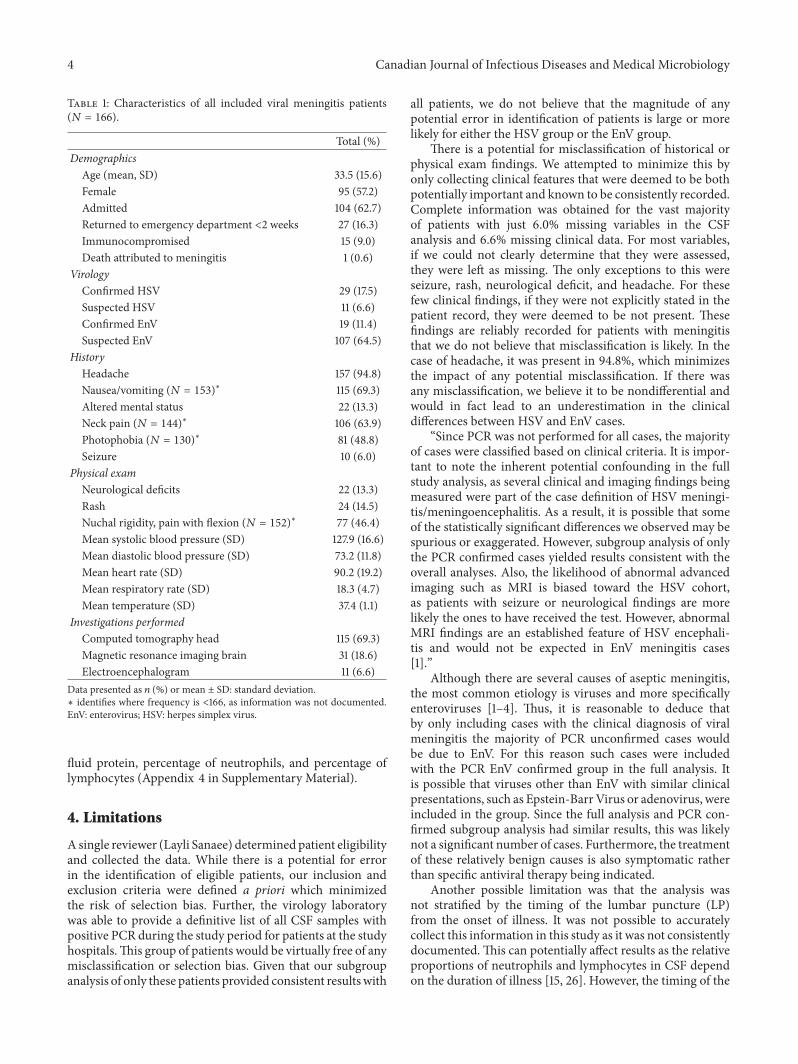
Table 1 presents baseline characteristics for the includedpatients. Just over half (57.2%)were female,mean agewas 33.5years, 62.7% were admitted, and 1 patient (0.6%) died due tomeningitis. We included 16 (9.6%) pediatric patients (age of 3months to 17 years) and 4 (2.4%) were under the age of 1 year.
Table 2 compares characteristics of HSV and EnVmenin-gitis patients. HSV meningitis patients were significantlyolder (40.4 versus 31.3 years, 𝑝 = 0.005) and more likelyto have had seizures (21.1 versus 1.6%, 𝑝 ≤ 0.001), historyof altered mental status (46.2% versus 3.2%, 𝑝 ≤ 0.001),or neurological deficits on examination (44.7 versus 3.9%,𝑝 ≤ 0.001). Initial CSF findings demonstrated no significant
difference in WBC counts (𝑝 = 0.448); however, neutrophilpercentages were significantly lower in HSV cases (3.0 versus9.5%, 𝑝 = 0.0002), while lymphocytes (87.0 versus 67.0%,𝑝 = 0.0004) and protein levels (0.9 versus 0.6 g/L,𝑝 = 0.0005)were significantly higher. Not surprisingly, when CT, MRI,and EEG were performed, HSV patients more frequentlyhad abnormal findings (CT head: 𝑝 = 0.001, MRI brain:𝑝 ≤ 0.001, and EEG: 𝑝 ≤ 0.001). Sensitivity analysisafter removing the 16 pediatric patients did not result insignificantly different results (Appendix 2 in SupplementaryMaterial). Analysis of just the 16 pediatric patients withonly one HSV patient was not conducted due to the smallnumbers.
Our planned subgroup analysis of only virology PCRconfirmed cases (HSV, 𝑁 = 29 versus EnV, 𝑁 = 19) hadfindings consistent with all patients in our study. Statisticallysignificant associations were detected, indicating higher age,more females, higher prevalence of altered mental status andneurological deficits, lower percentages of neutrophils, andhigher lymphocyte percentage and protein levels amongHSVpatients (Table 3). Likewise, our second subgroup analysisusing only immunocompetent cases with PCR confirmedviral meningitis (HSV, 𝑁 = 23 versus EnV, 𝑁 = 19)had results consistent with the full study population(Appendix 3 in Supplementary Material). Receiver operatorcharacteristic curves were calculated for cerebrospinal
4 Canadian Journal of Infectious Diseases and Medical Microbiology
Table 1: Characteristics of all included viral meningitis patients(𝑁 = 166).
Total (%)Demographics
Age (mean, SD) 33.5 (15.6)Female 95 (57.2)Admitted 104 (62.7)Returned to emergency department <2 weeks 27 (16.3)Immunocompromised 15 (9.0)Death attributed to meningitis 1 (0.6)
VirologyConfirmed HSV 29 (17.5)Suspected HSV 11 (6.6)Confirmed EnV 19 (11.4)Suspected EnV 107 (64.5)
HistoryHeadache 157 (94.8)Nausea/vomiting (𝑁 = 153)∗ 115 (69.3)Altered mental status 22 (13.3)Neck pain (𝑁 = 144)∗ 106 (63.9)Photophobia (𝑁 = 130)∗ 81 (48.8)Seizure 10 (6.0)
Physical examNeurological deficits 22 (13.3)Rash 24 (14.5)Nuchal rigidity, pain with flexion (𝑁 = 152)∗ 77 (46.4)Mean systolic blood pressure (SD) 127.9 (16.6)Mean diastolic blood pressure (SD) 73.2 (11.8)Mean heart rate (SD) 90.2 (19.2)Mean respiratory rate (SD) 18.3 (4.7)Mean temperature (SD) 37.4 (1.1)
Investigations performedComputed tomography head 115 (69.3)Magnetic resonance imaging brain 31 (18.6)Electroencephalogram 11 (6.6)
Data presented as 𝑛 (%) or mean ± SD: standard deviation.∗ identifies where frequency is <166, as information was not documented.EnV: enterovirus; HSV: herpes simplex virus.
fluid protein, percentage of neutrophils, and percentage oflymphocytes (Appendix 4 in Supplementary Material).
4. Limitations
Asingle reviewer (Layli Sanaee) determined patient eligibilityand collected the data. While there is a potential for errorin the identification of eligible patients, our inclusion andexclusion criteria were defined a priori which minimizedthe risk of selection bias. Further, the virology laboratorywas able to provide a definitive list of all CSF samples withpositive PCR during the study period for patients at the studyhospitals.This group of patients would be virtually free of anymisclassification or selection bias. Given that our subgroupanalysis of only these patients provided consistent resultswith
all patients, we do not believe that the magnitude of anypotential error in identification of patients is large or morelikely for either the HSV group or the EnV group.
There is a potential for misclassification of historical orphysical exam findings. We attempted to minimize this byonly collecting clinical features that were deemed to be bothpotentially important and known to be consistently recorded.Complete information was obtained for the vast majorityof patients with just 6.0% missing variables in the CSFanalysis and 6.6% missing clinical data. For most variables,if we could not clearly determine that they were assessed,they were left as missing. The only exceptions to this wereseizure, rash, neurological deficit, and headache. For thesefew clinical findings, if they were not explicitly stated in thepatient record, they were deemed to be not present. Thesefindings are reliably recorded for patients with meningitisthat we do not believe that misclassification is likely. In thecase of headache, it was present in 94.8%, which minimizesthe impact of any potential misclassification. If there wasany misclassification, we believe it to be nondifferential andwould in fact lead to an underestimation in the clinicaldifferences between HSV and EnV cases.
“Since PCR was not performed for all cases, the majorityof cases were classified based on clinical criteria. It is impor-tant to note the inherent potential confounding in the fullstudy analysis, as several clinical and imaging findings beingmeasured were part of the case definition of HSV meningi-tis/meningoencephalitis. As a result, it is possible that someof the statistically significant differences we observed may bespurious or exaggerated. However, subgroup analysis of onlythe PCR confirmed cases yielded results consistent with theoverall analyses. Also, the likelihood of abnormal advancedimaging such as MRI is biased toward the HSV cohort,as patients with seizure or neurological findings are morelikely the ones to have received the test. However, abnormalMRI findings are an established feature of HSV encephali-tis and would not be expected in EnV meningitis cases[1].”
Although there are several causes of aseptic meningitis,the most common etiology is viruses and more specificallyenteroviruses [1–4]. Thus, it is reasonable to deduce thatby only including cases with the clinical diagnosis of viralmeningitis the majority of PCR unconfirmed cases wouldbe due to EnV. For this reason such cases were includedwith the PCR EnV confirmed group in the full analysis. Itis possible that viruses other than EnV with similar clinicalpresentations, such as Epstein-Barr Virus or adenovirus, wereincluded in the group. Since the full analysis and PCR con-firmed subgroup analysis had similar results, this was likelynot a significant number of cases. Furthermore, the treatmentof these relatively benign causes is also symptomatic ratherthan specific antiviral therapy being indicated.
Another possible limitation was that the analysis wasnot stratified by the timing of the lumbar puncture (LP)from the onset of illness. It was not possible to accuratelycollect this information in this study as it was not consistentlydocumented. This can potentially affect results as the relativeproportions of neutrophils and lymphocytes in CSF dependon the duration of illness [15, 26]. However, the timing of the

Canadian Journal of Infectious Diseases and Medical Microbiology 5
Table 2: Comparison of demographic and clinical characteristics and investigations between patients with HSV and EnV (𝑁 = 166).
Total (%)𝑝 value
HSV (𝑁 = 40) EnV (𝑁 = 126)Demographics
Age (mean, SD) 40.4 (18.3) 31.3 (14.0) 0.005Female 25 (62.5) 70 (55.6) 0.439Admitted 40 (100) 64 (50.8) <0.001Returned to emergency department <2 weeks 8 (17.9) 19 (15.1) 0.467Immunocompromised 6 (15.0) 9 (7.1) 0.201
HistoryHeadache 35 (97.2) 122 (99.2) 0.403Nausea/vomiting (𝑛 = 153 (36,117)) 27 (75.0) 89 (75.2) 0.979Altered mental status 18 (46.2) 4 (3.2) <0.001Neck pain (𝑛 = 144 (32,112)) 25 (78.1) 81 (72.3) 0.511Photophobia (𝑛 = 130 (24,106)) 16 (66.7) 65 (61.3) 0.626Seizure 8 (21.1) 2 (1.6) <0.001
Physical examNeurological deficit 17 (44.7) 5 (3.9) <0.001Rash 6 (15.0) 18 (14.6) 0.955Nuchal rigidity or pain with flexion (𝑛 = 152 (33,119)) 19 (57.9) 58 (49.2) 0.489Mean systolic blood pressure (SD) 129.9 (17.0) 127.2 (16.5) 0.518Mean diastolic blood pressure (SD) 75.5 (12.8) 72.4 (11.4) 0.225Mean heart rate (SD) 93.3 (19.0) 89.1 (19.3) 0.175Mean respiratory rate (SD) 18.6 (4.4) 18.2 (4.8) 0.684Mean temperature (SD) 37.5 (1.2) 37.3 (1.1) 0.388
InvestigationsCT head abnormal 3 (7.5) 0 (0.0) 0.001MRI brain abnormal 13 (33.3) 3 (2.4) <0.001EEG abnormal 6 (15.0) 1 (0.8) <0.001CSF analysis (median, IQR∗)RBC (×106/L) 5.5 (1.0–16.5) 6.0 (1.0–22.0) 0.965WBC (×106/L) 199.0 (75.0–406.0) 156.5 (51.0–420.0) 0.448% neutrophils 3.0 (0.0–8.0) 9.5 (2.0–37.5) 0.0002% lymphocytes 87.0 (72.0–94.0) 67.0 (40.0–87.0) 0.0004%monocytes 7.0 (4.0–15.0) 10.0 (3.0–20.0) 0.767Glucose (mmol/L) 3.2 (2.8–4.4) 3.1 (2.7–3.5) 0.093Protein (g/L) 0.9 (0.6–1.2) 0.6 (0.5–0.9) 0.0005
Data presented as 𝑛 (%) or mean ± SD (standard deviation). ∗ identifies where frequency is <166, as information was not documented. EnV: enterovirus, HSV:herpes simplex virus.
LP was unlikely to be substantially different between theHSVand the EnV groups.
It is not known what proportion of the PCR confirmedHSV cases wereHSV-1 versus HSV-2. Althoughmanagementis the same for both, their clinical features and imagingfindings can be different as HSV-2 only accounts for 1.6% to6.5% of all herpes simplex encephalitis cases in adults [4].Thus, the ratio of HSV-1 : HSV-2 among the cases can affectthe results by influencing the proportion of meningitis andencephalitis cases in the study. It is therefore unknown inwhat direction the results are potentially skewed.
Finally, several other serious causes of encephalitis werenot included in this study, such as Varicella Zoster Virusand West Nile Virus. The purpose of the study was to
compare the most common serious causes of viral meningi-tis/meningoencephalitis, HSV, with the most common causeof viral meningitis, EnV. Assessing less common causes ofviral meningitis, while potentially worthwhile, was beyondthe scope of this study given that this study would have beengrossly underpowered to find any meaningful differencesbetween these very rare causes of seriousmeningitis and EnV.We chose not to combine all serious etiologies together, as wedo not know if the clinical or laboratory findings would besimilar for all etiologies of serious viral meningitis.
5. Discussion
Our six-year multicenter study identified that viral menin-gitis patients with HSV were more likely to be older and

6 Canadian Journal of Infectious Diseases and Medical Microbiology
Table 3: Subgroup analysis comparing demographic and clinical characteristics and investigations between patients with confirmed HSVand confirmed EnV (𝑁 = 48).
Total (%)𝑝 value
HSV (𝑁 = 29) EnV (𝑁 = 19)Demographics
Mean age ± SD 43.9 ± 18.4 13.1 ± 9.0 <0.001Female 21 (72.4) 5 (26.3) 0.002Admitted 29 (100) 17 (89.5) 0.152Returned to emergency department <2 weeks 5 (17.2) 3 (15.8) 1.000Immunocompromised 6 (15.0) 9 (7.1) 0.201
HistoryHeadache (𝑛 = 41 (25,16)) 24 (96.0) 16 (100) 1.000Nausea/vomiting (𝑛 = 43 (26,17)) 17 (65.4) 12 (70.6) 0.722Altered mental status 12 (41.4) 1 (5.6) 0.008Neck pain (𝑛 = 39 (23,16)) 17 (73.9) 13 (81.3) 0.711Photophobia (𝑛 = 30 (17,13)) 10 (58.8) 6 (46.2) 0.491Seizure 6 (21.4) 1 (5.3) 0.215
Physical examNeurological deficit 10 (35.7) 1 (5.3) 0.032Rash 3 (10.3) 8 (42.1) 0.016Nuchal rigidity, pain with flexion (𝑛 = 42 (24,18)) 14 (58.3) 9 (50.0) 0.591Mean systolic blood pressure (SD) 127.7 (17.6) 116.7 (15.2) 0.031Mean diastolic blood pressure (SD) 75.3 (14.3) 67.3 (13.7) 0.068Mean heart rate (SD) 96.9 (20.0) 99.1 (29.0) 0.775Mean respiratory rate (SD) 19.0 (5.1) 22.1 (10.3) 0.244Mean temperature (SD) 37.5 (1.2) 37.8 (1.10) 0.374
InvestigationsCT head abnormal 3 (10.3) 0 (0) <0.001MRI brain abnormal 9 (32.1) 1 (5.3) 0.031EEG abnormal 2 (6.9) 1 (5.3) 0.224CSF analysis (median, IQR∗)
RBC (×106/L) 5.0 (1.0–13.0) 9.0 (3.0–14.0) 0.386WBC (×106/L) 207.0 (39.0–403.0) 150.0 (42.0–365.0) 0.697% neutrophils 3.5 (0.0–0.08) 20.0 (3.0–70.0) 0.004% lymphocytes 87.0 (71.0–94.0) 55.0 (30.0–73.0) 0.001%monocytes 8.0 (4.0–19.0) 11.5 (7.0–20.0) 0.444Glucose (mmol/L) 3.3 (2.8–4.9) 3.3 (2.6–3.3) 0.141Protein (g/L) 0.7 (0.6–1) 0.4 (0.3–0.7) 0.001
Data presented as 𝑛 (%) or mean ± SD (standard deviation). ∗ identifies where frequency is <166, as information was not documented. EnV: enterovirus, HSV:herpes simplex virus.
have seizure, an altered mental status, neurological deficits,lower CSF neutrophil counts, higher CSF lymphocyte counts,or higher CSF protein levels than patients with EnV. Thesefindings are clinically important as physicians performing alumbar puncture to rule out central nervous system infection,who diagnose their patients with viral meningitis, need toconsider HSV meningitis. Patients with one or more of ourclinical or CSF findings ought to be started on intravenousantiviral agents (e.g., acyclovir) pending the results of PCRtesting.
Prior studies, including the study by Ihekwaba, havedemonstrated that HSV meningitis cases have greater CSF
white blood cell counts and protein levels [13, 14]. Our studydid not find a statistically significant difference in the overallwhite blood cell count; however, we did find significantdifferences in the neutrophil and lymphocyte differentials.This may be due to a difference in timing of the LP inthe course of illness between groups or study populations,as children tend to have LPs earlier during admission, andIhekwaba et al. excluded patients <16 years of age [15, 27].It may also be that the previous study sample sizes were toosmall and the statistical difference the researchers observedwas due to chance. Similar to these two studies, we found thatHSV meningitis cases had a higher protein level than EnV

Canadian Journal of Infectious Diseases and Medical Microbiology 7
cases. Our study covered three emergency departments andallowed for analysis of a larger number of HSV and EnV casesthan previous studies.
Another previous study, by Hanson and colleagues,developed a laboratory-screening tool for HSV meningitisusing 1,659 HSV PCR requests [17]. Their tool incorporatedCSF parameters along with immune status and age. Nocomparator group was used and they did not assess theassociation of clinical features with HSV in patients thoughtto have viral meningitis. In their study, 7.8% of patients whowere not tested by their criteria were treated with intravenousacyclovir due to concern for HSV. Our study’s objective wasto determine high-risk features of HSV versus the morecommon EnV meningitis.
The majority of signs and symptoms did not differamongst the groups, including vital signs, presence of nuchalrigidity, rash, or photophobia. We did, however, find a signif-icant difference in the frequency of seizure, history of alteredmental status, and neurological deficit on physical exam.These three features are well-known signs of encephalitis,and given that HSV has a greater propensity for progress-ing to encephalitis than EnV, these were not surprisingresults.
In the subgroup analysis of PCR confirmed cases, theHSV group had a significantly increased percentage offemales. This may be accounted by the fact that HSV-2meningitis seems to affect a greater proportion of femalescompared to males [24, 28, 29]. This trend was not seen per-haps as the full study analysis included a greater proportionof HSV-1 cases.
6. Clinical Implications
The clinical features that were statistically different betweenHSV and enteroviral meningitis included typical signs andsymptoms of encephalitis. However, these did not identifyall HSV cases. This suggests that in the absence of clinicalfindings we cannot absolutely rule out HSV meningitis.This has been previously found in children where HSVmeningitis andmeningoencephalitis can present without anyovert clinical signs ofmeningeal irritation [5, 11].The patientswith HSV CNS disease not identified in our study by clinicalor historical features ranged from 27 to 50 years of age. CSFresults in addition to clinical features can help us identifyhigh-risk patients earlier. Given that current practice inmanyinstitutions involves a significant delay to PCR testing ofCSF, our study supports empiric intravenous antiviral therapypatients with suspected HSV encephalitis, including thosewith any of the following features: altered mental status,seizure, neurological deficits, lower CSF neutrophil percent-ages, high CSF lymphocyte percentages, or high CSF proteinlevels. Furthermore, our study supports future prospec-tive observational studies to confirm the reproducibilityof these associations and also to derive cutoffs for CSFlymphocyte proportions and protein levels. Since no absolutecutoffs for CSF parameters were derived, the diagnosis ofHSV CNS disease remains clinical until PCR virology isobtained.
7. Conclusion
In summary, we found that patients with HSV meningitiswere more likely than patients with enteroviral meningitisto present with seizure, altered mental status, neurologicaldeficits, lower CSF neutrophils, higher CSF lymphocytes, orhigher CSF protein levels. We recommend that HSV directedintravenous antiviral treatment be strongly considered inpatients with one or more of these clinical or CSF featureswhile awaiting PCR results.
Disclosure
This paper was presented at (1) Society for Academic Emer-gencyMedicineAnnualMeeting,May 2013, Atlanta, Georgia,United States, and (2) Canadian Association of EmergencyPhysicians Annual Conference, June 2013, Vancouver, BritishColumbia, Canada.
Competing Interests
There are no competing interests to declare.
Authors’ Contributions
Layli Sanaee, TimKarnauchow, and Jeffrey J. Perry conceivedand designed the study. Layli Sanaee, Tim Karnauchow, andJeffrey J. Perry supervised the conduct of the study. LayliSanaee conducted the data collection. Monica Taljaard andJeffrey J. Perry provided statistical advice on study design;Monica Taljaard and Jeffrey J. Perry performed statisticaldata analysis. Layli Sanaee drafted the paper, and all authorscontributed substantially to its revision. Layli Sanaee takesresponsibility for the paper as a whole.
References
[1] L. Kupila, T. Vuorinen, R. Vainionpaa, V. Hukkanen, R. J. Mart-tila, and P. Kotilainen, “Etiology of aseptic meningitis andencephalitis in an adult population,” Neurology, vol. 66, no. 1,pp. 75–80, 2006.
[2] B. E. Lee and H. D. Davies, “Aseptic meningitis,” CurrentOpinion in Infectious Diseases, vol. 20, no. 3, pp. 272–277, 2007.
[3] H. Rubeiz and R. P. Roos, “Viral meningitis and encephalitis,”Seminars in Neurology, vol. 12, no. 3, pp. 165–177, 1992.
[4] E. Aurelius, B. Johansson, B. Skoldenberg, and M. Forsgren,“Encephalitis in immunocompetent patients due to herpes sim-plex virus type 1 or 2 as determined by type-specific polymerasechain reaction and antibody assays of cerebrospinal fluid,”Journal of Medical Virology, vol. 39, no. 3, pp. 179–186, 1993.
[5] S. A. E. Logan and E. MacMahon, “Viral meningitis,” BritishMedical Journal, vol. 336, article 36, 2008.
[6] T. Solomon, B. D. Michael, P. E. Smith et al., “Management ofsuspected viral encephalitis in adults—Association of BritishNeurologists and British Infection Association National Guide-lines,” Journal of Infection, vol. 64, no. 4, pp. 347–373, 2012.
[7] R. J.Whitley, C.A.Alford,M. S.Hirsch et al., “Vidarabine versusacyclovir therapy in herpes simplex encephalitis,” The NewEngland Journal of Medicine, vol. 314, no. 3, pp. 144–149, 1986.
8 Canadian Journal of Infectious Diseases and Medical Microbiology
[8] C. Alford Jr., R. Dolin, M. S. Hirsch, A. W. Karchmer, and R. J.Whitley, “Herpes simplex encephalitis and clinical trial design,”The Lancet, vol. 319, no. 8279, p. 1013, 1982.
[9] T. Bergstrom and K. Alestig, “Treatment of primary andrecurrent herpes simplex virus type 2 induced meningitis withacyclovir,” Scandinavian Journal of Infectious Diseases, vol. 22,no. 2, pp. 239–240, 1990.
[10] B. Skoldenberg, M. Forsgren, K. Alestig et al., “Acyclovir versusvidarabine in herpes simplex encephalitis. Randomised multi-centre study in consecutive Swedish patients,” The Lancet, vol.324, no. 8405, pp. 707–711, 1984.
[11] R. Kneen, B. D. Michael, E. Menson et al., “Management ofsuspected viral encephalitis in children—association of BritishNeurologists and British Paediatric Allergy, Immunology andInfection Group National Guidelines,” Journal of Infection, vol.64, no. 5, pp. 449–477, 2012.
[12] S. Yang and R. E. Rothman, “PCR-based diagnostics for infec-tious diseases: uses, limitations, and future applications inacute-care settings,” Lancet Infectious Diseases, vol. 4, no. 6, pp.337–348, 2004.
[13] R. J. Whitley and F. Lakeman, “Herpes simplex virus infectionsof the central nervous system: therapeutic and diagnosticconsiderations,” Clinical Infectious Diseases, vol. 20, no. 2, pp.414–420, 1995.
[14] I. Steiner, H. Budka, A. Chaudhuri et al., “Viral encephalitis: areview of diagnostic methods and guidelines for management,”European Journal of Neurology, vol. 12, no. 5, pp. 331–343, 2005.
[15] T. W. Smalling, S. E. Sefers, H. Li, and Y.-W. Tang, “Molecularapproaches to detecting herpes simplex virus and enterovirusesin the central nervous system,” Journal of Clinical Microbiology,vol. 40, no. 7, pp. 2317–2322, 2002.
[16] U. K. Ihekwaba, G. Kudesia, and M. W. McKendrick, “Clinicalfeatures of viral meningitis in adults: significant differences incerebrospinal fluid findings among herpes simplex virus, vari-cella zoster virus, and enterovirus infections,”Clinical InfectiousDiseases, vol. 47, no. 6, pp. 783–789, 2008.
[17] K. E. Hanson, B. D. Alexander, C. Woods, C. Petti, and L. B.Reller, “Validation of laboratory screening criteria for herpessimplex virus testing of cerebrospinal fluid,” Journal of ClinicalMicrobiology, vol. 45, no. 3, pp. 721–724, 2007.
[18] L. Srinivasan,M. C. Harris, and S. S. Shah, “Lumbar puncture inthe neonate: challenges in decision making and interpretation,”Seminars in Perinatology, vol. 36, no. 6, pp. 445–453, 2012.
[19] S. C. M. de Crom, M. A. M. van Furth, M. F. Peeters, J. W. A.Rossen, andC. C.Obihara, “Characteristics of pediatric patientswith enterovirus meningitis and no cerebral fluid pleocytosis,”European Journal of Pediatrics, vol. 171, no. 5, pp. 795–800, 2012.
[20] K. R. Rittichier, P. A. Bryan, K. E. Bassett et al., “Diagnosis andoutcomes of enterovirus infections in young infants,” PediatricInfectious Disease Journal, vol. 24, no. 6, pp. 546–550, 2005.
[21] W. S. Mulford, R. S. Buller, M. Q. Arens, and G. A. Storch,“Correlation of cerebrospinal fluid (CSF) cell counts and ele-vated CSF protein levels with enterovirus reverse transcription-PCR results in pediatric and adult patients,” Journal of ClinicalMicrobiology, vol. 42, no. 9, pp. 4199–4203, 2004.
[22] K. R. Jerome, M.-L. Huang, A. Wald, S. Selke, and L. Corey,“Quantitative stability of DNA after extended storage of clinicalspecimens as determined by real-time PCR,” Journal of ClinicalMicrobiology, vol. 40, no. 7, pp. 2609–2611, 2002.
[23] W. A. Verstrepen, P. Bruynseels, and A. H.Mertens, “Evaluationof rapid real time RT-PCR assay for detection of enterovirus
RNA in CSF,” Journal of Clinical Virology, vol. 25, supplement1, pp. S39–S43, 2002.
[24] J. R. Berger and S. Houff, “Neurological complications of herpessimplex virus type 2 infection,”Archives ofNeurology, vol. 65, no.5, pp. 596–600, 2008.
[25] J. P. Simko, A. M. Caliendo, K. Hogle, and J. Versalovic, “Differ-ences in laboratory findings for cerebrospinal fluid specimensobtained from patients with meningitis or encephalitis due toherpes simplex virus (HSV) documented by detection of HSVDNA,” Clinical Infectious Diseases, vol. 35, no. 4, pp. 414–419,2002.
[26] D. F. Gaieski, B. R. Nathan, S. D. Weingart, and W. S. Smith,“Emergency neurologic life support: meningitis and encephali-tis,” Neurocritical Care, vol. 17, supplement 1, pp. S66–S72, 2012.
[27] P. E. Jimenez Caballero, F. Munoz Escudero, S. Murcia Car-retero, andA. Verdu Perez, “Descriptive analysis of viralmenin-gitis in a general hospital: differences in the characteristicsbetween children and adults,”Neurologia, vol. 26, no. 8, pp. 468–473, 2011.
[28] G. J. Mertz, “Epidemiology of genital herpes infections,” Infec-tious Disease Clinics of North America, vol. 7, no. 4, pp. 825–839,1993.
[29] Y.-W. Tang, P. S. Mitchell, M. J. Espy, T. F. Smith, and D. H.Persing, “Molecular diagnosis of herpes simplex virus infectionsin the central nervous system,” Journal of Clinical Microbiology,vol. 37, no. 7, pp. 2127–2136, 1999.

Submit your manuscripts athttp://www.hindawi.com
Stem CellsInternational
Hindawi Publishing Corporationhttp://www.hindawi.com Volume 2014
Hindawi Publishing Corporationhttp://www.hindawi.com Volume 2014
MEDIATORSINFLAMMATION
of
Hindawi Publishing Corporationhttp://www.hindawi.com Volume 2014
Behavioural Neurology
EndocrinologyInternational Journal of
Hindawi Publishing Corporationhttp://www.hindawi.com Volume 2014
Hindawi Publishing Corporationhttp://www.hindawi.com Volume 2014
Disease Markers
Hindawi Publishing Corporationhttp://www.hindawi.com Volume 2014
BioMed Research International
OncologyJournal of
Hindawi Publishing Corporationhttp://www.hindawi.com Volume 2014
Hindawi Publishing Corporationhttp://www.hindawi.com Volume 2014
Oxidative Medicine and Cellular Longevity
Hindawi Publishing Corporationhttp://www.hindawi.com Volume 2014
PPAR Research
The Scientific World JournalHindawi Publishing Corporation http://www.hindawi.com Volume 2014
Immunology ResearchHindawi Publishing Corporationhttp://www.hindawi.com Volume 2014
Journal of
ObesityJournal of
Hindawi Publishing Corporationhttp://www.hindawi.com Volume 2014
Hindawi Publishing Corporationhttp://www.hindawi.com Volume 2014
Computational and Mathematical Methods in Medicine
OphthalmologyJournal of
Hindawi Publishing Corporationhttp://www.hindawi.com Volume 2014
Diabetes ResearchJournal of
Hindawi Publishing Corporationhttp://www.hindawi.com Volume 2014
Hindawi Publishing Corporationhttp://www.hindawi.com Volume 2014
Research and TreatmentAIDS
Hindawi Publishing Corporationhttp://www.hindawi.com Volume 2014
Gastroenterology Research and Practice
Hindawi Publishing Corporationhttp://www.hindawi.com Volume 2014
Parkinson’s Disease
Evidence-Based Complementary and Alternative Medicine
Volume 2014Hindawi Publishing Corporationhttp://www.hindawi.com