research article bioassay of prion-infected blood plasma in … · research article bioassay of...
TRANSCRIPT

Research Article
Bioassay of prion-infected blood plasma in PrPtransgenic DrosophilaAlana M. Thackray1, Olivier Andreoletti2 and Raymond Bujdoso11Department of Veterinary Medicine, University of Cambridge, Madingley Road, Cambridge CB3 OES, U.K. and 2UMR INRA ENVT 1225 Hôtes-Agents Pathogènes, Ecole NationaleVétérinaire de Toulouse, 23 Chemin des Capelles, 31076 Toulouse, France
Correspondence: Raymond Bujdoso ([email protected])
In pursuit of a tractable bioassay to assess blood prion infectivity, we have generatedprion protein (PrP) transgenic Drosophila, which show a neurotoxic phenotype in adult-hood after exposure to exogenous prions at the larval stage. Here, we determined thesensitivity of ovine PrP transgenic Drosophila to ovine prion infectivity by exposure ofthese flies to a dilution series of scrapie-infected sheep brain homogenate. Ovine PrPtransgenic Drosophila showed a significant neurotoxic response to dilutions of 10−2 to10−10 of the original scrapie-infected sheep brain homogenate. Significantly, we deter-mined that this prion-induced neurotoxic response in ovine PrP transgenic Drosophila wastransmissible to ovine PrP transgenic mice, which is indicative of authentic mammalianprion detection by these flies. As a consequence, we considered that PrP transgenicDrosophila were sufficiently sensitive to exogenous mammalian prions to be capable ofdetecting prion infectivity in the blood of scrapie-infected sheep. To test this hypothesis,we exposed ovine PrP transgenic Drosophila to scrapie-infected plasma, a blood fractionnotoriously difficult to assess by conventional prion bioassays. Notably, pre-clinicalplasma from scrapie-infected sheep induced neurotoxicity in PrP transgenic Drosophilaand this effect was more pronounced after exposure to samples collected at the clinicalphase of disease. The neurotoxic phenotype in ovine PrP transgenic Drosophila inducedby plasma from scrapie-infected sheep was transmissible since head homogenate fromthese flies caused neurotoxicity in recipient flies during fly-to-fly transmission. Our datashow that PrP transgenic Drosophila can be used successfully to bioassay prion infectiv-ity in blood from a prion-diseased mammalian host.
IntroductionPrion diseases are fatal neurodegenerative conditions that affect humans and a variety of other verte-brate species [1]. These conditions, which include scrapie of sheep, bovine spongiform encephalopathy(BSE) of cattle and Creutzfeldt–Jakob disease (CJD) of humans, are associated with the misfolding ofthe normal host protein PrPC into a disease-specific conformer PrPSc (abnormal disease-specificconformation of PrP) [2]. The disease-associated form of prion protein (PrP) is enriched in β-sheetstructure, and a proportion of the protein ensemble is partially resistant to proteolytic digestion [3,4].The accumulation of misfolded PrP in the brain of prion-diseased individuals is central to the patho-logical process of these conditions [5,6], which typically manifests as synaptic loss and neuronal dys-function, with resultant neurological signs and clinical symptoms. Prion diseases are unique amongneurodegenerative conditions since they are transmissible, and PrPSc is considered to be the infectiousprion agent [7].Prion diseases are a significant risk to public health through the potential for zoonotic transmission
of prions as evidenced by the BSE epizootic in U.K. cattle and subsequent emergence of variant CJD(vCJD) in humans [8,9]. The occurrence of vCJD prions in human lymphoid tissue, together with thedetection of prion infectivity in the blood of animals with asymptomatic experimental prion disease
Accepted Manuscript online:13 October 2016Version of Record published:25 November 2016
Received: 4 May 2016Revised: 3 October 2016Accepted: 11 October 2016
© 2016 The Author(s). This is an open access article published by Portland Press Limited on behalf of the Biochemical Society and distributed under the Creative Commons Attribution License 4.0 (CC BY). 4399
Biochemical Journal (2016) 473 4399–4412DOI: 10.1042/BCJ20160417

[10,11], raised the possibility for the potential of blood-borne vCJD transmission. These concerns were realisedwith the emergence of cases of vCJD in individuals within the U.K. who had received red blood cell concen-trates [12–15]. In addition, abnormal prion protein was detected in a post-mortem spleen sample from ahaemophilic patient who had received purified Factor VIII prepared from plasma batches that included dona-tions from individuals who later developed vCJD [12]. These particular cases of vCJD infection collectivelyprovide strong support for the view that this prion disease can be transmitted by blood transfusion.Consequently, much attention has focused on understanding the biology of infectious prions in blood throughthe analysis of prion infectivity and PrPSc within different blood fractions from hosts with prion disease.The only reliable method to detect prion infectivity is by bioassay in an appropriate indicator species. The
bioassay of blood-borne prion infectivity has been performed for a variety of different species, such as rodents,sheep, cervids and primates including humans, undergoing, collectively, either experimental or natural priondisease [10,15–21]. These bioassays have typically involved intracerebral inoculation of blood, or blood frac-tions, into a recipient indicator species, usually rodents including mice with a PrP transgene autologous for thedonor species. Collectively, these studies revealed that prion infectivity titres of blood samples harvested duringthe asymptomatic phase of prion disease were up to 10-fold less than those collected during the clinical phase[10,19,20,22]. Infectious prions were found to be associated with white and red blood cells, platelets andplasma, although interspecies variations existed with regard to the distribution of prion infectivity in these dif-ferent blood fractions [10,15–21]. Studies in ruminants have analysed bioassays of autologous whole or fractio-nated blood by the intravenous route [19,20]. These experimental transfusion studies have confirmed that allblood fractions prepared by protocols similar to those used in transfusion medicine can transmit prion disease[23,24]. In addition, prion transmission by transfusion of cellular blood fractions was not solely dependent ontheir infectious titre since prion disease was more efficiently transmitted with viable rather than non-viable leu-kocytes [20]. While these studies have collectively been immensely informative in providing evidence thatblood from prion-diseased hosts can harbour prion infectivity, the bioassays used to establish this are relativelycumbersome and time-consuming.We have developed a novel invertebrate system to begin to establish a tractable bioassay to assess blood
prion infectivity [25]. We have previously generated Drosophila transgenic for ovine PrP and have shown thatafter exposure to exogenous ovine prions at the larval stage these flies develop a neurotoxic phenotype in adult-hood, evidenced by decreased locomotor ability and the accumulation of Proteinase K (PK)-resistant PrPSc[26–28]. Here, we determined the sensitivity of ovine PrP transgenic Drosophila to prion infectivity by exposureof these flies to a dilution series of scrapie-infected sheep brain homogenate. Dilutions of scrapie-infected sheepbrain material in the range of 10−2–10−10 were capable of inducing neurotoxicity in ovine PrPtransgenic Drosophila. Furthermore, we have shown that ovine prion-induced neurotoxicity in ovine PrP trans-genic Drosophila was transmissible to ovine PrP transgenic mice. These observations suggested that PrP trans-genic Drosophila were sufficiently sensitive to detect the reportedly low level of prion infectivity in blood fromprion-diseased hosts. To test this hypothesis, we exposed ovine PrP transgenic Drosophila to plasma fromsheep with scrapie disease, a blood fraction notoriously difficult to assess by conventional prion bioassay inmore sentient experimental animals. Pre-clinical plasma from scrapie-infected sheep induced a neurotoxiceffect in PrP transgenic Drosophila, a response that was more pronounced after exposure to samples collectedat the clinical phase of disease. The neurotoxic phenotype induced in ovine PrP transgenic Drosophila byscrapie-infected plasma was transmissible, since head homogenate from these Drosophila caused neurotoxicityin recipient flies during fly-to-fly transmission. Our data show that PrP transgenic Drosophila can be used suc-cessfully for the bioassay of prion infectivity in blood samples from a prion-diseased mammalian host.
Materials and methodsFly stocksThe UAS-PrP fly line w; M{VRQ-PrP, 3xP3-RFP.attP}ZH-51D that is transgenic for ovine V136R154Q171 PrPwith an N-terminal leader peptide and a C-terminal glycosylphosphatidylinositol signal sequence [VRQ(GPI)]was generated as previously described [25]. The UAS-PrP fly line w; M{VRQcyt-PrP, 3xP3-RFP.attP}ZH-51Dthat is transgenic for cytosolic expression of ovine V136R154Q171 [VRQ(cyt)] was generated as recently described[28]. Cre-mediated removal of the red fluorescent protein (RFP) gene from the VRQ(GPI) and VRQ(cyt) PrPfly genome was performed by conventional fly crosses. In some experiments, VRQ(cyt) PrP or the relevantcontrol 51D flies that contained the RFP cassette were used as stated in Figures 3, 5 and 6 and Supplementary
4400 © 2016 The Author(s). This is an open access article published by Portland Press Limited on behalf of the Biochemical Society and distributed under the Creative Commons Attribution License 4.0 (CC BY).
Biochemical Journal (2016) 473 4399–4412DOI: 10.1042/BCJ20160417
Data S3. The Elav-GAL4 driver line (P{w[+mW.hs] = GawB}elav[C155]) and control 51D (w; M{3xP3-RFP.attP}ZH-51D) fly line were obtained from the Department of Genetics, University of Cambridge, U.K. All fly lineswere raised on standard cornmeal media [29] at 25°C and maintained at low-to-medium density, andpre-mated before experimental use.
Sheep brain material and plasmaScrapie-infected sheep brainThis was either a classical scrapie PG127-infected VRQ/VRQ sheep brain stem sample that has been endpointtitrated by intracerebral inoculation in tg338 mice and has a titre of 106.6 ID50/g [30], or cerebral cortex brainmaterial from a confirmed scrapie-positive VRQ/VRQ sheep (SE1848/0005) [31]. Plasma samples fromscrapie-infected sheep: these were obtained from classical scrapie-positive VRQ/VRQ sheep. Plasma samplesfrom sheep experimentally infected with classical scrapie were prepared from animals challenged by oral inocu-lation with PG127-infected sheep brain homogenate as previously described [19]. Plasma samples from sheepnaturally infected with classical scrapie were prepared from animals born and maintained in a flock endemicwith the disease [32] and that were confirmed positive for PrPSc through routine statutory surveillance (datanot shown). Control sheep brain material and plasma: these were obtained from New Zealand-derived scrapie-free VRQ/VRQ sheep [33].
Inoculation of Drosophila with sheep brain material or plasmaDrosophila at the larval stage of development were exposed to ovine brain material or plasma from confirmedclassical scrapie-positive or known scrapie-negative sheep. Two hundred and fifty microlitres of either 10%(v/v) plasma or 1% (v/v) sheep brain homogenate, or various dilutions of a 1/10 dilution series (v/v) of thesesamples prepared in PBS pH 7.4, were added to the top of the cornmeal that contained third instar Drosophilalarvae in 300 plastic vials. Following eclosion (i.e. hatching), flies were transferred to fresh non-treated vials.
Negative-geotaxis climbing assayThe locomotor ability of flies was assessed in a negative-geotaxis climbing assay [25] that was initiated with 45(3 × n = 15) age-matched, pre-mated female flies in each treatment group. Drosophila were placed in adaptedplastic 25 ml pipettes that were used as vertical climbing columns and allowed to acclimatise for 30 min priorto assessment of their locomotor ability. Flies were tapped to the bottom of the pipette (using the samenumber and intensity of taps on each occasion) and then allowed to climb for 45 s. At the end of the climbingperiod, the number of flies above the 25 ml mark, the number below the 2 ml mark and the number inbetween the 2 and 25 ml mark were recorded. This procedure was performed three times at each time point.The performance index (PI) was calculated for each group of 15 flies (average of three trials) using the formulaPI = 0.5 × (ntotal + ntop− nbottom)/ntotal, where ntotal is the total number of flies, ntop is the totalnumber of flies at the top and nbottom is the total number of flies at the bottom, as described previously [25].A PI value of 1 is recorded if all flies climb to the top of the tube whereas the value is 0 if no flies climb thetube past the 2 ml mark. The mean PI ± SD at individual time points for each treatment group was plotted,either as a fitted or regression line. In Figure 6, the PI endpoint was plotted and this represented the extrapo-lated time point for individual regression lines when the PI value was 0 (i.e. x-axis value when y = 0 in a regres-sion line analysis).
Preparation of Drosophila head homogenateWhole flies in an eppendorf tube were frozen in liquid nitrogen for 10 min and then vortexed for 2 min tocause decapitation. Individual fly heads were isolated and placed in clean eppendorf tubes using a fine paintbrush. PBS pH 7.4 was added to give 1 ml/head and homogenates were prepared by manual grinding of the flyheads with sterilised plastic pestles.
Mouse prion bioassayMouse prion bioassays were carried out in tg338 mice, which are transgenic for ovine VRQ PrP and are highlyefficient for the detection of sheep scrapie infectivity [34]. Mice (n = 6 per inoculum) were injected intracere-brally with 20 ml of diluted fly head homogenate (to give 2 fly head equivalents per mouse) and monitoreddaily until the occurrence of clinical signs of mouse prion disease. Inoculated mice were killed when they
© 2016 The Author(s). This is an open access article published by Portland Press Limited on behalf of the Biochemical Society and distributed under the Creative Commons Attribution License 4.0 (CC BY). 4401
Biochemical Journal (2016) 473 4399–4412DOI: 10.1042/BCJ20160417
started to show locomotor disorders and any impairment in their capacity to feed, or at a pre-defined endpointfor the assay (165 days post-inoculation) [30]. Brain tissue (cerebral cortex) was collected from killed mice andfrozen for PrPSc analysis by western blot (TeSeE, BioRad).
Fly-to-fly transmission of neurotoxicity induced by scrapie-infected sheepplasmaDrosophila head homogenates were prepared from 30-day-old flies that had been exposed at the larval stage toplasma samples from naturally infected scrapie-positive sheep. One hundred male and 100 female fly headswere homogenised in a hand-held plastic homogeniser in 50 ml of PBS and subsequently diluted to 2.5 ml inthe same buffer. Two hundred and fifty microlitres of the fly head homogenate were added to the top of thecornmeal that contained third instar Drosophila larvae in 300 plastic vials (20 fly head equivalents per vial).Following eclosion (i.e. hatching), flies were transferred to fresh non-treated vials and their locomotor abilityassessed in a negative-geotaxis climbing assay as described above [25].
Statistical analysisStatistical analysis of the data was performed by one-way analysis of variance (ANOVA) together with Tukey’shonestly significant difference (HSD) for post hoc analysis, or the Student’s t-test, using Prism (GraphPadSoftware, Inc., San Diego, U.S.A.).
ResultsHere, we have determined the ability of ovine PrP transgenic Drosophila to bioassay prion infectivity in bloodplasma from scrapie-infected sheep. To achieve this, we have used flies that express either GPI-anchored ovineV136R154Q171 [VRQ(GPI)] PrP [25,26] or cytosolic ovine V136R154Q171 [VRQ(cyt)] PrP [28]. We have alreadyestablished that both PrP transgenic fly lines are susceptible to ovine scrapie prion infectivity, which does notinduce neurotoxicity in non-transgenic 51D Drosophila [25,26,28].
Sensitivity of PrP transgenic Drosophila to ovine prionsWe first established the sensitivity of ovine PrP transgenic Drosophila to ovine scrapie prion infectivity. To doso, PrP transgenic Drosophila at the larval stage were exposed to a dilution series of PG127 scrapie-infectedsheep brain homogenate. The locomotor ability of prion-exposed Drosophila was assessed, after hatching fromthe larval stage, by a negative-geotaxis climbing assay and the data were expressed as a PI. Supplementary DataS1 shows that PrP transgenic Drosophila demonstrated an accelerated decline in locomotor ability after hatchingfollowing exposure to scrapie prions at the larval stage compared with the response seen after exposure toscrapie-free sheep brain material. This accelerated decline, which became apparent ∼10 days after hatching,diminished upon exposure to increasing dilution of scrapie-infected brain homogenate. This titration effect wasevident over the duration of the assay and was most clearly shown during the first half of the time course whenthe climbing ability of flies was assessed. Accordingly, the data in Figure 1 show linear regression analysis ofthe PI from Supplementary Data S1 for each fly line from day 1 to day 22 after hatching. These data demon-strate that VRQ(GPI) (Figure 1A) and VRQ(cyt) (Figure 1B) PrP transgenic Drosophila both showed a similarsensitivity to all dilutions of scrapie-infected sheep brain material, although the magnitude of the response bythe VRQ(cyt) fly line was somewhat greater than that of VRQ(GPI) flies. Supplementary Data S2 showsP-values for the statistical analysis of the PI for each fly line in response to dilutions of scrapie-infected sheepbrain material compared with the response induced by the control sheep brain sample. A statistically significantdecline in PrP transgenic Drosophila locomotor ability was induced in the VRQ(GPI) and VRQ(cyt) fly linesby dilutions of scrapie-infected sheep brain homogenate in the range of 10−2–10−10. The locomotor responseseen for both fly lines after exposure to scrapie-free sheep brain homogenate was not significantly differentfrom that seen after exposure to PBS (P > 0.05). The PG127 scrapie-infected sheep brain isolate used here hasan endpoint titration of 106.6 ID50/g in tg338 mice [30]. The data presented here show that our Drosophilaprion bioassay has a comparable, if not greater, sensitivity for ovine prions compared with a mammalian prionbioassay.
4402 © 2016 The Author(s). This is an open access article published by Portland Press Limited on behalf of the Biochemical Society and distributed under the Creative Commons Attribution License 4.0 (CC BY).
Biochemical Journal (2016) 473 4399–4412DOI: 10.1042/BCJ20160417

Prion infectivity in scrapie brain-exposed PrP transgenic DrosophilaCardinal features of mammalian prion diseases are the accumulation of PK-resistant PrPSc in the brains ofprion-infected individuals, concomitant with transmissible neurotoxicity [1,35]. We have previously shown thatovine PrP transgenic Drosophila accumulate disease-associated PrP after exposure to ovine prions [26].PK-resistant PrPSc in the form of PrP27-30 was detected following protein misfolding cyclic amplification(PMCA) using head homogenate from ovine prion-exposed VRQ(GPI) PrP transgenic Drosophila. This wasnot due to carry over of PG127-infected brain material at the time of exposure of the flies to sheep scrapiesince PrP27-30 was not detected using head homogenate from similarly treated non-transgenic 51D flies [26].To determine whether the scrapie brain-induced neurotoxicity observed in ovine PrP transgenic Drosophila
was transmissible, we performed a fly-to-mouse prion transmission study. Head homogenate from adult VRQ
Figure 1. Drosophila bioassay of scrapie-infected sheep brain tissue.
Adult (A) VRQ(GPI) or (B) VRQ(cyt) PrP transgenic Drosophila were assessed for their locomotor ability by a negative-geotaxis
climbing assay after exposure, at the larval stage, to various dilutions from a 1/10 (v/v) dilution series (as shown) of PG127
scrapie-infected or a 10−2 (v/v) scrapie-free sheep brain tissue. The data shown are linear regression plots of the mean PI ± SD
for three groups of flies per time point calculated as described in the Materials and Methods section.
Table 1 Transmission of neurotoxic fly phenotype to tg338 miceHead homogenate from 30-day-old VRQ(GPI) PrP transgenic or control 51D Drosophila, exposed at thelarval stage to scrapie-infected (SE1848/0005) or scrapie-free sheep brain homogenate, was inoculatedinto tg338 mice (n = 6 per sample). Mice were killed after the development of terminal signs of murineprion disease or at the assay endpoint of 165 days. The presence of PK-resistant PrPSc in the brains ofkilled mice was used to confirm prion disease.
Fly line Prion exposure
tg338 transmission
Attack rate PK-res PrPSc Incubation time (days)
VRQ(GPI) + 6/6 6/6 87 ± 2
51D + 0/6 0/6 <165
VRQ(GPI) − 0/6 0/6 <165
51D − 0/6 0/6 <165
© 2016 The Author(s). This is an open access article published by Portland Press Limited on behalf of the Biochemical Society and distributed under the Creative Commons Attribution License 4.0 (CC BY). 4403
Biochemical Journal (2016) 473 4399–4412DOI: 10.1042/BCJ20160417

(GPI) or VRQ(cyt) PrP transgenic Drosophila, exposed to scrapie-infected sheep brain homogenate at thelarval stage, was inoculated into tg338 mice by an intracerebral injection. Inoculated tg338 mice that exhibitedterminal clinical signs of mouse prion disease were killed and their brains examined for disease-associated PrP.Data given in Table 1 show that head homogenate from scrapie brain-exposed VRQ(GPI) PrP transgenicDrosophila induced terminal prion disease in tg338 mice with a 100% attack rate and a mean incubation timeof 87 ± 2 days. Prion disease in the brains of terminal tg338 mice was verified by the presence of PK-resistantPrPSc as shown by the western blot data in Figure 2. The transmission of prion disease to tg338 mice wasmediated by scrapie exposure of VRQ(GPI) Drosophila since head homogenate from similar flies exposed toscrapie-free sheep brain material failed to do so. In addition, prion disease in tg338 mice was not due to carryover of the original sheep inoculum from scrapie-exposed VRQ(GPI) Drosophila since similarly treated non-transgenic 51D flies did not induce prion disease in the tg338 mouse line. Clinical prion disease was notdetected in tg338 mice inoculated with head homogenate from scrapie-exposed VRQ(cyt) Drosophila (data notshown). However, our previous studies have shown that head homogenate from scrapie brain-exposed VRQ(cyt) PrP transgenic Drosophila can induce neurotoxicity in recipient PrP transgenic flies during fly-to-flytransmission studies [26].
Bioassay of prion-infected blood plasma in PrP transgenic DrosophilaWe reasoned that ovine PrP transgenic Drosophila showed a high sensitivity to ovine prions and that this ren-dered them sufficiently susceptible to be capable of detecting the apparent low titre of prion infectivity in theblood of scrapie-infected sheep. To test this hypothesis, we used ovine PrP transgenic Drosophila to bioassayblood samples from sheep experimentally infected with PG127 scrapie and that are known to contain prioninfectivity [19]. We chose to bioassay sheep plasma as this blood fraction is reported to contain a low level ofprion infectivity and is notoriously difficult to assess by conventional prion bioassay [19].We first investigated the locomotor ability of VRQ(cyt) PrP transgenic Drosophila after exposure to blood
plasma since this fly line typically showed the greatest magnitude of response to sheep scrapie prions in com-parison with other PrP transgenic flies. The data given in Figure 3 show that adult VRQ(cyt) Drosophila dis-played an accelerated decline in locomotor activity, after exposure at the larval stage, to plasma isolated fromPG127-infected donor sheep compared with similar flies exposed to control scrapie-free sheep plasma. Thedecline in locomotor activity induced by plasma prepared from sheep 30 days post-PG127 challenge(Figure 3A) was less marked than that seen in response to plasma isolated from the same sheep at the clinicalstage of scrapie disease (Figure 3B). The locomotor activity of VRQ(cyt) Drosophila was also assessed afterexposure to plasma isolated from sheep at 60, 90 and 120 days post-PG127 challenge. Analyses of these add-itional time points provided further evidence that plasma isolated later in the time course of PG127-induced
Figure 2. PrPSc detection in prion-diseased tg338 mouse brains.
Brain homogenates of tg338 mice previously inoculated with Drosophila head homogenate were digested with Proteinase K at
64 mg/ml and analysed by SDS–PAGE and western blot using anti-PrP monoclonal antibody Sha31 as described in the
Materials and Methods section. Track 1: PG127-infected sheep brain homogenate (positive control); Track 2:
non-prion-exposed tg338 mouse brain (negative control); Tracks 3–8: brain samples from prion disease-positive tg338 mice in
Table 1.
4404 © 2016 The Author(s). This is an open access article published by Portland Press Limited on behalf of the Biochemical Society and distributed under the Creative Commons Attribution License 4.0 (CC BY).
Biochemical Journal (2016) 473 4399–4412DOI: 10.1042/BCJ20160417

experimental scrapie of sheep caused a more marked decrease in the locomotor activity of VRQ(cyt) PrP trans-genic Drosophila compared with plasma prepared at earlier time points (data not shown). Non-transgenic 51DDrosophila were used as the control fly line and were similarly exposed to plasma from PG127 scrapie-infected
Figure 3. Drosophila bioassay of plasma from experimental scrapie-infected sheep.
Adult VRQ(cyt) PrP transgenic Drosophila (with RFP) were assessed for their locomotor ability by a negative-geotaxis climbing
assay after exposure, at the larval stage, to a 10−1 (v/v) dilution of plasma isolated from VRQ/VRQ sheep (A) 30 days or (B) 150
days post-PG127 scrapie inoculation. Red lines: plasma from PG127 scrapie-infected donor sheep. Black lines: plasma from
scrapie-free donor sheep. The data shown are linear regression plots of the mean PI ± SD for three groups of flies per time
point calculated as described in the Materials and Methods section.
Figure 4. Drosophila bioassay of clinical plasma from experimental scrapie-infected sheep.
Adult PrP transgenic Drosophila were assessed for their locomotor ability by a negative-geotaxis climbing assay after exposure,
at the larval stage, to various dilutions from a 1/10 (v/v) dilution series (as shown) of plasma from a clinically diseased PG127
scrapie-infected sheep or a 10−1 (v/v) dilution of scrapie-free sheep plasma. The data shown are linear regression plots of the
mean PI ± SD for three groups of flies per time point calculated as described in the Materials and Methods section.
© 2016 The Author(s). This is an open access article published by Portland Press Limited on behalf of the Biochemical Society and distributed under the Creative Commons Attribution License 4.0 (CC BY). 4405
Biochemical Journal (2016) 473 4399–4412DOI: 10.1042/BCJ20160417
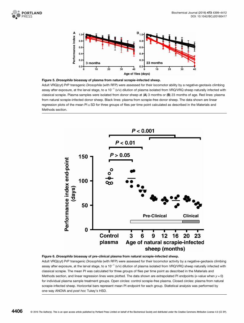
Figure 5. Drosophila bioassay of plasma from natural scrapie-infected sheep.
Adult VRQ(cyt) PrP transgenic Drosophila (with RFP) were assessed for their locomotor ability by a negative-geotaxis climbing
assay after exposure, at the larval stage, to a 10−1 (v/v) dilution of plasma isolated from VRQ/VRQ sheep naturally infected with
classical scrapie. Plasma samples were isolated from donor sheep at (A) 3 months or (B) 23 months of age. Red lines: plasma
from natural scrapie-infected donor sheep. Black lines: plasma from scrapie-free donor sheep. The data shown are linear
regression plots of the mean PI ± SD for three groups of flies per time point calculated as described in the Materials and
Methods section.
Figure 6. Drosophila bioassay of pre-clinical plasma from natural scrapie-infected sheep.
Adult VRQ(cyt) PrP transgenic Drosophila (with RFP) were assessed for their locomotor activity by a negative-geotaxis climbing
assay after exposure, at the larval stage, to a 10−1 (v/v) dilution of plasma isolated from VRQ/VRQ sheep naturally infected with
classical scrapie. The mean PI was calculated for three groups of flies per time point as described in the Materials and
Methods section, and linear regression lines were plotted. The data shown are extrapolated PI endpoints (x-value when y = 0)
for individual plasma sample treatment groups. Open circles: control scrapie-free plasma. Closed circles: plasma from natural
scrapie-infected sheep. Horizontal bars represent mean PI endpoint for each group. Statistical analysis was performed by
one-way ANOVA and post hoc Tukey’s HSD.
4406 © 2016 The Author(s). This is an open access article published by Portland Press Limited on behalf of the Biochemical Society and distributed under the Creative Commons Attribution License 4.0 (CC BY).
Biochemical Journal (2016) 473 4399–4412DOI: 10.1042/BCJ20160417
and scrapie-free sheep. Supplementary Data S3 shows that there was no difference in decline of locomotoractivity in the 51D fly line after exposure to prion-infected or prion-free plasma.We subsequently determined whether the neurotoxicity induced in ovine PrP transgenic Drosophila by
plasma from PG127 scrapie-affected sheep could be titrated. Accordingly, ovine PrP transgenic Drosophila wereexposed at the larval stage to a dilution series of plasma isolated from a sheep with PG127-induced clinicalscrapie disease. The locomotor ability of plasma-exposed Drosophila was assessed, after hatching from thelarval stage, by a negative-geotaxis climbing assay and the data were expressed as a PI. Supplementary Data S4shows that PrP transgenic Drosophila demonstrated an accelerated decline in locomotor ability soon afterhatching following exposure to PG127-infected sheep plasma at the larval stage compared with the responseseen after exposure to scrapie-free sheep plasma. The rate of decline in locomotor ability diminished withexposure to increasing dilution of scrapie-infected plasma, indicative of titration of a particulate transmissiblemoiety, a characteristic feature of the infectious scrapie agent [36]. The accelerated decline in locomotor abilitybecame apparent <10 days after hatching and was most evident during the first half of the time course whenthe fly climbing ability was assessed, in a similar manner to that seen in response to PG127 scrapie-infectedsheep brain material. Accordingly, the data given in Figure 4 show linear regression analysis of the PI fromSupplementary Data S4 for each fly line from day 1 to day 22 after hatching. These data demonstrate that VRQ(GPI) (Figure 4A) and VRQ(cyt) (Figure 4B) PrP transgenic Drosophila both showed a similar sensitivity tothe dilution series of scrapie-infected sheep plasma, although the VRQ(cyt) fly line showed a somewhat greatermagnitude of response compared with VRQ(GPI) flies. Supplementary Data S5 shows P-values for the statis-tical analysis of the PI for each fly line in response to dilutions of scrapie-infected sheep plasma compared withthe response induced by the control plasma. These data show that the limit of detection in both VRQ(GPI)and VRQ(cyt) PrP transgenic Drosophila was a dilution of ≤10−6 of plasma from sheep with PG127-inducedexperimental clinical scrapie. The locomotor response seen for both fly lines after exposure to scrapie-freesheep plasma was not significantly different from that seen after exposure to PBS (P > 0.05).We inoculated VRQ(GPI) PrP transgenic Drosophila at the larval stage with plasma from sheep with clinical
experimental scrapie and probed head homogenate from these flies for PK-resistant PrPSc and prion infectivity.While head homogenate from VRQ(GPI) PrP transgenic Drosophila exposed to clinical scrapie plasma was asuitable substrate to allow the detection of PK-resistant PrPSc by PMCA, although not routinely, it did notinduce clinical prion disease after intracerebral inoculation into tg338 mice (data not shown).
Detection of pre-clinical natural scrapie blood plasma by PrP transgenicDrosophilaWe next investigated whether PrP transgenic Drosophila could detect plasma from sheep with natural scrapie.The donor animals were VRQ/VRQ sheep born and maintained in a flock endemic for natural classical scrapie[32]. Blood samples were collected from sheep shortly after birth and then at regular intervals until the animalsshowed clinical signs of terminal scrapie disease when they were killed. All of the killed blood donor sheepwere shown to be positive for scrapie disease by routine testing for disease-associated PrP (data not shown),and the mean survival time of the animals was 695 ± 25 days. PrP transgenic Drosophila, at the larval stage,were exposed to plasma from sheep with natural scrapie and the locomotor ability of adult flies was assessed bya negative-geotaxis climbing assay with the data expressed as a PI.The data given in Figure 5 show the locomotor ability of adult VRQ(cyt) PrP transgenic Drosophila following
exposure at the larval stage to plasma prepared from sheep with natural scrapie. Figure 5A shows that adultVRQ(cyt) PrP transgenic Drosophila showed no accelerated decline in locomotor activity after exposure at thelarval stage to plasma prepared from natural scrapie-affected sheep aged 3 months compared with that seenfollowing exposure to control plasma. In contrast, Figure 5B shows that an accelerated decline in locomotoractivity was evident after exposure to plasma isolated from the same sheep at the terminal stage (23 months) ofnatural scrapie disease compared with the response seen to scrapie-free plasma. These observations show thatplasma from sheep with natural scrapie can induce toxicity in PrP transgenic Drosophila. The locomotor abilityof adult VRQ(cyt) PrP transgenic Drosophila was also assessed after exposure at the larval stage to plasmaprepared at regular intervals during the time course of natural scrapie in sheep. The data given in Figure 6show that plasma from sheep with natural scrapie aged ≥6 months induced a statistically accelerated decline inlocomotor ability compared with that seen with control scrapie-free plasma. The decline in locomotor activityof VRQ(cyt) PrP transgenic Drosophila became more pronounced when exposed to plasma samples isolated
© 2016 The Author(s). This is an open access article published by Portland Press Limited on behalf of the Biochemical Society and distributed under the Creative Commons Attribution License 4.0 (CC BY). 4407
Biochemical Journal (2016) 473 4399–4412DOI: 10.1042/BCJ20160417

during the later stages of the time course for natural scrapie disease in sheep. These observations demonstratedthe ability of PrP transgenic Drosophila to respond to plasma samples from asymptomatic scrapie-infected indi-viduals since clinical signs of the disease were not evident until these sheep were ≥20 months of age.We next investigated whether the neurotoxic fly phenotype induced by pre-clinical natural scrapie plasma
was transmissible through fly-to-fly transmission. Accordingly, we prepared head homogenate from 30-day-oldVRQ(cyt) PrP transgenic Drosophila that had been exposed at the larval stage to plasma isolated from sheepwith pre-clinical or clinical natural scrapie. These head homogenates were subsequently used to inoculate freshbatches of recipient VRQ(cyt) PrP transgenic Drosophila larvae. After hatching, the locomotor ability of flyhead homogenate-exposed Drosophila was assessed by a negative-geotaxis climbing assay. The PI data inFigure 7 show that head homogenate from natural scrapie plasma-exposed VRQ(cyt) PrP transgenic Drosophilainduced a significantly accelerated decline in locomotor ability in recipient flies compared with similar fliesexposed to control scrapie-free plasma. The magnitude of the decline in locomotor ability exhibited by recipientflies increased upon transmission of head homogenate prepared from flies exposed to plasma isolated increas-ingly later in the time course of natural scrapie in sheep. Collectively, these data are consistent with the accu-mulation of a transmissible moiety in the blood of sheep with natural scrapie, which can be detected at anearly pre-clinical time point by bioassay in ovine PrP transgenic Drosophila.
DiscussionIn our studies reported here, we have tested the ability of PrP transgenic Drosophila to act as a tractable bio-assay for the detection of infectious prions in the blood from individuals with prion disease. We have shownthat adult ovine PrP transgenic Drosophila exposed, at the larval stage, to scrapie-infected sheep plasma exhib-ited an accelerated decline in locomotor ability compared with the response seen after exposure to scrapie-freeplasma. The neurotoxic phenotype in PrP transgenic Drosophila declined upon exposure to increasing dilutionsof scrapie-infected plasma, indicative of titration of a particulate transmissible moiety, a characteristic feature ofthe infectious scrapie agent [36]. In addition, scrapie-infected sheep plasma does not induce toxicity in controlnon-PrP transgenic flies. These observations are compatible with a view that PrP transgenic Drosophila are sen-sitive to the neurotoxicity induced by a PrP-dependent transmissible moiety present in the blood ofscrapie-affected sheep.In mammalian hosts, prion-mediated neurotoxicity is coupled to prion replication, evidenced by the gener-
ation of prion infectivity, which may be accompanied by the accumulation of PK-resistant PrPSc, and these
Figure 7. Transmissibility of neurotoxic fly phenotype induced by plasma from pre-clinical scrapie sheep.
Adult VRQ(cyt) PrP transgenic flies were assessed for their locomotor activity by a negative-geotaxis climbing assay following
exposure at the larval stage to head homogenate from 30-day-old flies of the same genotype previously exposed to a
10−1 (v/v) dilution of plasma from sheep with natural scrapie at 6 months (green line); 12 months (blue line) or 23 months of
age (red line). Control flies were adult VRQ(cyt) Drosophila exposed at the larval stage to a 10−1 (v/v) dilution of scrapie-free
plasma (black line). The data shown are linear regression plots of the mean PI ± SD for three groups of flies per time point
calculated as described in the Materials and Methods section. Statistical analysis was performed by the unpaired Student’s
t-test for each scrapie-infected treatment group of flies versus the control group of flies (P≤ 0.025 in all cases).
4408 © 2016 The Author(s). This is an open access article published by Portland Press Limited on behalf of the Biochemical Society and distributed under the Creative Commons Attribution License 4.0 (CC BY).
Biochemical Journal (2016) 473 4399–4412DOI: 10.1042/BCJ20160417
events only occur in hosts that express PrP [1,6,37–39]. Accordingly, it was important to determine whetherany of these cardinal signs of mammalian prion infection were evident in PrP transgenic Drosophila exposed toprion-infected plasma to validate the specificity of our fly-based prion bioassay. For this purpose, we comparedthe response by PrP transgenic Drosophila to scrapie-infected plasma with that by these flies to a known sourceof infectious ovine prions, namely scrapie-infected sheep brain material. We have previously demonstrated thatPK-resistant PrPSc was detected following PMCA using head homogenate from PrP transgenic Drosophilaexposed to scrapie-infected sheep brain tissue [26] and as reported here, that the same fly head homogenatecontained prion infectivity by successful transmission studies in tg338 ovine PrP transgenic mice. In compari-son, our studies reported here have found that head homogenate from PrP transgenic Drosophila exposed toscrapie-infected plasma did not routinely show evidence of PK-resistant PrPSc by PMCA and did not induceclinical prion disease when inoculated into tg338 mice. However, ovine PrP transgenic Drosophila exposed toplasma from scrapie-infected sheep did show a transmissible neurotoxic phenotype, since head homogenatefrom these flies caused neurotoxicity when inoculated into recipient PrP transgenic flies during fly-to-fly trans-mission studies. We speculate that these observations indicate that scrapie-infected sheep brain tissue andblood plasma contain a common form of a PrP-dependent transmissible moiety, one that is responsible forinduction of the neurotoxic locomotor defect in PrP transgenic Drosophila. We further speculate thatscrapie-infected sheep brain tissue contains an additional form of PrP-dependent transmissible moiety, onecapable of inducing bona fide prion-mediated effects, detected by transmission in a mammalian host, and thatis absent or present at a sufficiently lower level in scrapie-infected sheep blood plasma to be undetectable byour fly bioassay. In support of these suggestions are the hypotheses that transmissible prions comprise anensemble of PrPSc conformers and that the neurotoxic prion moiety may be distinct from PK-resistant PrPSc[2,37]. Therefore, despite the absence of a molecular definition of the neurotoxic agent present in plasma fromblood of scrapie-infected sheep, this moiety would appear to bear the hallmarks of a transmissible prion, whichcan be efficiently detected by bioassay in PrP transgenic Drosophila.Blood plasma from prion-infected hosts is notorious for its poor transmission in conventional prion infectiv-
ity bioassays using sentient experimental animals [19,21] and does not act as a seed for the initiation of in vitroPMCA [40]. Furthermore, plasma from scrapie-affected sheep was less efficient than either whole blood orwhite blood cells in transmission of the disease when recipient scrapie-free sheep were inoculated with thesesamples by the intravenous route [19]. This was evident from the extended scrapie disease incubation periodsof sheep that received prion-infected plasma, which were considerably longer than those that received wholeblood. Our titration studies carried out here in Drosophila show that the fly prion bioassay is extremely sensi-tive to a PrP-dependent transmissible moiety present in scrapie-infected sheep plasma. The observed diversityin the neurotoxic potential of scrapie-infected sheep plasma identified by our Drosophila-based bioassay andthat seen during transfusion studies in the natural host may arise because of differences in the experimentalsystems used to analyse the samples on each occasion. Differences in experimental systems and protocols usedto assess prion infectivity are evident in the analysis of blood products by animal bioassay [41]. For example,transfusion of whole blood from scrapie-affected sheep into scrapie-free recipients transmits disease more effi-ciently than does scrapie-infected sheep brain homogenate, although the latter has a significantly higher prioninfectivity titre when measured by intracerebral inoculation in tg338 mice [19]. Our studies here support theview that the level of prion infectivity in blood from prion-diseased individuals may be underestimated whenassessed by intracerebral inoculation of rodents in comparison with bioassay of similar material in other experi-mental systems. This is particularly pertinent to our assessment here of prion-infected plasma that could bediluted by several orders of magnitude and still trigger a phenotypic response in the PrP transgenic Drosophila,but appears to contain a low level of infectivity when assayed in other systems [10,15–21]. One possibility forthe efficient detection of scrapie-infected sheep plasma by PrP transgenic Drosophila is that this invertebratehost does not normally express PrP and may therefore not have evolved suitable defence mechanisms that effi-ciently remove or sequester misfolded neurotoxic forms of this protein. The PrP transgenic Drosophila usedhere expressed either GPI-anchored or cytosolic ovine prion protein. While the response by VRQ(cyt) PrPtransgenic Drosophila was usually of greater magnitude than that shown by VRQ(GPI) PrP flies when assessedphenotypically by the climbing assay, only the latter fly line showed evidence of PK-resistant PrPSc uponexposure to scrapie-infected brain material [26]. These variations in response to ovine prions may reflect differ-ences in the topography and expression level of the PrP transgene in the different fly lines. Whichever the case,the fact that VRQ(cyt) transgenic Drosophila develop a neurotoxic phenotype in response to scrapie-infected
© 2016 The Author(s). This is an open access article published by Portland Press Limited on behalf of the Biochemical Society and distributed under the Creative Commons Attribution License 4.0 (CC BY). 4409
Biochemical Journal (2016) 473 4399–4412DOI: 10.1042/BCJ20160417

sheep plasma and brain material indicates that the neurotoxic moiety present in these samples can interactwith cytosolic PrP in the absence of a cell surface form of this protein.A significant finding in our study reported here was that PrP transgenic Drosophila developed a neurotoxic
phenotype in response to blood plasma isolated during the pre-clinical phase of experimental and naturalscrapie disease in sheep. This response was enhanced when plasma obtained during the clinical phase ofscrapie in sheep was bioassayed in the fly, which is consistent with the progressive accumulation of a neurotoxicmoiety in the blood of the donor animals during the time course of the disease. Transfusion experiments insheep have shown that whole blood isolated from asymptomatic ovine donors at ≥3 months of age can be usedto detect scrapie-infected animals [19]. Our studies here demonstrated that prion disease could be detected insheep ≥6 months of age when plasma from asymptomatic naturally infected donor sheep was bioassayed inPrP transgenic Drosophila. This was an early time point in the pre-clinical phase of natural scrapie in VRQ/VRQ sheep since these animals develop clinical signs at ∼20 months and terminal disease at ∼23 months ofage. In this context, the PrP transgenic Drosophila model we describe here could be considered to be of com-parable, if not greater, sensitivity than transfusion studies in the natural host since plasma from scrapie-affectedsheep is reported to contain less prion infectivity than whole blood [19]. In addition, the length of timerequired to bioassay plasma in PrP transgenic Drosophila was considerably less than that required by transfu-sion studies in the natural host [23].At the present time, vCJD imposes a significant burden on the human blood supply because of the risk of
human prion infection through iatrogenic use of blood products [12–14]. However, the occurrence of vCJDcases caused by administration of prion-infected human blood has highlighted the realistic opportunity for thedevelopment of a blood-based diagnostic test for this condition. Recent developments have shown promisewith the detection of vCJD-positive blood samples from clinical vCJD cases by immuno-biochemical selectionof disease-associated PrP, without the use of PK [42,43], and by detection of PK-resistant PrPSc followingPMCA [44]. Since transmissibility is a defining hallmark of prion diseases, it will be important to develop areasonably rapid and versatile confirmatory prion infectivity bioassay to supplement these biochemical-basedprion diagnostic assays. The relatively facile nature of our novel PrP transgenic Drosophila-based bioassay lendsitself to a detailed analysis of blood fractions, including plasma, from prion-affected individuals in pursuit of aconfirmatory blood test for prion infectivity. This ideal is enhanced by the relative ease of transgenesis inDrosophila that will allow the generation of flies that express different species forms of PrP in order to testblood samples from, for example, humans with vCJD or cattle with BSE. Such an approach would help deter-mine the general applicability of this novel invertebrate prion bioassay. The apparent sensitivity of our novelsystem also suggests that PrP transgenic Drosophila are a suitable host to assess the reduction in prion infectiv-ity mediated by proprietary devices aimed at causing its removal from blood products. Collectively, the datapresented here highlight a role for the development of PrP transgenic Drosophila in the assessment of prioninfectivity in blood from a prion-diseased mammalian host.
AbbreviationsANOVA, analysis of variance; BSE, bovine spongiform encephalopathy; CJD, Creutzfeldt–Jakob disease; GPI,glycosylphosphatidylinositol; HSD, honestly significant difference; PI, performance index; PK, Proteinase K;PMCA, protein misfolding cyclic amplification; PrP, prion protein; PrPC, normal cellular PrP; PrPSc, abnormaldisease-specific conformation of PrP; RFP, red fluorescent protein; UAS, upstream activating sequence; vCJD,variant Creutzfeldt–Jakob disease.
Author ContributionA.M.T., O.A. and R.B. designed the experiments. A.M.T. and O.A. performed the experiments. A.M.T., O.A. andR.B. analysed the experimental data. A.M.T. and R.B. wrote the manuscript.
FundingThis work was supported by the Isaac Newton Trust [Grant RG83070] and by an MRC Project Grant [NC/K000462/1] (NC3R’s) [RG66690].
AcknowledgementsWe thank support staff at the Animal and Plant Health Agency (APHA), Weybridge for the collection and supplyof blood samples from sheep with natural scrapie.
4410 © 2016 The Author(s). This is an open access article published by Portland Press Limited on behalf of the Biochemical Society and distributed under the Creative Commons Attribution License 4.0 (CC BY).
Biochemical Journal (2016) 473 4399–4412DOI: 10.1042/BCJ20160417

Competing InterestsThe Authors declare that there are no competing interests associated with the manuscript.
References1 Prusiner, S.B. (2004) Prion Biology and Diseases, 2nd edn, Cold Spring Harbor Laboratory Press, New York2 Collinge, J. and Clarke, A.R. (2007) A general model of prion strains and their pathogenicity. Science 318, 930–936 doi:10.1126/science.11387183 Caughey, B.W., Dong, A., Bhat, K.S., Ernst, D., Hayes, S.F. and Caughey, W.S. (1991) Secondary structure analysis of the scrapie-associated protein
PrP 27-30 in water by infrared spectroscopy. Biochemistry 30, 7672–7680 doi:10.1021/bi00245a0034 Pan, K.M., Baldwin, M., Nguyen, J., Gasset, M., Serban, A., Groth, D. et al. (1993) Conversion of alpha-helices into beta-sheets features in the
formation of the scrapie prion proteins. Proc. Natl. Acad. Sci. U.S.A. 90, 10962–10966 doi:10.1073/pnas.90.23.109625 Collinge, J. (2001) Prion diseases of humans and animals: their causes and molecular basis. Annu. Rev. Neurosci. 24, 519–550 doi:10.1146/annurev.
neuro.24.1.5196 Aguzzi, A., Baumann, F. and Bremer, J. (2008) The prion’s elusive reason for being. Annu. Rev. Neurosci. 31, 439–477 doi:10.1146/annurev.neuro.31.
060407.1256207 Prusiner, S.B. (1982) Novel proteinaceous infectious particles cause scrapie. Science 216, 136–144 doi:10.1126/science.68017628 Bruce, M.E., Will, R.G., Ironside, J.W., McConnell, I., Drummond, D., Suttie, A. et al. (1997) Transmissions to mice indicate that ‘new variant’ CJD is
caused by the BSE agent. Nature 389, 498–501 doi:10.1038/390579 Hill, A.F., Desbruslais, M., Joiner, S., Sidle, K.C.L., Gowland, I., Collinge, J. et al. (1997) The same prion strain causes vCJD and BSE. Nature 389,
448–450, 526 doi:10.1038/3892510 Brown, P., Rohwer, R.G., Dunstan, B.C., MacAuley, C., Gajdusek, D.C. and Drohan, W.N. (1998) The distribution of infectivity in blood components and
plasma derivatives in experimental models of transmissible spongiform encephalopathy. Transfusion 38, 810–816 doi:10.1046/j.1537-2995.1998.38998408999.x
11 Brown, P., Cervenakova, L., McShane, L.M., Barber, P., Rubenstein, R. and Drohan, W.N. (1999) Further studies of blood infectivity in an experimentalmodel of transmissible spongiform encephalopathy, with an explanation of why blood components do not transmit Creutzfeldt-Jakob disease in humans.Transfusion 39, 1169–1178 doi:10.1046/j.1537-2995.1999.39111169.x
12 Peden, A., McCardle, L., Head, M.W., Love, S., Ward, H.J.T., Cousens, S.N. et al. (2010) Variant CJD infection in the spleen of a neurologicallyasymptomatic UK adult patient with haemophilia. Haemophilia 16, 296–304 doi:10.1111/j.1365-2516.2009.02181.x
13 Peden, A.H., Head, M.W., Diane, L.R., Jeanne, E.B. and James, W.I. (2004) Preclinical vCJD after blood transfusion in a PRNP codon 129 heterozygouspatient. Lancet 364, 527–529 doi:10.1016/S0140-6736(04)16811-6
14 Llewelyn, C.A., Hewitt, P.E., Knight, R.S.G., Amar, K., Cousens, S., Mackenzie, J. et al. (2004) Possible transmission of variant Creutzfeldt-Jakobdisease by blood transfusion. Lancet 363, 417–421 doi:10.1016/S0140-6736(04)15486-X
15 Lefrère, J.-J. and Hewitt, P. (2009) From mad cows to sensible blood transfusion: the risk of prion transmission by labile blood components in theUnited Kingdom and in France. Transfusion 49, 797–812 doi:10.1111/j.1537-2995.2008.02044.x
16 Gregori, L., Gurgel, P.V., Lathrop, J.T., Edwardson, P., Lambert, B.C., Carbonell, R.G. et al. (2006) Reduction in infectivity of endogenous transmissiblespongiform encephalopathies present in blood by adsorption to selective affinity resins. Lancet 368, 2226–2230 doi:10.1016/S0140-6736(06)69897-8
17 Holada, K., Vostal, J.G., Theisen, P.W., MacAuley, C., Gregori, L. and Rohwer, R.G. (2002) Scrapie infectivity in hamster blood is not associated withplatelets. J. Virol. 76, 4649–4650 doi:10.1128/JVI.76.9.4649-4650.2002
18 Douet, J.Y., Zafar, S., Perret-Liaudet, A., Lacroux, C., Lugan, S., Aron, N. et al. (2014) Detection of infectivity in blood of persons with variant andsporadic Creutzfeldt-Jakob disease. Emerg. Infect. Dis. 20, 114–117 doi:10.3201/eid2001.130353
19 Lacroux, C., Vilette, D., Fernandez-Borges, N., Litaise, C., Lugan, S., Morel, N. et al. (2012) Prionemia and leukocyte-platelet-associated infectivity insheep transmissible spongiform encephalopathy models. J. Virol. 86, 2056–2066 doi:10.1128/JVI.06532-11
20 Andréoletti, O., Litaise, C., Simmons, H., Corbière, F., Lugan, S., Costes, P. et al. (2012) Highly efficient prion transmission by blood transfusion. PLoSPathog. 8, e1002782 doi:10.1371/journal.ppat.1002782
21 Mathiason, C.K., Hayes-Klug, J., Hays, S.A., Powers, J., Osborn, D.A., Dahmes, S.J. et al. (2010) B cells and platelets harbor prion infectivity in theblood of deer infected with chronic wasting disease. J. Virol. 84, 5097–5107 doi:10.1128/JVI.02169-09
22 Cervenakova, L., Yakovleva, O., McKenzie, C., Kolchinsky, S., McShane, L., Drohan, W.N. et al. (2003) Similar levels of infectivity in the blood of miceinfected with human-derived vCJD and GSS strains of transmissible spongiform encephalopathy. Transfusion 43, 1687–1694 doi:10.1046/j.0041-1132.2003.00586.x
23 McCutcheon, S., Alejo Blanco, A.R., Houston, E.F., de Wolf, C., Tan, B.C., Smith, A. et al. (2011) All clinically-relevant blood components transmit priondisease following a single blood transfusion: a sheep model of vCJD. PLoS ONE 6, e23169 doi:10.1371/journal.pone.0023169
24 Hunter, N., Foster, J., Chong, A., McCutcheon, S., Parnham, D., Eaton, S. et al. (2002) Transmission of prion diseases by blood transfusion. J. Gen.Virol. 83, 2897–2905 doi:10.1099/0022-1317-83-11-2897
25 Thackray, A.M., Muhammad, F., Zhang, C., Di, Y., Jahn, T.R., Landgraf, M. et al. (2012) Ovine PrP transgenic Drosophila show reduced locomotoractivity and decreased survival. Biochem. J. 444, 487–495 doi:10.1042/BJ20112141
26 Thackray, A.M., Di, Y., Zhang, C., Wolf, H., Pradl, L., Vorberg, I. et al. (2014) Prion-induced and spontaneous formation of transmissible toxicity in PrPtransgenic Drosophila. Biochem. J. 463, 31–40 doi:10.1042/BJ20140129
27 Thackray, A.M., Muhammad, F., Zhang, C., Denyer, M., Spiropoulos, J., Crowther, D.C. et al. (2012) Prion-induced toxicity in PrP transgenic Drosophila.Exp. Mol. Pathol. 92, 194–201 doi:10.1016/j.yexmp.2012.01.005
28 Thackray, A.M., Zhang, C., Arndt, T. and Bujdoso, R. (2014) Cytosolic PrP can participate in prion-mediated toxicity. J. Virol. 88, 8129–8138 doi:10.1128/JVI.00732-14
29 Lewis, E.B. (1960) A new standard food medium. Drosoph. Inf. Serv. 34, 117–11830 Andréoletti, O., Orge, L., Benestad, S.L., Beringue, V., Litaise, C., Simon, S. et al. (2011) Atypical/Nor98 scrapie infectivity in sheep peripheral tissues.
PLoS Pathog. 7, e1001285 doi:10.1371/journal.ppat.1001285
© 2016 The Author(s). This is an open access article published by Portland Press Limited on behalf of the Biochemical Society and distributed under the Creative Commons Attribution License 4.0 (CC BY). 4411
Biochemical Journal (2016) 473 4399–4412DOI: 10.1042/BCJ20160417

31 Thackray, A.M., Hopkins, L., Spiropoulos, J. and Bujdoso, R. (2008) Molecular and transmission characteristics of primary-passaged ovine scrapieisolates in conventional and ovine PrP transgenic mice. J. Virol. 82, 11197–11207 doi:10.1128/JVI.01454-08
32 Dexter, G., Tongue, S.C., Heasman, L., Bellworthy, S.J., Davis, A., Moore, S.J. et al. (2009) The evaluation of exposure risks for natural transmission ofscrapie within an infected flock. BMC Vet. Res. 5, 38 doi:10.1186/1746-6148-5-38
33 Simmons, H.A., Simmons, M.M., Spencer, Y.I., Chaplin, M.J., Povey, G., Davis, A. et al. (2009) Atypical scrapie in sheep from a UK research flockwhich is free from classical scrapie. BMC Vet. Res. 5, 8 doi:10.1186/1746-6148-5-8
34 Le Dur, A., Beringue, V., Andreoletti, O., Reine, F., Lai, T.L., Baron, T. et al. (2005) A newly identified type of scrapie agent can naturally infect sheepwith resistant PrP genotypes. Proc. Natl. Acad. Sci. U.S.A. 102, 16031–16036 doi:10.1073/pnas.0502296102
35 Oesch, B., Westaway, D., Wälchli, M., McKinley, M.P., Kent, S.B.H., Aebersold, R. et al. (1985) A cellular gene encodes scrapie PrP 27-30 protein. Cell40, 735–746 doi:10.1016/0092-8674(85)90333-2
36 Stamp, J.T. (1962) Scrapie: a transmissible disease of sheep. Vet. Rec. 74, 357–36237 Sandberg, M.K., Al-Doujaily, H., Sharps, B., Clarke, A.R. and Collinge, J. (2011) Prion propagation and toxicity in vivo occur in two distinct mechanistic
phases. Nature 470, 540–542 doi:10.1038/nature0976838 Brandner, S., Isenmann, S., Raeber, A., Fischer, M., Sailer, A., Kobayashi, Y. et al. (1996) Normal host prion protein necessary for scrapie-induced
neurotoxicity. Nature 379, 339–343 doi:10.1038/379339a039 Brandner, S., Raeber, A., Sailer, A., Blattler, T., Fischer, M., Weissmann, C. et al. (1996) Normal host prion protein (PrPC) is required for scrapie spread
within the central nervous system. Proc. Natl. Acad. Sci. U.S.A. 93, 13148–13151 doi:10.1073/pnas.93.23.1314840 Saa, P., Castilla, J. and Soto, C. (2005) Cyclic amplification of protein misfolding and aggregation. Methods Mol. Biol. 299, 53–65 PMID:1598059541 Douet, J.Y., Bujdoso, R. and Andréoletti, O. (2015) Leukoreduction and blood-borne vCJD transmission risk. Curr. Opin. Hematol. 22, 36–40 doi:10.
1097/MOH.000000000000010142 Jackson, G.S., Burk-Rafel, J., Edgeworth, J.A., Sicilia, A., Abdilahi, S., Korteweg, J. et al. (2014) A highly specific blood test for vCJD. Blood 123,
452–453 doi:10.1182/blood-2013-11-53923943 Jackson, G.S., Burk-Rafel, J., Edgeworth, J.A., Sicilia, A., Abdilahi, S., Korteweg, J. et al. (2014) Population screening for variant Creutzfeldt-Jakob
disease using a novel blood test: diagnostic accuracy and feasibility study. JAMA Neurol. 71, 421–428 doi:10.1001/jamaneurol.2013.600144 Lacroux, C., Comoy, E., Moudjou, M., Perret-Liaudet, A., Lugan, S., Litaise, C. et al. (2014) Preclinical detection of variant CJD and BSE prions in
blood. PLoS Pathog. 10, e1004202 doi:10.1371/journal.ppat.1004202
4412 © 2016 The Author(s). This is an open access article published by Portland Press Limited on behalf of the Biochemical Society and distributed under the Creative Commons Attribution License 4.0 (CC BY).
Biochemical Journal (2016) 473 4399–4412DOI: 10.1042/BCJ20160417