research article application of a target array comparative genomic hybridization to prenatal
TRANSCRIPT

Park et al. BMC Medical Genetics 2010, 11:102http://www.biomedcentral.com/1471-2350/11/102
Open AccessR E S E A R C H A R T I C L E
Research articleApplication of a target array Comparative Genomic Hybridization to prenatal diagnosisJi Hyeon Park†1, Jung Hoon Woo†2, Sung Han Shim3, Song-Ju Yang2, Young Min Choi4, Kap-Seok Yang*2 and Dong Hyun Cha*5
AbstractBackground: While conventional G-banded karyotyping still remains a gold standard in prenatal genetic diagnoses, the widespread adoption of array Comparative Genomic Hybridization (array CGH) technology for postnatal genetic diagnoses has led to increasing interest in the use of this same technology for prenatal diagnosis. We have investigated the value of our own designed DNA chip as a prenatal diagnostic tool for detecting submicroscopic deletions/duplications and chromosome aneuploidies.
Methods: We designed a target bacterial artificial chromosome (BAC)-based aCGH platform (MacArray™ M-chip), which specifically targets submicroscopic deletions/duplications for 26 known genetic syndromes of medical significance observed prenatally. To validate the DNA chip, we obtained genomic DNA from 132 reference materials generated from patients with 22 genetic diseases and 94 clinical amniocentesis samples obtained for karyotyping.
Results: In the 132 reference materials, all known genomic alterations were successfully identified. In the 94 clinical samples that were also subjected to conventional karyotyping, three cases of balanced chromosomal aberrations were not detected by aCGH. However, we identified eight cases of microdeletions in the Yq11.23 chromosomal region that were not found by conventional karyotyping. This region harbors the DAZ gene, and deletions may lead to non-obstructive spermatogenesis.
Conclusions: We have successfully designed and applied a BAC-based aCGH platform for prenatal diagnosis. This platform can be used in conjunction with conventional karyotyping and will provide rapid and accurate diagnoses for the targeted genomic regions while eliminating the need to interpret clinically-uncertain genomic regions.
BackgroundSpeed and precision are two major requirements in pre-natal chromosome analyses. Conventional G-bandedkaryotyping remains the gold standard in prenatal geneticdiagnosis, but it is time-consuming and labor-intensive.Routinely, about 10-14 days is required to obtain theresult and this may increase the patient's anxiety. To over-come these limitations, rapid fluorescent in situ hybrid-ization (FISH), quantitative fluorescent polymerase chainreaction (QF-PCR), and multiplex ligation-dependentprobe amplification (MLPA) have been developed and arewidely used as adjuncts to conventional methods for
detecting common chromosome aneuploidies such as tri-somies 21, 13, 18, X and Y [1-6]. However, only a few locimay be tested at a time, so all those methods can usuallybe performed only in a limited manner based on pheno-type.
In addition, conventional G-banded karyotyping canhardly detect small deletions and duplications that resultin serious clinical conditions such as congenital anoma-lies, mental retardation and developmental delay duringthe fetal stage as well as after birth. Recently, array com-parative genomic hybridization (aCGH) has been used inprenatal as well as postnatal clinical cytogenetics todetect submicroscopic chromosomal imbalances [7-9].This technique gives rapid results and multiplex detec-tion of both numerical and unbalanced structural abnor-malities with much higher resolution and wider coveragethan conventional karyotyping and other molecular cyto-
* Correspondence: [email protected], [email protected] Department of Bio Chip Service, Macrogen Inc., Seoul, Korea5 Department of Obstetrics and Gynecology, CHA Gangnam Medical Center, CHA University, Seoul, Republic of Korea, Seoul, Korea† Contributed equallyFull list of author information is available at the end of the article
© 2010 Park et al; licensee BioMed Central Ltd. This is an Open Access article distributed under the terms of the Creative Commons At-tribution License (http://creativecommons.org/licenses/by/2.0), which permits unrestricted use, distribution, and reproduction in anymedium, provided the original work is properly cited.
Park et al. BMC Medical Genetics 2010, 11:102http://www.biomedcentral.com/1471-2350/11/102
Page 2 of 8
genetic techniques. Several types of platform with differ-ent resolutions for aCGH have been developed. Morerecently, genome-wide oligonucleotide aCGH chips withseveral-kb resolutions have become commercially avail-able. However, for clinical purposes, especially prenataldiagnosis, these high-resolution whole-genome aCGHchips are not adequate because of difficulties of interpre-tation mainly resulting from benign copy number varia-tions (CNVs), hybridization quality and cost. On theother hand, targeted bacterial artificial chromosome(BAC)-based aCGH methods have been successfully usedin prenatal diagnosis [7,10-12].
In this study, we have developed and evaluated a lowdensity BAC-array prenatal DNA chip targeted to 26known genetic syndromes of medical significance, causedby 19 microdeletions/duplications as well as numericalchanges in chromosomes 13, 18, 21, X and Y.
MethodsSubjectsTo validate the array, because we could not obtain fourmicrodeletion syndromes - Sotos syndrome, monosomy1p36, SRY region of Yp and Kallmann syndrome - weused 132 reference materials of 22 genetic disorders, 15for microdeletions or duplications and 7 for chromosomeaneuploidies, as positive controls. The cell lines were pur-chased from the Coriell Cell Resource http://ccr.cori-ell.org/nigms/products/pdr.html.
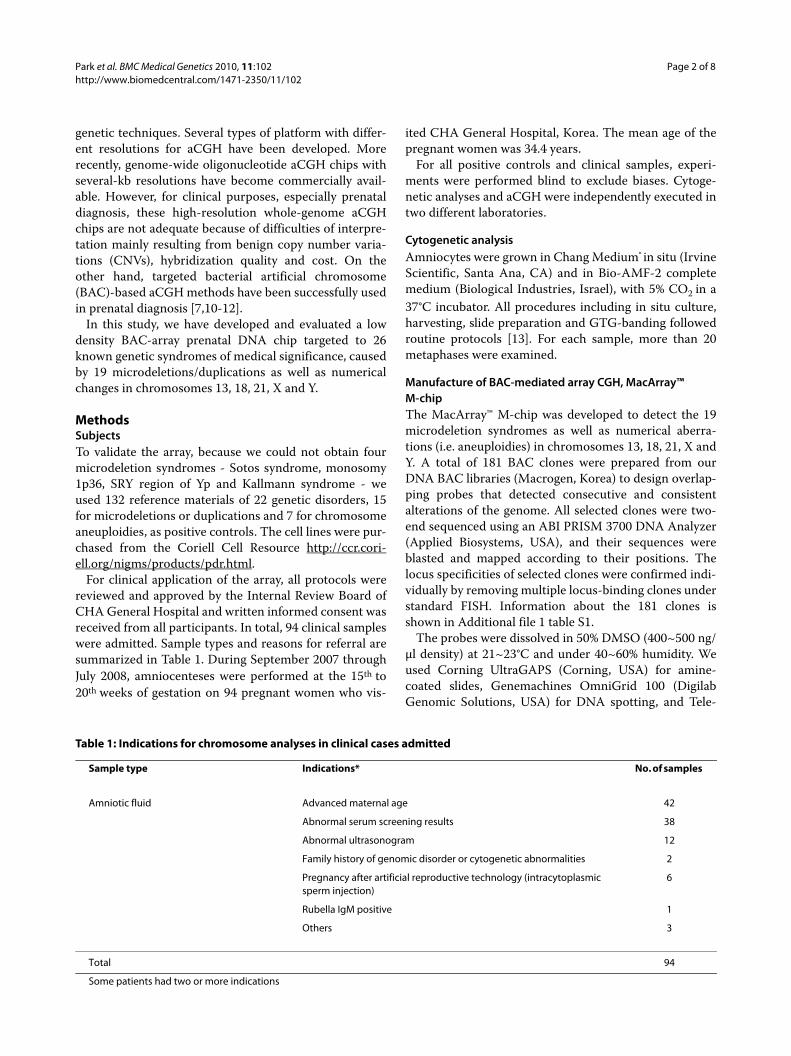
For clinical application of the array, all protocols werereviewed and approved by the Internal Review Board ofCHA General Hospital and written informed consent wasreceived from all participants. In total, 94 clinical sampleswere admitted. Sample types and reasons for referral aresummarized in Table 1. During September 2007 throughJuly 2008, amniocenteses were performed at the 15th to20th weeks of gestation on 94 pregnant women who vis-
ited CHA General Hospital, Korea. The mean age of thepregnant women was 34.4 years.
For all positive controls and clinical samples, experi-ments were performed blind to exclude biases. Cytoge-netic analyses and aCGH were independently executed intwo different laboratories.
Cytogenetic analysisAmniocytes were grown in Chang Medium® in situ (IrvineScientific, Santa Ana, CA) and in Bio-AMF-2 completemedium (Biological Industries, Israel), with 5% CO2 in a37°C incubator. All procedures including in situ culture,harvesting, slide preparation and GTG-banding followedroutine protocols [13]. For each sample, more than 20metaphases were examined.
Manufacture of BAC-mediated array CGH, MacArray™ M-chipThe MacArray™ M-chip was developed to detect the 19microdeletion syndromes as well as numerical aberra-tions (i.e. aneuploidies) in chromosomes 13, 18, 21, X andY. A total of 181 BAC clones were prepared from ourDNA BAC libraries (Macrogen, Korea) to design overlap-ping probes that detected consecutive and consistentalterations of the genome. All selected clones were two-end sequenced using an ABI PRISM 3700 DNA Analyzer(Applied Biosystems, USA), and their sequences wereblasted and mapped according to their positions. Thelocus specificities of selected clones were confirmed indi-vidually by removing multiple locus-binding clones understandard FISH. Information about the 181 clones isshown in Additional file 1 table S1.
The probes were dissolved in 50% DMSO (400~500 ng/μl density) at 21~23°C and under 40~60% humidity. Weused Corning UltraGAPS (Corning, USA) for amine-coated slides, Genemachines OmniGrid 100 (DigilabGenomic Solutions, USA) for DNA spotting, and Tele-
Table 1: Indications for chromosome analyses in clinical cases admitted
Sample type Indications* No. of samples
Amniotic fluid Advanced maternal age 42
Abnormal serum screening results 38
Abnormal ultrasonogram 12
Family history of genomic disorder or cytogenetic abnormalities 2
Pregnancy after artificial reproductive technology (intracytoplasmic sperm injection)
6
Rubella IgM positive 1
Others 3
Total 94
Some patients had two or more indications
Park et al. BMC Medical Genetics 2010, 11:102http://www.biomedcentral.com/1471-2350/11/102
Page 3 of 8
cam SMP4 (Arrayit Corporation, USA) for pin. We fol-lowed the general contact type spotter process. Each BACclone was represented on the array as triplicate spots.
DNA preparation and labeling for MacArray™ M-chipGenomic DNAs were extracted using a Gentra PuregenCell kit (Qiagen Inc., German) from cell lines and clinicalsamples. The DNAs in 85 of the amniotic fluid sampleswere directly extracted, while DNAs in nine other caseswere extracted after culture. DNA concentration andpurity were measured by 1% agarose gel electrophoresisand a NanoDrop ND-100 Full-spectrum UV/Vis Spectro-photometer (Thermo Scientific, USA). Only samples withboth 260/280 and 260/230 ratios >1.7 were used for theexperiment. The labeling and hybridization protocolsdescribed by Pinkel et al. [14] were followed with somemodifications, using a Bioprime labeling kit (InvitrogenCarlsberg, CA, USA). Briefly, 21 μl solution containing500 ng reference DNA or test DNA was mixed with ran-dom primers solution and water in a BioPrime ArrayCGH Labeling System Genomic labeling Module, incu-bated for 5 min at 95°C, and subsequently cooled on ice.After the addition of 5 μl 10 × dNTPs labeling mixture (1mM dCTP, 2 mM dATP, 2 mM dGTP, 2 mM dTTP), 3 μl1 mM Cy-3 or Cy-5 dCTP (GeneChem Inc., Daejeon,Korea), and 40 U Klenow fragment in the BioPrime ArrayCGH Labeling System Genomic labeling Module, themixture was gently mixed and incubated overnight at37°C. Addition of 5 μl Stop Buffer in BioPrime ArrayCGH Labeling System Genomic labeling Module termi-nated the reaction. After labeling, unincorporated fluo-rescent nucleotides were removed using the BioPrimeArray CGH Purification Module. In one tube, Cy3-labeled sample DNAs and Cy5-labeled reference DNAswere mixed and 50 μg human Cot-1 DNA (Invitrogen,Carlsberg, CA, USA), 20 μl 3 M sodium acetate and 600μl cold 100% ethanol were added to precipitate the DNA.
Array hybridization, imaging and data analysisThe pellet was resuspended in 30 μl hybridization solu-tion containing 50% formamide, 10% dextran sulfate, 2×SSC, 4% SDS and 200 μg yeast tRNA. The hybridizationsolution was denatured for 10 min at 72°C and subse-quently incubated for 1 h at 37°C to block repetitivesequences. Hybridization was performed in slide cham-bers for 48 h at 37°C. After post-hybridization washes,the arrays were rinsed, spin-dried and scanned into two16-bit TIFF image files using a GenePix 4200 a two-colorfluorescence scanner (MDS, Toronto, Canada). To deter-mine the intensity of each spot, the scanned images wereanalyzed with MacViewer™ M software (Macrogen, Inc.,Korea). The log2-transformed fluorescence ratios werecalculated from background-subtracted median intensityvalues. For each spot, we used 0.25 and -0.25 as thresh-olds of gain and loss, respectively.
Fluorescent in situ hybridization (FISH)Fluorescent in situ hybridization (FISH) was used to ver-ify the microdeletion of Yq11.23, which indicates non-obstructive spermatogenic failure, identified by the Mac-Array™ M-chip. FISH probes for the region were gener-ated from the human BAC clones RP11-214M24, RP11-140H23, RP11-26B12 and RP11-539D10, purchased fromAbbott Laboratories. Bacterial culture and DNA prepara-tions were performed according to the supplier's proto-col. Purified BAC clone DNA (1 μg) was labeled withspectrum-red dUTP using a Nick Translation LabelingKit (Abbott Laboratories, IL). FISH was performed usinga standard protocol.
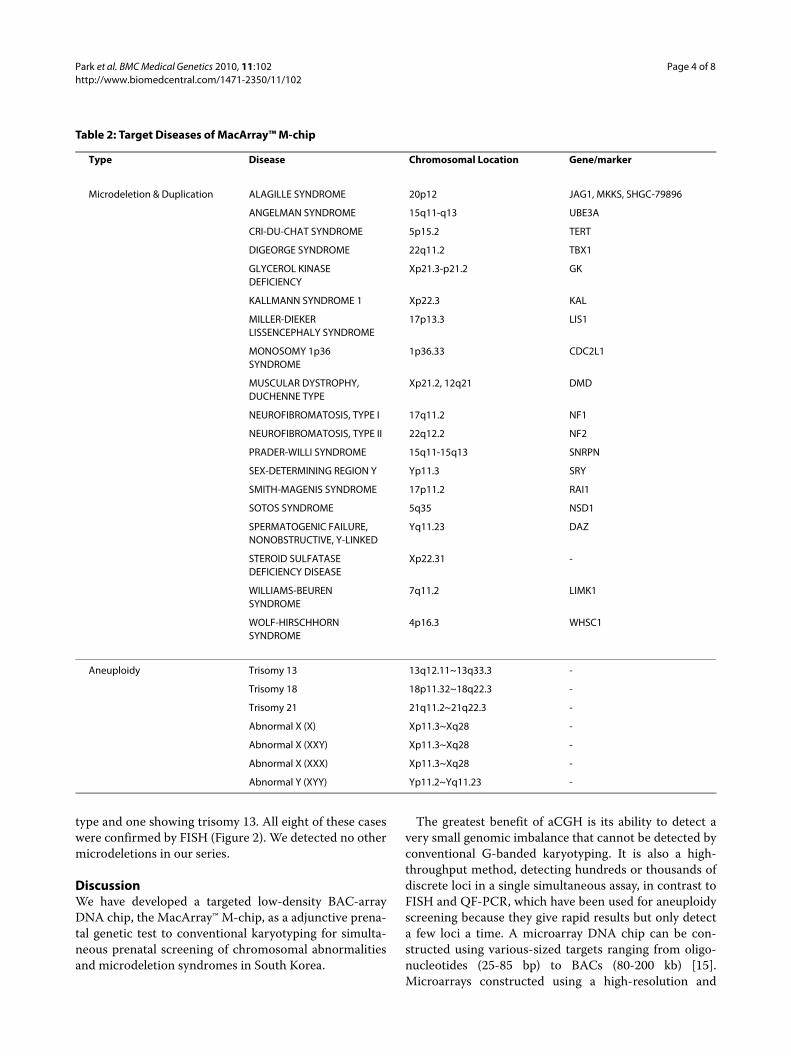
ResultsMacArray™ M-chip developmentWe designed a novel, low-density array CGH chip, theMACROGEN MacArray™ M-chip, to detect not only ane-uploidies but also micro-level chromosomal aberrationssensitively (see Materials and Methods). Table 2 specifiesthe characteristics of the 26 diseases (19 micro-deletionsand 7 aneuploidies) targeted by the MacArray™ M-chip.
Proficiency test using standard reference materialsWe evaluated the feasibility of the MacArray™ M-chipusing commercially available standard reference materi-als. We tested 132 samples bought from Coriell to evalu-ate the detection of the 15 micro-deletions/duplicationsand 7 aneuploidies. We successfully detected all theseaberrations. Four selected cases detected by the MacAr-ray™ M-chip - 15q11-15q13 micro-deletion causingPrader-Willi syndrome, 5p15.2 micro-deletion causingcri-du-chat syndrome, Xp21.2 micro-deletion causingDuchenne muscular dystrophy and 18p11.32~18q22.3aneuploidy causing trisomy18 are shown in figure 1. Thesample numbers for each target are different from eachother, and reference materials for Kallmann syndrome,Sotos syndrome, monosomy 1p36 and the SRY region ofYp were not available. The overall results are presented inAdditional file 2 table S2.
Clinical applications of MacArray™ M-chipBoth cytogenetic analyses and BAC-array CGH were per-formed for all 94 clinical samples. The overall results areshown in Additional file 3 table S3. One case showing the46,XX normal female karyotype failed in the MacArray™M-chip test because of poor hybridization. Genomicaberrations were detected in 20 cases (Table 3). Threebalanced rearrangements were identified in only conven-tional karyotyping. These balanced rearrangements weretwo cases of inv[9], which was considered a normal varia-tion, and one case of translocation, t[8,11].
On the other hand, we identified a microdeletion of theYq11.23 region harboring the DAZ gene by MacArray™M-chip in eight cases, seven showing normal male karyo-
Park et al. BMC Medical Genetics 2010, 11:102http://www.biomedcentral.com/1471-2350/11/102
Page 4 of 8
type and one showing trisomy 13. All eight of these caseswere confirmed by FISH (Figure 2). We detected no othermicrodeletions in our series.
DiscussionWe have developed a targeted low-density BAC-arrayDNA chip, the MacArray™ M-chip, as a adjunctive prena-tal genetic test to conventional karyotyping for simulta-neous prenatal screening of chromosomal abnormalitiesand microdeletion syndromes in South Korea.
The greatest benefit of aCGH is its ability to detect avery small genomic imbalance that cannot be detected byconventional G-banded karyotyping. It is also a high-throughput method, detecting hundreds or thousands ofdiscrete loci in a single simultaneous assay, in contrast toFISH and QF-PCR, which have been used for aneuploidyscreening because they give rapid results but only detecta few loci a time. A microarray DNA chip can be con-structed using various-sized targets ranging from oligo-nucleotides (25-85 bp) to BACs (80-200 kb) [15].Microarrays constructed using a high-resolution and
Table 2: Target Diseases of MacArray™ M-chip
Type Disease Chromosomal Location Gene/marker
Microdeletion & Duplication ALAGILLE SYNDROME 20p12 JAG1, MKKS, SHGC-79896
ANGELMAN SYNDROME 15q11-q13 UBE3A
CRI-DU-CHAT SYNDROME 5p15.2 TERT
DIGEORGE SYNDROME 22q11.2 TBX1
GLYCEROL KINASE DEFICIENCY
Xp21.3-p21.2 GK
KALLMANN SYNDROME 1 Xp22.3 KAL
MILLER-DIEKER LISSENCEPHALY SYNDROME
17p13.3 LIS1
MONOSOMY 1p36 SYNDROME
1p36.33 CDC2L1
MUSCULAR DYSTROPHY, DUCHENNE TYPE
Xp21.2, 12q21 DMD
NEUROFIBROMATOSIS, TYPE I 17q11.2 NF1
NEUROFIBROMATOSIS, TYPE II 22q12.2 NF2
PRADER-WILLI SYNDROME 15q11-15q13 SNRPN
SEX-DETERMINING REGION Y Yp11.3 SRY
SMITH-MAGENIS SYNDROME 17p11.2 RAI1
SOTOS SYNDROME 5q35 NSD1
SPERMATOGENIC FAILURE, NONOBSTRUCTIVE, Y-LINKED
Yq11.23 DAZ
STEROID SULFATASE DEFICIENCY DISEASE
Xp22.31 -
WILLIAMS-BEUREN SYNDROME
7q11.2 LIMK1
WOLF-HIRSCHHORN SYNDROME
4p16.3 WHSC1
Aneuploidy Trisomy 13 13q12.11~13q33.3 -
Trisomy 18 18p11.32~18q22.3 -
Trisomy 21 21q11.2~21q22.3 -
Abnormal X (X) Xp11.3~Xq28 -
Abnormal X (XXY) Xp11.3~Xq28 -
Abnormal X (XXX) Xp11.3~Xq28 -
Abnormal Y (XYY) Yp11.2~Yq11.23 -

Park et al. BMC Medical Genetics 2010, 11:102http://www.biomedcentral.com/1471-2350/11/102
Page 5 of 8
Figure 1 Detection of chromosomal aberrations by MACROGEN MacArray™ M-chip . (a) 15q11-15q13 deletion in DNA samples indicating Prad-er-Willi syndrome was detected by MACROGEN MacArray™ M-chip. The log 2-based test/reference intensity ratios of DNA clones located on chromo-some 15 were below -0.25, the threshold indicating chromosomal deletion. (b) Another 5q15.2 deletion in the DNA from cri-du-chat syndrome was detected. (c) An Xp21.2 deletion indicating Duchenne muscular dystrophy was also detected in the same way. An 12q21 deletion reported previously for the same disease was not detected by our platform. (d) Detection of Edward's syndrome (trisomy 18). The aberration was clearly detected by our array system.

Park et al. BMC Medical Genetics 2010, 11:102http://www.biomedcentral.com/1471-2350/11/102
Page 6 of 8
whole-genome approach can identify very many poly-morphisms with not only pathogenic deletions/duplica-tions but also uncharacterized genomic imbalances.About two thirds of genomic imbalances detected by pre-natal array CGH have been interpreted as probablybenign and of no clinical significance [12].
In contrast to genome-wide aCGH chips, this targetedBAC aCGH chip contains clones only for regions withknown clinical significance. While it had lower overalldetection rates than genome-wide arrays, it greatlyreduced the number of benign CNVs and other anoma-lies of unclear clinical significance that are otherwisedetected [15]. Shaffer et al. [16] reported that 5.6% ofcases showed clinically relevant abnormalities, with only2.4% and 0.9% showing familial variants and unclear sig-nificance, respectively. A targeted BAC array might there-fore be considered more applicable to clinical diagnosis.
Application of arrays to prenatal diagnosis requiresgreat care because even the detection of a familial variantraises maternal anxiety. Therefore, ruling out insignifi-cant CNVs and unclear results is critical. The MacArray™M-chip is specially designed for prenatal screening ofchromosomal abnormalities that should be detected in
the second trimester of pregnancy. Because the array onlycontains 181 clones with characterized clinical relevance,we greatly reduced the uncertainty of results and also thecost burden on the patients. In this study, we readilydetected 15 microdeletions and 7 aneuploidies in 132 ref-erence materials purchased from the Coriell CellResource. However, we identified no CNVs or inconclu-sive alterations in any of those 132 reference materials(Additional file 2 table S2). All chromosomal abnormali-ties found by conventional cytogenetic analysis were alsodetected by the MacArray™ M-chip except for the appar-ently balanced structural abnormalities inv[9] and t[8,11].The MacArray™ M-chip could not identify aneuploidiesof chromosomes 2, 3, 6, 8, 9, 10, 11, 12, 14, 16 and 19,which are rarely detected in amniocentesis. However,Additional clones for those chromosomes will be addedin the next upgraded platform. Our platform also couldnot be used to detect triploidy. The array CGH platformis not appropriate for detecting polyploidy because it isdifficult to separate polyploidy from systematic error (dyebias)
On the other hand, in eight prenatal cases, the MacAr-ray™ M-chip successfully identified the microdeletion on
Table 3: Abnormal cases detected by conventional karyotype analysis and MacArray™ M-chip test.
sample No. Karyotype analysis MacArray™ M-chip Test
S008 46,XY,inv(9) (q12q13) arr(1,4,5,7,13,15,17,18,20-22)x2,(XY)x1
S026 46,XY arr Yq11.223(23611993-25573091)x0
S031 46,XY arr Yq11.223(23611993-25573091)x0
S035 46,XX, inv(9)(q12q13) arr(1,4,5,7,13,15,17,18,20-22,X)x2
S044 46,XY,t[8;11](p21;p15.5) arr(1,4,5,7,13,15,17,18,20-22)x2,(XY)x1
S048 47,XX,+21 arr(21)x3
S049 47,XX,+18 arr(18)x3
S052 47,XY,+21 arr(21)x3
S055 47,XY,+21 arr(21x3)
S061 47,XY,+21 arr(21)x3,Yq11.223(23611993-25573091)x0
S065 47, XX,+21 arr(21x3)
S068 46, XY arr Yq11.223(23611993-25573091)x0
S069 46,XY arr Yq11.223(23611993-25573091)x0
S070 47,XY,+21 arr(21)x3
S073 47,XXY arr(X)x2,(Y)x1
S074 46,XY arr Yq11.223(23611993-25573091)x0
S075 46,XY arr Yq11.223(23611993-25573091)x0
S083 47,XY,+21 arr(21)x3
S086 47,XY,+13 arr(13)x2,Yq11.223(23611993-25573091)x0
S087 47,XXY arr(X)x2,(Y)x1

Park et al. BMC Medical Genetics 2010, 11:102http://www.biomedcentral.com/1471-2350/11/102
Page 7 of 8
the long arm of Y chromosome (Yq11) that is consideredto be associated with non-obstructive spermatogenic fail-ure. Because this is a prenatal diagnostic chip, our pur-pose is not to detect the DAZ microdeletion prenatally.The DAZ1 probe represents Yq and was used to identifyY chromosome aneuploidies such as 47,XXY.
Deletions in the Yq11 region are hard to distinguishfrom normal variations in routine cytogenetic analysisand are usually detected by polymerase chain reaction(PCR) using short tandem repeat (STR) markers. Four ofthe eight microdeletions were inherited from their fathersand the fetuses were fertilized by intracytoplasmic sperminjection (ICSI). The deletions in the fathers were identi-fied by PCR prior to in vitro fertilization procedure (IVF).We did not perform a paternal Y deletion test in the otherfour cases, so we did not know whether the deletion wasinherited. As shown in figure 2, deletions of Yq11 wereconfirmed by FISH in all eight cases. In this clinical appli-cation, we could detect only DAZ microdeletions in addi-tion to anueploidies.
It would be better to extend this study to many moresamples to explore the incidence of microdeletions andduplications for the given indications further. We plan torevise and upgrade the chip so that it will contain moreBAC clones for microdeletion syndromes and subtelo-meric regions for each chromosome. Of course, we willapply the chip to many more samples and extend itsusage, and hence be able to discuss the benefits of arrayCGH screening in comparison to FISH and QF-PCR.
ConclusionsWe have successfully designed and applied a BAC-basedarray CGH platform for prenatal diagnosis. Even thoughthe MacArray™ M-chip contains a very limited number ofclones, this system can provide a highly efficient andaccurate method for the prenatal screening of chromo-some aneuploidies and microdeletions, making it a clini-cally useful addition to conventional G-bandedkaryotyping.
Figure 2 Confirmation of Yq12 deletion by FISH . (a) The MacArray™ M-chip showed deletions in the Yq region. (b). In a patient with Yq deletion, the WCP Y signal (green) was detected but the DAZ locus signal (orange) was absent. (c). Normal male control for Yq12 deletion. The signal for the DAZ locus was detected in both chromosome and nucleus.
Park et al. BMC Medical Genetics 2010, 11:102http://www.biomedcentral.com/1471-2350/11/102
Page 8 of 8
Additional material
Competing interestsGrant sponsor: Korea Health 21 R&D Project, Ministry of Health & Welfare,Republic of Korea; Grant number: A050558
Authors' contributionsJHP participated in the design of the study, wrote and given final approval ofthe version to be submission. JHW integrated and analyzed array CGH data andwrote the manuscript. SHS analyzed and interpreted data and carried out draft-ing of the manuscript. SJY conceived and designed the study. YMC conceivedand designed the study. KSY conceived and designed the study and super-vised the project. DHC conceived and designed the study and carried out criti-cal revision of the manuscript for important intellectual content andsupervised the project.All authors read and approved the manuscript.
AcknowledgementsThe authors thank Mrs. Eun Hee Oh and Mr. Hyun Woong Kang (Macrogen, Inc.) for the design and production of the array CGH and Mrs. Myoung Joo Shin (Macrogen, Inc.) for the array CGH experiments.
Author Details1Department of Obstetrics and Gynecology, CHA Bundang Medical Center, CHA University, Seongnam-si, Korea, 2Department of Bio Chip Service, Macrogen Inc., Seoul, Korea, 3Genetic Laboratory, Fertility Center of CHA Gangnam Medical Center, CHA University, Seoul, Korea, 4Department of of Obstetrics and Gynecology, Seoul National University College of Medicine, Seoul, Korea and 5Department of Obstetrics and Gynecology, CHA Gangnam Medical Center, CHA University, Seoul, Republic of Korea, Seoul, Korea
References1. Kuo WL, Tenjin H, Segraves R, Pinkel D, Golbus MS, Gray J: Detection of
aneuploidy involving chromosomes 13, 18, or 21, by fluorescence in situ hybridization (FISH) to interphase and metaphase amniocytes. Am J Hum Genet 1991, 49:112-119.
2. Tepperberg J, Pettenati MJ, Rao PN, Lese CM, Rita D, Wyandt H, Gersen S, White B, Schoonmaker MM: Prenatal diagnosis using interphase fluorescence in situ hybridization (FISH): 2-year multi-center retrospective study and review of the literature. Prenat Diagn 2001, 21:293-301.
3. Mutter GL, Pomponio RJ: Molecular diagnosis of sex chromosome aneuploidy using quantitative PCR. Nucleic Acids Res 1991, 19:4203-4207.
4. Marongiu A, Ejarque M, Ordoñez E, Plaja A, Massobrio M, Todros T, Fuster C, Campogrande M, Egozcue J, Adinolfi M, Cirigliano V, Voglino G, Canadas MP: Rapid prenatal diagnosis of common chromosome aneuploidies by QF-PCR. Assessment on 18,000 consecutive clinical samples. Mol Hum Reprod 2004, 10:839-846.
5. Gerdes T, Kirchhoff M, Lind AM, Larsen GV, Schwartz M, Lundsteen C: Computer-assisted prenatal aneuploidy screening for chromosome 13, 18, 21, X and Y based on multiplex ligation-dependent probe amplification (MLPA). Eur J Hum Genet 2005, 13:171-175.
6. Van Opstal D, Boter M, de Jong D, van den Berg C, Brüggenwirth HT, Wildschut HI, de Klein A, Galjaard RJ: Rapid aneuploidy detection with multiplex ligation-dependent probe amplification: a prospective study of 4000 amniotic fluid samples. Eur J Hum Genet 2009, 17:112-121.
7. Sahoo T, Cheung SW, Ward P, Darilek S, Patel A, del Gaudio D, Kang SH, Lalani SR, Li J, McAdoo S, Burke A, Shaw CA, Stankiewicz P, Chinault AC, Van den Veyver IB, Roa BB, Beaud AL, Eng CM, et al.: Prenatal diagnosis of
chromosomal abnormalities using array-based comparative genomic hybridization. Genet Med 2006, 8:719-727.
8. Menten B, Maas N, Thienpont B, Buysse K, Vandesompele J, Melotte C, de Ravel T, Van Vooren S, Balikova I, Backx L, Janssens S, De Paepe A, De Moor B, Moreau Y, Marynen P, Fryns JP, Mortier G, Devriendt K, Speleman F, Vermeesch JR: Emerging patterns of cryptic chromosomal imbalance in patients with idiopathic mental retardation and multiple congenital anomalies: a new series of 140 patients and review of published reports. J Med Genet 2006, 43:625-633.
9. Shaffer LG, Bejjani BA, Torchia B, Kirkpatrick S, Coppinger J, Ballif BC: The identification of microdeletion syndromes and other chromosome abnormalities: cytogenetic methods of the past, new technologies for the future. Am J Med Genet C Semin Med Genet 2007, 145C:335-345.
10. Rickman L, Fiegler H, Shaw-Smith C, Nash R, Cirigliano V, Voglino G, Ng BL, Scott C, Whittaker J, Adinolfi M, Carter NP, Bobrow M: Prenatal detection of unbalanced chromosomal rearrangements by array CGH. J Med Genet 2006, 43:353-361.
11. Shaffer LG, Coppinger J, Alliman S, Torchia BA, Theisen A, Ballif BC, Bejjani BA: Comparison of microarray-based detection rates for cytogenetic abnormalities in prenatal and neonatal specimens. Prenat Diagn 2008, 28:789-795.
12. Van den Veyver IB, Patel A, Shaw CA, Pursley AN, Kang SH, Simovich MJ, Ward PA, Darilek S, Johnson A, Neill SE, Bi W, White LD, Eng CM, Lupski JR, Cheung SW, Beaud AL, et al.: Clinical use of array comparative genomic hybridization (aCGH) for prenatal diagnosis in 300 cases. Prenat Diagn 2009, 29:29-39.
13. Barch MJ, Knutsen T, Spurbeck JL: The AGT cytogenetics laboratory manual Philadelphia: Lippincott-Raven Publishers; 1997.
14. Pinkel D, Segraves R, Sudar D, Clark S, Poole I, Kowbel D, Collins C, Kuo WL, Chen C, Zhai Y, Dairkee SH, Ljung BM, Gray JW, Albertson DG: High resolution analysis of DNA copy number variation using comparative genomic hybridization to microarrays. Nat Genet 1998, 20:207-211.
15. Shaffer LG, Bui TH: Molecular cytogenetic and rapid aneuploidy detection methods in prenatal diagnosis. Am J Med Genet C Semin Med Genet 2007, 145C:87-98.
16. Shaffer LG, Kashork CD, Saleki R, Rorem E, Sundin K, Ballif BC, Bejjani BA, Shaffer LG, Kashork CD, Saleki R: Targeted genomic microarray analysis for identification of chromosome abnormalities in 1500 consecutive clinical cases. J Pediatr 2006, 149:98-102.
Pre-publication historyThe pre-publication history for this paper can be accessed here:http://www.biomedcentral.com/1471-2350/11/102/prepub
doi: 10.1186/1471-2350-11-102Cite this article as: Park et al., Application of a target array Comparative Genomic Hybridization to prenatal diagnosis BMC Medical Genetics 2010, 11:102
Additional file 1 Table S1. Contents of MACROGEN MacArray™ M-chip Additional file 2 Table S2. Performance of MacArray™ M-chip in detecting 26 diseases .Additional file 3 Table S3. Overall results of clinical applications using MacArray™ M-chip and karyotype analysis to detect chromosomal abnormalities .
Received: 14 September 2009 Accepted: 24 June 2010 Published: 24 June 2010This article is available from: http://www.biomedcentral.com/1471-2350/11/102© 2010 Park et al; licensee BioMed Central Ltd. This is an Open Access article distributed under the terms of the Creative Commons Attribution License (http://creativecommons.org/licenses/by/2.0), which permits unrestricted use, distribution, and reproduction in any medium, provided the original work is properly cited.BMC Medical Genetics 2010, 11:102