research article altered peptidase activities in thyroid...
TRANSCRIPT

Hindawi Publishing CorporationDisease MarkersVolume 35 (2013), Issue 6, Pages 825–832http://dx.doi.org/10.1155/2013/970736
Research ArticleAltered Peptidase Activities inThyroid Neoplasia and Hyperplasia
Gorka Larrinaga,1,2,3 Lorena Blanco,2,3 Peio Errarte,2,3 Maider Beitia,2,3
Begoña Sanz,2,3 Itxaro Perez,1,2,3 Amaia Irazusta,1,3 Clara E. Sánchez,1
Francisco Santaolalla,4 Leire Andrés,3,5 and José I. López3,5
1 Department of Nursing I, University School of Nursing, University of the Basque Country, P.O. Box 699, 48080 Bilbao, Bizkaia, Spain2Department of Physiology, Faculty of Medicine and Dentistry, University of the Basque Country, Bilbao, Spain3 BioCruces Research Institute, Spain4Department of Otolaryngology, Basurto University Hospital, Bilbao, Spain5 Department of Anatomic Pathology, Cruces University Hospital, Barakaldo, Bizkaia, Spain
Correspondence should be addressed to Gorka Larrinaga; [email protected]
Received 1 July 2013; Revised 29 September 2013; Accepted 1 October 2013
Academic Editor: Dinesh Kumbhare
Copyright © 2013 Gorka Larrinaga et al. This is an open access article distributed under the Creative Commons AttributionLicense, which permits unrestricted use, distribution, and reproduction in any medium, provided the original work is properlycited.
Background. Papillary thyroid carcinoma (PTC), follicular thyroid adenoma (FTA), and thyroid nodular hyperplasia (TNH) are themost frequent diseases of the thyroid gland. Previous studies described the involvement of dipeptidyl-peptidase IV (DPPIV/CD26)in the development of thyroid neoplasia and proposed it as an additional tool in the diagnosis/prognosis of these diseases. However,very little is known about the involvement of other peptidases in neoplastic and hyperplastic processes of this gland. Methods.The catalytic activity of 10 peptidases in a series of 30 PTC, 10 FTA, and 14 TNH was measured fluorimetrically in tumour andnontumour adjacent tissues. Results.The activity of DPPIV/CD26 was markedly higher in PTC than in FTA, TNH, and nontumourtissues. Aspartyl aminopeptidase (AspAP), alanyl aminopeptidase (AlaAP), prolyl endopeptidase, pyroglutamyl peptidase I, andaminopeptidase B activities were significantly increased in thyroid neoplasms when compared to nontumour tissues. AspAP andAlaAP activities were also significantly higher in PTC than in FTA and TNH. Conclusions.These data suggest the involvement ofDPPIV/CD26 and some cytosolic peptidases in the neoplastic development of PTC and FTA. Further studies will help to definethe possible clinical usefulness of AlaAP and AspAP in the diagnosis/prognosis of thyroid neoplasms.
1. Introduction
Solitary and multiple nodules of the thyroid gland are verycommon in clinical practice, with most of them beingbenign lesions, either thyroid nodular hyperplasia (TNH)or follicular thyroid adenoma (FTA). Roughly only 5%of thyroid nodules are malignant [1] and 70% of themare papillary thyroid carcinomas (PTC) [2]. The arrival ofultrasound examination to routine practice has discovereda large amount of nonpalpable and clinically silent thyroidnodules. TNH, FTA, and PTC can be distinguished fromeach other following well-established histopathologic criteria[3]. Interestingly, these lesions are able to be sampled with
recognized diagnostic success by sonographically guided fineneedle aspiration cytology [1] or by core biopsy [4].
TNH consists of multiple nodules of variable size com-posed of odd-shaped thyroid follicles with homogeneousbland cytology and partial encapsulation that may reverseand dissapear when the hormonal stimulus ceases. Thyroidhyperfunction is common.
FTA usually appears as asymptomatic solitary nodules.This lesion is composed of small size follicles lying in afibrous, sometimes edematous stroma and shows a welldefined fibrous capsule. There is no evidence of vascular orcapsular invasion in these neoplasms; otherwise, the lesion isconsidered carcinoma [3].

826 Disease Markers
PTC is the most common type of endocrine cancer andshows a varied clinical presentation. Most patients presentwith a thyroid nodule when they consult for the first time,but some others do not, and lymph node metastases in theneck are the solemanifestation of a tumor that remains occultin the thyroid. Histologically, PTC may display very differentgrowth patterns [5, 6], and its diagnosis relies on the iden-tification of characteristic nuclear features, such as nuclearenlargement, nuclear membrane irregularities, peripheralchromatin margination, and prominent micronucleoli [3].
Immunohistochemical markers like HBME-1, cytoker-atin 19, and galectin-3 may be of help in difficult cases [6].
Bioactive peptides are regulated through specific pep-tidases that hydrolyze them. These peptide-convertingenzymes are distributed in the main human tissues and havebeen originally considered to be only involved in protein andpeptide scavenging [7]. However, many studies have shownthat they are involved in several physiological functionsand play a key role in growth control, differentiation, andsignal transduction of many cell systems by modulating theactivity of bioactive peptides, degrading extracellular matrix,acting as adhesion molecules and directly participating inthe intracellular signaling [7–10]. So, altered expressionand catalytic function patterns of these enzymes maycontribute to several disease processes, including neoplastictransformation and tumour progression [8–10].
Increased understanding of the underlying pathophysiol-ogy of thyroid tumours has led to implicate several proteasesin its genesis, growth, and dissemination [11–16]. Metallo-proteases such as MMP-2 and 9 and peptidases such asdipeptidyl-peptidase IV/CD26 (DPPIV/CD26) change theirexpression and activity in these neoplasms and have beenrecognized as molecular markers in the diagnosis of thyroidtumours [11–18]. However, very little is known about theactivity pattern of other peptidases in thyroid neoplastic andhyperplastic diseases.
This study intends to analyse the activity of differentpeptidases in PTC, FTA, and TNH and in the adjacent non-tumour thyroid tissue, as we have performed before in otherneoplasms, such as renal, colorectal and head and necktumours [19–23]. For such a purpose, we have analyzedin these lesions the activity of 10 acid, basic, neutral, andomega peptidases covering the whole spectrum of peptide-converting activity. On the one hand, we selected five cell-surface peptidases—DPPIV/CD26, APN/CD13, NEP/CD10,APA and CAP—which have been described as tumour mark-ers in haematologic, kidney, skin, prostate, and gynaecologiccancers, among others [8–10, 24]. On the other, five cytosolicenzymes were selected—PEP, alaAP, AspAP, APB and PGI—for better know if these enzymes may also be involved inneoplastic development.”
2. Materials and Methods
Theauthors declare that all the experiments carried out in thisstudy complywith current Spanish and EuropeanUnion legalregulations.
2.1. Tissue Specimens. Representative tissue from 30 PTC,14 TNH and 10 FTA and their corresponding non tumourareas were adequately sampled in fresh at the Pathology Labwithin the first 30 minutes after surgical removal, immedi-ately frozen with isopentane, and stored at −80∘C until theenzyme assays were performed. Personal data of the patientswere appropriately encrypted to preserve the anonymoususe of the selected material accomplishing with internationalrecommendations.
2.2. Sample Preparation. Soluble andmembrane-bound frac-tions were obtained following a modified version of Zam-botti-Villela et al. [25]. Explanted tissue samples were homog-enized in 10mM Tris-HCl buffer at pH 7.4, for 30 secondsat 800 rpm using a Heidolph PZR 50 Selecta homogenizer,and ultracentrifuged in a Centrikon T-2070 Kontron Instru-ments apparatus at 100,000×g for 35min. The resultingsupernatants were used to measure soluble enzyme activities:APB, AspAP, PG I, PEP and AlaAP. To avoid contaminationwith soluble enzymes, the resulting pellets were washed threetimes by suspension in 10mM Tris-HCl buffer at pH 7.4 Thepellets were then homogenized in 10mM Tris-HCl bufferat pH 7.4, and centrifuged at low speed (800×g) for 3minto purify the samples. The supernatants thus obtained wereused to determinemembrane-bound enzyme activities: APA,APN/CD13, CAP, DPPIV/CD26, and NEP/CD10. All stepswere carried out at 4∘C.
2.3. Enzyme Assays. Peptidase activities were measured byincubating samples with saturating concentrations of flu-orogen-derived substrates following themethod described byYoshimoto et al. [26],Mantle et al. [27], and Silveira et al. [28].Selected peptidases, their substrates, and the reaction con-ditions for each enzymatic assay are summarized in Table 1.
Alanine aminopeptidase activities (AlaAP and APN/CD13) were measured in triplicate using Ala-𝛽-naph-thylamide as a substrate. Incubations with the specificAlaAP inhibitor puromycin (40 𝜇M) were performed inparallel to discriminate between the AlaAP and APN/CD13 forms of total alanine aminopeptidase activity. DPPIV/CD26 and PEP activities were assayed using H-Gly-Pro-𝛽-naphthylamide, and Z-Gly-Pro-𝛽-naphthylamide respec-tively. APB (basic) and acid aminopeptidase activities(AspAP, APA) were quantified with Arg-𝛽-naphthylamide,Asp-𝛽-naphthylamide, andGlu-𝛽-naphthylamide substrates,respectively. Omega peptidase (PGI) activity was mea-sured fluorometrically using pGlu-𝛽-naphthylamide as sub-strate. CAP activity was analyzed using L-cystine-di-𝛽-naphthylamide. NEP/CD10 assay was performed by incu-bating samples with a saturating concentration of N-dansyl-D-Ala-Gly-pNO
2-Phe-Gly (DAGNPG, a dansyl derivative).
These assays are based on the fluorescence of productsgenerated from substrate hydrolysis by the enzyme.
Reactions were started by adding 30–50𝜇L of sample to1mL of the appropriate incubationmixture depending on theenzyme and substrate analyzed as follows:AlaAP,APN/CD13,APB, PEP, and PGI activities (50mM phosphate buffer at pH7.4 for AlaAP, APN/CD13, and PGI activities, pH 6.5 for APB,

Disease Markers 827
Table 1: Substrates and the reaction conditions for each enzymatic assay.
Substrates (mM) pHa DTT(mM)
EDTA(mM)
CaCl2(mM)
NaCl(mM)
MnCl2(mM)
PEP (EC 3.4.21.26) Z-Gly-Pro-𝛽-NA 0,125 7,4 2 — — — —PGI (EC 3.4.19.3) pGlu-𝛽-NA 0,125 7,4 2 2 — — —APBb (EC 3.4.11.6) Arg-𝛽-NA 0,125 6,5 — — — 150 —APN/CD13b (EC 3.4.11.2) Ala-𝛽-NA 0,125 7,4 1,2 — — — —AlaAP (EC 3.4.11.14) Ala-𝛽-NA 0,125 7,4 1,2 — — — —APA (EC 3.4.11.7) H-Glu-𝛽-NA 0,125 7,4 0,65 — 0,5 — —AspAP (EC 3.4.11.21) Asp-𝛽-NA 0,125 7,4 — — — — 1DPPIV/CD26 (EC 3.4.14.5) H-Gly-Pro-𝛽-NA 0,2 8,3 — — — — —CAP (EC 3.4.11.3) L-Cys-di-𝛽-NA 0,125 5,9 — — — — —NEPc (EC 3.4.24.11) N-Dansyl-D-Ala-Gly-pNO2-Phe-Gly 0,125 7,4 — — — — —aReaction buffers: Phosphate buffer (50mM) was used for PEP, PGI, APB, APN/CD13, and AlaAP. Tris-HCl (50mM) was used for APA, AspAP, DPPIV/CD26,CAP, and NEP. b40𝜇M of the AlaAP inhibitor puromycin was added; c4.6 nM of the angiotensin-converting enzyme inhibitor captopril was added.
and 0.125mM aminoacyl-𝛽-naphthylamide); AspAP, APA,DPPIV/CD26, CAP, and NEP/CD10 activities (50mM Tris-HCl buffer at pH 7.4 for AspAP, NEP and APA, pH 8.3 forDPPIV/CD26, pH 5.9 for CAP, and 0.125mM aminoacyl-𝛽-naphthylamide or [D]AG (pN) PG).
After 30 min incubation at 37∘C, 1mL of 0.1M sodiumacetate buffer (pH 4.2) was added to the mixture to ter-minate the reaction. The released product was determinedby measuring the fluorescent intensity (at 412 nm withexcitation at 345 nm for 𝛽-naphthylamine and at 562 nmwith excitation at 342 nm for [D]AG) with a ShimadzuRF-540 Spectrofluorophotometer. Blanks consisted of buffer(instead the sample) and the same reactives and were usedto determine background fluorescence. Relative fluorescencewas converted into picomoles of product using a standardcurve constructed with increasing concentrations of 𝛽-naphthylamine or [D]AG.
In the case of DPPIV and PEP, to verify that the formationof𝛽-naphthylamine was specifically due to the action of thesepeptidases and not due to other enzymes which could cleavethe same substrates (H-Gly-Pro-𝛽-NA by DPP8 and DPP9and Z-Gly-Pro-𝛽-NA by seprase), we performed inhibitionassays with specific inhibitors (diprotin A for DPPIV andKYP-2047 for PEP). 87% of the DPPIV activity was inhibitedin PTC, 94% in TNH, and 100% in FTA and uninvolvedthyroid tissue. In the case of PEP, over 95% of the activitymeasured was sensitive to the inhibitor in both tumourand nontumour tissues. Therefore, we concluded that thereleasing of 𝛽-naphthylamine in both cases is mainly due toDPPIV and PEP.
2.4. Protein Determination. Protein concentration of solubleand membrane-bound fractions of thyroid tissues was mea-sured in triplicate by the Bradford method [29], using BSA(1mg/mL) as calibrator. Resultswere recorded as units of pep-tidase (UP) per milligram of protein (1 unit of peptidase (UP)is 1 pmol of naphthylamine released perminute). Fluorogenicassays were linear with respect to hydrolysis time and protein
content. Activity is expressed as pmol of product/min/mgprotein (UP/mg protein) and reported as mean ± SE.
2.5. Statistical Analysis. Shapiro-Wilk test was applied todata obtained from tumor samples to know if the numbersfollowed a normal distribution or not. Based on this informa-tion, nonparametric Mann-Whitney and Kruskal-Wallis testswere used to detect differences between two groups andmorethan two groups, respectively. SPSS 19.0 software was used forthe statistical analysis.
3. Results
Females predominated in the series: the PTC group included6 males and 24 females (average age: 53,7 years) and the FTA3 males and 7 females (35.4 years). The fourteen cases of theTNH group were all females (43.2 years).
Histopathological subtyping of the PTC group includedconventional PTC (16 cases, 53,3%), follicular variant PTC(10 cases, 33,3%), and papillary microcarcinomas (4 cases,13,3%). Organ confined disease (pT1/2) was detected in80% of the cases. Six cases presented minimal extracapsularextension (pT3) and two of them regional lymph nodemetastasis.
Table 2 shows all the peptidase activities measured inthe soluble and membrane-bound fractions of hyperplastic,neoplastic, and nontumour adjacent tissues.
The cytosolic fraction activity of AspAP, PG I, PEP,and AlaAP was significantly increased in PTC (Table 2(a))and FTA (Table 2(b)) when compared with the adjacentnontumour tissues. APB activity was also increased in bothneoplasms, although this change did not reach statisticalsignificance in PTC.There were no significant changes in theactivity of these peptidases in the TNH tissuewhen comparedwith the nonhyperplastic tissue (Table 2(c)).
The membrane-bound fraction activity of DPPIV/CD26was markedly higher in PTC (Table 2(a)) and lower in FTA

828 Disease Markers
Table 2: (a) Peptidase activities in papillary thyroid carcinoma(PTC) and in non neoplastic adjacent thyroid tissue. (b) Peptidaseactivities in follicular thyroid adenoma (FTA) and in non neoplasticadjacent thyroid tissue. (c) Peptidase activities in thyroid nodularhyperplasia (TNH) and in non neoplastic adjacent thyroid tissue.Values aremeans± SE of peptidase activity recorded as pmol of unitsof peptidase (UP) per milligram of protein. A value of 𝑃 ≤ 0.05 wasconsidered statistically significant (Significant results are in bold).
(a)
Peptidase PTC Non neoplastic PSoluble fraction
APB 7419 ± 1363 4259 ± 867 0.094AspAP 130 ± 15 87 ± 13 0.039PG I 206 ± 39 77 ± 18 0.008PEP 3836 ± 633 1977 ± 449 0.026AlaAP 8819 ± 1194 4122 ± 1111 0.007
Membrane-bound fractionAPA 1250 ± 280 1120 ± 258 0.96APN/CD13 2301 ± 538 2533 ± 516 0.554CAP 875 ± 253 665 ± 180 0.425DPPIV/CD26 4274 ± 1301 1347 ± 278 0.017NEP/CD10 3.1 ± 0.66 1.9 ± 0.45 0.12
(b)
Peptidase FTA Non neoplastic PSoluble fraction
APB 3772 ± 318 1969 ± 210 0.001AspAP 84 ± 7.2 54 ± 21 0.003PG I 245 ± 20 85 ± 12 0.001PEP 3056 ± 359 1482 ± 184 0.002AlaAP 2022 ± 206 792 ± 76 0.001
Membrane-bound fractionAPA 1513 ± 191 1270 ± 141 0.247APN/CD13 1670 ± 198 2542 ± 274 0.105CAP 453 ± 48 407 ± 47 0.354DPPIV/CD26 589 ± 87 1700 ± 546 0.037NEP/CD10 3.7 ± 0.63 2.8 ± 0.45 0.643
(c)
Peptidase TNH Nonhyperplastic PSoluble fraction
APB 3713 ± 688 5477 ± 721 0.054AspAP 67 ± 4.3 66 ± 14.4 0.519PG I 73 ± 7.8 81 ± 5.3 0.687PEP 2934 ± 448 3580 ± 478 0.295AlaAP 1834 ± 258 2087 ± 310 0.227
Membrane-bound fractionAPA 789 ± 140 475 ± 87 0.116APN/CD13 1775 ± 329 1856 ± 243 0.601CAP 317 ± 73 394 ± 107 0.687DPPIV/CD26 319 ± 44 804 ± 198 0.116NEP/CD10 1.34 ± 0.25 1.74 ± 0.29 0.414
(Table 2(c)), when compared with the reference of nontu-mour tissue samples. By contrast, no significant changes werefound in the rest of membrane-bound activities tested. TNHtissue did not yield significant changes.
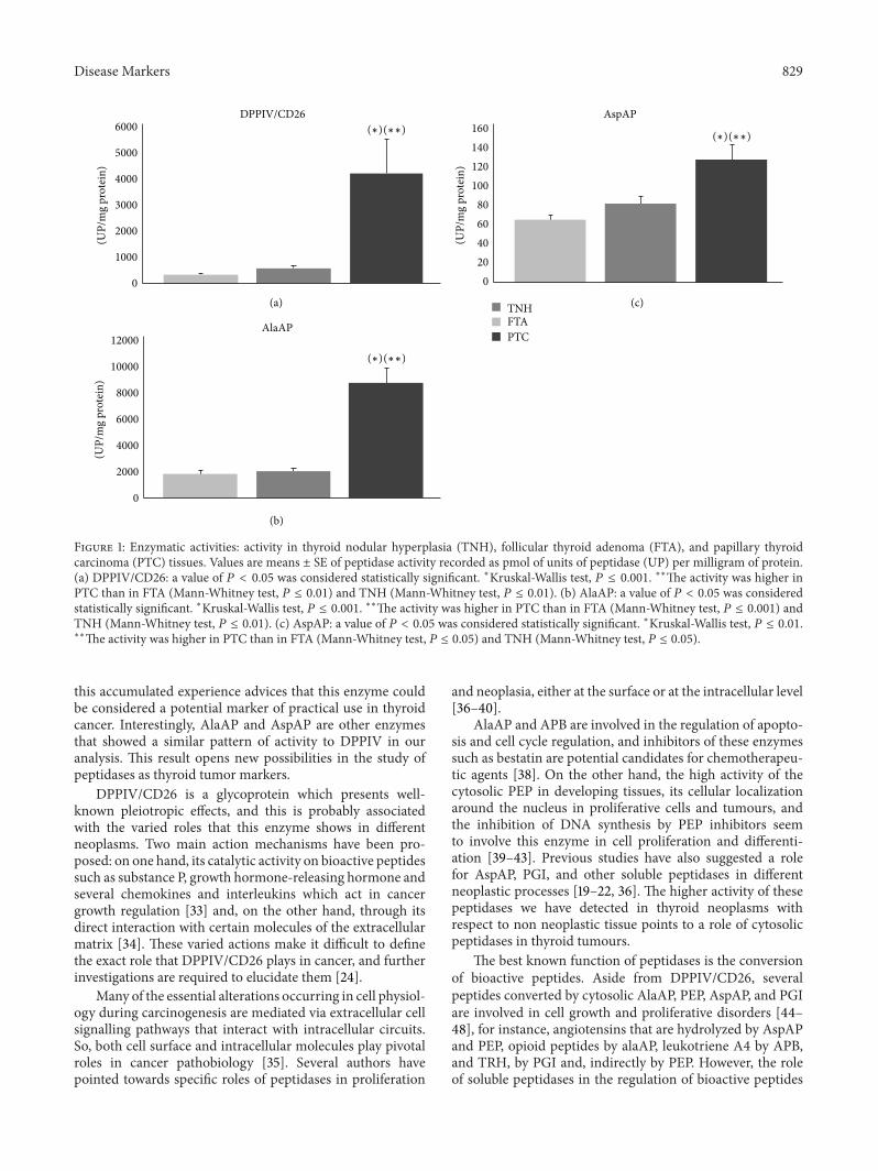
Finally, when the three thyroid lesions were comparedto each other, DPPIV/CD26, AlaAP, and AspAP showedsignificantly higher activities in PTC than in FTA and TNH(Figure 1). There were no significant results in the remaininganalyzed peptidases.
4. Discussion
It has been reported that expression and activity patternsof proteases, such as MMPs and peptidases, are altered inmalignant tumours, suggesting their implication in tumourcell growth, local invasion, and metastases [8–10]. It hasbeen demonstrated that MMP-2 and 9 are upregulated inthe tumour tissue when compared with nontumour tissuein thyroid neoplasms, suggesting that these enzymes couldhave a role in thyroid neoplastic transformation [14, 15].These MMPs and peptidases such as DPPIV/CD26 are dis-tinctly expressed in several thyroid neoplasms and have beenproposed as useful diagnostic/prognostic thyroid tumourmarkers [14–18].
In the same way, we observed that several peptidaseactivities are selectively modified in PTC and FTA whencompared with nontumour adjacent tissues. Thus, cytoso-lic peptidases showed higher activities in both neoplasms,whereas DPPIV/CD26 activity was markedly higher in PTCand lower in FTA. Since TNH did not show any changein peptidase activity, these results indicate a role for thesepeptidases in the development of thyroid neoplasia.
Interestingly, peptidase activity is distinctly modified inthyroid lesions. Some authors stress that peptidases mayeither promote or impede tumour development dependingon the specific type of tumour or on the phase of devel-opment where the tumour is, with this way suggesting thattumour growth regulation by peptides and their convertingpeptidases occur in a tumour specific manner [8–10]. Wehave shown in previous studies selective changes in peptidaseactivities of different neoplasms [19–23]. For example, whenthose were compared with uninvolved tissues, APN/CD13and AspAP activities were decreased in renal cell carcinomas[19] and increased in head and neck squamous cell carcinoma[20]. Evenmore, higher APN/CD13 activity is correlated withhigh grade renal cell carcinomas [19] and with worse survivalof these patients [22]. An exception to the observed tendencycould be PEP, whose activity is increased in most of theneoplastic tissues studied to date [23, 30–32].
Recent data show that DPPIV/CD26 is a useful markerof malignancy in thyroid pathology [16]. Previous studieshad already demonstrated that this serine ectopeptidase wasspecifically expressed in follicular-cell derived thyroid carci-nomas [17, 18]. Some of these studies have been performedby semiquantitative cytoenzymatic methods and showed thehighest activities in PTC [16–18]. Our quantitative fluorimet-ric assay yields similar results, showing that DPPIV/CD26activity is markedly higher in PTC than in FTA and TNH. All
Disease Markers 829
12000
10000
8000
6000
4000
2000
0
AlaAP
(UP/
mg
prot
ein)
(∗)(∗∗)
6000
5000
4000
3000
2000
1000
0
DPPIV/CD26
(UP/
mg
prot
ein)
(∗)(∗∗)
(a)
(b)
160
140
120
100
80
60
40
20
0
AspAP
TNHFTAPTC
(∗)(∗∗)
(UP/
mg
prot
ein)
(c)
Figure 1: Enzymatic activities: activity in thyroid nodular hyperplasia (TNH), follicular thyroid adenoma (FTA), and papillary thyroidcarcinoma (PTC) tissues. Values are means ± SE of peptidase activity recorded as pmol of units of peptidase (UP) per milligram of protein.(a) DPPIV/CD26: a value of 𝑃 < 0.05 was considered statistically significant. ∗Kruskal-Wallis test, 𝑃 ≤ 0.001. ∗∗The activity was higher inPTC than in FTA (Mann-Whitney test, 𝑃 ≤ 0.01) and TNH (Mann-Whitney test, 𝑃 ≤ 0.01). (b) AlaAP: a value of 𝑃 < 0.05 was consideredstatistically significant. ∗Kruskal-Wallis test, 𝑃 ≤ 0.001. ∗∗The activity was higher in PTC than in FTA (Mann-Whitney test, 𝑃 ≤ 0.001) andTNH (Mann-Whitney test, 𝑃 ≤ 0.01). (c) AspAP: a value of 𝑃 < 0.05 was considered statistically significant. ∗Kruskal-Wallis test, 𝑃 ≤ 0.01.∗∗The activity was higher in PTC than in FTA (Mann-Whitney test, 𝑃 ≤ 0.05) and TNH (Mann-Whitney test, 𝑃 ≤ 0.05).
this accumulated experience advices that this enzyme couldbe considered a potential marker of practical use in thyroidcancer. Interestingly, AlaAP and AspAP are other enzymesthat showed a similar pattern of activity to DPPIV in ouranalysis. This result opens new possibilities in the study ofpeptidases as thyroid tumor markers.
DPPIV/CD26 is a glycoprotein which presents well-known pleiotropic effects, and this is probably associatedwith the varied roles that this enzyme shows in differentneoplasms. Two main action mechanisms have been pro-posed: on one hand, its catalytic activity on bioactive peptidessuch as substance P, growth hormone-releasing hormone andseveral chemokines and interleukins which act in cancergrowth regulation [33] and, on the other hand, through itsdirect interaction with certain molecules of the extracellularmatrix [34]. These varied actions make it difficult to definethe exact role that DPPIV/CD26 plays in cancer, and furtherinvestigations are required to elucidate them [24].
Many of the essential alterations occurring in cell physiol-ogy during carcinogenesis are mediated via extracellular cellsignalling pathways that interact with intracellular circuits.So, both cell surface and intracellular molecules play pivotalroles in cancer pathobiology [35]. Several authors havepointed towards specific roles of peptidases in proliferation
and neoplasia, either at the surface or at the intracellular level[36–40].
AlaAP and APB are involved in the regulation of apopto-sis and cell cycle regulation, and inhibitors of these enzymessuch as bestatin are potential candidates for chemotherapeu-tic agents [38]. On the other hand, the high activity of thecytosolic PEP in developing tissues, its cellular localizationaround the nucleus in proliferative cells and tumours, andthe inhibition of DNA synthesis by PEP inhibitors seemto involve this enzyme in cell proliferation and differenti-ation [39–43]. Previous studies have also suggested a rolefor AspAP, PGI, and other soluble peptidases in differentneoplastic processes [19–22, 36]. The higher activity of thesepeptidases we have detected in thyroid neoplasms withrespect to non neoplastic tissue points to a role of cytosolicpeptidases in thyroid tumours.
The best known function of peptidases is the conversionof bioactive peptides. Aside from DPPIV/CD26, severalpeptides converted by cytosolic AlaAP, PEP, AspAP, and PGIare involved in cell growth and proliferative disorders [44–48], for instance, angiotensins that are hydrolyzed by AspAPand PEP, opioid peptides by alaAP, leukotriene A4 by APB,and TRH, by PGI and, indirectly by PEP. However, the roleof soluble peptidases in the regulation of bioactive peptides

830 Disease Markers
related to proliferative disorders is not so clear as it is inthe case of cell surface peptidases. Although the secretion ofPEP, APB, and other soluble peptidases into the extracellularspace has been suggested as a possible peptide regulatorymechanism [49–51], there is accumulating evidence in favorof intracellular trafficking and action of certain peptidesand proteolytic enzymes known as intracrine action [52–54].Thus, it has been suggested that the aforementioned bioactivepeptides may also act as intracrine factors in the intracellularspace inducing cell proliferation and angiogenesis in severaltissues [52–54]. Therefore, the idea of increasing cytosolicpeptidase activities as a result of intracrine peptide dysreg-ulation in thyroid neoplasms should not be ruled out.
In summary, this study analyses the activity of severalpeptidases in PTC, FTA, and TNH and suggests a potentialrole of a cell surface peptidase (DPPIV/CD26) and severalcytosolic peptidases (PEP, APB, AspAP, AlaAP, and PGI) inthyroid neoplasms. However, further studies are needed todefine the exact involvement of peptidases in the pathobiol-ogy of these tumours and their possible clinical usefulness.
Abbreviations
APA: Aminopeptidase AAPN/CD13: Aminopeptidase N/CD13AspAP: Aspartyl aminopeptidaseAPB: Aminopeptidase BDPPIV/CD26: Dipeptidyl peptidase IV/CD26NEP/CD10: Neutral endopeptidase/CD10PEP: Prolyl endopeptidasePGI: Pyroglutamyl peptidase IAlaAP: Puromycin-sensitive alanyl aminopeptidaseCAP: Cystinyl aminopeptidaseMMP: Matrix metalloproteinasePTC: Papillary thyroid carcinomaFTA: Follicular thyroid adenomaTNH: Thyroid nodular hyperplasia.
Conflict of Interests
The authors declare that there is no conflict of interests.
Ethical Approval
The ethic committee of the Basurto University hospitalapproved this study (CEIC 11/51).
Acknowledgments
The authors wish to thank Arantza Perez (University of theBasque Country) for her technical contribution to this study.This work was supported by a Grant from the Basque Gov-ernment (Saiotek SA-2010/00) and UPV/EHU (UFI 11/44).
References
[1] R. S. Bahn and M. R. Castro, “Approach to the patient withnontoxic multinodular goiter,” Journal of Clinical Endocrinologyand Metabolism, vol. 96, no. 5, pp. 1202–1212, 2011.
[2] J. Rosai and G. Tallini, “Thyroid gland,” in Rosai and Ackerman’sSurgical Pathology, vol. 1, chapter 9, pp. 487–564, Elsevier-Mosby, Edinburgh, UK, 10th edition, 2011.
[3] O. Mete and S. L. Asa, “Pitfalls in the diagnosis of follicularepithelial proliferations of the thyroid,” Advances in AnatomicPathology, vol. 19, no. 6, pp. 363–373, 2012.
[4] J. I. Lopez, R. Zabala, and J. L. del Cura, “Histological diagnosisof the thyroid disease using ultrasound-guided core biopsies,”European Thyroid Journal, vol. 2, pp. 29–36, 2013.
[5] R. V. Lloyd, D. Buehler, and E. Khanafshar, “Papillary thyroidcarcinoma variants,” Head and Neck Pathology, vol. 5, no. 1, pp.51–56, 2011.
[6] V. A. LiVolsi, “Papillary thyroid carcinoma: an update,”ModernPathology, vol. 24, supplement 2, pp. S1–S9, 2011.
[7] M. Hallberg, P. Le Greves, and F. Nyberg, “Neuropeptideprocessing,” in Proteases in the Brain, U. Lendeckel and N. M.Hooper, Eds., pp. 203–234, Springer Science+Business Media,New York, NY, USA, 2004.
[8] K. Ino, K. Shibata, H. Kajiyama, F. Kikkawa, and S. Mizu-tani, “Regulatory role of membrane-bound peptidases in theprogression of gynecologic malignancies,” Biological Chemistry,vol. 385, no. 8, pp. 683–690, 2004.
[9] S. Carl-McGrath, U. Lendeckel, M. Ebert, and C. Rocken,“Ectopeptidases in tumour biology: a review,” Histology andHistopathology, vol. 21, no. 12, pp. 1339–1353, 2006.
[10] C. Antczak, I. De Meester, and B. Bauvois, “Ectopeptidases inpathophysiology,” Bioessays, vol. 23, no. 3, pp. 251–260, 2001.
[11] Y. Ito, H. Yoshida, K. Kakudo, Y. Nakamura, K. Kuma, andA. Miyauchi, “Inverse relationships between the expressionof MMP-7 and MMP-11 and predictors of poor prognosis ofpapillary thyroid carcinoma,” Pathology, vol. 38, no. 5, pp. 421–425, 2006.
[12] Z. Kraiem and S. Korem, “Matrix metalloproteinases and thethyroid,”Thyroid, vol. 10, no. 12, pp. 1061–1069, 2000.
[13] H. Liang, Y. Zhong, Z. Luo et al., “Assessment of biomarkers forclinical diagnosis of papillary thyroid carcinoma with distantmetastasis,” International Journal of Biological Markers, vol. 25,no. 1, pp. 38–45, 2010.
[14] H. Maeta, S. Ohgi, and T. Terada, “Protein expression ofmatrix metalloproteinases 2 and 9 and tissue inhibitors ofmetalloproteinase 1 and 2 in papillary thyroid carcinomas,”Virchows Archiv, vol. 438, no. 2, pp. 121–128, 2001.
[15] I. Marecko, D. Cvejic, S. Tatic, V. Dragutinovic, I. Paunovic,and S. Savin, “Expression of matrix metalloproteinase-2 and itstissue inhibitor-2 in fetal and neoplastic thyroid tissue and theirsignificance as diagnostic and prognostic markers in papillarycarcinoma,” Cancer Biomark, vol. 11, no. 1, pp. 49–58, 2011-2012.
[16] C. De Micco, V. Savchenko, R. Giorgi, F. Sebag, and J.-F.Henry, “Utility of malignancymarkers in fine-needle aspirationcytology of thyroid nodules: comparison of Hector Batti-fora mesothelial antigen-1, thyroid peroxidase and dipeptidylaminopeptidase IV,” British Journal of Cancer, vol. 98, no. 4, pp.818–823, 2008.
[17] T. Kotani, J. Kawano, T. Suganuma et al., “Immunohistochem-ical localization of dipeptidyl aminopeptidase IV in thyroidpapillary carcinoma,” International Journal of ExperimentalPathology, vol. 73, no. 2, pp. 215–222, 1992.
[18] K.Umeki, T. Tanaka, I. Yamamoto et al., “Differential expressionof dipeptidyl peptidase IV (CD26) and thyroid peroxidase inneoplastic thyroid tissues,” Endocrine Journal, vol. 43, no. 1, pp.53–60, 1996.

Disease Markers 831
[19] L. Blanco, G. Larrinaga, I. Perez et al., “Acid, basic, and neutralpeptidases present different profiles in chromophobe renal cellcarcinoma and in oncocytoma,”American Journal of Physiology,vol. 294, no. 4, pp. F850–F858, 2008.
[20] I. Perez, A. Varona, L. Blanco et al., “Increased APN/CD13 andacid aminopeptidase activities in head and neck squamous cellcarcinoma,”Head and Neck, vol. 31, no. 10, pp. 1335–1340, 2009.
[21] A. Varona, L. Blanco, J. I. Lopez et al., “Altered levels of acid,basic, and neutral peptidase activity and expression in humanclear cell renal cell carcinoma,” American Journal of Physiology,vol. 292, no. 2, pp. F780–F788, 2007.
[22] G. Larrinaga, L. Blanco, B. Sanz et al., “The impact of peptidaseactivity on clear cell renal cell carcinoma survival,” AmericanJournal of Physiology, vol. 303, no. 12, pp. F1584–F1559, 2012.
[23] G. Larrinaga, I. Perez, L. Blanco et al., “Increased prolylendopeptidase activity in human neoplasia,” Regulatory Pep-tides, vol. 163, no. 1–3, pp. 102–106, 2010.
[24] F. Kikkawa, H. Kajiyama, K. Shibata, K. Ino, S. Nomura, andS. Mizutani, “Dipeptidyl peptidase IV in tumor progression,”Biochimica et Biophysica Acta, vol. 1751, no. 1, pp. 45–51, 2005.
[25] L. Zambotti-Villela, S. C. Yamasaki, J. S. Villarroel, R. F. Alponti,and P. F. Silveira, “Aspartyl, arginyl and alanyl aminopep-tidase activities in the hippocampus and hypothalamus ofstreptozotocin-induced diabetic rats,” Brain Research, vol. 1170,pp. 112–118, 2007.
[26] T. Yoshimoto, K. Ogita, and R. Walter, “Post-proline cleavingenzyme. Synthesis of a new fluorogenic substrate and distribu-tion of the endopeptidase in rat tissues and body fluids of man,”Biochimica et Biophysica Acta, vol. 569, no. 2, pp. 184–192, 1979.
[27] D.Mantle, B. Lauffat, J.McDermott, andA. Gibson, “Character-ization of aminopeptidases in human kidney soluble fraction,”Clinica Chimica Acta, vol. 187, no. 2, pp. 105–113, 1990.
[28] P. F. Silveira, J. Irazusta, J. Gil, N. Agirregoitia, and L.Casis, “Interactions among challenges of hydromineral balance,angiotensin-converting enzyme, and cystine aminopeptidase,”Peptides, vol. 22, no. 12, pp. 2137–2144, 2001.
[29] M. M. Bradford, “A rapid and sensitive method for the quanti-tation of microgram quantities of protein utilizing the principleof protein dye binding,”Analytical Biochemistry, vol. 72, no. 1-2,pp. 248–254, 1976.
[30] F. Goossens, I. De Meester, G. Vanhoof, and S. Scharpe,“Distribution of prolyl oligopeptidase in human peripheraltissues and body fluids,” European Journal of Clinical Chemistryand Clinical Biochemistry, vol. 34, no. 1, pp. 17–22, 1996.
[31] J. L. Liu, M. Kusinski, V. Ilic et al., “Overexpression of theangiogenic tetrapeptide AcSDKP in human malignant tumors,”Anticancer Research, vol. 28, no. 5, pp. 2813–2817, 2008.
[32] A. Sedo, E. Krepela, and E. Kasafirek, “Dipeptidyl peptidaseIV, prolyl endopeptidase and cathepsin B activities in primaryhuman lung tumours and lung parenchyma,” Journal of CancerResearch and Clinical Oncology, vol. 117, no. 3, pp. 249–253, 1991.
[33] M. D. Gorrell, “Dipeptidyl peptidase IV and related enzymes incell biology and liver disorders,” Clinical Science, vol. 108, no. 4,pp. 277–292, 2005.
[34] P. Busek, R. Malık, and A. Sedo, “Dipeptidyl peptidase IV activ-ity and/or structure homologues (DASH) and their substrates incancer,” International Journal of Biochemistry and Cell Biology,vol. 36, no. 3, pp. 408–421, 2004.
[35] M. Grujic and M. Renko, “Aminopeptidase inhibitors bestatinand actinonin inhibit cell proliferation of myeloma cells pre-dominantly by intracellular interactions,” Cancer Letters, vol.182, no. 2, pp. 113–119, 2002.
[36] J.M.Martınez, I. Prieto,M. J. Ramırez, C. Cueva, F. Alba, andM.Martınez, “Aminopeptidase activities in breast cancer tissue,”Clinical Chemistry, vol. 45, no. 10, pp. 1797–1802, 1999.
[37] D.M.Nanus, “Of peptides andpeptidases: the role of cell surfacepeptidases in cancer,” Clinical Cancer Research, vol. 9, no. 17, pp.6307–6309, 2003.
[38] M. W. Thompson and L. B. Hersh, “The puromycin-sensitiveaminopeptidase. Role in neurological, reproductive, inmuno-logical and proliferative disorders,” in Aminopeptidases in Biol-ogy and Disease, N. M. Hooper and U. Lendeckel, Eds., pp. 1–15,Kluwer Academic/Plenum Press, New York, NY, USA, 2004.
[39] T. Ishino, S. Ohtsuki, K. Homma, and S. Natori, “cDNA cloningof mouse prolyl endopeptidase and its involvement in DNAsynthesis by Swiss 3T3 cells,” Journal of Biochemistry, vol. 123,no. 3, pp. 540–545, 1998.
[40] M. J. Moreno-Baylach, V. Felipo, P. T. Mannisto, and J. A.Garcıa-Horsman, “Expression and traffic of cellular prolyloligopeptidase are regulated during cerebellar granule celldifferentiation, maturation, and aging,” Neuroscience, vol. 156,no. 3, pp. 580–585, 2008.
[41] T. T. Myohanen, J. I. Venalainen, E. Tupala, J. A. Garcia-Horsman, R. Miettinen, and P. T. Mannisto, “Distribution ofimmunoreactive prolyl oligopeptidase in human and rat brain,”Neurochemical Research, vol. 32, no. 8, pp. 1365–1374, 2007.
[42] M. J. Hannula, P. T. Mannisto, and T. T. Myohanen, “Sequentialexpression, activity and nuclear localization of prolyl oligopep-tidase protein in the developing rat brain,” British Journal ofPharmacology, vol. 166, no. 3, pp. 1097–1113, 2012.
[43] M. J. Moreno-Baylach, K. A. Puttonen, J. Tenorio-Laranga etal., “Prolyl endopeptidase is involved in cellular signalling inhuman neuroblastoma SH-SY5Y cells,”NeuroSignals, vol. 19, no.2, pp. 97–109, 2011.
[44] R. N. DuBois, “Leukotriene A4 signaling, inflammation, andcancer,” Journal of the National Cancer Institute, vol. 95, no. 14,pp. 1028–1029, 2003.
[45] X. Chen, N. Li, S. Wang et al., “Leukotriene A4 hydrolase in ratand human esophageal adenocarcinomas and inhibitory effectsof bestatin,” Journal of the National Cancer Institute, vol. 95, no.14, pp. 1053–1061, 2003.
[46] E. I. Ager, J. Neo, and C. Christophi, “The renin-angiotensinsystem andmalignancy,” Carcinogenesis, vol. 29, no. 9, pp. 1675–1684, 2008.
[47] A. Malaguti, C. Della Casa, S. Castorina et al., “Molecularmechanisms for pituitary thyrotroph cell growth,” Journal ofEndocrinological Investigation, vol. 27, no. 6, pp. 151–167, 2004.
[48] I. S. Zagon,M. F. Verderame, and P. J.McLaughlin, “The biologyof the opioid growth factor receptor (OGFr),” Brain ResearchReviews, vol. 38, no. 3, pp. 351–376, 2002.
[49] A. Balogh, S. Cadel, T. Foulon et al., “Aminopeptidase B: aprocessing enzyme secreted and associated with the plasmamembrane of rat pheochromocytoma (PC12) cells,” Journal ofCell Science, vol. 111, no. 2, pp. 161–169, 1998.
[50] J. A. Garcıa-Hornsman, P. T. Mannisto, and J. I. Venalainen,“On the role of prolyl oligopeptidase in health and disease,”Neuropeptides, vol. 41, no. 1, pp. 1–24, 2007.
[51] C. N. Shrimpton, A. I. Smith, and R. A. Lew, “Soluble met-alloendopeptidases and neuroendocrine signaling,” EndocrineReviews, vol. 23, no. 5, pp. 647–664, 2002.
[52] R. N. Re and J. L. Cook, “An intracrine view of angiogenesis,”BioEssays, vol. 28, no. 9, pp. 943–953, 2006.

832 Disease Markers
[53] R. N. Re and J. L. Cook, “The intracrine hypothesis: an update,”Regulatory Peptides, vol. 133, no. 1–3, pp. 1–9, 2006.
[54] R. N. Re and J. L. Cook, “Noncanonical intracrine action,”Journal of the American Society of Hypertension, vol. 5, no. 6,pp. 435–448, 2011.

Submit your manuscripts athttp://www.hindawi.com
Stem CellsInternational
Hindawi Publishing Corporationhttp://www.hindawi.com Volume 2014
Hindawi Publishing Corporationhttp://www.hindawi.com Volume 2014
MEDIATORSINFLAMMATION
of
Hindawi Publishing Corporationhttp://www.hindawi.com Volume 2014
Behavioural Neurology
EndocrinologyInternational Journal of
Hindawi Publishing Corporationhttp://www.hindawi.com Volume 2014
Hindawi Publishing Corporationhttp://www.hindawi.com Volume 2014
Disease Markers
Hindawi Publishing Corporationhttp://www.hindawi.com Volume 2014
BioMed Research International
OncologyJournal of
Hindawi Publishing Corporationhttp://www.hindawi.com Volume 2014
Hindawi Publishing Corporationhttp://www.hindawi.com Volume 2014
Oxidative Medicine and Cellular Longevity
Hindawi Publishing Corporationhttp://www.hindawi.com Volume 2014
PPAR Research
The Scientific World JournalHindawi Publishing Corporation http://www.hindawi.com Volume 2014
Immunology ResearchHindawi Publishing Corporationhttp://www.hindawi.com Volume 2014
Journal of
ObesityJournal of
Hindawi Publishing Corporationhttp://www.hindawi.com Volume 2014
Hindawi Publishing Corporationhttp://www.hindawi.com Volume 2014
Computational and Mathematical Methods in Medicine
OphthalmologyJournal of
Hindawi Publishing Corporationhttp://www.hindawi.com Volume 2014
Diabetes ResearchJournal of
Hindawi Publishing Corporationhttp://www.hindawi.com Volume 2014
Hindawi Publishing Corporationhttp://www.hindawi.com Volume 2014
Research and TreatmentAIDS
Hindawi Publishing Corporationhttp://www.hindawi.com Volume 2014
Gastroenterology Research and Practice
Hindawi Publishing Corporationhttp://www.hindawi.com Volume 2014
Parkinson’s Disease
Evidence-Based Complementary and Alternative Medicine
Volume 2014Hindawi Publishing Corporationhttp://www.hindawi.com