research article a new device to automate the monitoring...
TRANSCRIPT

Research ArticleA New Device to Automate the Monitoring ofCritical Patients’ Urine Output
Abraham Otero,1 Andrey Apalkov,2 Roemi Fernández,2 and Manuel Armada2
1 Department of Information and Communications Systems Engineering, University San Pablo CEU, 28668 Madrid, Spain2 Centre for Automation and Robotics CSIC-UPM, Carretera Campo Real Km 0,200, La Poveda, Arganda del Rey,28500 Madrid, Spain
Correspondence should be addressed to Abraham Otero; [email protected]
Received 23 April 2013; Revised 8 September 2013; Accepted 4 November 2013; Published 28 January 2014
Academic Editor: Judith Jacobi
Copyright © 2014 Abraham Otero et al.This is an open access article distributed under the Creative CommonsAttribution License,which permits unrestricted use, distribution, and reproduction in any medium, provided the original work is properly cited.
Urine output (UO) is usually measuredmanually each hour in acutely ill patients.This task consumes a substantial amount of time.Furthermore, in the literature there is evidence that more frequent (minute-by-minute) UO measurement could impact clinicaldecision making and improve patient outcomes. However, it is not feasible to manually take minute-by-minute UOmeasurements.A device capable of automatically monitoring UO could save precious time of the healthcare staff and improve patient outcomesthrough a more precise and continuous monitoring of this parameter. This paper presents a device capable of automaticallymonitoringUO. It providesminute byminutemeasures and it can generate alarms that warn of deviations from therapeutic goals. Ituses a capacitive sensor for themeasurement of theUOcollectedwithin a rigid container.When the container is full, it automaticallyempties without requiring any internal or external power supply or any intervention by the nursing staff. In vitro tests have beenconducted to verify the proper operation and accuracy in the measures of the device. These tests confirm the viability of the deviceto automate the monitoring of UO.
1. Introduction
Critical care unit staff has the support of multiple monitoringdevices capable of measuring most of the patient’s physio-logical parameters. More often than not, these devices alsocheck whether these parameters remain within acceptablevalues, and they alert the healthcare staff (usually via audiblewarnings) when the parameters take values that pose a riskto the patient’s life [1, 2]. Patient monitoring devices reducethe workload of the healthcare staff, since they need notcontinuously check the values of the physiological parametersof every patient [3, 4]. A physiological parameter that is stillmeasured manually and therefore has not benefited from theautomation of monitoring is urine output (UO). This para-meter is the best indicator of the state of the patient’s kidneys.When the kidneys are producing an adequate amount ofurine it means that they are well perfused and oxygenated.Otherwise, it indicates that the patient is suffering fromsome pathology. UO is used inmultiple therapeutic protocols
to assess how the patient reacts to treatment, such as theresuscitation of septic shock patients [5, 6] and the resusci-tation and early management of burn patients [7, 8].
Critical patients’ urine is collected in a graduated con-tainer which is often divided into several chambers with anoverall capacity of approximately 500mL. This container isconnected to a 1500–3000mL plastic bag. Every hour, thereading of the container of every patient must be manuallyrecorded. This requires walking to the patient’s bed, takingthe measure of UO production visually, writing it downon the nursing documentation sheet, opening the valvethat releases urine from the graduated container to theplastic bag, waiting for the urine to drain, closing the valve,checking that the valve is properly sealed, and checking ifthe plastic bag needs to be emptied. This entire process cantake up to two minutes [9]. In a critical care unit with 10patients, this means 20 minutes per hour, 8 hours a day.The automation of some steps of this process, or ideally ofall the steps, could save a considerable amount of work.
Hindawi Publishing CorporationBioMed Research InternationalVolume 2014, Article ID 587593, 8 pageshttp://dx.doi.org/10.1155/2014/587593

2 BioMed Research International
Electronic unit
Capacitive sensor
MetalMagnet
Input tube
Upper float
Motion limiter
Hollow cylindricalguide
Lower float
Stopper
Output tube
Bluetooth
Hollow cylindricalguide
MetalMagnet
Drain orifice
Moving rod
Plastic bag
Figure 1: Device design.
Furthermore, the frequency of measuring UO is determinednot by physiological reasons, but by the convenience of itsmeasurement over rather long periods of time such as onehour. A more continuous measurement of UO could permitthe identification of changes in UO at earlier stages, withthe associated potential for improving patient outcomes [10–14]. There is already preliminary evidence pointing to thisdirection [15–18].
This paper presents a patent pending device [19] thatcompletely automates most of the tasks related to the mon-itoring of the UO of critical patients. This device extends asolution previously developed by the authors to automate themeasurements of UO [20] by automating the emptying of thecontainerwhere urine collects, without requiring any internalor external power supply or actuators.
2. Materials and Methods
Our device has a container with a single 90mL chamber.Thiscontainer receives the urine through a flexible tube, which inturn is connected to a Foley catheter. In its outer wall it hasa capacitive sensor (see Figure 1). The longitudinal blade of
the capacitive sensormust be at least as long as the container’sheight to provide a correct measurement of the height of thecolumn of liquid (urine) inside the container. An evaluationof the use of capacitive sensors for measuring the amount ofurinewithin a rigid container can be found in [20]; when usedto measure minute-by-minute UO the error of these sensorsis below 5%.
When full, the container that collects the urine will beemptied automatically without requiring electrical power. Toachieve this, the device uses magnetic forces to prevent theactivation of the emptying mechanism of the container untilit is nearly full of urine. The drain orifice is closed by astopper placed at the end of a moving rod to which twofloats are coupled. In the absence of magnetic force, a smallamount of liquid in the container would be sufficient for thefloats to pull up the rod and the stopper and therefore totrigger the emptying. But the magnetic force prevents thisfrom happening until the container is almost full; only atthis point does the buoyancy force overcome the magneticforce.Then themagnetic force disappears because themagnetmoves away from themetal surface. Before themagnetic forcecan reappear, most of the liquid in the container has to bedrained.

BioMed Research International 3
AABB
A-AB-B
Upper float
MetalMagnet
Moving rod
Hollow cylindricalguide
MagnetMetal
guide (shoulder)
Upper float
Hollow cylindricalguide
Hollow cylindrical
Figure 2: Location of the magnets and metal pieces in the upperfloat and on the shoulder of the hollow cylindrical guide.
We shall describe now the balance of the different forcesthat come into play at different points during filling. Whenthe container is empty the exit hole is sealed by the stopperthat is on the end of a rod. The rod has a float located in itslower part and a motion limiter at the top (see Figure 1). Therod passes through the interior of a hollow cylindrical guideattached to the base of the container. Let𝑊 be the sum of theweights of the rod, the lower float, the stopper, and themotionlimiter. Note that the weight of all these elements is fallingover the stopper. If there is no liquid inside the container, thisis the only force acting on the stopper. There is a second floatsurrounding the top portion of the cylindrical guide that restson a shoulder of the cylindrical guide. At the bottom of theupper float there are two magnets located at opposite sides ofthe diameter of the float (see Figure 2). In the shoulder of thecylindrical guide there is a piece of metal. If the second floatrises, the rod’s motion limiter will come into contact with thisfloat.
When the container starts filling, two new forces comeinto play.There is the buoyancy force that equals theweight ofthe volume of liquid displaced by the rod, the lower float, andthe stopper (see Figure 3(b)).We shall call the buoyancy force𝐵(ℎ), where the (ℎ) indicates that it depends on the heightof the column of liquid. The second force is the one causedby the pressure of the column of liquid on the stopper. Weshall call this force, which also depends on the height of thecolumn of liquid, 𝑃(ℎ) (see Figure 3(b)). The rod, the lowerfloat, the stopper, and the motion limiter are designed so that
for any height of the column of liquid the buoyancy force(which pulls up the stopper) is lower than the pressure ofthe column of liquid on the stopper plus the weight of thesecomponents.Therefore, in the absence of any other forces, thestopper never opens, regardless of the height of the column ofliquid.
When the liquid reaches the second float, three differentforces are acting on it: the weight of the float (𝑊𝑓), thebuoyancy force that depends on the height of the column ofliquid (𝐵𝑓(ℎ)), and themagnetic force that helpsmaintain thefloat on the shoulder of the cylindrical guide (𝑀). Initiallythe weight of the float plus the magnetic force exceeds thebuoyancy force (see Figure 3(b)). However, 𝑊𝑓 and 𝑀 areconstant, while 𝐵𝑓(ℎ) increases with the height of the columnof liquid. For some value ℎ𝑐 of the height of the column ofliquid it holds that the buoyancy force is equal to themagneticforce plus the weight of the upper float (see Figure 3(c)).When more liquid enters the container, the buoyancy forcewill overcome the other two forces and the float will rise. Themagnetic force𝑀 falls to zero and the float rises rapidly andhits themotion limiter, pulling up the rod and the stopper (seeFigure 3(d)). At this point the force caused by the pressureof the column of liquid on the stopper also disappears.This permits most of the liquid from the container to bedrained (see Figure 3(e)) before it holds that 𝐵(ℎ) < 𝑊 (seeFigure 3(f)). At that time, the sum of the weights of the rod,the lower float, the stopper, and themotion limiter overcomesthe buoyancy force, and the stopper closes the drain orifice.The container begins to be filled again, repeating the samecycle, although this time it startswith a small amount of liquidinside the container.
In our prototype (see Figure 4) the total mass of theassembly formed by the rod, the stopper, the lower float, andthe motion limiter is 2.6 g, while the upper float has a mass of5.5 g. The force𝑊 (measured in Newtons) is 0.049N. For theforces 𝑃(ℎ) and 𝐴(ℎ) it holds that 0 < 𝑃(ℎ) < 0.0064N and0 < 𝐴(ℎ) < 0.0549N, depending on the height of the columnof liquid inside the container. But for any height, it holds that𝐴(ℎ) < 𝑃(ℎ) +𝑊. When the container is filled and the upperfloat overcomes the magnetic force (that is approximately0.09N), it rises with a kinetic energy approximately equal to0.1 N, thus opening the stopper.
The capacitive sensor we used was manufactured bySensortechnics GmbH. An interface circuit was built toenable communication between the sensor and a serial port toBluetooth adapter, which sends the readings to and receivesthe commands from the central PC (see Figure 1), althoughany other mechanisms of wired or wireless communicationwith the PC could have been used. The UO readings areacquired by a Java program running on a PC, which providesthe health care staff with minute-by-minute measurement ofthe patient’s UO and permits the triggering of alarms if thisproduction deviates from the therapeutic goals.
2.1. Some Additional Considerations. For the correct opera-tion of the stopper, it must have a highly polished smoothsurface, and it must bemade of a corrosion resistant material.The patient’s urine may contain hard inorganic and soft

4 BioMed Research International
(a) Empty
B(h) < P(h) + W
Bf(h) < Wf + M
(b) Filling
B(h) < P(h) + W
Bf(hc) = Wf + M
(c) Completely full
B(h) + Bf(h) >> W+Wf
(d) Starts draining
B(h) > W
(e) Draining
B(h) < W
(f) Closes again
Figure 3: Functioning of the device. Each subfigure shows the forces that come into play in each of the states. The solid black downwardspointing arrows correspond to forces caused by gravity; the solid blue upwards pointing arrows to buoyancy forces; the dashed red downwardspointing arrows to magnetic forces; and the green inclined arrows to forces caused by the pressure of the column of liquid.
organic sediments that may cause the stopper not to closeproperly. In order to avoid this, either the stopper or thewalls of the hole, or both, must be made of a soft materialsuch as silicone, so that the stopper can block the outflowof liquid even in the presence of sediments. To prevent therod from adhering to the hollow guide, the rod should nothave flat surfaces. It must have a conical or spherical surface.The top of the second float and the top of the motion limitermust not have a flat surface, since in this case fluid couldaccumulate on them and would change the balance of forces,which may cause the device to malfunction. To avoid this,
the top of the second float and of the motion limiter musthave a pyramidal or conical shape.
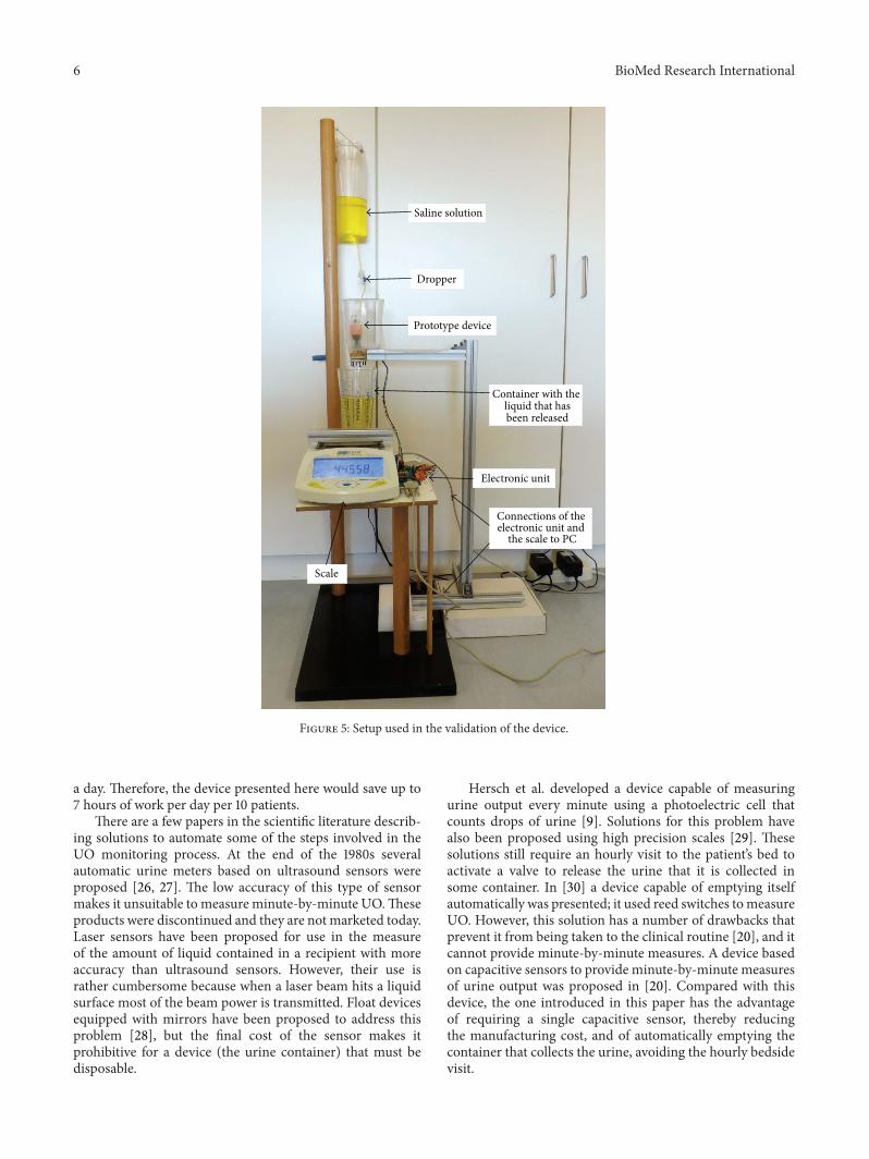
2.2. In Vitro Testing Setup. To verify the proper operationof the prototype we have used a saline solution with similarproperties to urine. A dropper was used to simulate theurine flow into the device (see Figure 5). The prototype waspositioned so that through automatic release the liquid insidethe rigid container would fall into a container located on theplate of a high-precision industrial scale, a PGW 4502e, builtby Adam Equipment Inc. This scale is used to determine if

BioMed Research International 5
Upper float
Base
Magnet
Moving rod
Shoulder of the hollow cylindrical
guide
Hollow cylindrical guide
Lower float
Stopper
Figure 4: On the left we show lateral and bottom views of our prototype. On the right we show the corresponding parts in the design diagram.
the liquid released from our device is released at the rightlevel of filling of the container and if the right amount offluid is released before the stopper closes the drain orifice.The scale is equipped with a serial port that permits queryingfor readings. We built a program that periodically takesmeasurements from the scale. This program was running ona PC which was connected with the scale through the serialport. We can determine the volume of liquid in the containerlocated on the plate of the scale at any time by subtracting theweight of the empty container from the weight measured bythe scale and dividing the result by the saline solution density;that is, from the scale’s measures we can calculate the amountof liquid that has been released each time.
This setup permits the automation of the process ofcarrying out multiple measures of the fluid drained by thedevice each time its content is released. Given that the PGW4502e scale has an accuracy guaranteed by the manufacturerof 0.01 g, we shall consider that measures obtained from thescale are the ground truth which we shall use to determinehow reliable the device is; that is, the release of fluid from thedevice always occurs when the liquid reaches the same leveland always drains the same amount of fluid before starting toaccumulate liquid again.
3. Results and Discussion
Using the in vitro testing setup described in Section 2.2, wemeasured a total of 50 discharges of the prototype, grouped
in 10 experiments of 5 discharges in each. In the experiments,three different rates of urine production were simulatedusing the dropper. The urine production rates correspondedapproximately to 500, 1500, and 3000mL/day. 10 dischargeswere performed at 500mL/day, while 20 discharges wereperformed at 1500 and 3000mL/day. The device released88.1 ± 1.4mL, 89.2 ± 1.7mL, and 88.3 ± 1.8mL at 500, 1500,and 3000mL/day, respectively. According to the theoreticalcalculations based on the dimensions and geometry of thedevice, it should release 90mL in each discharge.
3.1. Discussion. Automating the monitoring of UO can pro-vide the same benefits that the automation of the monitoringof many other physiological parameters has already broughtto critical care units: decreasing the workload of the healthcare staff, simplifying the construction of digital records ofthe patient, and providing more frequent measures of theparameter. As we have already argued, in a critical care unitwith 10 patients, up to 8 hours of health staff time a day areused in tasks related to the monitoring of UO. If these taskswere automated, there may be an improvement in patientoutcomes equivalent to an increase in the staffing of the unitproportional to the saved time [21–25]. The device we havepresented in this paper automates all tasks related to themonitoring of UO, except the changing of the plastic bagwhere urine accumulates. We estimate that this task in acritical care unit with 10 patients requires about 60 minutes
6 BioMed Research International
Saline solution
Dropper
Prototype device
Container with the liquid that hasbeen released
Electronic unit
Scale
Connections of the electronic unit and
the scale to PC
Figure 5: Setup used in the validation of the device.
a day. Therefore, the device presented here would save up to7 hours of work per day per 10 patients.
There are a few papers in the scientific literature describ-ing solutions to automate some of the steps involved in theUO monitoring process. At the end of the 1980s severalautomatic urine meters based on ultrasound sensors wereproposed [26, 27]. The low accuracy of this type of sensormakes it unsuitable to measure minute-by-minute UO.Theseproducts were discontinued and they are not marketed today.Laser sensors have been proposed for use in the measureof the amount of liquid contained in a recipient with moreaccuracy than ultrasound sensors. However, their use israther cumbersome because when a laser beam hits a liquidsurface most of the beam power is transmitted. Float devicesequipped with mirrors have been proposed to address thisproblem [28], but the final cost of the sensor makes itprohibitive for a device (the urine container) that must bedisposable.
Hersch et al. developed a device capable of measuringurine output every minute using a photoelectric cell thatcounts drops of urine [9]. Solutions for this problem havealso been proposed using high precision scales [29]. Thesesolutions still require an hourly visit to the patient’s bed toactivate a valve to release the urine that it is collected insome container. In [30] a device capable of emptying itselfautomatically was presented; it used reed switches tomeasureUO. However, this solution has a number of drawbacks thatprevent it from being taken to the clinical routine [20], and itcannot provide minute-by-minute measures. A device basedon capacitive sensors to provide minute-by-minute measuresof urine output was proposed in [20]. Compared with thisdevice, the one introduced in this paper has the advantageof requiring a single capacitive sensor, thereby reducingthe manufacturing cost, and of automatically emptying thecontainer that collects the urine, avoiding the hourly bedsidevisit.

BioMed Research International 7
A disadvantage of the device presented in this paper whencompared with the manual urine meters currently used incritical care units is its use of metals and magnets. Manualurine meters can be made entirely of plastic and therefore areMRI compatible.Our device should be disconnected from thepatient before performing an MRI.
4. Conclusion
We have designed and built a device capable of automaticallymonitoring the UO of critical care patients. This deviceautomates all tasks related to the monitoring of UO, withthe exception of emptying the plastic bag that collects theurine. Currently this parameter is measured and monitoredmanually by nursing staff, which requires at least one super-visory visit to the patient’s bedside every hour. We estimatethat this device could save up to 7 hours of nursing staffwork per day per 10 patients. Furthermore, with this devicemore frequent urine production measures can be taken (upto one per minute). In the literature there is evidence thatindicates that more frequent UO measurement can impactclinical decision making and improve patient outcomes [15–18] and hence the interest of the device presented in thispaper.
Conflict of Interests
None of the authors of this paper has received any money orany grants, owns shares, or has some other kinds of financialconflict of interests with the commercial entities mentionedin this paper.
Acknowledgments
This work was supported by the Spanish MEC and theEuropean FEDER under Grant TIN2009-14372-C03-03.
References
[1] A. Otero, P. Felix, S. Barro, and F. Palacios, “Addressing theflaws of current critical alarms: a fuzzy constraint satisfactionapproach,” Artificial Intelligence in Medicine, vol. 47, no. 3, pp.219–238, 2009.
[2] M. Shin, “Secure remote health monitoring with unreliablemobile devices,” Journal of Biomedicine and Biotechnology, vol.2012, Article ID 546021, 5 pages, 2012.
[3] A. Jungk, B.Thull, and G. Rau, “Intelligent alarms for anaesthe-sia monitoring based on fuzzy logic approach,” in Fuzzy Logicin Medicine, R. Marın and S. Barro, Eds., pp. 113–138, Physica,Heidelberg, Germany, 2002.
[4] F. A. Mora, G. Passariello, G. Carrault, and J-P. Le Pichon,“Intelligent patient monitoring and management systems: areview,” IEEE Engineering in Medicine and Biology Magazine,vol. 12, no. 4, pp. 23–33, 1993.
[5] E. Rivers, B. Nguyen, S. Havstad et al., “Early goal-directed ther-apy in the treatment of severe sepsis and septic shock,”TheNewEngland Journal ofMedicine, vol. 345, no. 19, pp. 1368–1377, 2001.
[6] M. F. Osuchowski, S. Drechsler, K. M. Weixelbaumer, H. Redl,M. Van Griensven, and S. Bahrami, “Experimentally approach-ing the ICU: monitoring outcome-based responses in thetwo-hit mouse model of posttraumatic sepsis,” Journal ofBiomedicine and Biotechnology, vol. 2011, Article ID 357926, 12pages, 2011.
[7] B.Mitra,M. Fitzgerald, P. Cameron et al., “Fluid resuscitation inmajor burns,”ANZ Journal of Surgery, vol. 76, no. 1-2, pp. 35–38,2006.
[8] M. Hersch, S. Einav, and G. Izbicki, “Accuracy and ease of useof a novel electronic urine output monitoring device comparedwith standard manual urinometer in the intensive care unit,”Journal of Critical Care, vol. 24, no. 4, pp. 629–633, 2009.
[9] M. Hersch, S. Einav, and G. Izbicki, “Accuracy and ease of useof a novel electronic urine output monitoring device comparedwith standard manual urinometer in the intensive care unit,”Journal of Critical Care, vol. 24, no. 4, pp. 629.e13–629.e17, 2009.
[10] I. J. Stewart, M. A. Tilley, C. L. Cotant et al., “Association of AKIwith adverse outcomes in burned military casualties,” ClinicalJournal of the American Society of Nephrology, vol. 7, no. 2, pp.199–206, 2012.
[11] R. Lombardi, N. Nin, J. A. Lorente et al., “An assessment ofthe acute kidney injury network creatinine-based criteria inpatients submitted to mechanical ventilation,” Clinical Journalof the American Society of Nephrology, vol. 6, no. 7, pp. 1547–1555, 2011.
[12] H. Gammelager, C. Christiansen, M. Johansen et al., “One-year mortality among danish intensive care patients with acutekidney injury: a cohort study,”Critical Care, vol. 16, no. 4, articleR124, 2012.
[13] A. Odutayo, N. Adhikari, J. Barton et al., “Epidemiology ofacute kidney injury in canadian critical care units: a prospectivecohort study,”Canadian Journal of Anesthesia, vol. 59, no. 10, pp.934–942, 2012.
[14] W. van Biesen, R. Vanholder, and N. Lameire, “Definingacute renal failure: RIFLE and beyond,” Clinical journal of theAmerican Society of Nephrology, vol. 1, no. 6, pp. 1314–1319, 2006.
[15] A. Panagiotou, F.Garzotto, S.Gramaticopolo et al., “Continuousreal-time urine output monitoring for early detection of acutekidney injury,” Contributions to Nephrology, vol. 171, pp. 194–200, 2011.
[16] M. Y. Shamir, L. Kaplan, R. S. Marans, D. Willner, and Y. Klein,“Urine flow is a novel hemodynamic monitoring tool for thedetection of hypovolemia,” Anesthesia & Analgesia, vol. 112, no.3, pp. 593–596, 2011.
[17] Y. Klein, M. Grinstein, S. M. Cohn, J. Silverman et al., “Minute-to-minute urine flow rate variability: a new renal physiologyvariable,” Anesthesia & Analgesia, vol. 115, no. 4, pp. 843–847,2012.
[18] A. Otero, P. Cardenal-Fernandez, Y. Rojas et al., “On theminuteby minute variations of urine output: a study in a porcinemodel,” Journal of Nephrology, 2014.
[19] R. Fenandez, A. Apalkov, M. Armada et al., “Device formeasuring the amount of liquid that flows and procedure for itsmeasurement and autonomous drain in function of the amountof accumulated fluid,” Patent Application. No. ESP201231159,2012.
[20] A. Otero, R. Fernandez, A. Apalkov et al., “An automatic criticalcare urine meter,” Sensors, vol. 12, no. 10, pp. 13109–13125, 2012.
[21] C. Kovner and P. J. Gergen, “Nurse staffing levels and adverseevents following surgery in U.S. hospitals,” Journal of NursingScholarship, vol. 30, no. 4, pp. 315–321, 1998.

8 BioMed Research International
[22] R. K. Amaravadi, J. B. Dimick, P. J. Pronovost, and P. A. Lipsett,“ICU nurse-to-patient ratio is associated with complicationsand resource use after esophagectomy,” Intensive CareMedicine,vol. 26, no. 12, pp. 1857–1862, 2000.
[23] R. Knauf, L. Lichtig, R. Risen-McCoy et al., Implementing Nurs-ing’s Report Card: A Study of RN Staffing, Length of Stay andPatient Outcomes, American Nurses Association, Washington,DC, USA, 1997.
[24] P. J. Pronovost, D. C. Angus, T. Dorman, K. A. Robinson, T. T.Dremsizov, and T. L. Young, “Physician staffing patterns andclinical outcomes in critically ill patients: a systematic review,”The Journal of the AmericanMedical Association, vol. 288, no. 17,pp. 2151–2162, 2002.
[25] J. B. Dimick, P. J. Pronovost, R. F. Heitmiller, and P. A. Lipsett,“Intensive care unit physician staffing is associated withdecreased length of stay, hospital cost, and complications afteresophageal resection,” Critical Care Medicine, vol. 29, no. 4, pp.753–758, 2001.
[26] M.M. Shabot,M. LoBue, and B. J. Leyerle, “An automatic PDMSinterface for the Urotrack Plus 220 urimeter,” InternationalJournal of Clinical Monitoring and Computing, vol. 5, no. 2, pp.125–131, 1988.
[27] J. F. Shotts and E. Hauf, “The hewlett-packard 1000—vital-metrics VM220 connection: a description of the automatedultrasonic urine output measurement system in the CICU ofgenolier clinic,” International Journal of Clinical Monitoring andComputing, vol. 3, no. 3, pp. 175–182, 1986.
[28] S. Ishida, “Liquid level indicator using laser beam,” US Patent4938590, 1990.
[29] A. Otero, F. Palacios, T. Akinfiev, and R. Fernandez, “A devicefor automatically measuring and supervising the critical carepatient’s urine output,” Sensors, vol. 10, no. 1, pp. 934–951, 2010.
[30] A. Otero, T. Akinfiev, A. Apalkov, F. Palacios, and J. Presedo,“Urine output monitoring: a simple and reliable device formonitoring critical patients’ urine output,” in Proceedings of theInternational Conference on Biomedical Electronics and Devices(BIODEVICES ’11), pp. 26–29, Roma, Italy, January 2011.

Submit your manuscripts athttp://www.hindawi.com
Stem CellsInternational
Hindawi Publishing Corporationhttp://www.hindawi.com Volume 2014
Hindawi Publishing Corporationhttp://www.hindawi.com Volume 2014
MEDIATORSINFLAMMATION
of
Hindawi Publishing Corporationhttp://www.hindawi.com Volume 2014
Behavioural Neurology
EndocrinologyInternational Journal of
Hindawi Publishing Corporationhttp://www.hindawi.com Volume 2014
Hindawi Publishing Corporationhttp://www.hindawi.com Volume 2014
Disease Markers
Hindawi Publishing Corporationhttp://www.hindawi.com Volume 2014
BioMed Research International
OncologyJournal of
Hindawi Publishing Corporationhttp://www.hindawi.com Volume 2014
Hindawi Publishing Corporationhttp://www.hindawi.com Volume 2014
Oxidative Medicine and Cellular Longevity
Hindawi Publishing Corporationhttp://www.hindawi.com Volume 2014
PPAR Research
The Scientific World JournalHindawi Publishing Corporation http://www.hindawi.com Volume 2014
Immunology ResearchHindawi Publishing Corporationhttp://www.hindawi.com Volume 2014
Journal of
ObesityJournal of
Hindawi Publishing Corporationhttp://www.hindawi.com Volume 2014
Hindawi Publishing Corporationhttp://www.hindawi.com Volume 2014
Computational and Mathematical Methods in Medicine
OphthalmologyJournal of
Hindawi Publishing Corporationhttp://www.hindawi.com Volume 2014
Diabetes ResearchJournal of
Hindawi Publishing Corporationhttp://www.hindawi.com Volume 2014
Hindawi Publishing Corporationhttp://www.hindawi.com Volume 2014
Research and TreatmentAIDS
Hindawi Publishing Corporationhttp://www.hindawi.com Volume 2014
Gastroenterology Research and Practice
Hindawi Publishing Corporationhttp://www.hindawi.com Volume 2014
Parkinson’s Disease
Evidence-Based Complementary and Alternative Medicine
Volume 2014Hindawi Publishing Corporationhttp://www.hindawi.com