research article 12-deoxyphorbols promote adult ... · humana, universidad de cádiz, cádiz, spain...
TRANSCRIPT

Received: December 10, 2014; Revised: July 16, 2015; Accepted: July 22, 2015
© The Author 2015. Published by Oxford University Press on behalf of CINP.
International Journal of Neuropsychopharmacology, 2015, 1–14
doi:10.1093/ijnp/pyv085Research Article
1This is an Open Access article distributed under the terms of the Creative Commons Attribution License (http://creativecommons.org/licenses/by/4.0/), which permits unrestricted reuse, distribution, and reproduction in any medium, provided the original work is properly cited.
research article
12-Deoxyphorbols Promote Adult Neurogenesis by Inducing Neural Progenitor Cell Proliferation via PKC ActivationNoelia Geribaldi-Doldán, Eugenia Flores-Giubi, Maribel Murillo-Carretero, Francisco García-Bernal, Manuel Carrasco, Antonio J. Macías-Sánchez, Jesús Domínguez-Riscart, Cristina Verástegui, Rosario Hernández-Galán, and Carmen Castro
Area de Fisiología, Facultad de Medicina, Universidad de Cadiz, Cádiz, Spain (Mrs Geribaldi-Doldán, Mrs Flores-Giubi, Dr Murillo-Carretero, Mr García-Bernal, Dr Carrasco, Dr Domínguez-Riscart, and Dr Castro); Departamento de Química Orgánica, Facultad de Ciencias, Universidad de Cádiz, Puerto Real, Cádiz, Spain (Mrs Flores-Giubi, Dr Macías-Sánchez, and Dr Hernández-Galán); Departamento de Anatomía y Embriología Humana, Universidad de Cádiz, Cádiz, Spain (Dr Verástegui).
N.G.-D. and E.F.-G. contributed equally to the work.
Correspondence: Carmen Castro, PhD, Area de Fisiología, Facultad de Medicina, Universidad de Cadiz, Cádiz, Spain ([email protected]).
Abstract
Background: Neuropsychiatric and neurological disorders frequently occur after brain insults associated with neuronal loss. Strategies aimed to facilitate neuronal renewal by promoting neurogenesis constitute a promising therapeutic option to treat neuronal death-associated disorders. In the adult brain, generation of new neurons occurs physiologically throughout the entire life controlled by extracellular molecules coupled to intracellular signaling cascades. Proteins participating in these cascades within neurogenic regions constitute potential pharmacological targets to promote neuronal regeneration of injured areas of the central nervous system.Methodology: We have performed in vitro and in vivo approaches to determine neural progenitor cell proliferation to understand whether activation of kinases of the protein kinase C family facilitates neurogenesis in the adult brain.Results: We have demonstrated that protein kinase C activation by phorbol-12-myristate-13-acetate induces neural progenitor cell proliferation in vitro. We also show that the nontumorogenic protein kinase C activator prostratin exerts a proliferative effect on neural progenitor cells in vitro. This effect can be reverted by addition of the protein kinase C inhibitor G06850, demonstrating that the effect of prostratin is mediated by protein kinase C activation. Additionally, we show that prostratin treatment in vivo induces proliferation of neural progenitor cells within the dentate gyrus of the hippocampus and the subventricular zone. Finally, we describe a library of diterpenes with a 12-deoxyphorbol structure similar to that of prostratin that induces a stronger effect than prostratin on neural progenitor cell proliferation both in vitro and in vivo.
International Journal of Neuropsychopharmacology Advance Access published August 18, 2015 by guest on January 25, 2016
http://ijnp.oxfordjournals.org/D
ownloaded from

2 | International Journal of Neuropsychopharmacology, 2015
Conclusions: This work suggests that protein kinase C activation is a promising strategy to expand the endogenous neural progenitor cell population to promote neurogenesis and highlights the potential of 12-deoxyphorbols as pharmaceutical agents to facilitate neuronal renewal.
Keywords: 12-deoxyphorbol, prostratin, PKC, neurogenesis, neural progenitor cells
IntroductionNeuropsychiatric and neurodegenerative diseases frequently occur after central nervous system (CNS) insults such as stroke or traumatic brain injury, characterized by an irreversible neu-ronal loss, for which there is no adequate treatment at present. Among novel treatment approaches, some intend to promote neuronal replacement in the injured tissue through the genera-tion of new neurons from neural stem cells (NSCs). In mammals, adult NSCs continuously undergoing physiological neurogene-sis are located fundamentally in the dentate gyrus of the hip-pocampus (DG) and the subventricular zone (SVZ). Glial-like NSCs within these regions produce multipotent undifferenti-ated neural progenitor cells (NPCs), which can commit to either a neuronal phenotype (through neuroblast and mature neuron production), or a glial phenotype, generating either astrocytes or oligodendrocytes (Alvarez-Buylla and Garcia-Verdugo, 2002; Goldman, 2003). Cells isolated from these areas, and cultured in the presence of growth factors such as epidermal growth factor (EGF) and/or basic fibroblast growth factor (bFGF or FGF-2), gen-erate cell aggregates known as neurospheres that contain NPCs, which exhibit stem cell properties, including the ability to pro-liferate, self-renew, and undergo multipotential differentiation, generating neurons, astrocytes, and oligodendrocytes (Pastrana et al., 2009). Newly generated neurons have been found in brain lesions of different origins (Parent et al., 1997; Liu et al., 1998; Jin et al., 2001; Luka et al., 2002), some of them resulting from the differentiation and maturation of neuroblasts generated in neu-rogenic regions that had migrated to the injured area and some others generated from nearby stem cells activated locally by the specific injury. However, the neuroregenerative capacity under any of these conditions is very limited (Arvidsson et al., 2002; Nakatomi et al., 2002; Teramoto et al., 2003); thus, to improve neuron replacement within an injured region of the CNS, it is necessary to find molecules that activate proliferation and/or neuronal differentiation of NPCs and NSCs.
Growth factors like EGF or bFGF promote proliferation and/or fate determination on NPC pools (Palmer et al., 1995; Doetsch et al., 2002) through interaction with receptor tyrosine kinases (Ullrich and Schlessinger, 1990; Annenkov, 2014). Receptor tyros-ine kinases activate intracellular signaling molecules, including the family of protein kinase C (PKC) (Annenkov, 2014); several members of this PKC family are expressed in neurogenic regions (Minami et al., 2000) and participate in various signaling cas-cades initiated by growth factors, often determining growth fac-tor specificity (Corbit et al., 2000). Ten serine-threonine kinases constitute the PKC family, which are divided into 3 subfamilies: the classical (α, β1, β2, and γ), the novel (δ, ε, θ, and η), and the atypical (ζ and λ). Atypical PKCs ζ and λ have been involved in NSC to neuron transition (Wang et al., 2012), and several other isoforms of PKC are expressed in NPCs isolated from the SVZ of newborn rats, among which PKCε seems to be relevant for astro-cytic differentiation (Steinhart et al., 2007). It is plausible that particular isoforms of PKC may be implicated in other specific aspects of adult neurogenesis, like NPC autoregeneration, prolif-eration, survival, or neuronal differentiation.
Phorbol esters are natural, plant-derived diterpenes known to activate novel and classical PKCs by binding to their reg-ulatory domain, mimicking their physiological activator diacylglicerol. The most common phorbol ester is phorbol-12-myristate-13-acetate (PMA) (Goel et al., 2007), whose major drawback is its strong tumor-promoting activity (Deleers and Malaisse, 1982). Other naturally occurring phorbols, like pros-tratin, activate PKC but lack the tumor-promoting activity that makes PMA unsuitable for biomedical research (Szallasi and Blumberg, 1991b); rather than that, prostratin has shown some tumor-supressing activity (Shen et al., 2015), but it is best known for its antiviral properties, particularly against HIV (see Miana et al., 2015 for a recent review). Prostratin is a 12-deoxyphorbol first isolated from the plant Pimelea prostrate, an endemic New Zealand shrub. Extracts from this and other plants containing 12-deoxyphorbols have been historically used in Asian medicine. Diterpenes are also abundant in plants of the Euphorbiaceae family.
We have studied herein the effect of PKC activation by PMA and prostratin on the proliferation and expansion of NPC in vitro and in vivo. In addition, we have isolated 6 natural prod-ucts from Euphorbia resinifera with a 12-deoxyphorbol structure similar to that of prostratin, and have comparatively tested this library of compounds on NPC proliferation, in an attempt to identify whether additional harmless molecules were capable of promoting neurogenesis and improving neuronal replacement. Our results show that prostratin as well as the related com-pound ER272 increase NPC proliferation in a PKC-dependent manner, both in vivo and in vitro, without affecting the neuronal differentiation potential of these NPCs.
Materials and Methods
Reagents
12-Deoxyphorbols were isolated and purified in our labora-tory. PMA (13-O-acetyl-12-O-tetradecanoylphorbol), prostratin (13-O-acetyl-12-deoxyphorbol), and other products unless oth-erwise indicated were purchased from Sigma-Aldrich (St. Louis, MO). The general PKC inhibitor bisindolylmaleimide I, or G06850, was from Calbiochem (Millipore, Billerica, MA), used at a final concentration of 0.5 µM, and added to the cells 30 minutes before addition of PKC activators. Stock solutions of all PKC interact-ing compounds were prepared in dimethyl sulfoxide (DMSO) and prediluted in culture medium before addition to cell cultures.
Plant Material
Latex from Euphorbia resinifera was collected in Tanat, Morocco, on December, 2007, by making cuts on stalks and collecting the milky secretion. Natural products with 12-deoxyphorbol struc-tures were isolated from this latex as described in the supple-mentary Methods.
by guest on January 25, 2016http://ijnp.oxfordjournals.org/
Dow
nloaded from

Geribaldi-Doldán et al. | 3
Animal Subjects
Seven-day postnatal CD1 mice were used for NPC isolation from the SVZ. Two-month-old adult male mice were used for intrac-erebroventricular (ICV) injections (in vivo experiments). Animals were housed under controlled conditions of temperature (21–23°C) and light (light:dark 12:12) with free access to food (AO4 standard maintenance diet; SAFE, Épinay-sur-Orge, France) and water. Care and handling of animals were performed according to the Guidelines of the European Union Council (2010/63/EU) and following the Spanish regulations (65/2012 and RD53/2013) for the use of laboratory animals.
SVZ Cell Isolation and Culture
NPCs were obtained from the SVZ of 7-day postnatal mice fol-lowing the same procedure described in Rabaneda et al. (2008). Neurosphere cultures were maintained in defined medium (DM) composed of Dulbecco’s modified Eagle’s medium/F12 medium (1:1 vol/vol) plus 1 mg/L gentamicin (GIBCO) and the B27 supple-ment (Invitrogen, Carlsbad, CA). EGF (20 ng/mL) and bFGF (10 ng/mL; both from PeproTech, Frankfurt, Germany) were added to DM for culture expansion, but only bFGF was present during experimental settings, unless otherwise indicated.
Neurosphere Assay
To test the effects of our compounds on NPC proliferation, sin-gle cells from mechanically disaggregated neurospheres were plated onto anti-adherent 96-well plates at a density of 20,000 cells/mL in DM + 10 ng/mL bFGF. 12-Deoxyphorbols and other pharmacological agents were added at the time of seeding, and all conditions were run in triplicates.
Neurosphere number and size were measured as previously described (Rabaneda et al., 2008). Results were obtained from a minimum of 3 independent experiments performed with tripli-cate samples.
Cell Viability Assay
Cells were seeded at a density of 20,000 cells/mL and cultured in the absence or presence of specific treatments. Then, cultures were disaggregated and mixed with trypan blue [0.04% wt/vol in phosphate-buffered saline (PBS)]. Viable cells (those exclud-ing the trypan blue dye) were counted using a hemocytometer under an inverted microscope and were expressed as percent-age of the total number of cells.
Immunocytochemistry
Neurosphere cells were disaggregated and adhered onto poly-L-ornithine-coated 8-well glass slide chambers (Lab-Tek) and were grown for 48 hours in the presence of bFGF plus specific treat-ments. Immunodetection was performed as previously described (Rabaneda et al., 2008). Antibodies used were rabbit polyclonal anti-Ki67 (1:1000; Vector) and goat anti-rabbit IgG labeled with Alexa Fluor 594 (1:1500; Invitrogen, Carlsbad, CA). Nuclei were counter-stained for 10 minutes with 0.1 mg/L DAPI (4',6-diamidino-2-phe-nylindole). Ki67+ cells were counted under a BX60 epifluorescence microscope (Olympus, Hamburg, Germany) and expressed as per-centage of the total number of cells, determined by DAPI staining. Quantification was performed in 12 predetermined visual fields/well and 3 wells/condition. Experiments were repeated a mini-mum of 3 times and results were expressed as the mean ± SEM.
Cell Death Measurement
Cell death was estimated by counting pyknotic nuclei in the same sample preparations and wells used for immunocytochemistry (see previous paragraph). Nuclei showing chromatin hypercon-densation (apoptotic and/or necrotic) were detected by DAPI stain-ing and expressed as percentage of the total number of nuclei.
Western Blot
Cells from disaggregated neurospheres were resuspended in defined medium without any added growth factor and seeded in 12-well plates (500 000 cells per well and condition). Immediately after, cells were pretreated with or without 0.5 µM G06850 for 30 minutes, then with or without 5 µM prostratin and, 1.5 hours later, stimulated with or without 10 ng/mL bFGF. Western blot was performed as previously described (Carrasco et al., 2014). Antibodies used were mouse anti-cyclin D1, rabbit anti-cyclin E (both at 1:500, Santa Cruz Biotechnologies, Santa Cruz, CA), and mouse anti-β-actin as loading control (1:1000, Sigma-Aldrich, St Louis, MO). Secondary antibodies were from commercial kits (Western Breeze, Invitrogen, Carlsbad, CA). Band density was quantified using the ImageJ software.
ICV Administration of Prostratin, ER272, and ER271
Adult male mice weighing 35 to 40 g were anaesthetized with ketamine/xylazine and placed on a stereotaxic frame (Kopf Instruments). A small trepanation was made 0.8 mm lateral to Bregma, where the needle of a 5-µL Hamilton syringe was introduced 2.4 mm below the brain surface. Ultra-filtered pros-tratin, diluted in PBS at a concentration of 5 µM (n = 12), or vehicle (n = 12), were injected ipsilaterally into the right lateral ventricles. Single injections of 2 µL per brain were performed through a time-frame of 10 minutes (corresponding to a total of 4 ng of prostratin in the case of prostratin-injected mice); afterwards, the Hamilton syringe was left another 5 minutes before removal. Animals received daily injections of bromod-eoxyuridine (BrdU) (120 mg/kg), during 3 days starting the day of the surgical procedure. Then, mice were deeply anaesthe-tized with pentobarbital and perfused with 4% parafolmade-hyde via ascending aorta. Brains were removed and sliced in 30-µm serial sections from Bregma 2.1 to -3.8 (Franklin and Paxinos, 2007), where the SVZ and hippocampus are contained. In a different set of experiments, the same procedure was fol-lowed except that mice were injected with 2 µL of 1 µM ER272 (n = 6) or 1 µM ER271 (n = 6), corresponding to a dosage of 0.8 ng of each compound per brain, and 6 vehicle-injected mice were processed in parallel.
Brain Processing and Immunohistochemistry
Brain removal, processing, and immunohistochemical tech-niques to detect BrdU and doublecortin (DCX) were performed as previously described (Rabaneda et al., 2008). Antibodies used were mouse monoclonal anti-BrdU (1:100) from Dako (Hamburg, Germany), goat polyclonal anti-DCX (1:200, Santa Cruz Biotech.), AlexaFluor 488 donkey anti-mouse and AlexaFluor 594 (both at 1:1000, from Life Tech.), and biotinylated anti-mouse IgG (1:250) from GE Healthcare (Albany, NY).
Quantification of Neurogenesis in Brain Sections
BrdU+ and DCX+ cells were counted in neurogenic areas as pre-viously described (Rabaneda et al., 2008; Carrasco et al., 2014).
by guest on January 25, 2016http://ijnp.oxfordjournals.org/
Dow
nloaded from

4 | International Journal of Neuropsychopharmacology, 2015
Statistical Analysis
When more than one treatment group were compared, statis-tical analyses were performed using 1-way ANOVA, and when normality failed a nonparametric Dunnett’s Method was per-formed. The Student’s t test was used when only one treatment group was compared with the control. Differences were consid-ered significant at values of P ≤ .05.
Results
PMA-Induced PKC Activation Promotes NPC Proliferation
NPCs divide in culture in the presence of growth factors, form-ing floating cell aggregates known as neurospheres (Bull and Bartlett, 2005). The size reached by neurospheres at specific time-points is indicative of NPC proliferation, while the num-ber of new neurospheres formed after subsequent subcultures depends on NPC self-renewal capacity (Ramirez-Castillejo et al., 2006) and survival (Torroglosa et al., 2007). We used neuro-sphere assays, in which the size and number of newly formed
neurospheres is estimated, to test whether PKC activators mod-ulated NPC proliferation. Experiments were performed in the presence of a single growth factor, bFGF, which should not mask other mitotic stimuli.
NPC were treated for 72 hours with increasing concentra-tions of PMA, a potent and widely known PKC activator. PMA treatment increased neurosphere size, being 16 nM, the most effective concentration (Figure 1 A-B), and this effect was reverted by the PKC inhibitor G06850 (Figure 1 C). PMA treatment did not modify neurosphere number (Figure 1D). These results suggested that PMA promoted NPC proliferation without affect-ing the viability of neurosphere-forming cells (as neurosphere number was unchanged). Neurosphere enlargement in cultures treated with 16 nM PMA for 72 hours was accompanied by an increment in the total number of cells (Figure 1F), while cell via-bility was not affected by this concentration of PMA (Figure 1E). The proliferative effect of PMA was confirmed by immunocyto-chemical detection of the cell cycle-marker Ki67 in neurosphere-derived cells that had been cultured onto an adherent surface for 48 hours. In the presence of 16 nM PMA, a 2-fold increase in the percentage of Ki67+ cells was observed compared with the control, and pretreatment of cultures with the PKC inhibitor
Figure 1. Effect of phorbol-12-myristate-13-acetate (PMA) on neural progenitor cell (NPC) proliferation in vitro. Proliferation was tested in NPC cultures grown in the
presence of the growth factor basic fibroblast growth factor (bFGF), unless otherwise indicated. (A) Phase-contrast microscopy images of neurospheres cultured for 72
hours with or without the protein kinase C (PKC) activator PMA (16 nM) alone or in combination with the PKC inhibitor G06850 (0.5 µM). Scale bar indicates 200 µm. (B)
Graph shows the effect of increasing concentrations of PMA on neurosphere area after 72 hours of culture. (C) Graph shows neurosphere area after 72 hours of treat-
ment with 16 nM PMA, alone or in combination with 0.5 µM G06850. (D) Effect of increasing concentrations of PMA on neurosphere number, counted after 72 hours
of culture. (E) Effect of 16 nM PMA on cell viability. (F) Effect of 16 nM PMA on total cell number, counted in a Neubauer chamber after neurosphere disaggregation. (G)
Fluorescence microscopy images of neurosphere-derived adhered cells grown for 48 h with or without 16 nM PMA and/or the PKC inhibitor G06850 (0.5 μM). Cells were
immunostained to detect the proliferation marker Ki67 and nuclei were counterstained with 4',6-diamidino-2-phenylindole. Scale bar indicates 100 µm. (H) Quantifica-
tion of Ki67+ cells in cultures described in G. (I) Quantification of pyknotic nuclei in cultures described in G. *P < .05 compared with control in a Student’s t test; #P < .05
compared with the other groups by 1-way ANOVA.
by guest on January 25, 2016http://ijnp.oxfordjournals.org/
Dow
nloaded from

Geribaldi-Doldán et al. | 5
G06850 (0.5 µM) abolished the effect of PMA (Figure 1 G-H); these results indicate that PMA induced NPC proliferation by activat-ing PKC. The percentage of pyknotic nuclei was not affected by any treatment (Figure 1I).
Effect of Prostratin-Induced PKC Activation on NPC Proliferation
In contrast to PMA, other phorbols like prostratin are nontu-morigenic PKC activators (Szallasi and Blumberg, 1991a, 1991b; Szallasi et al., 1992). Thus, the effect of this compound on NPC proliferation was tested. In the presence of 1 to 10 µM prostratin, neurosphere size increased in a concentration-dependent man-ner, reaching its maximum at 5 µM (Figure 2A-B). The effect of prostratin on neurosphere size occurred through PKC activation, because it was reverted by pretreatment with the PKC inhibi-tor G06850 (Figure 2A, C). In contrast to PMA, PKC activation mediated by prostratin increased the number of neurospheres formed (Figure 2D); this effect of prostratin could be due to
increased NPC survival but could also be indicative of a higher number of neurosphere-forming cells dividing actively. In fact, treatment with prostratin for 72 hours not only increased the total cell number present in neurospheres, but it also had a protective effect on cell viability (Figure 2E-F). Also, treatment of adhered NPC cultures with prostratin induced a 2.5-fold increase in the percentage of Ki67+ cells, which was reduced by pretreatment with the PKC inhibitor G06850 even below control levels (Figure 2G-H), confirming that prostratin-induced activa-tion of PKC was responsible for this rise in the number of cycling cells. No effect of prostratin on pyknotic nuclei percentage was observed (data not shown).
A major drawback of manipulating NPC proliferation may be the loss of self-renewal capacity, causing stem-cell pool exhaustion. In the context of NPC cultures, this would result in a decrease on neurosphere number as subcultures go on (Ramirez-Castillejo et al., 2006). To rule out that PKC activation was affecting NPC self-renewal, we disaggregated neurospheres that had been treated for 72 hours with prostratin, PMA, or none,
* #
F
Control Prostratin
G06850 Prostratin + G06850
FFFFFFFFFFF
*
Tota
l nu
mb
er o
f ce
lls
Control Prostratin
Acontrol prostratin prostratin + G06850
C
Neu
rosp
her
e ar
ea
(% o
f co
ntr
ol)
B*
*
*
Neu
rosp
her
e ar
ea
(% o
f co
ntr
ol)
0 5 1080
100
120
140
160
180
200
1.0
[Prostratin] (µM)
D *
*
Neu
rosp
her
e n
um
ber
(% o
f co
ntr
ol)
0
50
100
150
200
E
*
No
n-v
iab
le c
ells
(%
)
*
Nu
mb
er o
f se
con
dar
y n
euro
sph
eres
*I
* *
Ki6
7+ c
ells
(% o
f to
tal c
ells
)
0
10
20
30
40
* #
H
*
G
Figure 2. Effect of prostratin on neural progenitor cell (NPC) proliferation in vitro. Proliferation was tested in NPC cultures grown in the presence of basic fibroblast
growth factor (bFGF). (A) Phase-contrast microscopy images of neurospheres cultured for 72 hours in the absence or presence of prostratin (5 µM) with or without addi-
tion of the PKC inhibitor G06850 (0.5 µM). Scale bar indicates 200 µm. (B) Graph shows the effect of increasing concentrations of prostratin on neurosphere area after
72 hours of culture. (C) Graph shows neurosphere area after 72 hours of treatment with or without 5 µM prostratin and/or 0.5 µM G06850. (D) Effect of prostratin on
neurosphere number, counted 72 hours after seeding, in the absence or presence of 0.5 µM G06850. (E) Percentage of nonviable cells quantified by trypan blue exclusion
after 48 hours of culture in the presence or absence of 5 µM prostratin and/or 0.5 µM G06850. (F) Total cell number in NPC cultures after 72 hours in the presence or
absence of 5 µM prostratin. (G) Fluorescence microscopy images of neurosphere-derived adhered cells grown for 48 hours with or without 5 µM prostratin and/or 0.5 µM
G06850. Cells were immunostained to detect Ki67, and nuclei were counterstained with 4’,6-diamidino-2-phenylindole. Scale bar indicates 100 µm. (H) Quantification of
Ki67+ cells in cultures described in G. (I) Effect of a 72-hour pretreatment of NPC with prostratin or PMA (at the indicated concentrations) on the number of secondary
neurospheres formed in the following 72-hour subculture in the absence of drugs. *P < .05 compared with control in a Student’s t test; #P < .05 compared with the other
groups in ANOVA for repeated measurements, followed by Student’s t test.
by guest on January 25, 2016http://ijnp.oxfordjournals.org/
Dow
nloaded from

6 | International Journal of Neuropsychopharmacology, 2015
reseeded single cells at a density of 20 000 cells/mL in fresh con-trol medium, and cultured them for another 72 hours. The num-ber of newly formed secondary neurospheres was then counted. Surprisingly, pretreatment with 5 µM prostratin increased the number of secondary neurospheres formed in the subsequent subculture when compared with the control, while no effect of PMA was observed (Figure 2I).
The other concern that had to be ruled out was whether the capacity of NPCs to differentiate, particularly towards the neuronal lineage, would be compromised after treatment with PKC activators. To answer this, we disaggregated neurospheres that had been treated for 72 hours with prostratin, PMA, or none and seeded them onto adherent surfaces in differentiat-ing conditions for another 72 hours. Then, the neuronal marker β-III-tubulin and the glial marker GFAP were detected by immu-nocytochemistry. Prostratin did not change the percentage of cells committed to either neuronal or glial differentiation, while only a small effect of PMA on glial differentiation was observed (supplementary Figure S1; supplementary Methods).
Prostratin and bFGF Work in Synergy to Upregulate Cyclin Expression in NPC Cultures
Growth factors like EGF or bFGF induce NPC proliferation through activation of Akt and Erk1/2 signaling pathways (Sung et al., 2007; Torroglosa et al., 2007), which results in cyclin
upregulation and cell division (Rabaneda et al., 2008). To study whether these mechanisms were involved in the proliferative effect of prostratin, a neurosphere assay was performed in the absence or presence of the Akt pathway inhibitor LY294002 or the Erk1/2 pathway inhibitor UO126. The effect of prostratin on neurosphere growth was blocked by UO126 and, remarkably, completely abolished by LY294002, indicating that both Akt and Erk1/2 are essential downstream effectors of prostratin’s action (supplementary Figure S2).
Next, the effect of prostratin treatment on cyclin expres-sion was analyzed by Western blot. Cyclin E and cyclin D1 were upregulated in cultures stimulated by either bFGF or prostratin alone, and the combination of both treatments had a synergic effect on cyclin upregulation (Figure 3A-B). These results indi-cated a strong interaction between prostratin and bFGF signal-ing cascades on cyclin expression induction. But the fact that prostratin alone was able to induce the expression of cyclin D1 and E through PKC activation in growth factor-devoid cultures prompted us to consider whether this drug could, by itself, increase NPC proliferation. As shown in Figure 3C-D, prostra-tin alone, in the absence of any added growth factor, was able to induce a small but statistically significant increase on neu-rosphere area despite the fact that NPC are strictly depend-ent on growth factors for survival and proliferation in culture. A synergic effect of prostratin and bFGF on neurosphere growth was also evident in agreement with previous observations on
Figure 3. Effect of prostratin on the expression of cyclins. (A) Representative images of Western blots showing immunodetection of cyclin D1 and E, and beta-actin as
a loading control. Neural progenitor cell (NPC) cultures were pretreated with or without prostratin (5 μM) and/or the PKC inhibitor G06850 (0.5 μM) for 1.5 hours and
then stimulated with or without bFGF for 6.5 additional hours. (B) Densitometric quantification of cyclin D1 and cyclin E bands relative to the optical density of the
corresponding β-actin bands. Results are the mean ± SEM of 3 independent experiments; *P < .05 compared with none (basal levels of cyclins in the absence of basic
fibroblast growth factor [bFGF] and protein kinase C [PKC]-modulating drugs) in a Student’s t test; #P < .05 compared with the other groups by 1-way ANOVA. (C) Graph
shows the effect of 5 μM prostratin on both the basal proliferation of NPC in the absence of any added growth factors and on bFGF-induced NPC proliferation after 72
hours of culture. Prostratin alone significantly increased neurosphere area in the absence of added growth factors; also, a synergistic effect of prostratin and bFGF on
neurosphere area enlargement was observed. Results are the mean ± SEM of 3 independent experiments; ##P < .01 when compared with control by Student’s t test. (D)
Representative phase-contrast photomicrographs of the neurosphere culture conditions explained in C. Calibration bar indicates 200 µm.
by guest on January 25, 2016http://ijnp.oxfordjournals.org/
Dow
nloaded from
Geribaldi-Doldán et al. | 7
cyclin expression patterns induced by the combination of both molecules.
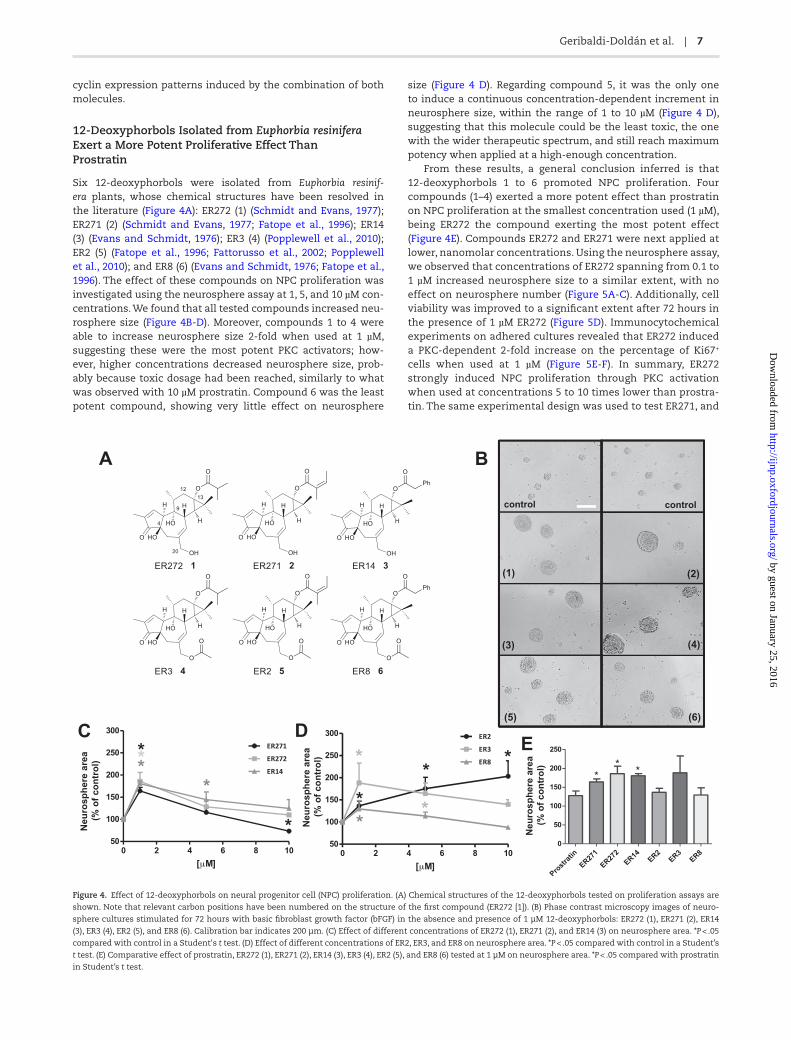
12-Deoxyphorbols Isolated from Euphorbia resinifera Exert a More Potent Proliferative Effect Than Prostratin
Six 12-deoxyphorbols were isolated from Euphorbia resinif-era plants, whose chemical structures have been resolved in the literature (Figure 4A): ER272 (1) (Schmidt and Evans, 1977); ER271 (2) (Schmidt and Evans, 1977; Fatope et al., 1996); ER14 (3) (Evans and Schmidt, 1976); ER3 (4) (Popplewell et al., 2010); ER2 (5) (Fatope et al., 1996; Fattorusso et al., 2002; Popplewell et al., 2010); and ER8 (6) (Evans and Schmidt, 1976; Fatope et al., 1996). The effect of these compounds on NPC proliferation was investigated using the neurosphere assay at 1, 5, and 10 µM con-centrations. We found that all tested compounds increased neu-rosphere size (Figure 4B-D). Moreover, compounds 1 to 4 were able to increase neurosphere size 2-fold when used at 1 µM, suggesting these were the most potent PKC activators; how-ever, higher concentrations decreased neurosphere size, prob-ably because toxic dosage had been reached, similarly to what was observed with 10 µM prostratin. Compound 6 was the least potent compound, showing very little effect on neurosphere
size (Figure 4 D). Regarding compound 5, it was the only one to induce a continuous concentration-dependent increment in neurosphere size, within the range of 1 to 10 µM (Figure 4 D), suggesting that this molecule could be the least toxic, the one with the wider therapeutic spectrum, and still reach maximum potency when applied at a high-enough concentration.
From these results, a general conclusion inferred is that 12-deoxyphorbols 1 to 6 promoted NPC proliferation. Four compounds (1–4) exerted a more potent effect than prostratin on NPC proliferation at the smallest concentration used (1 µM), being ER272 the compound exerting the most potent effect (Figure 4E). Compounds ER272 and ER271 were next applied at lower, nanomolar concentrations. Using the neurosphere assay, we observed that concentrations of ER272 spanning from 0.1 to 1 µM increased neurosphere size to a similar extent, with no effect on neurosphere number (Figure 5A-C). Additionally, cell viability was improved to a significant extent after 72 hours in the presence of 1 µM ER272 (Figure 5D). Immunocytochemical experiments on adhered cultures revealed that ER272 induced a PKC-dependent 2-fold increase on the percentage of Ki67+ cells when used at 1 µM (Figure 5E-F). In summary, ER272 strongly induced NPC proliferation through PKC activation when used at concentrations 5 to 10 times lower than prostra-tin. The same experimental design was used to test ER271, and
Figure 4. Effect of 12-deoxyphorbols on neural progenitor cell (NPC) proliferation. (A) Chemical structures of the 12-deoxyphorbols tested on proliferation assays are
shown. Note that relevant carbon positions have been numbered on the structure of the first compound (ER272 [1]). (B) Phase contrast microscopy images of neuro-
sphere cultures stimulated for 72 hours with basic fibroblast growth factor (bFGF) in the absence and presence of 1 µM 12-deoxyphorbols: ER272 (1), ER271 (2), ER14
(3), ER3 (4), ER2 (5), and ER8 (6). Calibration bar indicates 200 µm. (C) Effect of different concentrations of ER272 (1), ER271 (2), and ER14 (3) on neurosphere area. *P < .05
compared with control in a Student's t test. (D) Effect of different concentrations of ER2, ER3, and ER8 on neurosphere area. *P < .05 compared with control in a Student’s
t test. (E) Comparative effect of prostratin, ER272 (1), ER271 (2), ER14 (3), ER3 (4), ER2 (5), and ER8 (6) tested at 1 µM on neurosphere area. *P < .05 compared with prostratin
in Student’s t test.
by guest on January 25, 2016http://ijnp.oxfordjournals.org/
Dow
nloaded from

8 | International Journal of Neuropsychopharmacology, 2015
similar results were obtained, except that a slightly higher con-centration of the drug (0.25 µM) was needed to exert maximal effects (Figure 6A-F). These results suggested that ER272 was more effective at activating PKC than ER271, notwithstanding that both were more potent than prostratin in promoting NPC proliferation.
Prostratin, ER272, and ER271 Did Not Exert a Proliferative Effect on a Tumoral Neuroblastoma Cell Line
Tumor-promoting phorbols like PMA do not generate tumors by themselves but promote tumor growth following exposure to a subcarcinogenic dose of a carcinogen (they can thus be des-ignated as co-carcinogens) (Goel et al., 2007). In an attempt to demonstrate that not only prostratin but also other 12-deoxy-phorbols are nontumorigenic, we tested whether they were able to induce proliferation in cancerous cells. The effect of 8 nM PMA, 5 µM prostratin, and 1 µM ER272 and ER271 on the pro-liferation of a neuroblastoma cell line (NB69) was assayed by quantifying BrdU incorporation. Unlike PMA, which increased 2-fold the proliferation of NB69 cells, neither prostratin nor ER272 or ER271 induced proliferation when added to NB69 cul-tures (Figure 6G). These results indicated that 12-deoxyphorbols, unlike PMA, exerted their proliferative effect specifically on NPC, without affecting neuroblastoma cells.
In Vivo Administration of Prostratin, ER272, or ER271 Increases NPC Proliferation in Neurogenic Regions without Affecting Neuronal Differentiation
To study the relevance of PKC activation on NPC proliferation in vivo, we injected prostratin, ER272, and ER271 into adult mouse ventricles and studied their effect on neurogenesis. Single ICV injections of prostratin (4 ng), ER272 or ER271 (0.8 ng each), or vehicle were administered ipsilaterally. Proliferating cells were concurrently labeled by BrdU intraperitoneal injections for 3 days, when brains were fixed and subjected to immunohisto-chemical detection of BrdU in neurogenic areas. Proliferation was quantified as the number of BrdU+ cells found in the SVZ and DG, comparing the ipsilateral (injected) side of the brain with the contralateral side.
In the SVZ of vehicle-injected mice, a significant increase of BrdU+ cells was observed in the ipsilateral side compared with the contralateral side, indicating that the injection itself induced NPC proliferation (Figure 7A-B, M), probably due to the small lesion caused by the procedure. Treatment with pros-tratin significantly increased the total number of BrdU+ cells in both the ipsilateral and contralateral SVZs compared with their equivalent in vehicle-injected mice, demonstrating that in vivo administration of prostratin increased NPC proliferation in the SVZ of adult mice independently of, and further beyond, the increase due to procedural tissue damage (Figure 7J-K, M).
Figure 5. Effect of ER272 (1) on neural progenitor cell (NPC) proliferation. (A) Phase contrast microscopy images of neurosphere cultures stimulated with basic fibroblast
growth factor (bFGF) in the absence and presence of 1 µM ER272 (1). Calibration bar indicates 200 µm. (B) Effect of different concentrations of ER272 (1) on neurosphere
area. (C) Effect of different concentrations of ER272 (1) on neurosphere number. (D) Effect of 1 µM ER272 (1) on cell viability assayed by trypan blue exclusion. (E) Fluo-
rescence microscopy images of NPC cultures stimulated with bFGF in the presence and absence of 1 µM ER272 (1) and/or 0.5 µM of the protein kinase C (PKC) inhibitor
G06850. Cells were immunostained for Ki67 detection and nuclei were counterstained with 4',6-diamidino-2-phenylindole. (F) Quantification of Ki67+ cells in cultures
described in E. *P < .05 compared with control in a Student’s t test; #P < .05 compared with the other groups by 1-way ANOVA.
by guest on January 25, 2016http://ijnp.oxfordjournals.org/
Dow
nloaded from

Geribaldi-Doldán et al. | 9
Identically, treatment with ER272 significantly increased the number of BrdU+ cells in both the ipsilateral and contralateral SVZs (Figure 7D-E, M). However, ER271 injection did not induce a statistically significant increase on BrdU+ cells (Figure 7G-H, M).
To address whether in vivo treatment with 12-deoxyphor-bols would alter the capacity of NPC to differentiate towards neuronal lineage, double immunostaining was performed in the SVZ of prostratin-, ER272-, and ER271-injected mice to detect the early neuronal differentiation marker doublecortin (DCX) within BrdU+ cells. As shown in Figure 7N, the percentage of BrdU+ cells that coexpressed DCX was not changed by any of these treat-ments (Figure 7C, F, I, L, N).
In the DG, the injection procedure itself also had an effect on BrdU+ cell number (Figure 8A-B, M). Treatment with prostra-tin further increased NPC proliferation in the ipsilateral DG, but not in the contralateral DG (Figure 8J-K, M); the fact that pros-tratin’s effect did not reach the contralateral DG could be due to the wide distance between the site of injection (right lateral ventricle) and the DG on the other hemisphere. In contrast, ER272 increased the number of BrdU+ cells both in the ipsilat-eral and the contralateral DG (Figure 8D-E, M), indicating that this compound was not only the most potent in vitro, but also in vivo. ER271 did not have any effect on DG NPC proliferation
(Figure 8G-H, M). Finally, none of these treatments affected the neurogenic capacity of NPCs within the DG (Figure 8C, F, I, L, N).
Discussion
Neuronal replacement within the adult brain requires the gen-eration of new neurons from NSCs. This process involves several steps: (1) activation and division of NSCs, giving rise to undif-ferentiated NPCs; (2) dynamic proliferation of NPCs; and (3) dif-ferentiation of NPC towards the neuronal lineage, which firstly requires production of committed neuroblasts that maturate into postmitotic neurons. Here, we have explored the capacity of new drugs with 12-deoxyphorbol structure to reinforce step number 2, that is, to expand NPC populations.
PKC activation promotes proliferation in different cell lines by cyclin upregulation (Watanabe et al., 1992; Dempsey et al., 2000; Black and Black, 2012), so we hypothesized that PKC acti-vators would also promote NPC proliferation. Our results dem-onstrate that addition of PMA, a prototypical PKC activator, to bFGF-stimulated NPC cultures increased proliferation of NPC in a PKC-dependent manner.
Phorbol esters activate PKC by acting as diacylglicerol ana-logs, which help translocate PKC to the cell membrane, where
Figure 6. Effect of ER271 (2) on neural progenitor cell (NPC) proliferation. (A) Phase contrast microscopy images of neurosphere cultures stimulated with basic fibroblast
growth factor (bFGF) in the absence and presence of 1 µM ER271 (2). Calibration bar indicates 200 µm. (B) Effect of different concentrations of ER271 (2) on neurosphere
area. (C) Effect of different concentrations of ER271 (2) on neurosphere number. (D) Effect of 1 µM ER271 (2) on cell viability. (E) Fluorescence microscopy images of NPC
cultures stimulated with bFGF in the presence and absence of 1 µM ER271 (2) and/or 0.5 µM of the protein kinase C (PKC) inhibitor G06850. Cells were immunostained
for Ki67 detection and nuclei were counterstained with 4',6-diamidino-2-phenylindole. (F) Quantification of Ki67+ cells in cultures described in E. (G) Bromodeoxyur-
idine (BrdU) incorporation into NB69 human neuroblastoma cells (see supplementary file for procedure details) cultured for 48 hours in the presence of 8 nM phorbol-
12-myristate-13-acetate (PMA), 5 µM prostratin, 1 µM ER271 (2), 1 µM ER272 (1), or none (control). #P < .05 compared with the other groups by 1-way ANOVA.
by guest on January 25, 2016http://ijnp.oxfordjournals.org/
Dow
nloaded from

10 | International Journal of Neuropsychopharmacology, 2015
enzyme function takes place. However, some phorbol esters like PMA are hardly metabolized by the cells, making them strong, irreversible drugs that hyperactivate PKC and amplify
the efficacy of carcinogens (Goel et al., 2007). In contrast, other natural phorbols like prostratin are nontumorigenic PKC activa-tors (Szallasi and Blumberg, 1991a, 1991b; Szallasi et al., 1992).
LV
LV
LV
LV
LV
LV
LV
LV
LV
LV
LV
LV
AA B C
D E F
G H I
LJ K
M N
#
#
# #
†
contra ipsi
Brd
U+/
DC
X+
cells
(% o
f to
tal B
rdU
cel
ls)
†
Figure 7. Effect of intracerebroventricular (ICV) administration of 12-deoxyphorbols on subventricular zone (SVZ) neurogenesis. Mice received single unilateral ICV
injections of prostratin, ER271, ER272, or vehicle, and peritoneal injections of bromodeoxyuridine (BrdU) to label proliferating cells, as described in Methods. Photo-
micrographs show adult mouse brain coronal sections containing the SVZ. Left (A, D, G, J) and middle (B, E, H, K) column photomicrographs show contralateral and
ipsilateral brain sections, respectively, processed for immunohistochemical detection of BrdU to label proliferating neural progenitor cells (NPCs). Right column photo-
micrographs (C, F, I, L) show ipsilateral sections processed for double immunofluorescence detection of BrdU and doublecortin (DCX), the latter being a cell marker for
immature newly formed neurons; white arrows point to BrdU/DCX double-labeled cells, which represent a subpopulation of proliferating BrdU+ cells that are already
committed to neuronal differentiation. (A-C) Vehicle-injected control mice. (D-F) Mice injected with ER272. (G-I) Mice injected with ER271. (J-L) Mice injected with pros-
tratin. (M) Quantification of BrdU+ nuclei within the SVZ of mice that had received ICV injections of the indicated drugs; left bars correspond to the injected side of the
brain (ipsilateral) and right bars correspond to the non-injected side of the brain (contralateral). (N) Quantification of the percentage of BrdU+ cells that express the early
neuronal marker DCX; left bars (ipsilateral) and right bars (contralateral). contra, contralateral hemisphere; ipsi, ipsilateral hemisphere; LV, lateral ventricle. Statistics:
#P < .05 comparing ipsilateral vs contralateral sides in a Student’s t test; *P < .05 when comparing different treatments to control ipsilateral in 1-way ANOVA test; †P < .05
when comparing different treatments with control contralateral in 1-way ANOVA test. Scale bar = 100 μm.
by guest on January 25, 2016http://ijnp.oxfordjournals.org/
Dow
nloaded from

Geribaldi-Doldán et al. | 11
#
A CB
D FE
G IH
J LK
M N
†
#
#
#
DGDG
DGDG
DG
DG
DGDG
DG
DG
DG
DG
l ec+
XC
D/+U
drB
slf
o%(
) sll ecU
drBl at
ot
ipsicontra
GD
nisll ec
+U
drB
IntJNP-14-0444IntJNP-14-0444
DG DG
Figure 8. Effect of intracerebroventricular (ICV) administration of 12-deoxyphorbols on hippocampal neurogenesis. Mice received single unilateral ICV injections of
prostratin, ER271, ER272 or vehicle, and peritoneal injections of bromodeoxyuridine (BrdU) to label proliferating cells, as described in Methods. Photomicrographs show
adult mouse brain coronal sections containing the dentate gyrus of the hippocampus (DG). Left (A, D, G, J) and middle (B, E, H, K) column photomicrographs show con-
tralateral and ipsilateral brain sections, respectively, processed for immunohistochemical detection of BrdU to label proliferating neural progenitor cells (NPCs); black
arrows point to newly divided cells in the DG. Right column photomicrographs (C, F, I, L) show ipsilateral sections processed for double immunofluorescence detection
of BrdU and doublecortin (DCX), the latter being a cell marker for immature newly formed neurons; white arrows point to BrdU/DCX double-labeled cells, which rep-
resent a subpopulation of proliferating BrdU+ cells that are already committed to neuronal differentiation. (A-C) Vehicle-injected control mice. (D-F) Mice injected with
ER272. (G-I) Mice injected with ER271. (J-L) Mice injected with prostratin. (M) Quantification of BrdU+ nuclei within the DG of mice that had received ICV injections of
the indicated drugs; left bars correspond to the injected side of the brain (ipsilateral) and right bars correspond to the noninjected side of the brain (contralateral). (N)
Quantification of the percentage of BrdU+ cells that express the early neuronal marker DCX in the ipsilateral (left bars) or contralateral (right bars) side of the injection.
contra, contralateral hemisphere; ipsi, ipsilateral hemisphere. #P < .05 when comparing ipsilateral vs contralateral sides in a Student’s t test; *P < .05 when comparing
different treatments with control ipsilateral in 1-way ANOVA test; †P < .05 when comparing different treatments with control contralateral in 1-way ANOVA test. Scale
bar = 200 μm.
by guest on January 25, 2016http://ijnp.oxfordjournals.org/
Dow
nloaded from

12 | International Journal of Neuropsychopharmacology, 2015
Prostratin is a 12-deoxyphorbol currently investigated in pre-clinical trials for its potential use as a pharmacological agent. In our hands, prostratin was able to increase NPC proliferation in vitro to a similar extent as PMA without affecting the capacity of NPCs to differentiate towards the neuronal lineage right after treatment (supplementary Figure S1). Furthermore, prostratin was able to increase NPC proliferation even in the absence of any added growth factor (Figure 3C-D), but when added together with bFGF, a synergic effect on NPC proliferation was obtained. Prostratin not only increased NPC proliferation but also had a positive effect on cell survival, and both effects of prostratin disappeared when cells were co-incubated with the PKC inhibi-tor G06850 (Figure 2). In contrast to PKC activation, addition of G06850 alone had no decreasing effect on NPC proliferation (Figure 2C-D), suggesting that PKCs are not essential for basal bFGF-induced NPC proliferation, but, as demonstrated in here, these kinases can be pharmacologically activated to further increase NPC division.
We and others have previously shown that postnatal NPC proliferation is highly dependent on the activation of Erk1/2 and Akt signaling pathways (Sung et al., 2007; Torroglosa et al., 2007). Growth factors like EGF or bFGF can activate both pathways in these NPC, inducing a phospho-Erk1/2-driven upregulation of cyclins, like cyclin E, and a phospho-Akt-dependent phospho-rylation and degradation of cyclin-dependent kinase inhibitors like p27 (Torroglosa et al., 2007; Rabaneda et al., 2008). Thus, the combination of cyclin upregulation and cyclin-dependent kinase inhibitor degradation results in cellular division. It has been reported that PKC isoforms, some of which are also acti-vated by growth factors, can as well modulate the activity of Erk1/2 and Akt, although the type of modulation seems to differ depending on the cellular context and PKC subtypes involved (Black and Black, 2012; Kinehara et al., 2013). We have shown that Erk1/2 and Akt pathways were involved in prostratin-induced NPC proliferation and that both Erk1/2 and Akt are downstream effectors of prostratin-activated PKCs. This was inferred, since blockade of either pathway with specific inhibitors (UO126 and LY294002, respectively) ablated the effect of prostratin (supple-mentary Figure S2). Prostratin treatment also resulted in a PKC-dependent upregulation of cyclins D1 and E, both in the absence of growth factors and in synergy with bFGF (Figure 3A-B), sug-gesting an interaction between specific, still unidentified, PKC isoforms and proteins involved in the FGFR signaling cascade. An effect of prostratin on proliferation and cyclin expression in NPC had not been described before.
The effect of prostratin on adult brain neurogenesis was also analyzed in vivo. Our results showed that newly formed BrdU+ cells increased in both the DG and SVZ of adult mice after prostratin ICV administration. Endogenous neurogenesis is activated in response to brain damage (Parent et al., 1997; Liu et al., 1998; Jin et al., 2002; Nakatomi et al., 2002; Teramoto et al., 2003), generating NPCs and neuroblasts that eventually migrate to the damaged regions. In here, the small injury generated by the injections was enough to activate NPC proliferation in both the DG and SVZ, and treatment with 4 ng of prostratin increased even further the proliferative response to the small injury in both neurogenic regions ipsilaterally. The effect of prostratin also reached the contralateral SVZ, but not the contralateral DG. These results suggest that prostratin might constitute a new drug to increase endogenous neurogenesis and facilitate neu-ronal renewal after brain lesions. In fact, neuronal commitment of NPC was not affected in vivo following prostratin administra-tion (Figures 7N and 8N). The capacity of prostratin or related compounds to cross the blood brain barrier has not been tested,
but given the lipophilicity of 12-deoxyphorbols, they should easily cross lipid membranes; this issue will be object of future investigations of our group.
Excessive NSC/NPC proliferation may result in NSC pool exhaustion. For instance, in Alzheimer’s disease mouse mod-els, neurogenesis is highly increased at young ages (probably as a compensatory mechanism to neuronal damage) which, at last, results in neurogenic niche exhaustion at adult ages due to NSC depletion (Lopez-Toledano and Shelanski, 2007). Pharmacological expansion of NSC/NPC populations could simi-larly compromise their self-renewal capacity (Ramirez-Castillejo et al., 2006). However, we show in here that prostratin and PMA clearly have different effects on NPC self-renewal capacity. By analyzing the number of primary and secondary neurospheres formed we show that prostratin, but not PMA, may, on the long run, enhance self-renewal capacity increasing the pool of undif-ferentiated progenitors. Noteworthy, not all PKC activating drugs have the same outcome.
Nontumorigenic PKC activators, such as prostratin, have been enrolled as potentially safe drugs (Hecker, 1985). Therefore, we screened a library of natural compounds structurally similar to prostratin in the hope that new products with a more potent effect on neurogenesis than prostratin could be found. We were able to detect 2 compounds, ER271 and ER272, which were more potent than prostratin at inducing NPC proliferation. The lipo-philic domain of phorbol esters, largely comprised of the C12/C13 diacyl moiety, is thought to be responsible for anchoring the phorbol-PKC complex to the plasma membrane, an absolute requirement for PKC activation. The ability of phorbol analogues to membrane-recruit PKC can be gradually decreased or com-pletely abolished depending on the polarity and dimension of substitutions at these positions (Bertolini et al., 2003). There is a general agreement that only PKC agonists inducing irrevers-ible PKC translocation to the cell membrane are endowed with tumor-promoting activity (Sanchez-Duffhues et al., 2011). Like prostratin, our selected 12-deoxyphorbols (1–6) from E. resinifera have short lipophilic C13 acyl chains and have no substitution at C12 (Figure 4A). Therefore, these compounds should not be tumor promoters. In relation to this, 12-deoxyphorbols (1–6) were assayed for their capacity to induce proliferation in neuroblas-toma cells. In contrast to PMA, which was able to induce neuro-blastoma cell proliferation, none of the 12-deoxyphorbols tested exerted a proliferative effect on these tumor cells, indicating that 12-deoxyphorbols, including prostratin and ER272, exerted their proliferative effect specifically on NPC, without affecting cancer-ous cells. Thus, the use of this type of compound in vivo may specifically facilitate NPC expansion after a CNS lesion without causing undesirable proliferation of other cell types.
ER272 and ER271, the most potent 12-deoxyphorbols in vitro, were also analyzed in vivo. Unilateral ICV injections of 0.8 ng of ER271 did not significantly increase NPC proliferation in either the SVZ or DG, suggesting that this compound may be read-ily degraded or metabolized when injected in vivo. In contrast, unilateral injection of ER272 (0.8 ng) significantly increased NPC proliferation in the SVZ and DG of both brain hemispheres (Figure 8D-E, M). The largest effect obtained with ER272 in vivo is in agreement with its highest potency in vitro but may also reflect other pharmacokinetic properties such as half-life in vivo or diffusion capacity, since ER272 reached as far as the distant contralateral DG. None of these compounds affected the capacity of NPCs to differentiate towards neuroblasts (Figures 7N and 8N).
The development of strategies to promote neurogenesis in damaged regions may constitute a successful approach to solve the clinical problems associated with neuronal loss after brain
by guest on January 25, 2016http://ijnp.oxfordjournals.org/
Dow
nloaded from

Geribaldi-Doldán et al. | 13
injury. We show here that activating PKC promotes proliferation of NPCs in vitro as well as in vivo. Furthermore, we are describing at least 5 new PKC activators, and further characterizing 2 of them, among which ER272 exerts the most potent effect. Identifying the specific PKC isoform targeted by these compounds is a cru-cial step to facilitate the development of drugs with a more spe-cific and potent effect on adult brain neurogenesis.
Acknowledgments
We are thankful to Antonio Torres for his technical support.This work was funded by the Consejería de Innovación, Ciencia y Empleo, Junta de Andalucía, España (grant numbers P10CTS6639 and P07- FQM-02925).
Statement of Interest
None.
ReferencesAlvarez-Buylla A, Garcia-Verdugo JM (2002) Neurogenesis in
adult subventricular zone. J Neurosci 22:629–634.Annenkov A (2014) Receptor tyrosine kinase (RTK) signalling in
the control of neural stem and progenitor cell (NSPC) devel-opment. Mol Neurobiol 49:440–471.
Arvidsson A, Collin T, Kirik D, Kokaia Z, Lindvall O (2002) Neu-ronal replacement from endogenous precursors in the adult brain after stroke. Nat Med 8:963–970.
Bertolini TM, Giorgione J, Harvey DF, Newton AC (2003) Protein kinase C translocation by modified phorbol esters with func-tionalized lipophilic regions. J Org Chem 68:5028–5036.
Black AR, Black JD (2012) Protein kinase C signaling and cell cycle regulation. Front Immunol 3:423.
Bull ND, Bartlett PF (2005) The adult mouse hippocampal pro-genitor is neurogenic but not a stem cell. J Neurosci 25:10815–10821.
Carrasco M, Rabaneda LG, Murillo-Carretero M, Ortega-Mar-tinez S, Martinez-Chantar ML, Woodhoo A, Luka Z, Wagner C, Lu SC, Mato JM, Mico JA, Castro C (2014) Glycine N-meth-yltransferase expression in the hippocampus and its role in neurogenesis and cognitive performance. Hippocampus 24:840–852.
Corbit KC, Soh JW, Yoshida K, Eves EM, Weinstein IB, Rosner MR (2000) Different protein kinase C isoforms determine growth factor specificity in neuronal cells. Mol Cell Biol 20:5392–5403.
Deleers M, Malaisse WJ (1982) Binding of tumor-promoting and biologically inactive phorbol esters to artificial membranes. Cancer Lett 17:135–140.
Dempsey EC, Newton AC, Mochly-Rosen D, Fields AP, Reyland ME, Insel PA, Messing RO (2000) Protein kinase C isozymes and the regulation of diverse cell responses. Am J Physiol Lung Cell Mol Physiol 279:L429–438.
Doetsch F, Petreanu L, Caille I, Garcia-Verdugo JM, Alvarez-Buylla A (2002) EGF converts transit-amplifying neurogenic precur-sors in the adult brain into multipotent stem cells. Neuron 36:1021–1034.
Evans FJ, Schmidt RJ (1976) 2 New toxins from latex of euphor-bia-poisonii. Phytochemistry 15:333–335.
Fatope MO, Zeng L, Ohayaga JE, Shi G, McLaughlin JL (1996) Selec-tively cytotoxic diterpenes from Euphorbia poisonii. J Med Chem 39:1005–1008.
Fattorusso E, Lanzotti V, Taglialatela-Scafati O, Tron GC, Appen-dino G (2002) Bisnorsesquiterpenoids from Euphorbia
resinifera Berg. and an expeditious procedure to obtain resin-iferatoxin from its fresh latex. Eur J Org Chem 71–78.
Franklin KBJ, Paxinos G. (2007) The mouse brain in stereotaxic coordinates. Third Edition. San Diego: Elsevier/Academic Press.
Goel G, Makkar HPS, Francis G, Becker K (2007) Phorbol esters: structure, biological activity, and toxicity in animals. Int J Toxicol 26:279–288.
Goldman S (2003) Glia as neural progenitor cells. Trends Neuro-sci 26:590–596.
Hecker E (1985) Cell-membrane associated protein-kinase-C as receptor of diterpene ester cocarcinogens of the tumor pro-moter type and phenotypic-expression of tumors. Arzneim-ittel-Forsch 35-2:1890–1903.
Jin K, Minami M, Lan JQ, Mao XO, Batteur S, Simon RP, Greenberg DA (2001) Neurogenesis in dentate subgranular zone and ros-tral subventricular zone after focal cerebral ischemia in the rat. Proc Natl Acad Sci U S A 98:4710–4715.
Jin K, Zhu Y, Sun Y, Mao XO, Xie L, Greenberg DA (2002) Vascular endothelial growth factor (VEGF) stimulates neurogenesis in vitro and in vivo. Proc Natl Acad Sci U S A 99:11946–11950.
Liu J, Solway K, Messing RO, Sharp FR (1998) Increased neurogen-esis in the dentate gyrus after transient global ischemia in gerbils. J Neurosci 18:7768–7778.
Lopez-Toledano MA, Shelanski ML (2007) Increased neurogen-esis in young transgenic mice overexpressing human APP(Sw, Ind). J Alzheimers Dis 12:229–240.
Luka Z, Cerone R, Phillips JA 3rd, Mudd HS, Wagner C (2002) Mutations in human glycine N-methyltransferase give insights into its role in methionine metabolism. Hum Genet 110:68–74.
Miana GA, Riaz M, Shahzad-Ul-Hussan S, Paracha RZ, Paracha UZ (2015) Prostratin: an overview. Mini Rev Med Chem. 15:1–10. doi:10.2174/1389557515666150511154108
Minami H, Owada Y, Suzuki R, Handa Y, Kondo H (2000) Locali-zation of mRNAs for novel, atypical as well as conventional protein kinase C (PKC) isoforms in the brain of developing and mature rats. J Mol Neurosci 15:121–135.
Nakatomi H, Kuriu T, Okabe S, Yamamoto S, Hatano O, Kawa-hara N, Tamura A, Kirino T, Nakafuku M (2002) Regeneration of hippocampal pyramidal neurons after ischemic brain injury by recruitment of endogenous neural progenitors. Cell 110:429–441.
Palmer TD, Ray J, Gage FH (1995) FGF-2-responsive neuronal pro-genitors reside in proliferative and quiescent regions of the adult rodent brain. Mol Cell Neurosci 6:474–486.
Parent JM, Yu TW, Leibowitz RT, Geschwind DH, Sloviter RS, Lowenstein DH (1997) Dentate granule cell neurogenesis is increased by seizures and contributes to aberrant network reorganization in the adult rat hippocampus. J Neurosci 17:3727–3738.
Pastrana E, Cheng LC, Doetsch F (2009) Simultaneous prospec-tive purification of adult subventricular zone neural stem cells and their progeny. Proc Natl Acad Sci U S A 106:6387–6392.
Popplewell WL, Marais EA, Brand L, Harvey BH, Davies-Coleman MT (2010) Euphorbias of South Africa: two new phorbol esters from Euphorbia bothae. S Afr J Chem-S-Afr T 63:175–179.
Rabaneda LG, Carrasco M, Lopez-Toledano MA, Murillo-Carretero M, Ruiz FA, Estrada C, Castro C (2008) Homocysteine inhibits proliferation of neuronal precursors in the mouse adult brain by impairing the basic fibroblast growth factor signaling cas-cade and reducing extracellular regulated kinase 1/2-depend-ent cyclin E expression. Faseb J 22:3823–3835.
by guest on January 25, 2016http://ijnp.oxfordjournals.org/
Dow
nloaded from

14 | International Journal of Neuropsychopharmacology, 2015
Ramirez-Castillejo C, Sanchez-Sanchez F, Andreu-Agullo C, Ferron SR, Aroca-Aguilar JD, Sanchez P, Mira H, Escribano J, Farinas I (2006) Pigment epithelium-derived factor is a niche signal for neural stem cell renewal. Nat Neurosci 9:331–339.
Sanchez-Duffhues G, Vo MQ, Perez M, Calzado MA, Moreno S, Appendino G, Munoz E (2011) Activation of latent HIV-1 expression by protein kinase C agonists. A novel therapeu-tic approach to eradicate HIV-1 reservoirs. Curr Drug Targets 12:348–356.
Schmidt RJ, Evans FJ (1977) Succulent Euphorbias of Nigeria .2. Aliphatic diterpene esters of lattices of Euphorbia-poisonii pax and Euphorbia-Unispina Ne Br. Lloydia 40:225–229.
Shen X, Xiong GL, Jing Y, Xiao H, Cui Y, Zhang YF, Shan YJ, Xing S, Yang M, Liu XL, Dong B, Wang LS, Luo QL, Yu ZY, Cong YW (2015) The protein kinase C agonist prostratin induces dif-ferentiation of human myeloid leukemia cells and enhances cellular differentiation by chemotherapeutic agents. Cancer Lett 356:686–696.
Steinhart R, Kazimirsky G, Okhrimenko H, Ben-Hur T, Brodie C (2007) PKCepsilon induces astrocytic differentiation of multi-potential neural precursor cells. Glia 55:224–232.
Sung SM, Jung DS, Kwon CH, Park JY, Kang SK, Kim YK (2007) Hypoxia/reoxygenation stimulates proliferation through PKC-dependent activation of ERK and Akt in mouse neural progenitor cells. Neurochemical research 32:1932–1939.
Szallasi Z, Blumberg PM (1991a) Prostratin, a nonpromoting phorbol ester, inhibits induction by phorbol 12-myristate 13-acetate of ornithine decarboxylase, edema, and hyperpla-sia in Cd-1 mouse skin. Cancer Res 51:5355–5360.
Szallasi Z, Blumberg PM (1991b) Prostratin, a nonpromoting phorbol ester, inhibits induction by phorbol 12-myristate 13-acetate of ornithine decarboxylase, edema, and hyperpla-sia in CD-1 mouse skin. Cancer Res 51:5355–5360.
Szallasi Z, Krausz KW, Blumberg PM (1992) Non-promoting 12-deoxyphorbol 13-esters as potent inhibitors of phorbol 12-myristate 13-acetate-induced acute and chronic biological responses in CD-1 mouse skin. Carcinogenesis 13:2161–2167.
Teramoto T, Qiu J, Plumier JC, Moskowitz MA (2003) EGF ampli-fies the replacement of parvalbumin-expressing striatal interneurons after ischemia. The Journal of clinical investiga-tion 111:1125–1132.
Torroglosa A, Murillo-Carretero M, Romero-Grimaldi C, Matarre-dona ER, Campos-Caro A, Estrada C (2007) Nitric oxide decreases subventricular zone stem cell proliferation by inhibition of epidermal growth factor receptor and phosphoi-nositide-3-kinase/Akt pathway. Stem Cells 25:88–97.
Ullrich A, Schlessinger J (1990) Signal transduction by receptors with tyrosine kinase activity. Cell 61:203–212.
Wang J, Gallagher D, DeVito LM, Cancino GI, Tsui D, He L, Kel-ler GM, Frankland PW, Kaplan DR, Miller FD (2012) Metformin activates an atypical PKC-CBP pathway to promote neuro-genesis and enhance spatial memory formation. Cell Stem Cell 11:23–35.
Watanabe T, Ono Y, Taniyama Y, Hazama K, Igarashi K, Ogita K, Kikkawa U, Nishizuka Y (1992) Cell division arrest induced by phorbol ester in CHO cells overexpressing pro-tein kinase C-delta subspecies. Proc Natl Acad Sci U S A 89:10159–10163.
by guest on January 25, 2016http://ijnp.oxfordjournals.org/
Dow
nloaded from