requirement of ryanodine receptors for pacemaker ca...
TRANSCRIPT

IntroductionMany missing links in the pathway that generates spontaneousrhythmicity in the gastro-intestinal tract remain, despite itsimportance. Like the heart beat, spontaneous movement of thegastro-intestinal tract is essential to mix, digest and transportthe luminal contents. Recent studies have suggested that, as insino-atria node cells in the heart, specialised pacemaker cellsexist in the gastro-intestinal wall, and drive smooth musclecells to achieve their functions (Suzuki, 2000). The pacemakercells of the gastro-intestinal wall express a specific tyrosinekinase, c-Kit, and are characterised from their histologicalfeatures as interstitial cells of Cajal (ICCs).
L-type Ca2+ channels play a central role in the excitation-contraction coupling of smooth muscle (Nakayama et al.,1996; Beech and McHugh, 1996). Electrical pacemakeractivity in the form of slow waves, is, however, hardlyaffected by dihydropyridine (DHP) L-type Ca2+ channelblockers. This fact suggests that other Ca2+ sources playimportant roles in gastro-intestinal pacemaking. Severalgroups have suggested that Ca2+-activated Cl– channels inICCs are responsible for electrical pacemaker potentials(Tokutomi et al., 1995; Edwards et al., 1999; Huizinga et al.,2002). It can therefore be deduced that oscillations ofcytosolic Ca2+ concentration ([Ca2+]i), presumably throughrelease of Ca2+ from intracellular stores, are the primarypacemaker activity. This putative underlying mechanism
also accounts for the low voltage-sensitivity of pacemakerfrequency (Tomita, 1981).
Two families of intracellular Ca2+ release channels, such asinositol 1,4,5-trisphosphate (InsP3) receptor types 1-3 andryanodine receptor types 1-3, are known along with severalintracellular messengers and modulators (e.g. cyclic adenosinediphosphate ribose and nicotinic acid adenine dinucleotidephosphate) (Galione and Churchill, 2002; Masgrau et al.,2003). Several lines of evidence suggest that the InsP3 receptor(IP3R) plays a role in generating spontaneous electrical activityin gastro-intestinal pacemaker cells (Suzuki et al., 2000; Hirstand Edwards, 2001; Malysz et al., 2001; Sergeant et al., 2001),and it has been suggested that RyR is involved in NO-inducedCa2+ transients in colonic ICCs (Publicover et al., 1993).However, the contribution of the ryanodine receptor (RyR)family to the pacemaker activity is still unclear. Recently,we have developed a new preparation to study mechanismsunderlying gastro-intestinal pacemaker activity: small cellclusters containing c-Kit-immunopositive interstitial cells(ICCs), smooth muscle cells and enteric neurones, which showstable spontaneous rhythmicity in terms of mechanical,electrical and intracellular Ca2+ activities (Nakayama andTorihashi, 2002; Torihashi et al., 2002). In the present study,using this preparation, we directly demonstrate that RyR aswell as IP3R contribute to intracellular Ca2+ oscillation inICCs, the putative pacemaker cells.
2813
Intracellular Ca 2+ ([Ca2+]i) oscillations seen in interstitialcells of Cajal (ICCs) are considered to be the primarypacemaker activity in the gut. Here, we show evidence thatperiodic Ca2+ release from intracellular Ca2+ storesproduces [Ca2+]i oscillations in ICCs, using cell clusterpreparations isolated from mouse ileum. The pacemaker[Ca2+]i oscillations in ICCs are preserved in the presence ofdihydropyridine Ca 2+ antagonists, which suppress Ca2+
activity in smooth muscle cells. However, applications ofdrugs affecting either ryanodine receptors or inositol 1,4,5-trisphosphate receptors terminated [Ca2+]i oscillations atrelatively low concentrations. RT-PCR analyses revealed a
predominant expression of type 3 RyR (RyR3) in isolatedc-Kit-immunopositive cells (ICCs). Furthermore, wedemonstrate that pacemaker-like global [Ca2+]i oscillationactivity is endowed by introducing RyR3 into HEK293cells, which originally express only IP3Rs. Thereconstituted [Ca2+]i oscillations in HEK293 cells possessessentially the same pharmacological characteristics asseen in ICCs. The results support the functional role ofRyR3 in ICCs.
Key words: Ryanodine receptor, Pacemaker, Calcium oscillation, c-Kit-immunopositive cells, Interstitial cells of Cajal
Summary
Requirement of ryanodine receptors for pacemakerCa2+ activity in ICC and HEK293 cellsMasahiro Aoyama 1,*, Aki Yamada 4,*, Jing Wang 2, Susumu Ohya 4, Shinji Furuzono 4, Takayo Goto 4,Shingo Hotta 4, Yasushi Ito 1, Tatsuaki Matsubara 3, Kaoru Shimokata 1, S. R. Wayne Chen 5, Yuji Imaizumi 4,‡
and Shinsuke Nakayama 2,‡
1Department of Physiological Medicine, 2Department of Cell Physiology and 3Department of Metabolic Diseases, Nagoya University GraduateSchool of Medicine, Nagoya 466-8550, Japan4Department of Molecular and Cellular Pharmacology, Graduate School of Pharmaceutical Sciences, Nagoya City University, Nagoya 467-8603,Japan5Department of Physiology and Biophysics, Faculty of Medicine, University of Calgary, Calgary, Alberta T2N 4N1, Canada*These authors contributed equally to this work‡Authors for correspondence (e-mail: [email protected]; [email protected])
Accepted 2 February 2004Journal of Cell Science 117, 2813-2825 Published by The Company of Biologists 2004doi:10.1242/jcs.01136
Research Article
2814
RT-PCR reveals that ICCs predominantly express ryanodinereceptor type 3 (RyR3). Among the three types of RyRs, RyR3has been proved to be widely expressed in brain, smoothmuscles, skeletal muscle and non-excitable cells (Bennett et al.,1996; Ogawa et al., 2002; Sorrentino and Volpe, 1993).Elucidation of the physiological impact of RyR3 on cellularfunctions has, however, been hampered by lower expressionlevels of RyR3 in comparison with other isoforms of RyRs co-expressed in many types of cells. The contribution to [Ca2+]ioscillations in ICCs is, so far as we know, the first evidencefor an obligatory role of RyR3. Furthermore, we havereconstituted pacemaker-like [Ca2+]i oscillations bytransfecting an expression vector for RyR3 in HEK293 cells,which originally express IP3Rs, but not RyRs, and do notdisplay spontaneous [Ca2+]i oscillation. Interestingly, RyR orIP3R inhibitors had essentially the same effects on [Ca2+]ioscillations seen in ICCs and the HEK293 cells. Takentogether, these observations suggest that RyR3 can be a keymolecule to generate, or at least modulate, pacemaker [Ca2+]ioscillations in both cell clusters containing ICCs andreconstituted cell systems.
Materials and MethodsPreparation of cell clusters and cell dispersionThe preparation of cell clusters has been described previously(Nakayama and Torihashi, 2002). The mice were treated ethicallyaccording to the Guidelines for the Care and Use of Animals approvedby the Physiological Society of Japan. BALB/c mice (10-20 d afterbirth) were killed by cervical dislocation, and small pieces of the smallintestine were incubated in Ca2+-free Hanks’ solution containingdigestive enzymes as previously reported (Nakayama and Torihashi,2002). After being rinsed with Ca2+-free Hanks’ solution, the musclepieces were triturated with fire-blunted glass pipettes of decreasing tipdiameter. The resultant small cell clusters were plated onto a lab-madeculture dish (a sterile silicone ring approximately 20 mm in diameteron a pig collagen-coated sterile 25 mm glass cover slip). After 2-3days of incubation, the cultured cell clusters were used for Ca2+
imaging.The resultant cell suspension was incubated with ‘normal’ solution
containing phycoerythrin-conjugated anti-mouse CD117 (c-Kit)antibody (PE-ACK2, eBioscience, San Diego, CA) in 1/100 v/v for10 minutes. About 5-10 isolated smooth muscle cells and c-Kit-immunopositive cells were separately collected with glass pipettes of10-20 µm tip diameter under a fluorescent microscope and kept at–80°C until the use of RT-PCR. Smooth muscle cells and c-Kit-immunopositive cells (ICCs) were judged by their characteristicspindle shape and immunofluorescence.
Cell culture and DNA transfectionHEK293 cells (Japanese Health Science Research Resources Bank,Tokyo, Japan) were maintained as previously reported (Imaizumi etal., 2002). Cells were transfected with DNA encoding RyR2 or RyR3,using the Ca2PO4 co-precipitation method. Cells were plated on coverslips about 24 hours before transfection and were transfected with 2-4 µg of DNA per dish. Control cells were treated in the same waywith no DNA.
Ca2+ imagingThe cultured cell clusters were incubated for 3-4 hours in normalsolution containing 8 µM fluo-3/AM and detergents (0.02% PluronicF-127, Dojindo, Kumamoto, Japan; 0.02% cremophor EL, Sigma).A CCD camera system (Argus HiSCA, Hamamatsu Photonics,
Hamamatsu, Japan) combined with an inverted microscope (AxiovertS100TV, Zeiss, Germany) was used to monitor continuously digitalimages of fluo-3 emission light. The cell clusters were illuminated at488 nm, and emission light of 515-565 nm was detected. Digitalimages were normally collected at 300 millisecond intervals. Changesin fluorescence emission intensity (F) were expressed as Ft /F0, whereF0 is the basal fluorescence intensity obtained at the start of theexperiment. During Ca2+ imaging, the temperature of the recordingchamber was kept at 35°C using a micro-warm plate system (DC-MP10DM, Kitazato Supply, Fujinomiya, Japan). When the amplitudeof [Ca2+]i oscillations fell below 10% of the control value or withinthe noise level, we judged it to cease.
Average of [Ca2+]i in transfected and non-transfected HEK293 cellswas measured with fura-2/AM. About 48 hours after transfection,cells were loaded with 10 µM fura-2/AM for 20 minutes. [Ca2+]i wasmeasured with a Ca2+ imaging system (ARGUS-50/CA, Hamamatsu,Japan) under constant flow of KRH solution at room temperature. Theintensity of emission fluorescence at 500 nm was measuredsynchronously to the alternate excitation (F340 and F380). The ratioof fluorescence emitted at 340 nm and 380 nm was converted to Ca2+
concentration using methods introduced previously (Grynkiewicz etal., 1985).
Confocal Ca2+ images were obtained using a fast laser scanningconfocal microscope (RCM 8000, Nikon, Japan) and Ratio3 software(Nikon), as reported previously (Imaizumi et al., 1998). Cells wereloaded with 10 µM fluo-4/AM for 30 minutes. Images were analyzedusing Ratio3 software and GLOBAL LAB image (Data Translation,Marlboro, MA).
Immunohistochemistry and immunocytochemistrySmooth muscle layers (including the myenteric plexus) isolatedfrom mouse small intestine were fixed with 4% paraformaldehydeand permeabilized with 0.5% Triton X-100 for 10 minutes. Thetissue was cut into small segments (~10 mm), and was doublestained sequentially with anti-RyR antibody (clone 34C produced inmouse, Sigma, St Louis, MO) and anti-c-Kit (mouse CD117)antibody for 1.5 hours. This was followed by incubation withsecondary antibodies, Alexa-conjugated anti-mouse or rat IgG(Molecular Probes, Eugene, OR) at the concentration of 15 µg/mlfor 1 hour. Controls were prepared by omitting the primaryantibodies. Double-stained small segments were mounted on a slideglass with an anti-fading agent (ProLong, Molecular Probes) andscanned using a confocal microscope (MRC-1024: Bio-Rad,Hercules, CA).
The level of RyR protein expression in HEK293 transfectants wasexamined using similar protocols. After the measurements of [Ca2+]i,the coverslips were fixed, permeabilized, and treated with anti-RyRantibody specific for RyR2 or RyR3 for 2 hours. After washing, thecover slips were incubated with Alexa-conjugated anti-rabbit ormouse IgG for 60 minutes. Finally, the cover slips were mounted onslides after washing, and used for the analyses with the confocalmicroscope (RCM 8000; Nikon, Japan).
Total RNA extraction and RT-PCRTotal RNA extraction and reverse-transcription were performed asreported previously (Ohya et al., 1997). The resultant cDNA productswere amplified by PCR with gene-specific primers (Table 1). Theamplification profile was as follows: 15 seconds at 95°C and60 seconds at 60°C. In the tissue and cell-based RT-PCR, theamplification was performed for 35 and 45 cycles, respectively. TheRT-PCR products were separated by electrophoresis on a 2% agarosegel, and documented on a FluorImager 595 (Amersham Biosciences,Piscataway, NJ). The no template control (NTC: Fig. 5) was an RT-PCR product in which no sample RNA was added in order to monitornon-specific amplification and spurious primer-dimer fragments. Each
Journal of Cell Science 117 (13)
2815Role of RyR3 and IP3R in pacemaker [Ca2+]i oscillation
amplicon was sequenced using a DSQ-1000L sequencer (Shimadzu,Kyoto, Japan).
Solutions and drugsThe composition of the standard solution used for cell clusters was(mM): 125 NaCl; 5.9 KCl; 1.2 MgCl2; 2.4 CaCl2; 11 glucose; 11.8Tris-HEPES (pH 7.4). The standard solution used for HEK293 cellscontained (mM): 125 NaCl, 5 KCl, 1.2 KH2PO4, 6 glucose, 1.2MgCl2, 2 CaCl2 and 25 Hepes; pH 7.4, adjusted with NaOH. Ca2+-free KRH solution was prepared by omitting Ca2+ in the preparationof standard KRH solution and the addition of 0.01 mM EGTA.
The source of pharmacological agents were as follows: nifedipineand ryanodine (Sigma); caffeine (anhydrous, Kanto Kagaku, Tokyo,Japan); xestospongin C and 2APB (2-aminoethoxydiphenyl borate)(Calbiochem, San Diego, CA); fura-2/AM, fluo-4/AM, and Alexa™-labelled secondary antidodies (Molecular Probes); fluo-3/AM(Dojindo).
StatisticsNumerical data are expressed as means±s.d. in the text. Statisticalsignificance (P<0.05) was evaluated using ANOVA or Student’s t-test.
ResultsIntracellular Ca2+ oscillations in ICCsCultured cell clusters from mouse small intestine were loadedwith fluo3-AM, and fluo-3 emission light was monitored as anindex of intracellular Ca2+ using a CCD camera system. In a‘normal’ solution containing 2.4 mM Ca2+, many cell clustersshowed spontaneous contractions at a frequency of 8-20cycles/minute at 35°C (Nakayama and Torihashi, 2002). It isknown that DHP L-type Ca2+ channel blockers abolishspontaneous contraction in gastro-intestinal smooth musclepreparations, but have little effect on slow waves (thepacemaker potentials) (e.g. Dickens et al., 1999; Huang et al.,1999). In the present study, nifedipine was thus used todistinguish pacemaker Ca2+ activity in cultured cell clusters.(We also checked that spontaneous contractions in cell clusterpreparations were accompanied by such pacemaker Ca2+
activity, and cell clusters contracted like a functional syncytiumin the absence of Ca2+ channel blockers; S.N., unpublished.)In the presence of 1-2 µM nifedipine, [Ca2+]i oscillations were
observed in one or more regions (e.g. Fig. 1Aa,Ba) of the cellclusters which had shown spontaneous contraction beforenifedipine application. In the majority of cell clusters in which[Ca2+]i oscillations were observed in multiple regions, theperiodic increases in [Ca2+]i were synchronized. The meanfrequency of [Ca2+]i oscillations in the presence of nifedipinewas 18.4±5.1 cycles/minute (n=26).
Ryanodine receptors are involved in generation of Ca2+
oscillationsUsing cell cluster preparations in the presence of a DHP Ca2+
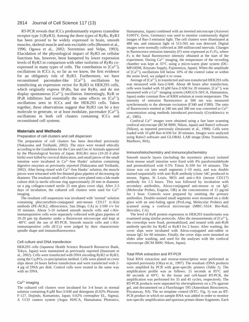
channel blocker, we examined drugs that could affectintracellular Ca2+ release channels. Fig. 1 shows examples ofthe effects of ryanodine on cell clusters with single (Fig. 1A)or multiple regions of [Ca2+]i oscillations (Fig. 1B). The tracesin Fig. 1Ab show changes in the fluorescence intensity (Ft/F0)measured in the square region indicated in Fig. 1Aa, panel 1.The Ca2+ images of Fig. 1Aa, panels 1-3 were recorded at thetimes indicated by lines 1-3 in Fig. 1Ab, respectively. In thepresence of 2 µM nifedipine, application of 1 µM ryanodinesignificantly reduced the amplitude and frequency of [Ca2+]ioscillations after 5 minutes. An increase in the ryanodineconcentration to 10 µM completely terminated [Ca2+]ioscillations after 5 minutes, and the subsequent washout ofryanodine for 5 minutes did not restore it. After examinationof the effects of ryanodine, the cell cluster was stained usingACK2. c-Kit-immunoreactivity was detected in the squareregion, indicating that [Ca2+]i oscillations in ICCs weremodulated by ryanodine. Similar effects of ryanodine wereobserved in three other cell clusters.
The cell cluster preparation shown in Fig. 1Ba containedmultiple regions of [Ca2+]i oscillations. The time courses ofchanges in [Ca2+]i in Fig. 1Bb were monitored in the boxedregions x (blue line) and y (red line). The [Ca2+]i oscillationsseen in the two regions of the cell cluster were wellsynchronised (in the presence of nifedipine). Application of 10µM ryanodine reduced both [Ca2+]i oscillation amplitude andfrequency in a time-dependent manner. As shown in the secondand third traces of Fig. 1Bb, [Ca2+]i in the x and y regions wereaffected similarly in terms of amplitude and frequency. Fig.1Bc shows the correlation between Ft/F0 in regions x and y for
Table 1. Primers for PCRPrimer sequence Product length GenBank
Clones (+) sense, (–) antisense Primer site (bp) accession no.
RyR1 (+) 5 ′-ATTACAGAGCAGCCCGAGGAT-3′ 450-470 113 X83932(–) 5 ′-AGAACCTTCCGCTTGACAAACT-3′ 541-562
RyR2 (+) 5 ′-CTTCGATGTTGGCCTTCAAGAG-3′ 432-453 100 NM_023868(–) 5 ′-CCAACACGCACTTTTTCTCCTT-3′ 512-533
RyR3 (+) 5 ′-GGCCAAGAACATCAGAGTGACTAA-3′ 385-408 101 AF111166(–) 5 ′-TCACTTCTGCCCTGTCAGTTTC-3′ 464-485
c-Kit (+) 5 ′-CAATGGAAGGTTGTCGAGGA-3′ 370-389 101 X58687(–) 5 ′-GCCTGTTTCTGGGAAACTCC-3′ 451-470
FKBP12 (+) 5 ′-ACTAGGCAAGCAGGAGGTGA-3′ 247-266 104 NM_008019(–) 5 ′-CTCCATAGGCATAGTCTGAGGAGAT-3′ 326-350
FKBP12.6 (+) 5 ′-AGAAGGCACTGCCCAGATGA-3′ 287-306 116 NM_016863(–) 5 ′-AAAGATGAGGGTGGCATTGG-3′ 385-404
GAPDH (+) 5 ′-CATGGCCTTCCGTGTTCCT-3′ 730-749 104 M32599(–) 5 ′-CCTGCTTCACCACCTTCTTGA-3′ 814-833

2816 Journal of Cell Science 117 (13)
2817Role of RyR3 and IP3R in pacemaker [Ca2+]i oscillation
the three traces shown in Fig. 1Bb. For each pair of Ft/F0, thecross correlation function was maximal (0.93-0.95) at t=0 (Fig.1Bd). When the [Ca2+]i oscillation amplitude was reducedsignificantly, the frequency also tended to decrease. The timefor [Ca2+]i oscillations to cease ranged from 3 to 18 minutesafter 10 µM ryanodine application (6.6±4.9 minutes). In theFt/F0 trace recorded before cessation, the mean amplitude andfrequency were 54±32% and 71±21% of the control,respectively.
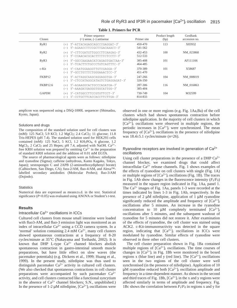
It is well known that caffeine modulates the properties ofryanodine receptors and amplifies Ca2+-induced Ca2+ release(Endo, 1977; Ogawa et al., 2002). In Fig. 2A, panels 1 and 2the Ca2+ images were obtained at resting and peak times of[Ca2+]i oscillations, respectively (indicated by lines 1 and 2 inFig. 2Ba). In the presence of nifedipine (2 µM), application of10 mM caffeine significantly suppressed [Ca2+]i oscillations.In all clusters examined (n=5), [Ca2+]i oscillations ceasedwithin 5 minutes, while in four out of the five clusters, as theamplitude decreased, the frequency also decreased. Theamplitude and frequency of [Ca2+]i oscillations in the tracesbefore cessation were 42±23% and 40±24% of the control,respectively (n=4). As shown in Fig. 2Bb, caffeine prominentlyreduced the [Ca2+]i oscillation frequency, but the [Ca2+]ioscillations seen over the pacemaker area were wellsynchronised (blue line from region x; red line from region y).The trace Fig. 2Bc was recorded 3 minutes after 10 mMcaffeine application, and the Ca2+ image in Fig. 2A, panel 3was obtained at the time indicated by line 3 in Fig. 2Bc. Theinhibitory effect of caffeine on [Ca2+]i oscillations was
reversible as shown in Fig. 2Bd. Lower concentrations (∼ 1mM) of caffeine produced similar inhibitory effects, but [Ca2+]ioscillations ceased within 5 minutes in only two out of fivepreparations.
In order to further confirm the involvement of RyR, weexamined effects of other drugs affecting RyR, (becausecaffeine is also known to interact with type 1 IP3R) (Maeset al., 1999; Maes et al., 2000; Bezprozvanny et al., 1994).Tetracaine and procine are known RyR blockers (e.g.Lukyanenko et al., 2001; Kimball et al., 1996). In the presenceof nifedipine (1 µM), application of 30 µM tetracaineterminated [Ca2+]i oscillations within 3-5 minutes (n=5) (Fig.2C). Also, 30 µM procaine terminated it within 5 minutes (n=4,not shown). By contrast, FK506 is known to modulate the RyRchannel function by binding to the 12 kDa FK506-bindingproteins (FKBP12 and FKBP12.6) (Masumiya et al., 2003;Bultynck et al., 2001). As shown in Fig. 2D, [Ca2+]ioscillations very rapidly ceased (1-2 minutes) after applicationof 10 µM FK506 (n=5).
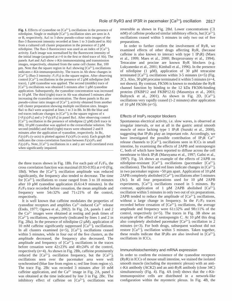
Effects of InsP3 receptor blockersSpontaneous electrical activity, i.e. slow waves, is observed atirregular intervals, or not at all in the gastric antral smoothmuscle of mice lacking type 1 IP3R (Suzuki et al., 2000),suggesting that IP3Rs play an important role. Accordingly, wehave investigated the possible contribution of IP3R Ca2+
release channels to [Ca2+]i oscillations seen in ICCs in smallintestine, by examining the effects of 2APB and xestosponginC, both of which have been reported to diffuse across the cellmembrane to block IP3R (Maruyama et al., 1997; Gafni et al.,1997). Fig. 3A shows an example of the effects of 2APB onnifedipine-resistant [Ca2+]i oscillations (pacemaker [Ca2+]ioscillations). The blue and red lines indicate changes in [Ca2+]iin two pacemaker regions ~50 µm apart. Application of 10 µM2APB completely abolished [Ca2+]i oscillations after 5 minutes(Ac). In all four preparations used for 10 µM 2APBexperiments, [Ca2+]i oscillations ceased in 5 minutes. Bycontrast, application of 1 µM 2APB abolished [Ca2+]ioscillation within 5 minutes in only two out of six preparations.2APB tended to reduce the amplitude of [Ca2+]i oscillations,without a large change in frequency. In the Ft /F0 tracesrecorded before cessation of [Ca2+]i oscillations, the averageamplitude and frequency were 61±32% and 98±11% of thecontrol, respectively (n=5). The traces in Fig. 3B show anexample of the effect of xestospongin C. At 10 µM this drugalso completely abolished pacemaker [Ca2+]i oscillation in 2minutes (n=3). For both drugs, subsequent washout did notrestore [Ca2+]i oscillation within 5 minutes. Taken together,these results indicate that IP3Rs are also involved in [Ca2+]ioscillations in ICCs.
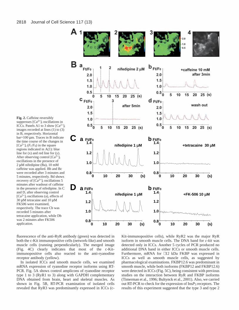
Immunohistochemistry and mRNA expressionIn order to confirm the existence of the ryanodine receptors(RyR) in ICCs of mouse small intestine, we stained the isolatedsmooth muscle (including the myenteric plexus) with an anti-c-Kit antibody (ACK2) and an anti-RyR antibody (clone 34C)simultaneously (Fig. 4). Fig. 4A (red) shows that the c-Kit-immunopositive cells are distributed in a network-likeconfiguration within the myenteric plexus. In Fig. 4B, the
Fig. 1.Effects of ryanodine on [Ca2+]i oscillations in the presence ofnifedipine. Single or multiple [Ca2+]i oscillation sites are seen in Aor B, respectively. Aa1 to 3 show pseudo-colour ratio images of thefluo-3 fluorescent intensity recorded at lines 1 to 3 (indicated in Ab)from a cultured cell cluster preparation in the presence of 2 µMnifedipine. The fluo-3 fluorescence was used as an index of [Ca2+]iactivity. Each image was normalised by the fluorescent intensity ofthe initial image (acquired at t=0 in the first or third trace of Ab). Thepanels Aa4 and Aa5 show c-Kit-immunostaining and transmissionimages, respectively, obtained from the same cell cluster. Bar, 100µm. Note that the square region in Aa1 showing [Ca2+]i oscillationsindicates c-Kit-immunopositivity. The traces in Ab show changes in[Ca2+]i (fluo-3 intensity: Ft /F0) in the square region. After observingcontrol [Ca2+]i oscillations in the presence of 2 µM nifedipine (lefttrace), 1 µM ryanodine was applied. The second (middle) trace of[Ca2+]i oscillations was obtained 5 minutes after 1 µM ryanodineapplication. Subsequently, the ryanodine concentration was increasedto 10 µM. The third (right) trace in Ab was obtained 5 minutes afterincreasing the ryanodine concentration. The three panels in Ba arepseudo-colour ratio images of [Ca2+]i activity obtained from anothercell cluster preparation showing multiple oscillation sites. ImagesBa1 to Ba3 were acquired at lines 1 to 3 in Bb. In Bb the blue andred traces indicate changes in [Ca2+]i in the square regions of x[=Ft(x)/F0] and y [=Ft(y)/F0] in panel Ba1. After observing control[Ca2+]i oscillation in the presence of nifedipine (2 µM) (left trace inBb), 10 µM ryanodine was applied to the extracellular solution. Thesecond (middle) and third (right) traces were obtained 2 and 8minutes after the application of ryanodine, respectively. In Bc,Ft(y)/F0 (y-axis) is plotted against Ft(x)/F0 (x-axis). Each graph Bdindicates the cross-correlation function between Ft(x)/F0 andFt(y)/F0. Note, [Ca2+]i oscillations in x and y are well correlated evenwhen significantly impaired.
2818
fluorescence of the anti-RyR antibody (green) was detected inboth the c-Kit immunopositive cells (network-like) and smoothmuscle cells (running perpendicularly). The merged image(Fig. 4C) clearly indicates that most of the c-Kit-immunopositive cells also reacted to the anti-ryanodinereceptor antibody (yellow).
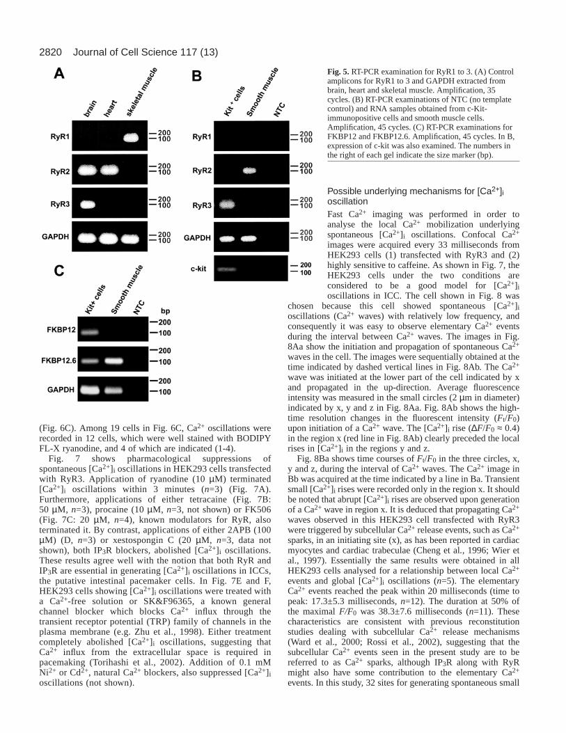
In isolated ICCs and smooth muscle cells, we examinedmRNA expression of ryanodine receptor isoforms using RT-PCR. Fig. 5A shows control amplicons of ryanodine receptortype 1 to 3 (RyR1 to 3) along with GAPDH complementaryDNA obtained from brain, heart and skeletal muscles. Asshown in Fig. 5B, RT-PCR examination of isolated cellsrevealed that RyR3 was predominantly expressed in ICCs (c-
Kit-immunopositive cells), while RyR2 was the major RyRisoform in smooth muscle cells. The DNA band for c-kit wasdetected only in ICCs. Another 5 cycles of PCR produced noadditional DNA band in either ICCs or smooth muscle cells.Furthermore, mRNA for ∼ 12 kDa FKBP was expressed inICCs as well as smooth muscle cells, as suggested bypharmacological examinations. FKBP12.6 was predominant insmooth muscle, while both isoforms (FKBP12 and FKBP12.6)were detected in ICCs (Fig. 5C), being consistent with previousstudies on the interaction between RyR and FKBP isoforms(Timerman et al., 1996; Bultynck et al., 2001). Also, we carriedout RT-PCR to check for the expression of InsP3 receptors. Theresults of this experiment suggested that the type 3 and type 2
Journal of Cell Science 117 (13)
010
C F/tF 0
8.0
0.1
2.1
02
D
4.1 1 enipidefin µM
)s(03
03 eniacartet+ µM
F/tF 0a b
a b 1 enipidefin µ 01 605-KF+M µM
F/tF 0F/tF 0
8.0
0.1
2.1
4.1
8.0
0.1
2.1
4.1
8.0
0.1
2.1
4.1
01 )s(03020
01 01)s(03020 )s(03020
Fig. 2.Caffeine reversiblysuppresses [Ca2+]i oscillations inICCs. Panels A1 to 3 show [Ca2+]iimages recorded at lines (1) to (3)in B, respectively. Horizontalbar=100 µm. Traces in B indicatethe time course of the changes in[Ca2+]i (Ft /F0) in the squareregions indicated in A(1): blueline for (x) and red line for (y).After observing control [Ca2+]ioscillations in the presence of2 µM nifedipine (Ba), 10 mMcaffeine was applied. Bb and Bcwere recorded after 3 minutes and5 minutes, respectively. Bd showsrecovery of [Ca2+]i oscillations 5minutes after washout of caffeinein the presence of nifedipine. In Cand D, after observing control[Ca2+]i oscillations (a), effects of30 µM tetracaine and 10 µMFK506 were examined,respectively. The trace Cb wasrecorded 5 minutes aftertetracaine application, while Dbwas 2 minutes after FK506application.

2819Role of RyR3 and IP3R in pacemaker [Ca2+]i oscillation
isoforms of IP3Rs were the major components in smoothmuscle and ICCs, respectively (data not shown).
Reconstituted pacemaker-like [Ca2+]i oscillations inHEK293 cellsIn order to confirm the contribution of RyR3 to [Ca2+]ioscillations seen in ICCs, we examined the reconstitution offunctional RyR3 in HEK293 cells, which originally onlyexpress IP3R. According to RT-PCR results, mRNAs of allthree IP3R types were expressed in native HEK293 cells in theorder of IP3R3 > IP3R2 > IP3R1, but mRNAs of RyR subtypeswere not detected after 45 cycles of amplification (data notshown).
[Ca2+]i was measured with a fluorescent Ca2+ indicator(Fura-2) 2 days after the transfection. Approximately 10% ofHEK293 cells showed spontaneous global [Ca2+]i oscillations.Fig. 6Aa (black line) demonstrates spontaneous oscillations of[Ca2+]i over the whole cell area and the elevation of [Ca2+]i inresponse to 1 mM caffeine. Further addition of caffeine did notinduce marked response. Approximately 30% of HEK293 cellsresponded to 1 mM caffeine, suggesting that the efficacy forthe functional expression of RyR3 was 30%. As shown by thered line in Fig. 6Aa, the remaining HEK cells (~70%) did notrespond to caffeine in the range of 1 and 10 mM, but did
respond to 1 µM ACh. Fig. 6Ab shows Ca2+ images acquiredunder resting conditions and in the presence of 1 mM caffeineor caffeine plus 1 µM ACh and an image followingimmunocytochemical staining with anti-RyR antibody.Application of 1 mM caffeine increased [Ca2+]i in two cellsindicated by white arrowheads, but not in other cells.Substantial expression of RyR3 proteins was identified in cellssensitive to caffeine without exception (n>200), indicating adirect correlation between high sensitivity to caffeine and thesuccessful expression of RyR3 proteins in transfected cells.Application of 1 µM ACh in the presence of caffeine evoked[Ca2+]i transients in caffeine-insensitive cells. The resultssuggest that RyR3 expressed in the HEK293 cells shares theCa2+ storage sites with IP3R.
Fig. 6B demonstrates typical spontaneous [Ca2+]ioscillations in HEK293 cells transfected with RyR3. Two cells(1 and 2) in the confocal Ca2+ images exhibited Ca2+
oscillations, which occurred as Ca2+ waves in each cell. Ca2+
waves started from a particular initiation point in each cell, asindicated by white arrowheads in cell 1. The frequency of Ca2+
oscillations was 1.3±0.6 cycles/minute (n=39 from separate 8groups of transfection). The average value of the peak [Ca2+]iwas 1.6±1.4 µM (n=24). In some experiments, the relationbetween RyR3 protein expression and the generation of Ca2+
oscillation was also examined using BODIPY FL-X ryanodine
Fig. 3.Examples of the effectsof InsP3 receptor blockers. Thetraces in A and B were obtainedfrom regions showing [Ca2+]ioscillations in different cellcluster preparations. In A, afterobserving control [Ca2+]ioscillations in the presence ofnifedipine (2 µM) (Aa), 10 µM2APB was applied. The traceAb was recorded 3 minutes after10 µM 2APB application. Blueand red lines indicate changes in[Ca2+]i measured in twodifferent regions at a distance of~50 µm. This distance iscomparable to that for regions xand y shown in Fig. 1Ba1 andFig. 2A1. In B, after observing a control in the presence of nifedipine (Ba), 10 µM xestospongin C was applied. The trace Bb was obtained 2minutes after the application of xestospongin C.
2.1
2 enipidefin µMa
030201 05040 )s(8.00.1
F/tF 0A
4.1
b 01 BPA2+ µM
01 C nignopsotsex+ µM 2 enipidefin µMa
51015 52020 )s(6.08.0
F/tF 0B
0.12.1
b4.1
51015 )s(520206.08.0
F/tF 0
0.12.14.1
6.1
030201 05040 )s(8.00.1
F/tF 0
2.14.16.1
Fig. 4. Immunohistochemistry for c-Kit andryanodine receptors. Smooth muscle (including themyenteric plexus) of mouse small intestine wasdouble-labelled with anti-c-Kit antibody (A: ACK2with Alexa 488, red) and anti-ryanodine receptorantibody (B: C34 with Alexa 594, green). C shows amerged image. Note that network-like cells weremostly stained in yellow, indicating that ICCs containryanodine receptors.
2820
(Fig. 6C). Among 19 cells in Fig. 6C, Ca2+ oscillations wererecorded in 12 cells, which were well stained with BODIPYFL-X ryanodine, and 4 of which are indicated (1-4).
Fig. 7 shows pharmacological suppressions ofspontaneous [Ca2+]i oscillations in HEK293 cells transfectedwith RyR3. Application of ryanodine (10 µM) terminated[Ca2+]i oscillations within 3 minutes (n=3) (Fig. 7A).Furthermore, applications of either tetracaine (Fig. 7B:50 µM, n=3), procaine (10 µM, n=3, not shown) or FK506(Fig. 7C: 20 µM, n=4), known modulators for RyR, alsoterminated it. By contrast, applications of either 2APB (100µM) (D, n=3) or xestospongin C (20 µM, n=3, data notshown), both IP3R blockers, abolished [Ca2+]i oscillations.These results agree well with the notion that both RyR andIP3R are essential in generating [Ca2+]i oscillations in ICCs,the putative intestinal pacemaker cells. In Fig. 7E and F,HEK293 cells showing [Ca2+]i oscillations were treated witha Ca2+-free solution or SK&F96365, a known generalchannel blocker which blocks Ca2+ influx through thetransient receptor potential (TRP) family of channels in theplasma membrane (e.g. Zhu et al., 1998). Either treatmentcompletely abolished [Ca2+]i oscillations, suggesting thatCa2+ influx from the extracellular space is required inpacemaking (Torihashi et al., 2002). Addition of 0.1 mMNi2+ or Cd2+, natural Ca2+ blockers, also suppressed [Ca2+]ioscillations (not shown).
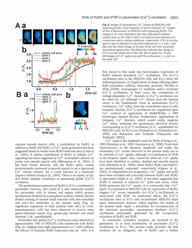
Possible underlying mechanisms for [Ca2+]ioscillationFast Ca2+ imaging was performed in order toanalyse the local Ca2+ mobilization underlyingspontaneous [Ca2+]i oscillations. Confocal Ca2+
images were acquired every 33 milliseconds fromHEK293 cells (1) transfected with RyR3 and (2)highly sensitive to caffeine. As shown in Fig. 7, theHEK293 cells under the two conditions areconsidered to be a good model for [Ca2+]ioscillations in ICC. The cell shown in Fig. 8 was
chosen because this cell showed spontaneous [Ca2+]ioscillations (Ca2+ waves) with relatively low frequency, andconsequently it was easy to observe elementary Ca2+ eventsduring the interval between Ca2+ waves. The images in Fig.8Aa show the initiation and propagation of spontaneous Ca2+
waves in the cell. The images were sequentially obtained at thetime indicated by dashed vertical lines in Fig. 8Ab. The Ca2+
wave was initiated at the lower part of the cell indicated by xand propagated in the up-direction. Average fluorescenceintensity was measured in the small circles (2 µm in diameter)indicated by x, y and z in Fig. 8Aa. Fig. 8Ab shows the high-time resolution changes in the fluorescent intensity (Ft/F0)upon initiation of a Ca2+ wave. The [Ca2+]i rise (∆F/F0 ≈ 0.4)in the region x (red line in Fig. 8Ab) clearly preceded the localrises in [Ca2+]i in the regions y and z.
Fig. 8Ba shows time courses of Ft/F0 in the three circles, x,y and z, during the interval of Ca2+ waves. The Ca2+ image inBb was acquired at the time indicated by a line in Ba. Transientsmall [Ca2+]i rises were recorded only in the region x. It shouldbe noted that abrupt [Ca2+]i rises are observed upon generationof a Ca2+ wave in region x. It is deduced that propagating Ca2+
waves observed in this HEK293 cell transfected with RyR3were triggered by subcellular Ca2+ release events, such as Ca2+
sparks, in an initiating site (x), as has been reported in cardiacmyocytes and cardiac trabeculae (Cheng et al., 1996; Wier etal., 1997). Essentially the same results were obtained in allHEK293 cells analysed for a relationship between local Ca2+
events and global [Ca2+]i oscillations (n=5). The elementaryCa2+ events reached the peak within 20 milliseconds (time topeak: 17.3±5.3 milliseconds, n=12). The duration at 50% ofthe maximal F/F0 was 38.3±7.6 milliseconds (n=11). Thesecharacteristics are consistent with previous reconstitutionstudies dealing with subcellular Ca2+ release mechanisms(Ward et al., 2000; Rossi et al., 2002), suggesting that thesubcellular Ca2+ events seen in the present study are to bereferred to as Ca2+ sparks, although IP3R along with RyRmight also have some contribution to the elementary Ca2+
events. In this study, 32 sites for generating spontaneous small
Journal of Cell Science 117 (13)
Fig. 5.RT-PCR examination for RyR1 to 3. (A) Controlamplicons for RyR1 to 3 and GAPDH extracted frombrain, heart and skeletal muscle. Amplification, 35cycles. (B) RT-PCR examinations of NTC (no templatecontrol) and RNA samples obtained from c-Kit-immunopositive cells and smooth muscle cells.Amplification, 45 cycles. (C) RT-PCR examinations forFKBP12 and FKBP12.6. Amplification, 45 cycles. In B,expression of c-kit was also examined. The numbers inthe right of each gel indicate the size marker (bp).

2821Role of RyR3 and IP3R in pacemaker [Ca2+]i oscillation
subcelluar [Ca2+]i rises were observed in 29 cells prepared by9 separate transfections, and all of these sites tested (n=20)showed RyR3 immunofluorescence reactivity. The size ofelementary [Ca2+]i rises at the peak amplitude was similar tothat of punctuated staining patterns of RyR3 protein (2.1±0.3µm vs. 1.9±0.3 µm, n=13). Small [Ca2+]i rises occurredrepetitively in the same spatial location of a cell with a meanfrequency of 2.9±1.2 events/second (n=18). These resultsstrongly suggest that the sites initiating [Ca2+]i oscillationscorrespond to regions where RyR3 are expressed in a clusteredfashion.
DiscussionThe cultured cell cluster preparation that we have recentlydeveloped (Nakayama and Torihashi, 2002) contains smoothmuscle, enteric neurones and c-Kit-immunopositive interstitial
cells (ICCs). The last cell member is the putative pacemakerof the gastro-intestinal tract (Suzuki, 2000; Thuneberg,1982; Torihashi et al., 1995; Komuro, 1999). In addition,neurotransmission is well known to modulate the spontaneousrhythmicity and contractility in gastro-intestinal smoothmuscle tissues (although nervous activity did not seem tocontribute basal [Ca2+]i oscillations in our preparation becausetetrodotoxin had no effect). This cell cluster preparation istherefore considered to consist of the essential minimum cellmembers necessary to investigate mechanisms underlyinggastro-intestinal motility and pacemaker function.
In the present study, we demonstrate that reasonably lowconcentrations (1-10 µM) of ryanodine suppressed andeventually terminated [Ca2+]i oscillations in ICCs. This isprobably because the small size of the cell cluster preparationenables a sufficient amount of ryanodine molecules to enterICCs relatively rapidly. Further evidence is provided by the
Fig. 6. Spontaneous Ca2+ oscillations weredetected in HEK293 cells transfected withRyR3. (Aa) The time-course of [Ca2+]ichanges in HEK293 cell expressing RyR3(black line) and a non-expressing cell (redline). Cells were sequentially treated with1, 2, 5 and 10 mM caffeine and 1 µM ACh.(Ab) Ca2+ images in cells loaded with fura-2AM were obtained during restingconditions and in the presence of 1 mMcaffeine or 1 µM ACh. The bottom-rightpanel in Ab indicates theimmunofluorescence staining of RyR. It isnoted that cells responding to 1 mMcaffeine were stained withimmunofluorescent RyR3 antibody. Bars inAb, 20 µm. (B) The time courses ofspontaneous [Ca2+]i oscillations (a) wereobtained from cells (1 and 2) as indicatedin a series of confocal images (b). TheCa2+ images were obtained every 3seconds, and cells were loaded with fluo-4for 15 minutes. (C) Confocal imagesfollowing fluo-4 loading (a) and stainingwith BODIPY FL-X ryanodine (b) inHEK293 cells transfected with RyR3.(a) Cells were loaded with fluo-4. [Ca2+]ioscillations were then recorded in fourindicated cells (1-4) among the 19 in thisframe. (b) After recording [Ca2+]ioscillation, cells were stained withBODIPY FL-X ryanodine for 5 minutesand washed. Among the 19 cells in theframe, 12 cells, including all four cellsexhibiting [Ca2+]i oscillations were wellstained with BODIPY FL-X ryanodine.(c) The time courses of [Ca2+]i oscillationsof four cells in the frame.

2822
effects of other drugs affecting RyRs, intracellular Ca2+ releasechannels. As shown in Fig. 2C,D and Fig. 3, application ofthese drugs (i.e. tetracaine, FK506) completely abolished[Ca2+]i oscillations at relatively low concentrations (~10 µM).By contrast, IP3R blockers also eliminated [Ca2+]i oscillationsin ICCs (Fig. 3), as described below. Taken together, the resultsare the first pharmacological evidence for involvement of bothRyR3 and IP3R in terms of pacemaker [Ca2+]i oscillations inICCs. The fact that many drugs, including ryanodine, areeffective suggests that use of this preparation is advantageousin pharmacological investigations of [Ca2+]i oscillations. [Inprevious reports, greater concentrations (>50 µM) of ryanodinefor longer exposure times compared with the presentexperiments failed to terminate pacemaker potentials in thegastro-intestinal smooth muscle tissues (e.g. Malysz et al.,2001).]
Periodic Ca2+ release from the intracellular Ca2+ store isconsidered to be the fundamental event in ICC pacemaking,because Ca2+-dependent ion permeability, i.e. Ca2+-activatedCl– channels, has been suggested to underlie electricaloscillation (Tokutomi et al., 1995; Edwards et al., 1999;Huizinga et al., 2002). The present experiments indicate thatboth RyR and IP3R occur in ICCs. It has been proposed thatsome smooth muscles have distinct types of ER (endoplasmicreticulum) in terms of the distribution of RyR and IP3R (Iino,1991; Yamazawa et al., 1992), but that other smooth musclesdo not. Since application of either drugs related to RyR or IP3Rterminated [Ca2+]i oscillations, both receptors appear to existin the ER responsible for the periodic Ca2+ release in ICCs.
2APB and xestospongin C are known to affect othermechanisms for [Ca2+]i regulation (Bootman et al., 2002). Forexample, 100 µM xestospongin C largely inhibits Ca2+ influx
through endoplasmic reticulum Ca2+ pumps (SERCA). At theconcentration (10 µM) used in the present study, this drug,however, has little effect on SERCA (De Smet et al., 1999). Asdescribed above, lines of evidence have been shown for theinvolvement of IP3R in spontaneous rhythmicity in ICCs (e.g.Suzuki et al., 2000). The present results with 2APB andxestospongin C appear to support the notion for the importanceof IP3R. However, we have previously shown that Ca2+ influxfrom the extracellular space (presumably via TRP homologuechannels) is also required to maintain pacemaker activity inICCs (Nakayama and Torihashi, 2002; Torihashi et al., 2002).In the light of the essential similarity between 2APB andxestospongin C (although the evidence for inhibition of store-operated Ca2+ entry has not yet been reported for xestosponginC) (Bootman et al., 2002), we can also speculate that IP3Rmight additionally contribute to [Ca2+]i oscillations in ICCs viastore-operated types of Ca2+ entry processes.
RT-PCR examinations in the present study revealed thatICCs in murine small intestine predominantly express RyR3,while the major RyR isoform in smooth muscle is RyR2. RyR3is known as a brain type isoform, because of its characteristic,but faint, expression in several brain regions, i.e. the corpusstriatum, hippocampus and thalamus (Ogawa et al., 2002).RyR3 is also expressed in skeletal and smooth muscles, albeitat low levels. RyR3 in skeletal muscle may amplify Ca2+
release originally elicited by activation of RyR1 (Ogawa et al.,2002; Rossi and Sorrentino, 2002; Yang et al., 2001). Althoughthe presence of the RyR3 isoform is recognised in numeroustissues and organs, predominant expression over RyR1 orRyR2 has been shown only in non-pregnant uterus, whereRyR3 may contribute Ca2+ signalling only when SR isoverloaded with Ca2+ (Mironneau et al., 2002). In arterial
Journal of Cell Science 117 (13)
Fig. 7.Pharmacologicalcharacterisation of spontaneousCa2+ oscillations in HEK293 cellstransfected with RyR3. In A-F,effects of the following drugs ortreatments were examined:(A) 10 µM ryanodine;(B) 10-50µM tetracaine;(C) 20µM FK506 after transientapplication of caffeine;(D) 100µM 2APB; (E) removal ofexternal Ca2+; (F) application of40 µM SK&F96365.
2823Role of RyR3 and IP3R in pacemaker [Ca2+]i oscillation
vascular smooth muscle cells, a contribution by RyR3, inaddition to RyR1 and RyR2, to Ca2+ spark generation has beensuggested based on results from RyR3-deficient mice (Lohn etal., 2001). A similar contribution of RyR3 to cellular Ca2+
signalling has been suggested in Ca2+-overloaded cultured ratportal vein smooth muscle cells (Mironneau et al., 2001). Ithas been found, however, that the RyR3 splice variantpredominantly expressed in smooth muscle is not a functionalCa2+ release channel, but it could function in a dominantnegative fashion (Jiang et al., 2003). There is no report, so far,that RyR3 subtype contributes to generating global [Ca2+]ioscillation.
The predominant expression of RyR3 in ICCs contributed topacemaker function, and could be a new molecular markerfor pacemaker cells in tissues and organs which possessspontaneous rhythmicity in peripheral systems. Indeed, like thedouble staining of murine small intestine with anti-ryanodineand anti-c-Kit antibodies in the present study (Fig. 4),significant expression of RyR (stained with 34C) in c-Kit-immunopositive cells has been observed in several othergastro-intestinal tissues (e.g. guinea-pig stomach and smallintestine; J.W., unpublished).
Pacemaker-like global [Ca2+]i oscillations were observed inapproximately 10% of HEK293 cells transfected with RyR3(Fig. 6). Judging from high responsiveness to 1 mM caffeine,the efficacy of transient RyR3 expression may be ~30%. It is
first shown in this study that heterologous expression ofRyR3 induced periodical Ca2+ oscillations. The [Ca2+]ioscillations seen in the HEK293 cells and ICCs share thefollowing features. (1) Applications of drugs affecting eitherRyR (ryanodine, caffeine, tetracaine, procaine, FK506) orIP3R (2APB, xestospongin C) modulate and/or terminate[Ca2+]i oscillations. In both cases, the contribution ofvoltage-dependent Ca2+ channels to [Ca2+]i oscillation canbe ruled out. (2) Although Ca2+ release from the internalstores is the fundamental event in spontaneous [Ca2+]ioscillations, Ca2+ influx from the extracellular space is alsoessential. Namely, [Ca2+]i oscillations are suppressed uponCa2+ removal or application of SK&F96365, a TRPhomologue channel blocker. Furthermore, applications ofinorganic Ca2+ blockers, which would totally suppressCa2+ influx, terminate the spontaneous electrical activitycorresponding to [Ca2+]i oscillation [A.Y., unpublished forHEK293 cells; for ICCs, see Torihashi et al. (Torihashi et al.,2002) and Nakayama and Torihashi (Nakayama andTorihashi, 2002)].
Ca2+ spark is the landmark event for RyRs (Cheng et al.,1993; Bootman et al., 1995; Imaizumi et al., 1998). From theircharacteristics in the duration, amplitude and width, theelementary Ca2+ events observed in the present study are tobe referred to Ca2+ sparks, although co-contribution of IP3Rin the frequent ‘spark’ sites, cannot be ruled out. Ca2+ sparkshave been identified in cardiac, skeletal and smooth musclecells (Bootman et al., 1995), where RyR2, RyR1 and RyR2/3are predominantly expressed, respectively (Ogawa et al.,2002). In oligodendrocyte progenitors, Ca2+ sparks and puffshave been recorded and cross-talk between RyR3 and IP3R2is speculated (Haak et al., 2001). It has been suggested thatIP3R2 is essential to induced Ca2+ puffs and waves, whileRyR3 generates the Ca2+ sparks. It is noteworthy that ‘Ca2+
spark’ reconstituted in HEK293 cells by expression of RyR3,triggers Ca2+ waves repeatedly (i.e.=[Ca2+]i oscillations) inassociation with IP3R. As mentioned above, the [Ca2+]ioscillations seen in ICCs and reconstituted HEK293 sharemany characteristic features. Taken together, the results ofthe present study imply a possibility for reconstruction ofthe intestinal pacemaker: spontaneous rhythmic [Ca2+]ioscillations presumably generated by the co-operativeactivation of RyR3 and IP3R.
In conclusion, ryanodine receptors are involved in thegeneration, or at least modulation, of pacemaker [Ca2+]ioscillations in ICCs. The present study provides the firstevidence for an obligatory role of RyR3, and is further
Fig. 8. Images of spontaneous Ca2+ release in HEK293 cellsexpressing RyR3. (Aa) Sequence of seven confocal Ca2+ imagesof fluo-4 fluorescence in HEK293 cells expressing RyR3. Theimages in Aa were obtained at the time indicated by dashedvertical lines in Ab. (Ab) F and F0 are fluorescence intensity at acertain time and at resting conditions, respectively. F/F0 in circularspots (2 µm in diameter) at the point indicated by x, y and z, andthat over the whole image of the part of the cell were measuredand plotted against time. The black line indicates the change inF/F0 over the whole area of the cell. Bar in panel Aa, 10 µm.(B) Images of Ca2+ sparks (a) and F/F0 in the areas x, y and z inthe same cell.

2824
supported by the global [Ca2+]i oscillations reconstituted withRyR3 in HEK293 cells.
This work was supported by grants-in-aid for scientific researchfrom the Japan Society for the Promotion of Science, research grantsfrom the ministry of Health and Welfare (to S.N. and Y.I.) and byresearch grants from the Canadian Institutes of Health Research (toS.R.W.C.). The authors are grateful to Alison F. Brading and JeffBolstad for critical reading of the manuscript.
ReferencesBeech, D. J. and McHugh, D. (1996). Regulation of opening of voltage-gated
Ca channels in smooth muscle cells. In Smooth Muscle Excitation(ed. T. B.Bolton and T. Tomita), pp. 39-54. London, UK: Academic Press
Bennett, D. L., Cheek, T. R., Berridge, M. J., de Smedt, H., Parys, J. B.,Missiaen, L. and Bootman, M. D. (1996). Expression and function ofryanodine receptors in nonexcitable cells. J. Biol. Chem. 271, 6356-6362.
Bezprozvanny, I., Bezprozvannaya, S. and Ehrlich, B. E. (1994). Caffeineinduced inhibition of inositol 1,4,5-trisphosphate-gated calcium channelsfrom cerebellum. Mol. Biol. Cell 5, 97-103.
Bootman, M. D. and Berridge, M. J. (1995). The elemental principles ofcalcium signaling. Cell 83, 675-678.
Bootman, M. D., Colins, T. J., Mackenzie, H., Roderick, H. L., Berridge,M. J. and Peppiatt, C. M. (2002). 2-Aminoethoxydiphenyl borate (2-APB)is a reliable blocker of store-operated Ca2+ entry but an inconsistent inhibitorof InsP3-induced Ca2+ release FASEB J. 16, 1145-1150.
Bultynck, G., Rossi, D., Callewaert, G., Missiaen, L., Sorrentino, V.,Parys, J. B. and de Smedt, H. (2001). The conserved sites for the FK506-binding proteins in ryanodine receptors and inositol 1,4,5-trisphosphatereceptors are structurally and functionally different. J. Biol. Chem. 276,47715-47724.
Cheng, H., Lederer, W. J. and Cannell, M. B. (1993). Calcium sparks:elementary events underlying excitation-contraction coupling in heartmuscle. Science262, 740-744.
Cheng, H., Lederer, M. R., Lederer, W. J. and Cannell, M. B. (1996).Calcium sparks and [Ca2+]i waves in cardiac myocytes. Am. J. Physiol. 270,C148-C159.
De Smet, P., Parys, J. B., Callewart, G., Weidema, A. F., Hill, E., de Smet,H., Erneux, C., Sorrentino, V. and Messiaen, L. (1999). Xestospongin Cis an equally potent inhibitor of the inositol 1,4,5-trisphosphate receptor andthe endoplasmic reticulum. Cell Calcium26, 9-13.
Dickens, E. J., Hirst, G. D. S. and Tomita, T. (1999). Identification ofrhythmically active cells in guinea-pig stomach. J. Physiol. 514, 515-531.
Edwards, F. R., Hirst, G. D. S. and Suzuki, H. (1999). Unitary nature ofregenerative potentials recorded from circular muscle of guinea-pig antrum.J. Physiol. 519, 235-250.
Endo, M. (1977). Calcium release from sarcoplasmic reticulum. Physiol. Rev.57, 71-108.
Gafni, J., Munsch, J. A., Lam, T. H., Catlin, M. C., Costa, L. G., Molinski,T. F. and Pessah, I. N. (1997). Xestospongins: potent membrane permeableblockers of the inositol 1,4,5-trisphosphate receptor. Neuron19, 723-733.
Galione, A. and Churchill, G. C. (2002). Interactions between calciumrelease pathways: multiple messengers and multiple stores. Cell Calcium32, 343-354.
Grynkiewicz, G., Poenie, M. and Tsien, R. Y. (1985). A new generation ofCa2+ indicators with greatly improved fluorescence properties. J. Biol.Chem. 260, 3440-3450.
Haak, L. L., Song, L. S., Molinski, T. F., Pessah, I. N., Cheng, H. andRussell, J. T. (2001). Sparks and puffs in oligodendrocyte progenitors: crosstalk between ryanodine receptors and inositol trisphosphate receptors. J.Neurosci. 21, 3860-3870.
Hirst, G. D. S. and Edwards, F. R. (2001). Generation of slow waves in theantral region of guinea-pig stomach – a stochastic process. J. Physiol. 535,165-180.
Huang, S. M., Nakayama, S., Iino, S. and Tomita, T. (1999). Voltagesensitivity of slow wave frequency in isolated circular muscle strips fromguinea pig gastric antrum. Am. J. Physiol. 276, G518-G528.
Huizinga, J. D., Zhu, Y., Ye, J. and Molleman, A. (2002). High-conductancechloride channels generate pacemaker currents in interstitial cells of CajalGastroenterology123, 1627-1636.
Iino, M. (1991). Effects of adenine nucleotides on inositol 1,4,5-trisphosphate-
induced calcium release in vascular smooth muscle. J. Gen. Physiol. 98,681-698.
Imaizumi, Y., Torii, Y., Ohi, Y., Nagano, N., Atsuki, K., Yamamura, H.,Muraki, K., Watanabe, M. and Bolton, T. B. (1998). Ca2+ images and K+
current during depolarization in smooth muscle cells of the guinea-pig vasdeferens and urinary bladder. J. Physiol. 510, 705-719.
Imaizumi, Y., Sakamoto, K., Yamada, A., Hotta, A., Ohya, S., Muraki, K.,Uchiyama, M. and Ohwada, T. (2002). Molecular basis of pimaranecompounds as novel activators of large-conductance Ca2+-activated K+
channel alpha-subunit. Mol. Pharmacol. 62, 836-846.Jiang, D., Xiao, B., Li, X. and Chen, S. R. (2003). Smooth muscle tissues
express a major dominant negative splice variant of the type 3 Ca2+ releasechannel (ryanodine receptor).J. Biol. Chem. 278, 4763-4769.
Kimball, B. C., Yule, D. I. and Mulholland, M. W. (1996). Caffeine- andryanodine-sensitive Ca2+ stores in cultured guinea pig myenteric neurons.Am. J. Physiol. 270, G594-G603.
Koh, S. D., Kim, T. W., Jun, Y. J., Glasgow, N. J., Ward, S. M. and Sanders,K. M. (2000). Regulation of pacemaker currents in interstitial cells of Cajalfrom murine small intestine by cyclic nucleotides. J. Physiol.527, 149-162.
Komuro, T. (1999). Comparative morphology of interstitial cells of Cajal:ultrastructural characterization. Microsc. Res. Tech. 47, 267-285.
Lohn, M., Jessner, W., Furstenau, M., Wellner, M., Sorrentino, V., Haller,H., Luft, F. C. and Gollasch, M. (2001). Regulation of calcium sparks andspontaneous transient outward currents by RyR3 in arterial vascular smoothmuscle cells. Circ. Res. 89, 941-943.
Lukyanenko, V., Viatchenko-Karpinski, S., Smirnov, A., Wiesner, T. F. andGyorke, S. (2001). Dynamic regulation of sarcoplasmic reticulum Ca2+
content and release by luminal Ca2+-sensitive leak in rat ventricularmyocytes. Biophys. J. 81, 785-798.
Maes, K., Missiaen, L., Parys, J. B., Sienaert, I., Bultynck, G., Zizi, M.,De Smet, P., Casteels, R. and De Smedt, H. (1999). Adenine-nucleotidebinding sites on the inositol 1,4,5-trisphosphate receptor bind caffeine, butnot adenophostin A or cyclic ADP-ribose. Cell Calcium 25, 143-152.
Maes, K., Missiaen, L., De Smet, P., Vanlingen, S., Callewaert, G., Parys,J. B. and De Smedt, H. (2000). Differential modulation of inositol 1,4,5-trisphosphate receptor type 1 and type 3 by ATP. Cell Calcium 27, 257-267.
Malysz, J., Donnelly, G. and Huizinga, J. D. (2001). Regulation of slow wavefrequency by IP3-sensitive calcium release in the murine small intestine. Am.J. Physiol. 280, G439-G448.
Maruyama, T., Kanaji, T., Nakade, S., Kanno, T. and Mikoshiba, K.(1997). 2APB, 2-aminoethoxydiphenyl borate, a membrane-penetrablemodulator of Ins(1,4,5)P3-induced Ca2+ release. J. Biochem.122, 498-505.
Masgrau, R., Churchill, G. C., Morgan, A. J., Ashcroft, S. J. and Galione,A. (2003). NAADP: a new second messenger for glucose-induced Ca2+
responses in clonal pancreatic beta cells. Curr. Biol. 13, 247-251.Masumiya, H., Wang, R., Zhang, J., Xiao, B. and Chen, S. R. (2003).
Localization of the 12.6-kDa FK506-binding protein (FKBP12.6) bindingsite to the NH2-terminal domain of the cardiac Ca2+ release channel(ryanodine receptor). J. Biol. Chem. 278, 3786-3792.
Mironneau, J., Coussin, F., Jeyakumar, L. H., Fleischer, S., Mironneau, C.and Macrez, N. (2001). Contribution of ryanodine receptor subtype 3 toCa2+ responses in Ca2+-overloaded cultured rat portal vein myocytes. J. Biol.Chem. 276, 11257-11264.
Mironneau, J., Macrez, N., Morel, J. L., Sorrentino, V. and Mironneau,C. (2002). Identification and function of ryanodine receptor subtype 3 innon-pregnant mouse myometrial cells. J. Physiol. 538, 707-716.
Nakayama, S., Smith, L. M., Tomita, T. and Brading, A. F. (1996). Multipleopen states of calcium channels and their possible kinetic schemes. InSmooth Muscle Excitation.(ed. T. B. Bolton, and T. Tomita), pp. 13/25.London, UK: Academic Press.
Nakayama, S. and Torihashi, S. (2002). Spontaneous rhythmicity in culturedcell clusters isolated from mouse small intestine. Jpn. J. Physiol. 52, 217-227.
Ogawa, Y., Murayama, T. and Kurebayashi, N. (2002). Ryanodine receptorisoforms of non-mammalian skeletal muscle. Front. Biosci. 7, d1187-d1194.
Ohya, S., Tanaka, M. Oku, T., Asai, Y., Watanabe, M., Giles, W. R. andImaizumi, Y. (1997). Molecular cloning and tissue distribution of analternatively spliced variant of an A-type K+ channel alpha-subunit, Kv4.3in the rat. FEBS Lett. 420, 47-53.
Publicover, N. G., Hammond, E. M. and Sanders, K. M. (1993).Amplification of nitric oxide signaling by interstitial cells isolated fromcanine colon. Proc. Natl. Acad. Sci. USA 90, 2087-2091.
Rossi, D. and Sorrentino, V. (2002). Molecular genetics of ryanodinereceptors Ca2+-release channels. Cell Calcium32, 307-319.
Journal of Cell Science 117 (13)
2825Role of RyR3 and IP3R in pacemaker [Ca2+]i oscillation
Rossi, D., Simeoni, I., Micheli, M., Bootman, M., Lipp, P., Allen, P. D. andSorrentino, V. (2002). RyR1 and RyR3 isoforms provide distinctintracellular Ca2+ signals in HEK293 cells. J. Cell Sci. 115, 2497-2504.
Sergeant, G. P., Hollywood, M. A., McCloskey, K. D., McHale, N. G. andThornbury, K. D. (2001). Role of IP3 in modulation of spontaneous activityin pacemaker cells of rabbit urethra. Am. J. Physiol. 280, C1349-1356.
Sorrentino, V. and Volpe, P. (1993). Ryanodine receptors: how many, whereand why? Trends Pharmacol. Sci. 14, 98-103.
Suzuki, H. (2000). Cellular mechanisms of myogenic activity in gastricsmooth muscle. Jpn. J. Physiol. 50, 289-301.
Suzuki, H., Takano, H., Yamamoto, Y., Komuro, T., Saito, M., Kato, K.and Mikoshiba, K. (2000). Properties of gastric smooth muscles obtainedfrom mice which lack inositol trisphosphate receptor. J. Physiol. 525, 563-573.
Thuneberg, L. (1982). Interstitial cells of Cajal: intestinal pacemaker cells?Adv. Anat. Embryol. Cell Biol. 71, 1-130.
Timerman, A. P., Onoue, H., Xin, H. B., Barg, S., Copello, J., Widerrecht,G. and Fleischer, S. (1996). Selective binding of FKBP12.6 by cardiacryanodine receptor. J. Biol. Chem. 271, 20381-20391.
Tokutomi, N., Maeda, H., Tokutomi, Y., Sato, D., Sugita, M., Nishikawa,S., Nishikawa, S., Nakao, J., Imamura, T. and Nishi, K. (1995). RhythmicCl– current and physiological roles of the intestinal c-kit-positive cells.Pflügers Arch. 431, 169-177.
Tomita, T. (1981). Electrical activity (spikes and slow waves) ingastrointestinal smooth muscle. In Smooth muscle: an assessment of current
knowledge(ed. E. Bülbring, A. F. Brading, A. W. Jones and T. Tomita), pp.127-156. London, UK: Edward Arnold.
Torihashi, S., Ward, S. M., Nishikawa, S. I., Nishi, K., Kobayashi, S. andSanders, K. M. (1995). C-kit-dependent development of interstitial cellsand electrical activity in the murine gastrointestinal tract. Cell Tissue Res.280, 97-111.
Torihashi, S., Fujimoto, T., Trost, C. and Nakayama, S. (2002). Calciumoscillation linked to pacemaking of interstitial cells of Cajal. J. Biol. Chem.277, 19191-19197.
Ward, C. W., Schneider, M. F., Castillo, D., Protasi, F., Wang, Y., Chen, S.R. W. and Allen, P. D. (2000). Expression of ryanodine receptor RyR3produces Ca2+ sparks in dyspedic myotubes. J. Physiol. 525, 91-103.
Wier, W. G., ter Keurs, H. E., Marban, E., Gao, W. D. and Balke, C. W.(1997). Ca2+ ‘sparks’ and waves in intact ventricular muscle resolved byconfocal imaging. Circ. Res. 81, 462-469.
Yamazawa, T., Iino, M. and Endo, M. (1992). Presence of functionallydifferent compartments of the Ca2+ store in single intestinal smooth musclecells. FEBS Lett. 301, 181-184.
Yang, D., Pan, Z., Takeshima, H., Wi, C., Nagaraj, R. Y., Ma, J. andCheng, H. (2001). RyR3 amplifies RyR1-mediated Ca2+-induced Ca2+
release in neonatal mammalian skeletal muscle. J. Biol. Chem. 276, 40210-40214.
Zhu, X., Jiang, M. and Birnbaumer, L. (1998). Receptor-activated Ca2+
influx via human trp3 stably expressed in human embryonic kidney(HEK)293 cells. J. Biol. Chem. 273, 133-142.