replicative senescence as a model for osteoblast dysfunction with aging in vivo andrew rosenzweig 1,...
TRANSCRIPT

Replicative Senescence as a Model for Osteoblast Dysfunction
with Aging In vivo
Andrew Rosenzweig1, Robin K. Suda1, F. Brad Johnson2, and Robert J. Pignolo1
Departments of Medicine1, Division of Geriatric Medicine, and Pathology and Laboratory Medicine2,
University of Pennsylvania School of Medicine, Philadelphia, PA 19104 USA

Age-related Bone Loss
• Decreased bone formation by osteoblasts
• Relative increase in osteoclastic resorption
• Uncoupling of bone formation and resorption

Evidence for human osteoblast dysfunction with aging
• Mean wall thickness of trabecular bone decreases with age
• Inadequate formation response to increased resorption during bone remodeling
• Increased bone formation that normally occurs in response to mechanical loading is diminished
• Inverse relationship between donor age and in vitro lifespan
Lips, P Calcif Tiss Intl 26: 13-17 (1978); Kohrt, WM Intl J Sport Nutr Exer Metab 11:S137-42 (2001); Jager, A J Anat 189:257-64 (1996); Parfitt, AM Calcif Tiss Intl 36:S123-8 (1984); Clarke, BL et al J Clin Endocrinol Metab 81:2264-70 (1996); Yudoh, K et al JBMR 16:1453-64 (2001)

StemCell
MesenchymalStem Cell
Osteoprogenitor Pre-osteoblast Osteoblast
ChondrocytesMyocytesFibroblasts
Bone-Liningcell
Osteocyte
Adipocyte
BMPsTGFβ BMPs
Runx2 Osx
PTH
IGF-I, PGE2 Vitamin D Steroids
Histone Collagen TGFβ1Osteopontin
Alk Phos BSP Collagen
BMPs Collagen Osteocalcin Osteopontin Collagenase Other NCPsMineralization
Based on: R. Pignolo and F. Kaplan, Chapter 40: Bone Biology in Inverventional Spine (2008)
Possible cellular mechanisms of age-related bone loss
Osteoblast senescenceMSC senescenceLineage switchingTransdifferentiation

What is Cellular Senescence?

Cellular Senescence• Hayflick 1961- Normal, somatic cells do not divide
indefinitely but have a finite replicative lifespan.
• Senescent cells are characterized by an inability to progress through the cell cycle, usually with a DNA content consistent with late G1.
• Cells remain metabolically active but fail to initiate DNA replication.
• Apoptosis resistance.
www2.mrc-lmb.cam.ac.uk/.../CellCycle.gif

Background- Cellular Senescence

Replicative Senescence as a Model for Osteoblast Aging
• Finite in vitro life span
• Decreased osteoblast responsiveness to extracellular signals, including 1,25 (OH)2 vitamin D3, IGF-I, PTH, and prostaglandin E2 in both primary cultures from old donors as well as in osteoblasts aged in vitro by serial passage
• Loss of function with in vitro and in vivo age
Battmann A et al Exp Clin Endocrinol Diabeetes105:98-102 (1997); Kassem M et al Osteoporos Int 7:514-24 (1997); Kveiborg M et al Exp Gerontol 35:1061-74 (2000); Kveiborg M et al J Cell Physiol 186:298-306 (2001); Martinez ME et al Bone 24:203-9 (1999); Martinez P et al Bone 29:35-41 (2001); Yudoh, K et al JBMR 16:1453-64 (2001)
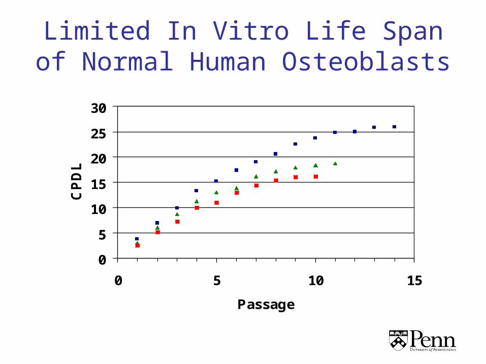
Limited In Vitro Life Span of Normal Human Osteoblasts
0
5
10
15
20
25
30
0 5 10 15
Passage
CP
DL

Replicative Senescence as a Model for Osteoblast Aging
• Finite in vitro life span
• Decreased osteoblast responsiveness to extracellular signals, including 1,25 (OH)2 vitamin D3, IGF-I, PTH, and prostaglandin E2 in both primary cultures from old donors as well as in osteoblasts aged in vitro by serial passage
• Loss of function with in vitro and in vivo age
Battmann A et al Exp Clin Endocrinol Diabeetes105:98-102 (1997); Kassem M et al Osteoporos Int 7:514-24 (1997); Kveiborg M et al Exp Gerontol 35:1061-74 (2000); Kveiborg M et al J Cell Physiol 186:298-306 (2001); Martinez ME et al Bone 24:203-9 (1999); Martinez P et al Bone 29:35-41 (2001); Yudoh, K et al JBMR 16:1453-64 (2001)

EarlyPassage
LatePassage
Alk Phos Mineral
Impaired Differentiated Function of Human Osteoblasts with In Vitro Age

Markers of Osteoblast Replicative Senescence
• Telomere dysfunction
• Senescence-associated heterochromatin (HIRA-PML nuclear bodies)
• SA-β-galactosidase activity
• Nucleolar association- Acridine Orange

Hypothesis
• In vitro replicative senescence can serve as a model for osteoblast dysfunction, which may recapitulate aspects of age-related bone loss. – Osteoblast cell strains derived from young individuals
have limited in vitro lifespans. – With advancing replicative age these cell strains
display impairment of differentiated function. – Loss of osteoblast differentiated function occurs
concomitantly with characteristics of senescence in culture.

Markers of Osteoblast Replicative Senescence
• Telomere dysfunction
• Senescence-associated heterochromatin (HIRA-PML nuclear bodies)
• SA-β-galactosidase activity
• Nucleolar association- Acridine Orange

Senescence Associated β-Galactosidase (SA β-gal)
• β-galactosidase is a eukaryotic hydrolase enzyme
• β-gal at pH 6.0 has been reported to increase during replicative senescence and may reflect replicative age of cells
• Limited application because not specific to senescence- – Also increased in quiescent, immortalized and
serum starved cells– Reversible under other conditions– May actually be lysosomal enzyme releases
at suboptimal pH (4.0)

SA β-gal
Early Passage Late Passage

Acridine Orange
• Nucleic acid selective fluorescent cationic dye.• Tips of 5 pairs of chromosomes fuse into fewer
and larger fragments as they approach S phase.• As cells progress through the cell cycle the
fraction of cells containing 1 or 2 nucleolar fragments increase while those containing 3 or more fragments decrease.
• Up to 90% of senescent cells in culture may contain only 1 to 2 nucleolar fragments.
Pignolo R et al Exp Geron 33:67-80 (1998)
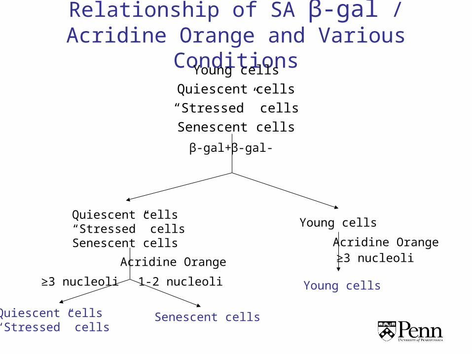
Relationship of SA β-gal / Acridine Orange and Various Conditions
Young cells
Quiescent cells
“Stressed” cells
Senescent cells
β-gal+ β-gal-
Quiescent cells“Stressed” cellsSenescent cells
Young cells
Acridine Orange
Acridine Orange
1-2 nucleoli
Quiescent cells“Stressed” cells
Senescent cells
≥3 nucleoli
Young cells≥3 nucleoli
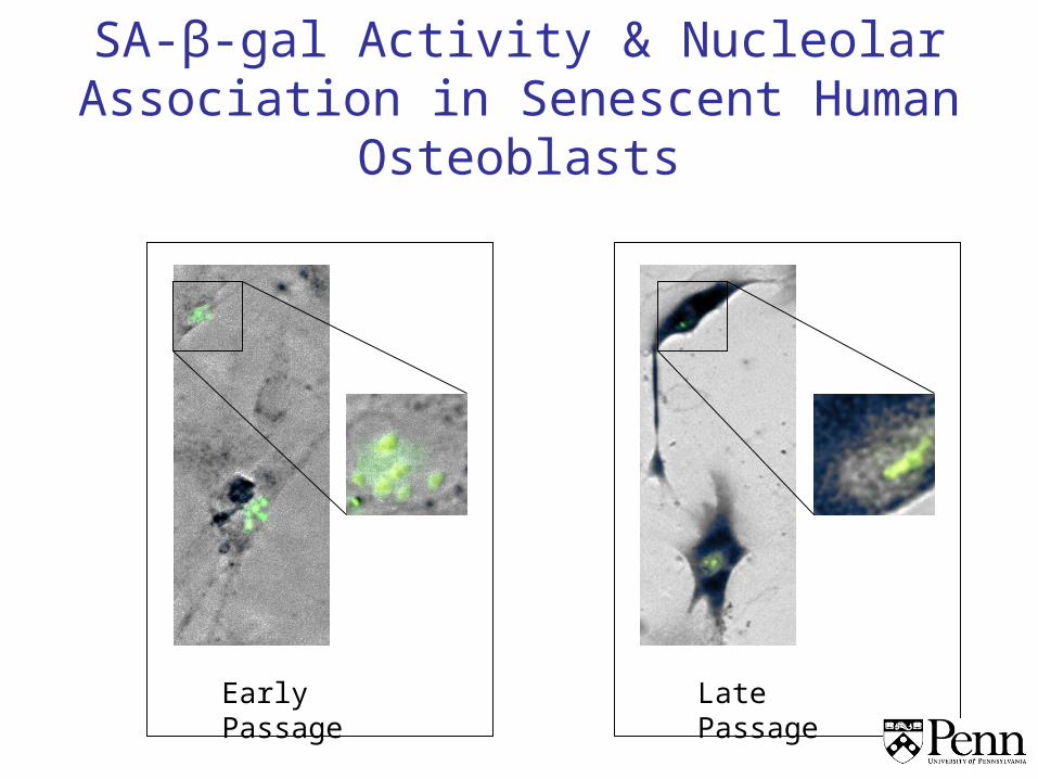
SA-β-gal Activity & Nucleolar Association in Senescent Human Osteoblasts
Early Passage Late Passage

SA-β-gal Activity & Nucleolar Association in Senescent Human Osteoblasts
Early passage/Toxic Early passage/Quiescent
40x

HIRA
• Histone Regulatory homolog A • Histone chaperone involved in assembly of
histones onto DNA• Senescent cells exhibit a specific pattern of
nucleolar foci with increased heterochromatin• Senescence-Associated Heterochromatin
Foci (SAHF)• Reorganized chromatin structure leads to a
loss of transcription activity by silencing of growth-promoting genes in SAHF

Senescence-Associated Heterochromatin in Aging Osteoblasts
Heterochromatin formation in senescent cells
Early Passage Late Passage

HIRA-associated Nuclear Foci in Aging Osteoblasts
0
5
10
15
20
HIRA-associated PML nuclear bodies/cell
# o
f c
ells
early passage
late passage

Markers of Osteoblast Replicative Senescence
• Telomere dysfunction
• Senescence-associated heterochromatin (HIRA-PML nuclear bodies)
• SA-β-galactosidase activity
• Nucleolar association- Acridine Orange

Telomere Dysfunction
Tel
omer
e dy
sfun
ctio
n/un
capp
ing
Persistent DNA damage Response
53BP153BP1
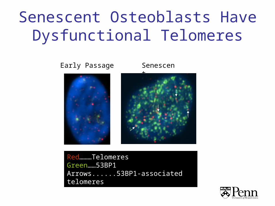
Senescent Osteoblasts Have Dysfunctional Telomeres
Red………TelomeresGreen……53BP1Arrows......53BP1-associated telomeres
SenescentEarly Passage

Conclusions
• With in vitro replicative senescence human osteoblasts display a loss of differentiated phenotype concomitant with limited life span
• Markers of in vitro osteoblast senescence exist which can potentially be used to detect aging osteoblasts in situ
• Replicative senescence may serve as a model for osteoblast dysfunction that occurs with aging

Acknowledgements
• PI/Mentor– Robert J Pignolo
• Laboratory– Kevin Egan– Alec Richardson– Emily McMillan– Robin Suda
• Collaborators– F. Brad Johnson (U of PA, Phila)– Peter Adams (FCCC, Phila)

Questions?