replacement of related pou transcription factors leads to severe defects in mouse forebrain...
TRANSCRIPT

Developmental Biology 332 (2009) 418–428
Contents lists available at ScienceDirect
Developmental Biology
j ourna l homepage: www.e lsev ie r.com/deve lopmenta lb io logy
Genomes & Developmental Control
Replacement of related POU transcription factors leads to severe defects in mouseforebrain development
Michael Wolf a, Petra Lommes a, Elisabeth Sock a, Simone Reiprich a, Ralf P. Friedrich a, Jana Kriesch a,C. Claus Stolt a, John R. Bermingham Jr. b, Michael Wegner a,⁎a Institut für Biochemie, Emil-Fischer-Zentrum Universität Erlangen-Nürnberg, Fahrstrasse 17, 91054 Erlangen, Germanyb McLaughlin Research Institute, Great Falls, Montana, USA
⁎ Corresponding author. Fax: +49 9131 85 22484.E-mail address: [email protected]
0012-1606/$ – see front matter © 2009 Elsevier Inc. Aldoi:10.1016/j.ydbio.2009.06.011
a b s t r a c t
a r t i c l e i n f oArticle history:Received for publication 26 November 2008Revised 19 May 2009Accepted 9 June 2009Available online 13 June 2009
Keywords:Oct-6Brn-1HomeodomainTranscription factorFunctional equivalenceRedundancyBrain malformationWnt signalling
Related transcription factors of the POU protein family show extensive overlap of expression in vivo andexhibit very similar biochemical properties in vitro. To study functional equivalence of class III POU proteinsin vivo, we exchanged the Oct-6 gene by Brn-1 in the mouse. Brn-1 can fully replace Oct-6 in Schwann cellsand rescue peripheral nervous system development in these mice. The same mice, however, exhibit severedefects in forebrain development arguing that Oct-6 and Brn-1 are not functionally equivalent in the centralnervous system. The cause of the observed forebrain phenotype is complex, but anteriorly expanded Wnt1expression contributes. Oct-6 normally represses Wnt1 expression in the early diencephalon andreplacement by Brn-1 as a weaker inhibitor is no longer sufficient to maintain the necessary level ofrepression in the mouse mutant. The extent of functional equivalence between related transcription factors isthus strongly dependent on the analyzed tissue.
© 2009 Elsevier Inc. All rights reserved.
Introduction
The POU family represents a large group of transcription factorsthat possess as their hallmark a composite DNA-binding domain thatconsists of a variant homeodomain preceded by a POU-specificdomain (Ryan and Rosenfeld, 1997; Wegner et al., 1993). The POUfamily can furthermore be subdivided in groups according to thesimilarity of its members to each other. One such group, the class III,consists of Brn-1, Brn-2, Brn-4 and Oct-6 (Hara et al., 1992; He et al.,1989). These class III POUproteins exhibit not only extensive sequencesimilarities on the amino acid level, they also show overlappingexpression patterns (Alvarez-Bolado et al., 1995; He et al., 1989) aswell as similar DNA binding, subcellular localization and transactiva-tion characteristics in vitro (Baranek et al., 2005; Schreiber et al., 1997;Sock et al., 1996).
As a consequence, substantial functional redundancy should existbetween different class III proteins. Evidence for such a redundancyhas for instance been obtained in mice in which deletions of multipleclass III POU proteins led to defects not observed in mice with singlegene deletions. Production, migration and positioning of corticalneurons are, for instance, strongly affected in Brn-1/Brn-2 double-
(M. Wegner).
l rights reserved.
deficient mice, but not in mice in which only Brn-1 or Brn-2 is deleted(McEvilly et al., 2002; Sugitani et al., 2002).
Nevertheless, developmental defects are also observed in micewith deletions of single class III genes. Brn-1 deficient mice, forinstance, suffer from kidney malformations (Nakai et al., 2003), anddevelopment of the endocrine hypothalamus is selectively affected inBrn-2 deficient mice (Nakai et al., 1995; Schoneman et al., 1995). Oct-6deficient mice, on the other hand, present with a glial defect inperipheral nervous system (PNS) development, as Schwann cells arearrested at the promyelin stage and peripheral myelination is stronglydelayed (Bermingham et al., 1996; Jaegle et al., 1996). Otherphenotypes include migration defects of select neuronal populationsof the central nervous system (CNS) that lead to mislocalization ofbrain nuclei including some involved in breathing control (Berming-ham et al., 1996).
These phenotypes in mice with single gene deletions correlatewith the unique or at least preferential expression of one class III POUprotein in the tissue or cell type that is affected. Schwann cells, forinstance, express high levels of Oct-6, much lower Brn-2 levels, andnone of the other class III proteins. As a consequence Oct-6 deficientmice exhibit a Schwann cell defect, whereas loss of Brn-2 iscompensated and thus inapparent in Schwann cells of Brn-2 deficientmice. The fact that loss of both Oct-6 and Brn-2 strongly aggravates theSchwann cell defect, further substantiates the notion that class III POUproteins act in a highly similar manner (Jaegle et al., 2003).

419M. Wolf et al. / Developmental Biology 332 (2009) 418–428
To directly address the question of functional equivalence betweenclass III POU proteins, we have previously generatedmice inwhich wereplaced the continuous Oct-6 open reading frame by the Brn-1 openreading frame, thereby generating the Oct-6brn-1 allele (Friedrich et al.,2005). This allele allows expression of Brn-1 in all cells and tissuesthat normally express Oct-6, at comparable levels. Analysis on amixedC57Bl/6J-129Sv background revealed that Brn-1 was capable ofcompletely rescuing the Schwann cell defect in Oct-6brn-1/brn-1 aswell as in Oct-6brn-1/− mice (Friedrich et al., 2005). This stronglysuggested that Oct-6 and Brn-1 are functionally very similar, if notequivalent at least with respect to their role in PNS development.
When backcrossing the Oct-6brn-1 allele on a C57Bl/6J background,we noticed, however, that Oct-6brn-1/brn-1 animals were born inincreasingly lower numbers, whereas Oct-6brn-1/− mice continued tobe born at Mendelian ratios, were phenotypically inapparent, fertileand still without an obvious Schwann cell phenotype. This pointed toselective embryonic lethality in the Oct-6brn-1/brn-1 genotype on aclean genetic background.
Here we report that these Oct-6brn-1/brn-1 mice suffer from severeforebrain truncations that are neither commonly observed inOct-6−/−
nor in Oct-6brn-1/− mice. We furthermore provide evidence that thedifferential influence of Brn-1 versus Oct-6 on Wnt1 expression leadsto an anteriorly expandedWnt1 expression inOct-6brn-1/brn-1mice thatis at least in part responsible for the observed phenotype. Our resultsthus provide evidence that class III POU proteins are functionallyequivalent only during PNS, but not during CNS development.
Materials and methods
Animal husbandry, genotyping, tissue preparation, histological staining,in situ hybridization, proliferation and apoptosis assays
Mice with an Oct-6brn-1 (Friedrich et al., 2005) or an Oct-6–
(Bermingham et al., 1996) allele were bred on a C57Bl/6J backgroundand kept as heterozygotes. Genotyping was performed by PCR asdescribed (Berminghamet al.,1996; Friedrich et al., 2005). Homozygousmutants were generated by intercrosses of Oct-6+/brn-1 or Oct-6+/–
animals, respectively. For some experiments, the Sox8lacZ allele (Sock etal., 2001)was additionally crossed into themice to provide a lacZmarkerthat labels the cranial neural crest in the head region. Embryos wereobtained from 9.5 to 18.5 days post coitum (dpc), photographed andprocessed.
For whole mount in situ hybridizations, embryos were fixedovernight at 4°C in 4% paraformaldehyde, dehydrated, bleached, andrehydrated. In situ hybridization was performed essentially asdescribed (Britsch et al., 2001) with antisense riboprobes for Brn-1,Oct-6 (Schreiber et al., 1997), Brn-2, Six3 (gifts of Q. Ma, Dana FarberCancer Institute, Boston), Emx1, Emx2 (gifts of J. Rubenstein, UCSF, SanFrancisco), En1 (gift ofW.Wurst, Helmholtz-Zentrum, München), Fgf8(gift of G. Martin, UCSF, San Francisco), HoxB1, Wnt1 and Wnt3a (giftsof A. Joyner, Memorial Sloan-Kettering Cancer Center, New York),Mash1 (gift of F. Guillemot, NIMR, Mill Hill), Shh (gift of A. MacMahon,Harvard University, Cambridge), Bmp4 (gift of B. Hogan, DukeUniversity Medical Center, Durham) and Pax6 (gift of D. Engelkamp,Erlangen). Riboprobes were routinely DIG-labelled. For double in situhybridization, fluorescein-labelled probes were additionally used. Allsteps except probe hybridization and final colorimetric detection usingNBT/BCIP or Fast Red (Roche) were performed automatically on aBiolane HTI (Hölle and Hüttner AG, Tübingen, Germany).
Detection of β-galactosidase activity in whole mount embryosfollowed standard procedures (Britsch et al., 2001). After overnightfixation in 1% paraformaldehyde, embryos were incubated for severalhours at 37°C in 1% X-gal until blue precipitates were detectable.
For HE staining, embryos were thoroughly and repeatedly washedin tapwater after fixation, dehydrated in a series of graded alcohol andembedded in paraffin. HE staining was carried out on 2 μm-thick
sagittal paraffin sections. Analysis and documentation was with aLeica MZFLIII stereomicroscope equipped with an Axiocam (Zeiss,Oberkochem, Germany).
For proliferation studies, pregnant mice were injected intraper-itoneally with 100 μg BrdU (Sigma) per gram body weight 1 h beforeembryo preparation (Stolt et al., 2003). Embryos underwent fixationin 4% paraformaldehyde, were frozen at −80 °C in Jung TissueFreezing Medium (Leica, Nussloch, Germany) and sectioned sagitallyon a cryotome at 14-μm thickness. Incorporated BrdU was visualizedby an Alexa-488 coupledmousemonoclonal antibody directed againstBrdU (Molecular Probes) at a 1:20 dilution as a measure ofproliferation. TUNEL assays were performed on cryosections accord-ing to the manufacturer`s protocol (Chemicon).
Cell culture, transfection, extract preparation, electrophoretic mobilityshift assays and luciferase assays
HEK 293 cells were maintained in DMEM containing 10% FCS andtransfected on 10-cm dishes with 10 μg pCMV5-based expressionplasmids for full length Oct-6 and Brn-1 as well as truncated versionsOct-6ΔNC (Sock et al., 1996) and Brn-1ΔN (Schreiber et al., 1997) usingSuperfect reagent (Qiagen). Transfected cells were harvested 48 hposttransfection and used to prepare whole cell protein extracts aspreviously described (Baranek et al., 2005). These extracts wereincubated for electrophoretic mobility shift assays with 32P-labelledoligonucleotides containing the sequence of the putative Oct-6 bindingsite at −1466 or at −955 of the Wnt1 promoter and poly(dIdC) asunspecific competitor. Samples were loaded on to native 5% polyacry-lamide gels and electrophoresed in 45 mM Tris, 45 mM boric acid and1mMEDTA (pH 8.3) at 120 V for 1.5 h. Gels were dried and exposed forautoradiography.
For luciferase assays, HEK 293 cells were transfected transiently induplicates in 24-well plates with 500 ng of luciferase reporter plasmidand 100 ng of effector plasmids per well. Luciferase reporterscontained the Wnt1 promoter region in a short (positions −1234 to+34, psWnt-luc) or a long (positions −1566 to +34, plWnt-luc)version and were based on the pGL2 backbone (Promega, Madison,WI). The short version of the Wnt1 promoter was additionallycombined in psWnt-luc-enh with the 3′ neural plate enhancer ofthe Wnt1 gene (positions +7744 to +8837) (Rowitch et al., 1998)which was inserted behind the luciferase reporter gene. Mutationswere additionally introduced into plWnt-luc to destroy both Oct-6binding sites, leading to the plWnt-delOct-luc reporter plasmid.Effector plasmids corresponded to pCMV5-based expression plasmidsfor Brn-1, Brn-2 and Oct-6 and have been described before (Schreiberet al., 1997; Sock et al., 1996). Cells were harvested 48 h posttransfec-tion and activities were determined as described (Renner et al., 1994).
Chromatin immunoprecipitation
Chromatin immunoprecipitation assays on embryonic brain wasperformed as described (Stolt et al., 2006). Briefly, whole brain tissueof 10.5 dpc-old embryos was dissociated by trituration. Consecutively,cellular protein and genomic DNAwere crosslinked by treatment with1% formaldehyde before chromatin extraction and sonification to anaverage fragment length of 300 to 600 bp. Immunoprecipitationswereperformed overnight at 4°C using polyclonal IgG against Oct-6(Baranek et al., 2005; Sock et al., 1996) or Brn-1 (Friedrich et al.,2005) as well as control IgG fractions. DNA was purified fromprecipitates after crosslink reversal and subjected to PCR. Fordetection of a fragment containing the Oct-6 binding site in theWnt1 promoter at position −1466, 5′-ACTGCAGAAACCTGGGAGAA-3′(position −1566 to −1586) and 5′-GTTCTCAGCACCCAAGTGGT-3′(position −1312 to −1332) were used as primers in 33 cycles ofstandard PCR using an annealing temperature of 60°C. The fragmentaround the second Oct-6 binding site at −955 was detected with 5′-

420 M. Wolf et al. / Developmental Biology 332 (2009) 418–428
CTACTCGGCTGGTGGTCCTA-3′ (position −1002 to −1022) and 5′-GACAGGAGACGTCCAGAAGG-3′ (position −768 to −788) as primers.
Results
Oct-6brn-1/brn-1 mice suffer from severe forebrain truncations
When backcrossing the Oct-6brn-1 allele on a C57Bl/6J background,Oct-6brn-1/brn-1 newborns became increasingly underrepresentedarguing for embryonic lethality in this genotype. To identify thepotential cause for this embryonic lethality, we performed timedmatings. Oct-6brn-1/brn-1 embryos exhibited severe brain malforma-tions already at 10.5 dpc (Figs. 1A–D). These brain malformationscontinued to be seen throughout embryonic development (Figs. 1E–T)until 18.5 dpc (Figs. 1U–X). The percentage of Oct-6brn-1/brn-1 embryos,however, became lower with increasing ages. The severity of theobserved brain malformations also varied substantially (compare forinstance Figs. 1C, D with Figs. 1G, H). In general, the phenotypeworsened with increased backcrossing. Intriguingly, these defectswere not commonly observed in Oct-6−/− or in Oct-6brn-1/− mice(Bermingham et al., 1996; Friedrich et al., 2005).
In common to all affected embryos was a truncation of theforebrain. By histology, diencephalon and telencephalon appeared to
Fig. 1. Appearance of Oct-6brn-1/brn-1 embryos. Compared to wildtype littermates (A, B, E, F, I,forebrain malformations that are visible on 10.5 dpc (A–D), 11.5 dpc (E–H), 12.5 dpc (I–L), 14.an external lateral view of the embryo, whereas panels B, C, F, G, J, K, N, O, R, S, V, W repre
be absent in the severe cases. Other facial structures were con-comitantly malformed. In the milder cases, only the anterior part ofthe telencephalon was affected. Other parts of the body appearedsuperficially normal. We therefore concentrated on the brainmalformation for further analyses.
Expression of forebrain markers is strongly reduced in Oct-6brn-1/brn-1
embryos
To characterize the defect in further detail, we performed wholemount in situ hybridizations with several region-specific brain markers.At 9.5 dpc, Pax6 is expressed throughout the complete forebrain(Walther and Gruss, 1991) as evident for the wildtype (Fig. 2A). InOct-6brn-1/brn-1 embryos, by contrast, the Pax6 expressing region isvariably, but severely reduced (Figs. 2B, C).
Emx1 as a telencephalic marker (Simeone et al., 1992) starts to beexpressed in wildtype embryos at 9.5 dpc (Fig. 2D), but not in themajority of affected Oct-6brn-1/brn-1 embryos (Figs. 2E, F) arguing thatthe telencephalon is most severely impaired in its development by themutation. Similar results were also obtained in whole mount in situhybridizations with Emx2 as probe (data not shown). Despite thestrong forebrain defect in Oct-6brn-1/brn-1 embryos, we did not detectany significant alteration in the proliferation rate at 9.5 dpc in BrdU
J, M, N, Q, R, U, V), Oct-6brn-1/brn-1 embryos (C, D, G, H, K, L, O, P, S, T, W, X) exhibit severe5 dpc (M–P), 16.5 dpc (Q–T) and 18.5 dpc (U–X). Panels A, D, E, H, I, L, M, P, Q, T, U, X givesent HE-stained midsagittal sections.
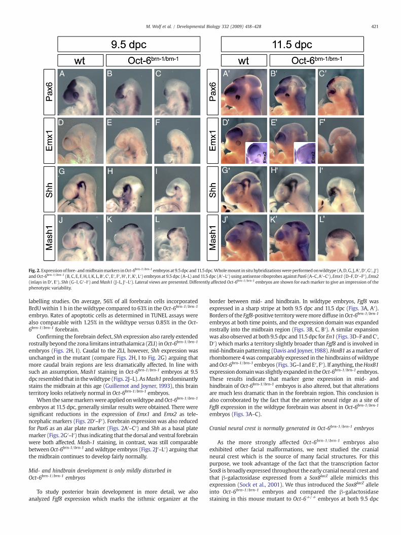
Fig. 2. Expression of fore- andmidbrainmarkers inOct-6brn-1/brn-1 embryos at 9.5 dpc and11.5dpc.Wholemount in situ hybridizationswereperformedonwildtype (A,D,G, J, A′, D′, G′, ,J′)andOct-6brn-1/brn-1 (B, C, E, F, H, I, K, L, B′, C′, E′, F′, H′, I′, K′, L′) embryos at 9.5 dpc (A–L) and 11.5 dpc (A′–L′) using antisense riboprobes against Pax6 (A–C, A′–C′), Emx1 (D–F, D′–F′), Emx2(inlays in D′, E′), Shh (G–I, G′–I′) andMash1 (J–L, J′–L′). Lateral views are presented. Differently affected Oct-6brn-1/brn-1 embryos are shown for each marker to give an impression of thephenotypic variability.
421M. Wolf et al. / Developmental Biology 332 (2009) 418–428
labelling studies. On average, 56% of all forebrain cells incorporatedBrdUwithin 1 h in thewildtype compared to 63% in the Oct-6brn-1/brn-1
embryo. Rates of apoptotic cells as determined in TUNEL assays werealso comparable with 1.25% in the wildtype versus 0.85% in the Oct-6brn-1/brn-1 forebrain.
Confirming the forebrain defect, Shh expression also rarely extendedrostrally beyond the zona limitans intrathalamica (ZLI) inOct-6brn-1/brn-1
embryos (Figs. 2H, I). Caudal to the ZLI, however, Shh expression wasunchanged in the mutant (compare Figs. 2H, I to Fig. 2G) arguing thatmore caudal brain regions are less dramatically affected. In line withsuch an assumption, Mash1 staining in Oct-6brn-1/brn-1 embryos at 9.5dpc resembled that in thewildtype (Figs. 2J–L). AsMash1predominantlystains the midbrain at this age (Guillemot and Joyner, 1993), this brainterritory looks relatively normal in Oct-6brn-1/brn-1 embryos.
When the samemarkerswere appliedonwildtype andOct-6brn-1/brn-1
embryos at 11.5 dpc, generally similar results were obtained. There weresignificant reductions in the expression of Emx1 and Emx2 as tele-ncephalic markers (Figs. 2D′–F′). Forebrain expressionwas also reducedfor Pax6 as an alar plate marker (Figs. 2A′–C′) and Shh as a basal platemarker (Figs. 2G′–I′) thus indicating that the dorsal and ventral forebrainwere both affected. Mash-1 staining, in contrast, was still comparablebetweenOct-6brn-1/brn-1 andwildtype embryos (Figs. 2J′–L′) arguing thatthe midbrain continues to develop fairly normally.
Mid- and hindbrain development is only mildly disturbed inOct-6brn-1/brn-1 embryos
To study posterior brain development in more detail, we alsoanalyzed Fgf8 expression which marks the isthmic organizer at the
border between mid- and hindbrain. In wildtype embryos, Fgf8 wasexpressed in a sharp stripe at both 9.5 dpc and 11.5 dpc (Figs. 3A, A′).Borders of the Fgf8-positive territoryweremore diffuse inOct-6brn-1/brn-1
embryos at both time points, and the expression domainwas expandedrostrally into the midbrain region (Figs. 3B, C, B′). A similar expansionwas also observed at both 9.5 dpc and 11.5 dpc for En1 (Figs. 3D–F andC′,D′) which marks a territory slightly broader than Fgf8 and is involved inmid-hindbrainpatterning (Davis and Joyner,1988).HoxB1 as amarker ofrhombomere 4was comparably expressed in the hindbrains of wildtypeandOct-6brn-1/brn-1 embryos (Figs. 3G–I and E′, F′). If anything, theHoxB1expression domainwas slightly expanded in theOct-6brn-1/brn-1 embryos.These results indicate that marker gene expression in mid- andhindbrain of Oct-6brn-1/brn-1 embryos is also altered, but that alterationsare much less dramatic than in the forebrain region. This conclusion isalso corroborated by the fact that the anterior neural ridge as a site ofFgf8 expression in the wildtype forebrain was absent in Oct-6brn-1/brn-1
embryos (Figs. 3A–C).
Cranial neural crest is normally generated in Oct-6brn-1/brn-1 embryos
As the more strongly affected Oct-6brn-1/brn-1 embryos alsoexhibited other facial malformations, we next studied the cranialneural crest which is the source of many facial structures. For thispurpose, we took advantage of the fact that the transcription factorSox8 is broadly expressed throughout the early cranial neural crest andthat β-galactosidase expressed from a Sox8lacZ allele mimicks thisexpression (Sock et al., 2001). We thus introduced the Sox8lacZ alleleinto Oct-6brn-1/brn-1 embryos and compared the β-galactosidasestaining in this mouse mutant to Oct-6+/+ embryos at both 9.5 dpc

Fig. 3. Expression of isthmic organizer and hindbrainmarkers in Oct-6brn-1/brn-1 embryos. Wholemount in situ hybridizations were performed onwildtype (A, D, G, A′, C′, E′) and Oct-6brn-1/brn-1 (B, C, E, F, H, I, B′, D′, F′) embryosat 9.5 dpc (A–I) and11.5dpc (A′–F′) using antisense riboprobes against Fgf8 (A–C, A′, B′),En1 (D–F, C′, D′) andHoxB1 (G–I, E′, F′). Lateral viewsare presented at 9.5 dpc, top views at 11.5 dpc. Differently affected Oct-6brn-1/brn-1 embryos are shown for each marker at 9.5 dpc to give an impression of the phenotypic variability.
422 M. Wolf et al. / Developmental Biology 332 (2009) 418–428
(Figs. 4A–F) and 11.5 dpc (Figs. 4A′–F′). Cranial neural crest cellspredominantly originate in the hind- and midbrain regions (Fig. 4A).There was no significant difference in cranial neural crest appearancebetween Oct-6+/+ and Oct-6brn-1/brn-1 embryos with regard totiming or location (Figs. 4A–C). Cranial neural crest furthermoremigrated normally into the branchial arches (Figs. 4A–C and A′–C′).Rostral migration of cranial neural crest cells was, however, stalled inOct-6brn-1/brn-1 embryos at 11.5 dpc, resulting in an accumulation ofcranial neural crest cells in the midbrain region and a reduction of
Fig. 4. Development of the cranial neural crest in Oct-6brn-1/brn-1 embryos. To follow cranialembryos at 9.5 dpc (A–F) and 11.5 dpc (A′–F′), a Sox8lacZ allele was additionally introducedA′–C′ represent lateral views of the stained head regions, panels D–F and D′–F′ are maDifferently affected Oct-6brn-1/brn-1 embryos are shown to give an impression of the pheno
cranial neural crest cell numbers in the frontonasal process(compare Figs. 4B′, C′ to Fig. 4A′). From the fact that cranialneural crest is generated normally (Fig. 4) and does not expresssignificant levels of Oct-6 (Figs. 7A, A′), it appears likely that theselatter defects are secondary to the forebrain defect.
Interestingly, β-galactosidase also stained brain cells that inOct-6+/+ embryos were exactly localized at the border betweenforebrain and midbrain (Figs. 4D and D′). This staining was stronglyreduced or completely absent in all analyzed Oct-6brn-1/brn-1 embryos
neural crest cells in Oct-6+/+ (A, D, A′, D′) and Oct-6brn-1/brn-1 (B, C, E, F, B′, C′, E′, F′)into the mice and β-galactosidase staining performed on the embryos. Panels A–C andgnifications from the respective region between diencephalon and mesencephalon.typic variability.

423M. Wolf et al. / Developmental Biology 332 (2009) 418–428
both at 9.5 dpc (Figs. 4E, F) and at 11.5 dpc (Figs. 4E′, F′) arguing thatprocesses at this border may be disturbed.
Wnt1 expression is anteriorly expanded in Oct-6brn-1/brn-1 embryos
With forebrain defects being already pronounced at 9.5 dpc, weextended our studies to earlier stages of development. At 8.25 dpc, theanterior region of the closing neural tube still appeared morphologi-cally normal and Shh expression was comparable between wildtypeand Oct-6brn-1/brn-1 embryos (Figs. 5A, B). Pax6 expression, in contrast,was already strongly reduced and restricted to the anterior mostregion (Figs. 5C, D). At the same time,Wnt1 expressionwas anteriorlyexpanded (Figs. 5E, F).
The expanded Wnt1 expression caught our attention as previousstudies had shown an essential role for Wnt signalling in thespecification of posterior-to-anterior fates within the neural plate.Compared to the midbrain, Wnt1 levels must be downregulated in theforebrain. As a consequence, the diencephalon will develop from theregion with reduced Wnt1 levels rostrally adjacent to the midbrainand the telencephalon from the Wnt-free rostralmost zone (Kieckerand Niehrs, 2001; Niehrs, 1999). Considering the alterations at theborder between midbrain and forebrain and the expanded Wnt1expression at 8.25 dpc, we also investigated Wnt1 expression in Oct-6brn-1/brn-1 embryos at 9.5 dpc (Figs. 6A–G) and 10.5 dpc (Figs. 6A′–E′).At both embryonic ages,Wnt1 is strongly expressed in the wildtype intwo regions of the midbrain, its dorsal midline and a caudal stripebordering the hindbrain (Figs. 6A, D, A′, D′). In Oct-6brn-1/brn-1
embryos, the caudal stripe appeared broadened (Figs. 6B, C, and B′, C′)in line with results from Fgf8 and En1 in situ hybridizations (Fig. 3). Incontrast, dorsal midline staining was not significantly altered in themidbrain (Figs. 6B, C, E, and B′, C′, E′).
Inspection of wildtype embryos furthermore revealed thatexpression at the dorsal midline continued at reduced levels into
Fig. 5. Expression of fore- and midbrain markers in Oct-6brn-1/brn-1 embryos at 8.25dpc. Whole mount in situ hybridizations were performed on wildtype (A, C, E) andOct-6brn-1/brn-1 (B, D, F) embryos at 8.25 dpc using antisense riboprobes against Shh(A, B) Pax6 (C, D), and Wnt1 (E, F). Ventral (A, B, E, F) and lateral views (C, D) arepresented.
the forebrain and disappeared at the transition from diencephalon totelencephalon (Figs. 6A, A′, D, D′). As a consequence, there was co-expression of Wnt1 and Pax6 in the diencephalon, whereas thetelencephalon was only stained for Pax6 (Fig. 6F). In Oct-6brn-1/brn-1
embryos, however, the dorsal midline expression extended signifi-cantly farther rostrally than in the wildtype and at higher levels (Figs.6B, B′, E, E′). In the most affected animals, Wnt1 expression in thedorsal midline continued to the rostral tip of the brain (Figs. 6C, C′) sothat the remaining Pax6 expression overlapped completely withWnt1(Figs. 6G, G′). Wnt1 dysregulation was not observed in Oct-6−/− orOct-6-/brn-1 embryos (data not shown) in line with the absence of aforebrain defect in these genotypes.
In situ hybridizations detected a similar expansion of Wnt3aexpression into the anterior most brain regions at 9.5 dpc andconfirmed the overall disturbance of Wnt signalling (Figs. 6H, I).Bmp4 expression in the dorsal midline was similarly expanded,whereas ventral expression was strongly reduced (Figs. 6J, K). Thisindicates that several signalling pathways involved in forebraindevelopment are affected and that the cause of the forebrain defectin Oct-6brn-1/brn-1 embryos is complex. Nevertheless, altered Wntsignalling due to anteriorly expanded Wnt1 expression likely con-tributes as a major factor.
Spatial Brn-1 expression is strongly altered in Oct-6brn-1/brn-1 brains
As the forebrain defect in Oct-6brn-1/brn-1 embryos was clearly not aconsequence of missing Oct-6 expression, but rather caused by ectopicBrn-1 expression, we investigated in what respect the Brn-1 expres-sion differed between wildtype and Oct-6brn-1/brn-1 embryos. In thewildtype, Brn-1 was expressed throughout the developing brain. At9.5 dpc, Brn-1 expression decreased in a caudal to rostral gradient(Fig. 7B), whereas there was strong Brn-1 expression throughout allbrain regions at 11.5 dpc (Fig. 7B′). Oct-6 transcripts, on the otherhand, were strongly enriched at 9.5 dpc in the diencephalon and in anoval patch in the ventral telencephalon (Fig. 7A) (Alvarez-Bolado etal., 1995). Strong Oct-6 expression in the diencephalon also persistedat 11.5 dpc (Fig. 7A′). In embryos carrying an Oct-6brn-1 allele, the Brn-1 expression pattern equals the combined Oct-6 and Brn-1 expressionin the wildtype at both 9.5 dpc and 11.5 dpc (Figs. 7C, C′).
The most conspicuous difference between Brn-1 expression inwildtype and Oct-6+/brn-1 or Oct-6brn-1/brn-1 brains are the stronglyincreased expression levels in the diencephalon (compare Figs. 7, B′to Figs. 7C, C′) that overlap with the Wnt1 expression domain (Figs.7D, D′). Ectopic Brn-1 expression from a single Oct-6brn-1 allelealready exceeds endogenous Brn-1 expression in the diencephalonsignificantly.
To get a more general impression of class III POU expression in thedeveloping brain, we also performed in situ hybridizations with a probespecific for Brn-2 (Figs. 8A–C, and A′–C′). These studies revealed thatthere is additional expression of Brn-2 throughout the developing brain.At 9.5 dpc, Brn-2 was expressed at significant levels in wildtypediencephalon, mesencephalon and hindbrain, but only weakly in theforming telencephalon (Fig. 8A). At 10.5 dpc, Brn-2 expression was alsoclearly visible in the telencephalon (Fig. 8A′). Similar data were alsoobtained in age-matchedOct-6brn-1/brn-1 embryos (Figs. 8B, C, and B′, C′).These studies furthermore gave no indication that Brn-2 (or Brn-4)expression was significantly altered in Oct-6brn-1/brn-1 embryos com-pared to the wildtype (Figs. 8A–C, and A′–C′ and data not shown).
Oct-6 and Brn-1 differentially influence Wnt1 expression
The upregulation of Brn-1 in the diencephalon of Oct-6brn-1/brn-1
embryos correlated with the rostrally expanded Wnt1 expression andargued for a causal relationship. Both Emx2 and Six3 have previouslybeen shown to function as negative regulators of Wnt1 expression inthe forebrain (Lagutin et al., 2003; Ligon et al., 2003). As Emx2

Fig. 6. Expression of Wnt and Bmp genes in Oct-6brn-1/brn-1 embryos. Whole mount in situ hybridizations were performed on wildtype (A, D, A′, D′, F, H, J) and Oct-6brn-1/brn-1 (B, C,E, B′, C′, E′, G, G′, I, K) embryos at 9.5 dpc (A–K) and 10.5 dpc (A′–G′) using antisense riboprobes against Wnt1 (A–G, A′–E′), Pax6 (F, G, G′ signal in red), Wnt3a (H, I) and Bmp4 (J,K). Lateral (A–C, A′–C′, F–K) and top (D, E, D′, E′) views are presented. The picture in panel G′ is a magnification of panel G. The arrowheads in panels F, G, G′ mark the area inwhich Pax6 and Wnt-1 are co-expressed. Arrowheads in panels H,I indicate the anterior boundary of Wnt3 expression, whereas they demarcate dorsal Bmp4 expression in panels J,K. Differently affected Oct-6brn-1/brn-1 embryos are shown in B, C, B′ and C′ to give an impression of the phenotypic variability.
424 M. Wolf et al. / Developmental Biology 332 (2009) 418–428
expression is strongly affected in Oct-6brn-1/brn-1 embryos (Figs. 2D′, E′and data not shown), it is conceivable that expandedWnt1 expressionis at least partially caused by the loss of Emx2.
To address the role of Six3, we studied whether the altered class IIIPOU expression would affect Six3 expression. At 9.5 dpc, we detectedSix3 predominantly in the ventral telencephalon of wildtype embryos(Fig. 8D). At 10.5 dpc, Six3 was expressed throughout the telencepha-lon, andweaker expressionwas nowalso detected in the diencephalon(Fig. 8D′). In agreement with the general phenotype, Six3 expressionwas clearly reduced in the forebrain of Oct-6brn-1/brn-1 embryos at bothages (Figs. 8E, F, and E′, F′). However, diencephalic Six3 expressionpersisted in all Oct-6brn-1/brn-1 embryos, and the relative rate of Six3expression in telencephalon, diencephalon and mesencephalon wascomparable in wildtype embryos and those Oct-6brn-1/brn-1 embryoswith a milder phenotype (compare Figs. 8D, D′ to Figs. 8E, E′). As thelatter embryos already exhibited an alteredWnt1 expression, it seemsunlikely that the Oct-6brn-1 allele exerts its effects via Six3.
We also considered the possibility that Oct-6 and Brn-1 directlyaffect Wnt1 expression. Both the promoter region as well as a 3′enhancer are known to regulate neuroectodermal Wnt1 expression(Lagutin et al., 2003; Rowitch et al., 1998). Sequence inspection ofboth regions revealed the presence of two octamer sequences aspotential binding sites for POU proteins in the promoter region of theWnt1 gene, whereas no obvious binding motif could be identified inthe enhancer region. The promoter sites were localized at positions−1466 and −955 relative to the transcriptional start site (Fig. 9A).Both the distal site at position−1466 and the proximal site at postion
−955 bound Oct-6 and Brn-1 strongly in electrophoretic mobilityshift assays independent of whether the class III POU protein wassupplied as full length protein or truncated version (Fig. 9B). Theproximal site furthermore appeared to have a somewhat higherbinding affinity for Brn-1 than for Oct-6.
To address whether these sites indeed interact with Brn-1 and Oct-6 in vivo, we performed immunoprecipitation experiments oncrosslinked chromatin of 10.5 dpc-old whole embryonic brains fromwildtype mice with antibodies directed against Brn-1 and Oct-6. Incontrast to control IgG, both specific antibodies immunoprecipitatedfragments from crosslinked chromatin that encompassed the octamerelements (Fig. 9C), thus indicating that both octamer elements areindeed bound in vivo by Brn-1 and Oct-6.
To analyze the effect of both class III POU proteins on Wnt1expression, luciferase reporter gene assays were performed intransiently transfected HEK 293 cells (Figs. 9D– G). In all experiments,we consistently observed a negative effect of class III POU proteins onWnt1 expression. This repressive effect was milder, when luciferasereporter gene expression was driven by a short version of the Wnt1promoter that contained only the proximal octamer site (Fig. 9D), andmore pronounced, when the luciferase reporter was under the controlof a long version of the Wnt1 promoter that contained both octamerelements (Fig. 9E). It was furthermore lost when both binding sites forclass III POU proteins were destroyed by site directed mutagenesis(Fig. 9F). The additional presence of the Wnt1 enhancer in theluciferase reporter construct did not influence the repressive POUprotein effect qualitatively or quantitatively (Fig. 9G), consistent with

Fig. 7. Expression of Brn-1 from the Oct-6brn-1 allele. Whole mount in situ hybridizationswere performed onwildtype (A, B, A′, B′), Oct-6+/brn-1 (C, C′) and Oct-6brn-1/brn-1 (D, D′)embryos at 9.5 dpc (A–D, D′) and 11.5 dpc (A′–C′) using antisense riboprobes againstOct-6 (A, A′), Brn-1 (B, C, B′, C′ signal in black, D, D′ signal in red) and Wnt1 (D, D′).Lateral (A–D, A′–C′) and top (D′) views are presented.
Fig. 8. Expression of Brn-2 and Six3 in Oct-6brn-1/brn-1 embryos. Whole mount in situ hybridizaembryos at 9.5 dpc (A–F) and 10.5 dpc (A′–F′) using antisense riboprobes against Brn-2 (A–C, Aembryos are shown to give an impression of the phenotypic variability.
425M. Wolf et al. / Developmental Biology 332 (2009) 418–428
the absence of POU binding motifs in the enhancer region. Interest-ingly, Brn-1 was a weaker repressor than either Oct-6 or Brn-2 on allreporter constructs and in every experiment (Figs. 9D–G), arguingthat the repressive effect of class III POU proteins on neural Wnt1expression is less in Oct-6brn-1/brn-1 embryos than in the wildtype. Thiseffect is particularly relevant in those regions that normally expresshigh levels of Oct-6 such as the diencephalon at 9.5 dpc.
Discussion
The transcription factor Brn-1, when introduced into Schwanncells, fully rescues the Oct-6 dependent differentiation defect(Friedrich et al., 2005). Oct-6 and the highly related Brn-1 thusfunction very similarly during Schwann cell development in the PNSand may very well be functionally equivalent in this cell type. CNSdevelopment, in contrast, is severely disturbedwhen Oct-6 is replacedby Brn-1 in the mouse. The two related class III POU proteins aretherefore not functionally equivalent in the CNS. This argues fortissue-specific differences in functional equivalence of closely relatedtranscription factors.
The observed phenotype is characterized by truncation of theforebrain. It had previously gone unnoticed because the phenotypebecame obvious only after backcrossing on a clean C57Bl/6J back-ground. Similar phenotypes have been observed in several mousemutants. These include not only mouse mutants in which genesintrinsically required for forebrain development are affected (Lagutinet al., 2003; Martinez-Barbera and Beddington, 2001), but also mousemutants in which genes involved in the development of the anteriorvisceral endoderm such as Otx2, Lim1 and Hex are mutated (Martinez-Barbera and Beddington, 2001; Perea-Gomez et al., 2001; Shawlot etal., 1999). Missing inhibition of BMP or Wnt signalling also leads tosevere forebrain truncations as evident from mice homozygously forboth BMP antagonists chordin and noggin (Bachiller et al., 2000) orDkk1-deficient mice (Mukhopadhyay et al., 2001).
That derepression of Wnt signalling causes forebrain truncations(Heisenberg et al., 2001; Kiecker and Niehrs, 2001; Lagutin et al.,2003), was particularly interesting in the context of Oct-6brn-1/brn-1
mice, as we observed rostrally expanded Wnt1 and Wnt3a expressionin this mouse mutant. Considering that we also obtained evidence foraltered Bmp signalling, we have to assume that multiple signalling
tions were performed on wildtype (A, D, A′, D′) and Oct-6brn-1/brn-1 (B, C, E, F, B′, C′, E′, F′)′–C′), and Six3 (D–F, D′–F′). Lateral views are presented. Differently affectedOct-6brn-1/brn-1

Fig. 9. Differential effect of Oct-6 and Brn-1 on Wnt1 expression. (A) Schematic representation of the Wnt1 gene locus including promoter, coding (black part of the bar) anddownstream region with the neural plate enhancer (enh). The short (sWnt1) and long (lWnt1) versions of the Wnt1 promoter that were used to generate luciferase reporterconstructs psWnt1-luc and plWnt1-luc are marked. Distal (−1466) and proximal (−955) octamer sites in theWnt1 promoter region are depicted as boxes. They are mutated in theplWnt1-delOct-luc luciferase reporter. (B) Electrophoretic mobility shift assays with radiolabelled oligonucleotides identical in sequence to the distal and proximal octamer sites oftheWnt1 promoter and extracts containing full length (fl) or shortened versions (ΔC, ΔNC) of Oct-6 and Brn-1. Protein–DNA complexes of characteristic mobility were obtained in allcases. −, no extract added; c, control extract. (C) Immunoprecipitation was performed on formaldehyde-fixed chromatin from whole mouse embryonic brain at 10.5 dpc in thepresence of antibodies. In addition to control IgGs (IgG), antisera specifically directed against Brn-1 (α-Brn-1) and Oct-6 (αOct-6) were employed. PCR was applied on theimmunoprecipitate to detect fragments containing either the distal (−1566 to −1312) or the proximal (−1002 to −768) octamer element of the Wnt1 promoter. The samefragments were also detected in 1/20 of the material used for immunoprecipitation (input). (H2O), water control. (D–G) Transient transfections were performed in HEK 293 cellsusing the psWnt1-luc (D), the plWnt1-luc (E), the plWnt1-delOct-luc (F) and an additional psWnt1-luc-enh reporter plasmid that carried both the short version of the Wnt1promoter as well as the 3′ neural plate enhancer (G). Expression plasmids for Oct-6, Brn-1 and Brn-2 were co-transfected as indicated below the bars. Transactivation rates arepresented as fold inductions±SEM. Luciferase activities were determined in three experiments each performed in duplicates. (H) A model is presented to explain different Wnt1expression levels in the diencephalon of wildtype, Oct-6−/−, Oct-6-/brn-1 and Oct-6brn-1/brn-1 embryos as a consequence of differential promoter occupancy and ensuing repression bythe respectively occurring class III POU proteins Oct-6 (cyan rhomboid), Brn-1 (red triangle) and Brn-2 (yellow circle).
426 M. Wolf et al. / Developmental Biology 332 (2009) 418–428
pathways are disturbed and contribute to the forebrain defect in Oct-6brn-1/brn-1 mice. Altered Wnt signalling is clearly one of them.
Wnt signalling in the forebrain both depends on the activity ofantagonistic factors as well as on the rate of Wnt gene expression. Weobserved that the Wnt1 and Wnt3a expression territories wereexpanded and that expression levels were increased in the forebrainof Oct-6brn-1/brn-1 embryos. Brain expression of Wnt1 and Wnt3aoverlaps with the expression domain of several class III POUproteins including Brn-1, Brn-2 and Oct-6. It is thus possible thatclass III POU proteins exert their influence directly on Wntexpression, although indirect mechanisms such as the loss ofEmx2 expression in Oct-6brn-1/brn-1 forebrains may also contribute.In linewith adirect influenceof class III POUproteinsonWntexpression,two octamer elements were identified in the Wnt1 promoter andverified as potential binding sites for class III POU proteins in
electrophoretic mobility shift assays. Chromatin immunoprecipitationfurthermore confirmed the actual presence of both Brn-1 and Oct-6 ontheWnt1 promoter in the region where the octamer sites were found.
Taking the highly similar DNA binding characteristics of class IIIPOU proteins into account (Schreiber et al., 1997), it is not surprisingthat Wnt1 promoter binding is not specific to one particular POUprotein. Promoter occupancy will thus strongly depend on the relativeamounts of the various class III POU proteins present in a particularbrain region. In the posterior forebrain region, i.e. the diencephalon,Oct-6 is normally present at high levels at 9.5 dpc to 10.5 dpc similar toBrn-2. Amounts of Brn-1, in contrast, are relatively low, arguing thatthe Wnt-1 promoter will predominantly be occupied by Oct-6 andBrn-2 (Fig. 9H).
In Oct-6brn-1/brn-1 embryos, however, occupancy will be alteredsuch that Brn-1 is now prominently bound to the Wnt1 promoter in

427M. Wolf et al. / Developmental Biology 332 (2009) 418–428
addition to Brn-2. It is also clear that promoter occupancy in otherOct-6mousemutants, such as Oct-6-/brn-1 and Oct-6-/-will be differentfrom that in the Oct-6brn-1/brn-1 genotype. Importantly, none of theothermutantswill have asmuch Brn-1 bound to theWnt1 promoter inthe posterior forebrain as the Oct-6brn-1/brn-1 mutant (Fig. 9H).
Although class III POU proteins predominantly activate transcrip-tion (Bermingham et al., 2002; Ghislain and Charnay, 2006; Renner etal., 1994; Schreiber et al., 1997; Tanaka et al., 2004), several data pointto additional repressive functions (He et al., 1991; Monuki et al., 1990)(JRB, unpublished data). Thus, class III POU proteins likely functionboth as transcriptional activators and repressors. The Wnt1 geneappears to belong to the repressed target genes, as the activity of theWnt1 promoter is downregulated in transient transfections by thepresence of class III POU proteins.
Compared to Oct-6 and Brn-2, Brn-1 is a rather weak repressor ofWnt1 expression and this difference in the repressive activity may beimportant during brain development in Oct-6brn-1/brn-1 embryos. Incells of the posterior forebrain of these mice, the Wnt1 promoter ispredominantly occupied by Brn-1 instead of Oct-6, and the resultinglower level of repressionwill lead to derepression of Wnt1 expressionand forebrain truncation (Fig. 9H). Oct-6−/− embryos, in contrast, willnot be affected, as Brn-2 predominantly occupies the Wnt1 promoterand in contrast to Brn-1 represses promoter activity efficiently (Fig.9H). The importance of the exact amount of Brn-1 in relation to otherclass III POU proteins thus nicely explains why the forebrain defectoccurs in the Oct-6brn-1/brn-1 genotype, but not in Oct-6-/brn-1 andOct-6−/− mice.
Independent of the exact mode of action, our results clearly showthat Oct-6 and Brn-1 are not as functionally equivalent during braindevelopment as they were during Schwann cell development.Whether this has anything to do with a predominantly activatingrole during Schwann cell development versus a repressive actionduring brain development will have to be analyzed in future studies.
Acknowledgments
We thank Drs. D. Engelkamp, F. Guillemot, B. Hogan, A. Joyner, Q.Ma, G. Martin, A. McMahon, J. Rubenstein and W. Wurst for the gift ofin situ hybridization probes. Funded by a grant from the DeutscheForschungsgemeinschaft to M.W (1326/5-4).
References
Alvarez-Bolado, G., Rosenfeld, M.G., Swanson, L.W., 1995. Model of forebrainregionalization based on spatiotemporal patterns of POU-III homeobox geneexpression, birthdates, and morphological features. J. Comp. Neurol. 355, 237–295.
Bachiller, D., Klingensmith, J., Kemp, C., Belo, J.A., Anderson, R.M., May, S.R., McMahon,J.A., McMahon, A.P., Harland, R.M., Rossant, J., De Robertis, E.M., 2000. Theorganizer factors Chordin and Noggin are required for mouse forebraindevelopment. Nature 403, 658–661.
Baranek, C., Sock, E., Wegner, M., 2005. The POU protein Oct-6 is a nucleocytoplasmicshuttling protein. Nucleic Acids Res 33, 6277–6286.
Bermingham, J.R., Scherer, S.S., O'Connell, S., Arroyo, E., Kalla, K.A., Powell, F.L.,Rosenfeld, M.G., 1996. Tst-1/Oct-6/SCIP regulates a unique step in peripheralmyelination and is required for normal respiration. Genes Dev. 10, 1751–1762.
Bermingham Jr., J.R., Shumas, S., Whisenhunt, T., Sirkowski, E.E., O'Connell, S., Scherer,S.S., Rosenfeld, M.G., 2002. Identification of genes that are downregulated in theabsence of the POU domain transcription factor pou3f1 (Oct-6, Tst-1, SCIP) insciatic nerve. J Neurosci 22, 10217–10231.
Britsch, S., Goerich, D.E., Riethmacher, D., Peirano, R.I., Rossner, M., Nave, K.A.,Birchmeier, C., Wegner, M., 2001. The transcription factor Sox10 is a key regulatorof peripheral glial development. Genes Dev. 15, 66–78.
Davis, C.A., Joyner, A.L., 1988. Expression patterns of the homeo box-containing genesEn-1 and En-2 and the proto-oncogene int-1 diverge during mouse development.Genes Dev 2, 1736–1744.
Friedrich, R., Schlierf, B., Tamm, E.R., Bösl, M.R., Wegner, M., 2005. The class III POUdomain protein Brn-1 can fully replace the related Oct-6 during Schwann celldevelopment and myelination. Mol. Cell. Biol. 25, 1821–1829.
Ghislain, J., Charnay, P., 2006. Control of myelination in Schwann cells: a Krox20 cis-regulatory element integrates Oct6, Brn2 and Sox10 activities. EMBO Rep. 7,52–58.
Guillemot, F., Joyner, A.L., 1993. Dynamic expression of the murine Achaete–Scutehomologue Mash-1 in the developing nervous system. Mech Dev 42, 171–185.
Hara, Y., Rovescalli, A.C., Kim, Y., Nirenberg, M., 1992. Structure and evolution of fourPOU domain genes expressed in mouse brain. Proc. Natl. Acad. Sci. USA 89,3280–3284.
Heisenberg, C.P., Houart, C., Take-Uchi, M., Rauch, G.J., Young, N., Coutinho, P., Masai, I.,Caneparo, L., Concha, M.L., Geisler, R., Dale, T.C., Wilson, S.W., Stemple, D.L., 2001. Amutation in the Gsk3-binding domain of zebrafish Masterblind/Axin1 leads to afate transformation of telencephalon and eyes to diencephalon. Genes Dev. 15,1427–1434.
He, X., Treacy, M.N., Simmons, D.M., Ingraham, H.A., Swanson, L.W., Rosenfeld, M.G.,1989. Expression of a large family of POU domain regulatory genes in mammalianbrain development. Nature 340, 35–42.
He, X., Gerrero, R., Simmons, D.M., Park, R.E., R.L.C., Swanson, L.W., Rosenfeld, M.G., 1991.Tst-1, a member of the POU domain gene family, binds the promoter of the geneencoding the cell surface adhesion molecule Po. Mol. Cell. Biol. 11, 1739–1744.
Jaegle, M., Mandemakers, W., Broos, L., Zwart, R., Karis, A., Visser, P., Grosveld, F., Meijer,D., 1996. The POU factor Oct-6 and Schwann cell differentiation. Science 273,507–510.
Jaegle, M., Ghazvini, M., Mandemakers, W., Piirsoo, M., Driegen, S., Levavasseur, F.,Raghoenath, S., Grosveld, F., Meijer, D., 2003. The POU proteins Brn-2 and Oct-6share important functions in Schwann cell development. Genes Dev. 17,1380–1391.
Kiecker, C., Niehrs, C., 2001. A morphogen gradient of Wnt/beta-catenin signallingregulates anteroposterior neural patterning in Xenopus. Development 128,4189–4201.
Lagutin, O.V., Zhu, C.C., Kobayashi, D., Topczewski, J., Shimamura, K., Puelles, L., Russell,H.R.C., McKinnon, P.J., Solnica-Krezel, L., Oliver, G., 2003. Six3 repression of Wntsignaling in the anterior neuroectoderm is essential for vertebrate forebraindevelopment. Genes Dev. 17, 368–379.
Ligon, K.L., Echelard, Y., Assimacopoulos, S., Danielian, P.S., Kaing, S., Grove, E.A.,McMahon, A.P., Rowitch, D.H., 2003. Loss of Emx2 function leads to ectopicexpression of Wnt1 in the developing telencephalon and cortical dysplasia.Development 130, 2275–2287.
Martinez-Barbera, J.P., Beddington, R.S., 2001. Getting your head around Hex and Hesx1:forebrain formation in mouse. Int. J. Dev. Biol. 45, 327–336.
McEvilly, R.J., de Diaz, M.O., Schonemann, M.D., Hooshmand, F., Rosenfeld, M.G., 2002.Transcriptional regulation of cortical neuron migration by POU domain factors.Science 295, 1528–1532.
Monuki, E.S., Kuhn, R., Weinmaster, G., Trapp, B., Lemke, G., 1990. Expression andactivity of the POU transcription factor SCIP. Science 249, 1300–1303.
Mukhopadhyay, M., Shtrom, S., Rodriguez-Esteban, C., Chen, L., Tsukui, T., Gomer, L.,Dorward, D.W., Glinka, A., Grinberg, A., Huang, S.P., Niehrs, C., Belmonte, J.C.,Westphal, H., 2001. Dickkopf1 is required for embryonic head induction and limbmorphogenesis in the mouse. Dev. Cell 1, 423–434.
Nakai, S., Kawano, H., Yudate, T., Nishi, M., Kuno, J., Nagata, A., Jishage, K., Hamada, H.,Fujii, H., Kawamura, K., 1995. The POU domain transcription factor Brn-2 is requiredfor the determination of specific neuronal lineages in the hypothalamus of themouse. Genes Dev. 9, 3109–3121.
Nakai, S., Sugitani, Y., Sato, H., Ito, S., Miura, Y., Ogawa, M., Nishi, M., Jishage, K., Minowa,O., Noda, T., 2003. Crucial roles of Brn1 in distal tubule formation and function inmouse kidney. Development 130, 4751–4759.
Niehrs, C., 1999. Head in the WNT: the molecular nature of Spemann's head organizer.Trends Genet. 15, 314–319.
Perea-Gomez, A., Lawson, K.A., Rhinn, M., Zakin, L., Brulet, P., Mazan, S., Ang, S.L., 2001.Otx2 is required for visceral endoderm movement and for the restriction ofposterior signals in the epiblast of the mouse embryo. Development 128, 753–765.
Renner, K., Leger, H., Wegner, M., 1994. The POU-domain protein Tst-1 and papovaviralT-antigen function synergistically to stimulate glia-specific gene expression of JCvirus. Proc. Natl. Acad. Sci. USA 91, 6433–6437.
Rowitch, D.H., Echelard, Y., Danielian, P.S., Gellner, K., Brenner, S., McMahon, A.P., 1998.Identification of an evolutionary conserved 110 base-pair cis-acting regulatorysequence that governs Wnt-1 expression in the murine neural plate. Development125, 2735–2746.
Ryan, A.K., Rosenfeld, M.G., 1997. POU domain family values: flexibility, partnerships,and developmental codes. Genes Dev. 11, 1207–1225.
Schoneman, M.D., Ryan, A.K., McEvilly, R.J., O'Connell, S.M., Arias, C.A., Kalla, K.A., Li, P.,Sawchenko, P.E., Rosenfeld, M.G., 1995. Development and survival of the endocrinehypothalamus and posterior pituitary gland requires the neuronal POU domainfactor Brn-2. Genes Dev. 9, 3122–3135.
Schreiber, J., Enderich, J., Sock, E., Schmidt, C., Richter-Landsberg, C., Wegner, M., 1997.Redundancy of class III POU proteins in the oligodendrocyte lineage. J. Biol. Chem.272, 32286–32293.
Shawlot, W., Wakamiya, M., Kwan, K.M., Kania, A., Jessell, T.M., Behringer, R.R., 1999.Lim1 is required in both primitive streak-derived tissues and visceral endoderm forhead formation in the mouse. Development 126, 4925–4932.
Simeone, A., Acampora, D., Gulisano, M., Stornaiuolo, A., Boncinelli, E., 1992. Nestedexpression domains of four homeobox genes in developing rostral brain. Nature358, 687–690.
Sock, E., Enderich, J., Rosenfeld, M.G., Wegner, M., 1996. Identification of the nuclearlocalization signal of the POU domain protein Tst-1/Oct6. J. Biol. Chem. 271,17512–17518.
Sock, E., Schmidt, K., Hermanns-Borgmeyer, I., Bösl, M.R., Wegner, M., 2001. Idiopathicweight reduction in mice deficient in the high-mobility-group transcription factorSox8. Mol. Cell. Biol. 21, 6951–6959.
Stolt, C.C., Lommes, P., Sock, E., Chaboissier, M.-C., Schedl, A., Wegner, M., 2003. The Sox9transcription factor determines glial fate choice in the developing spinal cord.Genes Dev. 17, 1677–1689.

428 M. Wolf et al. / Developmental Biology 332 (2009) 418–428
Stolt, C.C., Schlierf, A., Lommes, P., Hillgärtner, S., Werner, T., Kosian, T., Sock, E., Kessaris,N., Richardson, W.D., Lefebvre, V., Wegner, M., 2006. SoxD proteins influencemultiple stages of oligodendrocyte development and modulate SoxE proteinfunction. Dev. Cell 11, 697–710.
Sugitani, Y., Nakai, S., Minowa, O., Nishi, M., Jishage, K., Kawano, H., Mori, K., Ogawa, M.,Noda, T., 2002. Brn-1 and Brn-2 share crucial roles in the production andpositioning of mouse neocortical neurons. Genes Dev. 16, 1760–1765.
Tanaka, S., Kamachi, Y., Tanouchi, A., Hamada, H., Jing, N., Kondoh, H., 2004. Interplay ofSOX and POU factors in regulation of the nestin gene in neural primordial cells. Mol.Cell. Biol. 24, 8834–8846.
Walther, C., Gruss, P., 1991. Pax-6, a murine paired box gene, is expressed in thedeveloping CNS. Development 113, 1435–1449.
Wegner, M., Drolet, D.W., Rosenfeld, M.G., 1993. POU-domain proteins: structure andfunction of developmental regulators. Curr. Opin. Cell Biol. 5, 488–498.