reorganization of the nuclear lamina and … of the nuclear lamina and cytoskeleton ... (fcs) and...
TRANSCRIPT

ORIGINAL PAPER
Reorganization of the nuclear lamina and cytoskeletonin adipogenesis
Valerie L. R. M. Verstraeten • Johan Renes • Frans C. S. Ramaekers •
Miriam Kamps • Helma J. Kuijpers • Fons Verheyen • Martin Wabitsch •
Peter M. Steijlen • Maurice A. M. van Steensel • Jos L. V. Broers
Accepted: 7 February 2011 / Published online: 25 February 2011
� The Author(s) 2011. This article is published with open access at Springerlink.com
Abstract A thorough understanding of fat cell biology is
necessary to counter the epidemic of obesity. Although
molecular pathways governing adipogenesis are well
delineated, the structure of the nuclear lamina and nuclear-
cytoskeleton junction in this process are not. The identifi-
cation of the ‘linker of nucleus and cytoskeleton’ (LINC)
complex made us consider a role for the nuclear lamina in
adipose conversion. We herein focused on the structure of
the nuclear lamina and its coupling to the vimentin net-
work, which forms a cage-like structure surrounding indi-
vidual lipid droplets in mature adipocytes. Analysis of a
mouse and human model system for fat cell differentiation
showed fragmentation of the nuclear lamina and sub-
sequent loss of lamins A, C, B1 and emerin at the nuclear
rim, which coincides with reorganization of the nesprin-3/
plectin/vimentin complex into a network lining lipid
droplets. Upon 18 days of fat cell differentiation, the
fraction of adipocytes expressing lamins A, C and B1 at the
nuclear rim increased, though overall lamin A/C protein
levels were low. Lamin B2 remained at the nuclear rim
throughout fat cell differentiation. Light and electron
microscopy of a subcutaneous adipose tissue specimen
showed striking indentations of the nucleus by lipid drop-
lets, suggestive for an increased plasticity of the nucleus
due to profound reorganization of the cellular infrastruc-
ture. This dynamic reorganization of the nuclear lamina in
adipogenesis is an important finding that may open up new
venues for research in and treatment of obesity and nuclear
lamina-associated lipodystrophy.
Keywords LINC � Fat � Lamin � Adipose tissue �Nesprin-3 � Vimentin
Introduction
The incidence of obesity has increased dramatically in the
past few decades, urging a thorough understanding of
Electronic supplementary material The online version of thisarticle (doi:10.1007/s00418-011-0792-4) contains supplementarymaterial, which is available to authorized users.
V. L. R. M. Verstraeten (&) � M. Kamps �P. M. Steijlen � M. A. M. van Steensel
Department of Dermatology, Maastricht University
Medical Centre, P. Debyelaan 25, PO Box 5800,
6202 AZ Maastricht, The Netherlands
e-mail: [email protected];
V. L. R. M. Verstraeten � F. C. S. Ramaekers � M. Kamps �P. M. Steijlen � M. A. M. van Steensel
GROW - School for Oncology and Developmental Biology,
Maastricht University Medical Centre, Maastricht,
The Netherlands
J. Renes
Department of Human Biology, Maastricht University
Medical Centre, Maastricht, The Netherlands
J. Renes
NUTRIM - School for Nutrition, Toxicology and Metabolism,
Maastricht University Medical Centre, Maastricht,
The Netherlands
F. C. S. Ramaekers � M. Kamps � H. J. Kuijpers � F. Verheyen �J. L. V. Broers
Department of Molecular Cell Biology, Maastricht University
Medical Centre, Maastricht, The Netherlands
F. C. S. Ramaekers � J. L. V. Broers
CARIM - School for Cardiovascular Diseases, Maastricht
University Medical Centre, Maastricht, The Netherlands
M. Wabitsch
Department of Pediatrics and Adolescent Medicine,
University Hospital of Ulm, Ulm, Germany
123
Histochem Cell Biol (2011) 135:251–261
DOI 10.1007/s00418-011-0792-4
adipocyte biology to counter this problem. Although the
signaling pathways governing fat cell differentiation or
adipogenesis are well-defined [reviewed in (Rosen and
MacDougald, 2006)], the organization of nuclear infra-
structure, i.e. the nuclear lamina and nuclear-cytoskeleton
junction, in this process are not. The major building blocks
of the nuclear lamina are lamins, nuclear intermediate fil-
ament proteins forming a fibrous meshwork lining the inner
side of the nuclear envelope. Humans have three lamin
genes: LMNA encoding A-type lamins A, AD10, C and C2,
LMNB1 encoding lamin B1, and LMNB2 encoding lamins
B2 and B3. Lamins C2 and B3 are exclusively expressed in
spermatocytes. Although the exact role of nuclear lamins is
unknown, they are essential for maintenance of nuclear and
cellular integrity, genomic integrity and gene expression
[reviewed in (Broers et al. 2006; Verstraeten et al. 2007)].
Their role in cellular integrity is likely mediated through
the proposed ‘linker of nucleus and cytoskeleton’ (LINC)
complex (Crisp et al. 2006) comprising an interaction of
lamins with the inner nuclear membrane (INM)-bound
Sun proteins that associate with nuclear-cytoskeleton linker
proteins in the outer nuclear membrane, i.e. nesprins. These
proteins contain a binding domain for direct interaction
with actin such as nesprin-1 and -2, or in the case of
nesprin-3 for plectin, an intermediate filament binding
protein. Interestingly, mutations in LMNA and LMNB2 are
associated with generalized and (acquired) partial lipo-
dystrophy syndromes [reviewed in (Broers et al. 2006;
Verstraeten et al. 2007)]. Therefore, we hypothesized that
the nuclear lamina network, LINC and cytoskeleton are an
integral part of adipogenesis and studied their structure in
fat cell differentiation. We used two model systems, i.e.
mouse 3T3-L1 and human preadipocytes derived from a
patient with Simpson-Golabi-Behmel syndrome (SGBS)
(Wabitsch et al. 2001).
Materials and methods
Cell culture and cellular Oil Red O accumulation
Mouse 3T3-L1 preadipocyte cell line was purchased from
the American Type Culture Collection and cultured in
DMEM-F12 (Cambrex, East Rutherford, USA) containing
10% fetal calf serum (FCS) and antibiotics in a 1:100
dilution (penicillin–streptomycin; GIBCO, Cat. No.15140-
148). Two days after reaching confluence, preadipocytes
were induced to differentiate into adipocytes by culturing
in DMEM-F12 containing 10% FCS, 0.5 mM 3-isobutyl-1-
methyl-xanthine (Sigma, St. Louis, USA), 1 lM dexa-
methasone (Sigma) and 5 lM troglitazone (VWR,
Amsterdam, the Netherlands) for 2 days, followed by
18 days in DMEM-F12 containing 10% FCS and 1 lM
insulin (Sigma). A human preadipocyte cell line derived
from a patient with SGBS was provided and characterized
by Dr. Martin Wabitsch. SGBS cells were cultured and
differentiated according to existing protocols (Fischer-
Posovszky et al. 2008; Wabitsch et al. 2001). Differentia-
tion was monitored by the visual appearance of fat droplets
in the cells. At all time points, cells were fixed with 3.7%
formaldehyde in DMEM-F12 for 10 min at room temper-
ature (RT). The fixative was discarded and cells were
washed with H2O followed by an additional wash with
70% ethanol and incubation with a filtered Oil Red O
(ORO, Merck, Darmstadt, Germany) solution (1% in iso-
propanol) for 30 min at RT. Images of the cells were taken
with a Leica phase contrast microscope equipped with
digital image acquisition.
Immunocytochemical analysis of cultured
3T3-L1 and SGBS cells
3T3-L1 and SGBS cells were cultured and grown on glass
coverslips in 12-well culture plates. Before induction of
differentiation and 3, 10, 18 days after induction of 3T3-L1
cells and, 5 and 10 days after induction of SGBS cells,
cells were fixed with 4% formaldehyde in phosphate buf-
fered saline (PBS) pH 7.4 for 15 min, followed by per-
meabilization in 0.1% Triton X-100 for 15 min at RT.
Cells stained with the b-actin and BV-1118 antibodies
were fixed in methanol at -20�C for 10 min. Primary
antibodies (view list below) diluted in PBS containing 3%
bovin serum albumin (BSA) were applied onto the cells for
1 h at RT. After extensive washing in PBS, secondary
antibodies were applied for 1 h at RT. Secondary anti-
bodies used are fluorescein isothiocyanate (FITC)-conju-
gated rabbit anti-mouse Ig (1:100, DAKO, Glostrup,
Denmark), FITC-conjugated swine anti-rabbit Ig (1:80,
DAKO), Texas-Red-conjugated goat anti-mouse Ig (1:80,
ITK-SouthernBiotech, Uithoorn, the Netherlands). Sec-
ondary antibodies were also diluted in PBS with 3% BSA.
After three final washing steps (each 5 min) in PBS, slides
were mounted in 90% glycerol, 0.02 M Tris–HCl pH 8.0,
0.8% NaN3 and 2% 1, 4-di-azobicyclo-(2,2,2)-octane
(DABCO; Merck) containing 0.5 lg/ml propidium iodide
(PI) or 0.5 lg/ml 40-6-diamidino-2-phenylindole (DAPI) in
case of double stainings. Hereafter, 3T3-L1 and SGBS cells
were analyzed by confocal laser scanning microscopy. The
applied settings of the Bio-Rad MRC600 confocal micro-
scope (Bio-Rad Laboratories Ltd, Hemel Hempstead, UK)
have been previously described (Verstraeten et al. 2006).
Each fat cell differentiation experiment of 3T3-L1 and
SGBS cells was performed at least three independent times,
and in each experiment over 300 cells were imaged/eval-
uated for a specific protein labeling at the aforementioned
time points in differentiation. For quantification purposes,
252 Histochem Cell Biol (2011) 135:251–261
123

two independent investigators studied the nuclear lamina
organization of lamins A, C, B1 and emerin in 3T3-L1
preadipocytes (i.e. cells that lack any lipid accumulation)
and adipocytes before and 3, 10 and 18 days following
induction of differentiation. The labeling of the nuclear rim
was scored as strong/moderate/weak-to-absent or frag-
mented. The nuclear lamina of adipocytes could be scored
upon 10 and 18 days of differentiation. To compare frag-
mentation versus non-fragmentation or lack versus pres-
ence of nuclear rim expression (a binomial event) between
different conditions, results were analyzed by a Fischer’s
exact test. Data were expressed as the sample proportion
±S.E. A two-tailed value P B 0.05 was considered
significant.
Primary antibodies used for immunofluorescence
studies
Detailed description of the antibodies 133A2 (to lamin A
and AD10), RalC (to lamin C), Lamin B1, NCL-Emerin
and b-actin (dilution for immunocytochemistry: 1/500) has
been provided in (Verstraeten et al. 2006).
Additional antibodies
– X223 (mouse IgG1) directed against lamin B2 (Hoger
et al. 1990), kindly provided by Dr. G. Krohne
(Wurzburg, Germany). Dilution used for immunocy-
tochemistry: 1/5.
– Nesprin-3 is an affinity-purified rabbit polyclonal
antibody to nesprin-3, raised against the seventh
spectrin repeat common to both nesprin-3a and -3b,
kindly provided by Dr A. Sonnenberg (Netherlands
Cancer Institute, Amsterdam, the Netherlands)
(Wilhelmsen et al. 2005). Dilution used for immuno-
cytochemistry: 1/500.
– HD-121 is a mouse monoclonal antibody against
plectin/HD1. It was kindly provided by W.H. Irwin
McLean (University of Dundee, Dundee, UK) and
Katsushi Owaribe (University of Nagoya, Nagoya,
Japan) (Hieda et al. 1992). Dilution used for immuno-
cytochemistry: 1/100.
– P62 (mouse IgG2b) (Transduction Laboratories,
Lexington, KY, USA) directed to nucleoporin p62
(Carmo-Fonseca et al. 1991). Dilution used for immu-
nocytochemistry: 1/300.
– E7 (mouse IgG1) (Developmental Studies Hybridoma
Bank, Iowa City, USA) against b-tubulin. Dilution used
for immunocytochemistry: 1/25.
– BV-1118 (mouse IgM) kindly supplied by Dr C.
Viebahn (Bonn, Germany) directed against vimentin.
Dilution used for immunocytochemistry: 1/10.
– Texas Red-X phalloidin (Molecular Probes, Eugene,
OR 97402 USA) binding F-actin. Dilution used for
actin labeling: 1/100.
– GTU-88 (mouse IgG1) (Sigma-Aldrich) against
c-tubulin for staining of the microtubule-organizing
center (MTOC). Dilution used for immunocytochem-
istry: 1/3000.
– IID8 (mouse IgG1) (Affinity BioReagents, Golden,
Colorado, USA) recognizing the sarco/endoplasmic
reticulum (ER) Ca2?-ATPase SERCA2. Dilution for
immunocytochemistry: 1/100.
2D gel electrophoresis, mass spectrometry
and western blot analysis
Mouse 3T3-L1 preadipocytes were kept in DMEM-F12
containing 10% FCS and penicillin–streptomycin antibi-
otics in a 1:100 dilution. Two days after reaching conflu-
ence, preadipocytes were induced to differentiate into
adipocytes by culturing in DMEM-F12 containing 10%
FCS, 0.5 mM 3-isobutyl-1-methyl-xanthine, 1 lM dexa-
methasone and 10 lM prostaglandin I2 (Biomol, Plymouth
Meeting, PA, USA) for 1 day, followed by 18 days in
DMEM-F12 containing 10% FCS and 1 lM insulin
(Sigma). Whole cell protein lysates were obtained from
mouse 3T3-L1 cells before and 18 days following fat cell
differentiation as previously described (Renes et al. 2005).
The protein samples were processed for 2D protein gel
electrophoresis and differentially expressed protein spots
were subsequently analyzed by mass spectrometry as pre-
viously described (Bouwman et al. 2004). Western blot
analysis using the aforementioned primary antibodies RalC
and b-actin was performed as previously described
(Verstraeten et al. 2006).
Light and electron microscopy of a human adipose
tissue specimen
For light and transmission electron microscopy, a skin
biopsy specimen was fixed in 3% glutaraldehyde buf-
fered with 90 mM KH2PO4 (pH 7.4). After fixation with
2% OsO4 (Agar Scientific, Stansted, UK) in 0.1 M
veronalacetate buffer followed by impregnation in 1%
veronalacetate (pH 5.2) (Ladd Research Industries, Bur-
lington, VT), the samples were dehydrated in graded
series of ethanol and routinely embedded in Epon (Ladd
Research Industries). 1 lm thick sections were stained
with toluidine blue for light microscopy. Ultrathin sec-
tions were counterstained with uranium acetate and lead
citrate prior to examination in a Philips CM100 electron
microscope.
Histochem Cell Biol (2011) 135:251–261 253
123

Results
Reorganization of the nuclear lamina and nuclear-
cytoskeleton coupling in fat cell differentiation
We used Oil-Red O staining of the intracellular fat content
to monitor fat cell differentiation (Fig. 1a–d). 3T3-L1 cells
were evaluated before and 3, 10 and 18 days after induc-
tion of fat cell differentiation. SGBS cells were studied
before, and 5 and 10 days following induction. Lipid
accumulation occurred in small or large lipid droplets
(Fig. 1c, d). Labeling of the intermediate filament protein
vimetin showed previously identified well-defined cages
around small lipid droplets (Fig. 1e, arrow) (Franke et al.
1987). Adipocytes with large droplets revealed a rather
cortical organization of vimentin lining the plasma mem-
brane (Fig. 1e, top cell). Careful analysis of the lamin A
network in 3T3-L1 cells uncovered a dynamic reorgani-
zation of the lamina network upon induction of fat cell
differentiation (Fig. 1f). Lamin A expression was observed
within the nucleoplasm and particularly, at the nuclear
periphery of preadipocytes that were primed to differenti-
ate (Fig. 1f1). Hereupon, the lamin A network showed
fragmentation (Fig. 1f2, 3), resulting in loss of lamin A
expression at the nuclear periphery of up to 50% of adi-
pocytes at 10 days of fat cell differentiation (Fig. 1f 4–6;
1g).
In 3T3-L1 and SGBS preadipocytes all lamin subtypes
were expressed at the nuclear rim and in the nucleoplasm,
whereas the integral INM-bound emerin was organized at
the nuclear rim (Fig. 2a, panels 1, 5, 9, 13, 17 and Fig. 2d,
panels 1, 3, 5, 7, 9). Nesprin-3 was mostly defined to the
ER in 85% of 3T3-L1 preadipocytes (Figs. 2a21, 3a2), as
concluded from its co-localization with SERCA2, an ER
Ca2?-ATPase (Fig. 3c3). Plectin, which binds nesprin-3,
partially co-localized with vimentin (Figs. 3d3, 3e3). Three
days upon induction of differentiation, a stage at which
lipid accumulation could not yet be detected, nucleoplas-
mic expression for lamins A, C and B1 was reduced in
favor of a stronger nuclear lamina network that however,
subsequently fragmented (Fig. 2a, panels 2, 6, 10, 18).
Nuclear counterstaining with PI was normal in nuclei with
a fragmented nuclear lamina network (data not shown).
The lamin B2 network was not affected (Fig. 2a14). Up to
20% of preadipocytes primed to differentiate for 3–10 days
showed fragmentation of the nuclear rim for lamin A, 10%
for lamin C and *2% for lamin B1 and emerin (Fig. 1h,
Suppl. Table 1). Hereafter, emerin reorganized away from
the nuclear rim to the ER (Fig. 2a19). Upon 10 days of fat
cell differentiation, *50% of adipocytes eventually
showed lack of lamin A (Fig. 2a4), *30% lack of lamin C
(Fig. 2a8), *50% lack of lamin B1 (Fig. 2a12) and *70%
lack of emerin (Fig. 2a20) at the nuclear rim (Fig. 1i,
Suppl. Table 1). Double labeling of 3T3-L1 cells showed
initial loss of emerin, followed by that of lamin A and
subsequently, of lamin C (Fig. 2b). Upon 18 days of dif-
ferentiation, the fraction of adipocytes expressing lamins
A, C and B1 at the nuclear lamina increased significantly
(Fig. 1i; Suppl. Table 1). However, 2D gel electrophoresis
studies with subsequent mass spectrometry showed that the
overall lamin A protein level at this time point of differ-
entiation remained low (Fig. 2e). In previous 2D gels, we
already demonstrated a reduced expression of the other
Lmna gene product lamin C upon 18 days of fat cell dif-
ferentiation (Renes et al. 2005), which we now confirmed
by western blot analysis (Fig. 2f).
Nuclear pore complexes seem unaffected as evidenced
by the organization of nucleoporin NUP62 at the nuclear
rim of adipocytes (Fig. 2c2). Nesprin-3 moved away from
the ER to the nuclear rim upon confluency (Fig. 2a21,
insert) and, to the largest extent, upon induction of differ-
entiation, with 85% of 3T3-L1 cells expressing nesprin-3
solely at the nuclear envelope (Figs. 2a22, 3a5). Consistent
with its binding affinity for nesprin-3, plectin localized to
the nuclear envelope upon induction of differentiation
(Figs. 2d11 insert, 3d5 and 3e5), suggestive for recruitment
of plectin to the nuclear rim by nesprin-3 as previously
reported (Wilhelmsen et al. 2005). Hereafter, nesprin-3
reorganized away from the nuclear envelope to the ER
surrounding lipid droplets in 85% of 3T3-L1 adipocytes
(Figs. 2a23, 24 and 3a8). Co-localization studies with
SERCA2 confirmed the redistribution of nesprin-3 to the
ER (Fig. 3c6). Franke et al. (1987) previously reported a
lipid droplet-associated ER network and described a cage-
like meshwork of vimentin surrounding individual lipid
droplets in 3T3-L1 adipocytes. We found that nesprin-3
and vimentin formed an intricate network around each lipid
droplet, without co-localization (Fig. 3b, panels 12 and
18). As expected, plectin, which connects nesprin-3 to
vimentin, co-localized with vimentin at the lipid droplets
in 3T3-L1 adipocytes (Fig. 3d9). In SGBS cells, this
co-localization was less obvious (Fig. 3e9) with more
plectin remaining at the nuclear envelope of most adipo-
cytes (Figs. 2d12, 3e8, f2).
Reorganization of the cytoskeleton in adipogenesis
Aside from vimentin, which reorganized from a filamen-
tous network (Fig. 4a4, b3) to a network surrounding lipid
droplets in adipocytes (Fig. 4a5, b4) and a cortical network
in adipocytes with large lipid droplets (Fig. 4a6), actin and
microtubules also reorganized profoundly. Induction of fat
cell differentiation made actin organization change from
mostly stress-fibers (Fig. 4a1, b1) to a cortical network
surrounding wide, deep plasma membrane invaginations
(Fig. 4a2), previously described as ‘caves’ (Parton et al.
254 Histochem Cell Biol (2011) 135:251–261
123

Fig. 1 Reorganization of the lamin A network during adipose
conversion in 3T3-L1 cells. ORO staining of preadipocytes (a) and
upon 9 days of differentiation (b–d) showed lipid accumulation in
small (c) and large (d) lipid droplets. Vimentin formed a filamentous
cage-like structure surrounding small lipid droplets (e, Z-projection of
confocal stack; arrow indicates ‘‘cages’’) and reorganized to a
predominantly cortical network in adipocytes with large lipid droplets
(position of the nucleus is indicated by N). Lamin A was gradually
lost upon stimulation to fat cell differentiation [f1–6 green epifluo-
rescence indicating lamin A; nuclear counterstaining using PI (red)].
3T3-L1 preadipocytes showed a homogenous distribution of lamin A
at the nuclear rim and in the nucleoplasm (f1). Shortly upon induction
of differentiation the lamin A network became fragmented (f2, 3).
Some lipid-containing adipocytes showed a cytoplasmic lamin A
expression (f4, 5), and in about 50% of the adipocytes at 10 days of
differentiation, the nuclear rim was devoid of lamin A (f4-6).
g Z-projection of a confocal image stack through adipocytes double-
labelled for lamin A (green) and microtubules (red) revealed a
cortical organization of microtubules and loss of lamin A in mature
adipocytes (the nuclei are indicated by N). h Analysis of the nuclear
lamina in 3T3-L1 preadipocytes that were primed to differentiate for
3–10 days showed up to 20% fragmentation of the nuclear rim for
lamin A, 10% for lamin C and *2% for lamin B1 and emerin.
i Mouse 3T3-L1 adipocytes showed a weak-to-absent nuclear rim
expression of lamin A (*50%), lamin C (*30%), lamin B1 (*50%)
and emerin (*70%) upon 10 days of differentiation. After 18 days of
fat cell differentiation, significantly more adipocytes expressed lamins
A, C and B1 at the nuclear rim. More details provided in Suppl.
Table 1. Scale bar indicates 10 lm. **P \ 0.005; ***P \ 0.001
Histochem Cell Biol (2011) 135:251–261 255
123
256 Histochem Cell Biol (2011) 135:251–261
123

2002), protruding into the perinuclear region. In adipo-
cytes, the number of perinuclear caves decreased and a
mostly cortical actin network remained (Fig. 4a3, b2). The
microtubule network emanating from the centrosomes in
preadipocytes (Fig. 4a7, b5), reorganized into a network in
between lipid droplets (Fig. 4a8, b6) and under the plasma
membrane (Fig. 4a9) in adipocytes. Interestingly, this
profound reorganization of the nuclear lamina and cyto-
skeleton did not affect the distribution of the MTOC
(Fig. 4a10, 11, b7, 8).
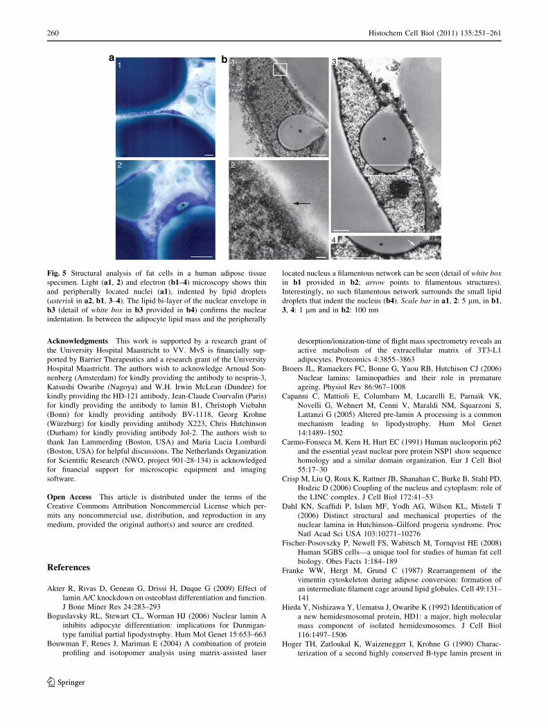
Structural analysis of adipocytes in a human adipose
tissue specimen
Light (Fig. 5a1, 2) and electron (Fig. 5b1–4) microscopy of
a subcutaneous adipose tissue specimen showed peripherally
located thin ovoid-shaped nuclei that were often indented by
lipid droplets (Fig. 5a2, b1, 3), as concluded from the lipid
bi-layer of the nuclear envelope that surrounds indenting
lipid droplets (arrow in Fig. 5b4). A filamentous network
could be detected in between the nucleus and the lipid mass
of the adipocyte (arrow in Fig. 5b2). Interestingly, no such
filamentous network could be seen surrounding small lipid
droplets indenting the nucleus (Fig. 5b4).
Discussion
Since the identification of the LINC complex (Crisp et al.
2006), researchers have tried to find more evidence for the
nuclear-cytoskeleton coupling. The herein described
dynamic organization of nuclear lamina structure, which
coincides with profound changes in the cytoskeleton
throughout fat cell differentiation further supports the
importance of the LINC complex in physiology. Although
our findings do not directly illustrate the necessity of
nuclear lamina breakdown in reorganization of the cyto-
skeleton, other groups have shown that over-expression of
wild-type or mutant lamin A disrupts fat cell differentiation
and lipid accumulation, whereas complete lack of lamin A
conversely promotes adipogenesis (Akter et al. 2009;
Boguslavsky et al. 2006; Capanni et al. 2005). These
reports underscore our results and hint towards the neces-
sity of nuclear lamina reorganization in adipogenesis.
Moreover, at 10 days of fat cell differentiation, a large
fraction of adipocytes showed lack of lamins A (*50%), C
(*30%), B1 (*50%) and emerin (*70%) at the nuclear
rim. Interestingly, however, upon 18 days of differentia-
tion, significantly more adipocytes expressed lamins A, C
and B1 at the nuclear lamina. It is conceivable that upon
continued differentiation the cells exhibit a different
adhesion capacity and tend to detach more easily, partic-
ularly those adipocytes lacking the above nuclear envelope
proteins. On the other hand, the loss of lamins A, C and B1
could be a temporary one needed for the initial steps of
adipose conversion and cells might regain nuclear rim
localization hereafter. Yet, protein analysis studies using
samples obtained at 18 days of differentiation [Fig. 2e, f
(Renes et al. 2005)] and studies performed by others
(Tilgner et al. 2009) showed an evident decrease in protein
levels of lamins A/C and emerin upon adipose conversion.
Emerin is thought to be a key player in adipogenesis
because of its role in nuclear-cytoplasmic shuttling of
b-catenin (Markiewicz et al. 2006). Moreover, cells defi-
cient in emerin have an increased adipogenic potential
(Tilgner et al. 2009). Therefore, it is not surprising that our
studies show an initial loss of emerin upon induction of
adipogenesis, preceding the reorganization/loss of lamins
A and C at the nuclear rim. Except for lamin B2, all lamin
subtypes and emerin dissociated away from the nuclear
envelope upon induction of differentiation. We therefore
hypothesize that the absence of most lamin subtypes and
particularly, that of lamins A and C, i.e. the major con-
tributors to nuclear stiffness (Lammerding et al. 2006),
results in enhanced plasticity of the nucleus. The latter may
add to the multitude of nuclear indentations by lipid
droplets detected in our human adipose tissue specimen.
Mutations in LMNA are associated with partial or gen-
eralized forms of lipodystrophy such as Dunnigan-type
familial partial lipodytsrophy (FPLD) and Hutchinson–
Gilford progeria syndrome (HGPS), respectively [reviewed
in (Broers et al. 2006; Verstraeten et al. 2007)]. In contrast
to FPLD, HGPS is mostly caused by a de novo 1824C [ T
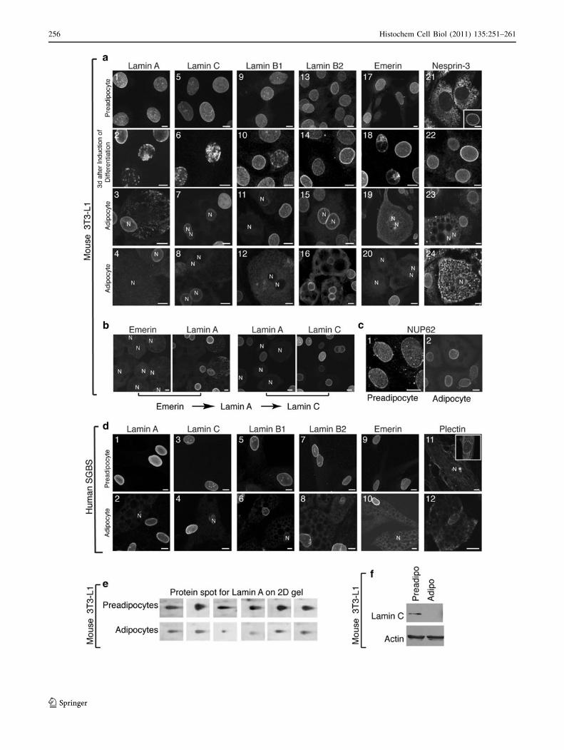
Fig. 2 Nuclear lamina breakdown in fat cell differentiation. Preadi-
pocytes show a homogenous distribution for lamins and emerin at the
nuclear envelope (a1, a5, a9, a13, a17; d1, d3, d5, d7, d9). Upon
induction of differentiation, lamins A, C and B1 accumulate at the
nuclear rim, followed by lamina fragmentation (a2, a6, a10, a18),
except for lamin B2 (a14). Emerin reorganizes away from the nuclear
rim to the ER (a19). Nesprin-3 reorganizes from the ER (a21) to the
nuclear rim upon confluency (a21, insert) and upon induction of
differentiation (a22), followed by dissociation from the lamina to
lipid-bound ER (a23, 24). From double-labeling studies we learned
that emerin reorganizes first, followed by lamin A, and then lamin C
(b). Nucleoporin p62 remains at the rim throughout differentiation
(c). Adipocytes show reduced expression of (a3, 7, 11) and eventually
lack lamin A (a4, d2), C (a8, d4), B1 (a12, d6) and emerin (a20,
d10). Plectin reorganizes from a more diffuse cytoskeletal distribution
(d11) to the nuclear rim upon induction of differentiation (d11, insert)and redistributes into a network in between fat droplets in adipocytes
(d12). e 2D protein gel electrophoresis and subsequent mass
spectrometry of 3T3-L1 cells before treatment (preadipocytes) and
18 days upon induction of fat cell differentiation (adipocytes) showed
a reduced expression of lamin A (protein spot with accession number
p11516 in the Swissprot database) in adipocytes (six replicates
derived from three independent experiments). f Western blot analysis
confirmed reduced expression of lamin C in 3T3-L1 mouse adipo-
cytes upon 18 days of fat cell differentiation. Bar, 10 lm. N nucleus.
All images result from Z-projection of the corresponding confocal
image stack
b
Histochem Cell Biol (2011) 135:251–261 257
123

mutation in LMNA that results in a permanently farnesy-
lated protein called progerin, which affects the polymeri-
zation of the nuclear lamina resulting in stiffer nuclei (Dahl
et al. 2006; Verstraeten et al. 2008). Breakdown of the
nuclear lamina is an early event in adipogenesis and
henceforward, may be a prerequisite for adipose
258 Histochem Cell Biol (2011) 135:251–261
123
conversion. It is conceivable that progerin impairs the
essential reorganization of the nuclear lamina network and
thereby, abolishes fat cell differentiation causing general-
ized lipodystrophy in HGPS. This hypothesis is supported
by the fact that human mesenchymal stem cells expressing
progerin showed markedly reduced differentiation along
the adipogenic lineage, with lipid accumulation only in
those hMSCs expressing very low levels of progerin
(Scaffidi and Misteli 2008).
In conclusion, our findings show a dramatic reorgani-
zation of the nuclear lamina and its coupling to the
vimentin intermediate filament network during adipose
conversion. We suggest that adipogenesis may therefore
serve as a model system in which to examine the LINC
complex. This dynamic reorganization of the nuclear
lamina network in the process of fat cell differentiation is
an important insight that may provide new venues for
research in and treatment of LMNA-associated lipodystro-
phy and other adipose tissue diseases such as obesity.
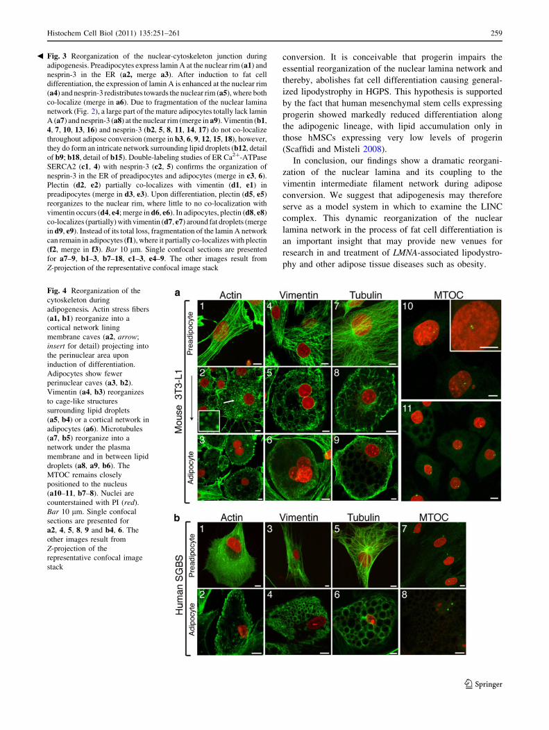
Fig. 3 Reorganization of the nuclear-cytoskeleton junction during
adipogenesis. Preadipocytes express lamin A at the nuclear rim (a1) and
nesprin-3 in the ER (a2, merge a3). After induction to fat cell
differentiation, the expression of lamin A is enhanced at the nuclear rim
(a4) and nesprin-3 redistributes towards the nuclear rim (a5), where both
co-localize (merge in a6). Due to fragmentation of the nuclear lamina
network (Fig. 2), a large part of the mature adipocytes totally lack lamin
A (a7) and nesprin-3 (a8) at the nuclear rim (merge in a9). Vimentin (b1,
4, 7, 10, 13, 16) and nesprin-3 (b2, 5, 8, 11, 14, 17) do not co-localize
throughout adipose conversion (merge in b3, 6, 9, 12, 15, 18), however,
they do form an intricate network surrounding lipid droplets (b12, detail
of b9; b18, detail of b15). Double-labeling studies of ER Ca2?-ATPase
SERCA2 (c1, 4) with nesprin-3 (c2, 5) confirms the organization of
nesprin-3 in the ER of preadipocytes and adipocytes (merge in c3, 6).
Plectin (d2, e2) partially co-localizes with vimentin (d1, e1) in
preadipocytes (merge in d3, e3). Upon differentiation, plectin (d5, e5)
reorganizes to the nuclear rim, where little to no co-localization with
vimentin occurs (d4, e4; merge in d6, e6). In adipocytes, plectin (d8, e8)
co-localizes (partially) with vimentin (d7, e7) around fat droplets (merge
in d9, e9). Instead of its total loss, fragmentation of the lamin A network
can remain in adipocytes (f1), where it partially co-localizes with plectin
(f2, merge in f3). Bar 10 lm. Single confocal sections are presented
for a7–9, b1–3, b7–18, c1–3, e4–9. The other images result from
Z-projection of the representative confocal image stack
Fig. 4 Reorganization of the
cytoskeleton during
adipogenesis. Actin stress fibers
(a1, b1) reorganize into a
cortical network lining
membrane caves (a2, arrow;
insert for detail) projecting into
the perinuclear area upon
induction of differentiation.
Adipocytes show fewer
perinuclear caves (a3, b2).
Vimentin (a4, b3) reorganizes
to cage-like structures
surrounding lipid droplets
(a5, b4) or a cortical network in
adipocytes (a6). Microtubules
(a7, b5) reorganize into a
network under the plasma
membrane and in between lipid
droplets (a8, a9, b6). The
MTOC remains closely
positioned to the nucleus
(a10–11, b7–8). Nuclei are
counterstained with PI (red).
Bar 10 lm. Single confocal
sections are presented for
a2, 4, 5, 8, 9 and b4, 6. The
other images result from
Z-projection of the
representative confocal image
stack
b
Histochem Cell Biol (2011) 135:251–261 259
123
Acknowledgments This work is supported by a research grant of
the University Hospital Maastricht to VV. MvS is financially sup-
ported by Barrier Therapeutics and a research grant of the University
Hospital Maastricht. The authors wish to acknowledge Arnoud Son-
nenberg (Amsterdam) for kindly providing the antibody to nesprin-3,
Katsushi Owaribe (Nagoya) and W.H. Irwin McLean (Dundee) for
kindly providing the HD-121 antibody, Jean-Claude Courvalin (Paris)
for kindly providing the antibody to lamin B1, Christoph Viebahn
(Bonn) for kindly providing antibody BV-1118, Georg Krohne
(Wurzburg) for kindly providing antibody X223, Chris Hutchinson
(Durham) for kindly providing antibody Jol-2. The authors wish to
thank Jan Lammerding (Boston, USA) and Maria Lucia Lombardi
(Boston, USA) for helpful discussions. The Netherlands Organization
for Scientific Research (NWO, project 901-28-134) is acknowledged
for financial support for microscopic equipment and imaging
software.
Open Access This article is distributed under the terms of the
Creative Commons Attribution Noncommercial License which per-
mits any noncommercial use, distribution, and reproduction in any
medium, provided the original author(s) and source are credited.
References
Akter R, Rivas D, Geneau G, Drissi H, Duque G (2009) Effect of
lamin A/C knockdown on osteoblast differentiation and function.
J Bone Miner Res 24:283–293
Boguslavsky RL, Stewart CL, Worman HJ (2006) Nuclear lamin A
inhibits adipocyte differentiation: implications for Dunnigan-
type familial partial lipodystrophy. Hum Mol Genet 15:653–663
Bouwman F, Renes J, Mariman E (2004) A combination of protein
profiling and isotopomer analysis using matrix-assisted laser
desorption/ionization-time of flight mass spectrometry reveals an
active metabolism of the extracellular matrix of 3T3-L1
adipocytes. Proteomics 4:3855–3863
Broers JL, Ramaekers FC, Bonne G, Yaou RB, Hutchison CJ (2006)
Nuclear lamins: laminopathies and their role in premature
ageing. Physiol Rev 86:967–1008
Capanni C, Mattioli E, Columbaro M, Lucarelli E, Parnaik VK,
Novelli G, Wehnert M, Cenni V, Maraldi NM, Squarzoni S,
Lattanzi G (2005) Altered pre-lamin A processing is a common
mechanism leading to lipodystrophy. Hum Mol Genet
14:1489–1502
Carmo-Fonseca M, Kern H, Hurt EC (1991) Human nucleoporin p62
and the essential yeast nuclear pore protein NSP1 show sequence
homology and a similar domain organization. Eur J Cell Biol
55:17–30
Crisp M, Liu Q, Roux K, Rattner JB, Shanahan C, Burke B, Stahl PD,
Hodzic D (2006) Coupling of the nucleus and cytoplasm: role of
the LINC complex. J Cell Biol 172:41–53
Dahl KN, Scaffidi P, Islam MF, Yodh AG, Wilson KL, Misteli T
(2006) Distinct structural and mechanical properties of the
nuclear lamina in Hutchinson–Gilford progeria syndrome. Proc
Natl Acad Sci USA 103:10271–10276
Fischer-Posovszky P, Newell FS, Wabitsch M, Tornqvist HE (2008)
Human SGBS cells—a unique tool for studies of human fat cell
biology. Obes Facts 1:184–189
Franke WW, Hergt M, Grund C (1987) Rearrangement of the
vimentin cytoskeleton during adipose conversion: formation of
an intermediate filament cage around lipid globules. Cell 49:131–
141
Hieda Y, Nishizawa Y, Uematsu J, Owaribe K (1992) Identification of
a new hemidesmosomal protein, HD1: a major, high molecular
mass component of isolated hemidesmosomes. J Cell Biol
116:1497–1506
Hoger TH, Zatloukal K, Waizenegger I, Krohne G (1990) Charac-
terization of a second highly conserved B-type lamin present in
Fig. 5 Structural analysis of fat cells in a human adipose tissue
specimen. Light (a1, 2) and electron (b1–4) microscopy shows thin
and peripherally located nuclei (a1), indented by lipid droplets
(asterisk in a2, b1, 3–4). The lipid bi-layer of the nuclear envelope in
b3 (detail of white box in b3 provided in b4) confirms the nuclear
indentation. In between the adipocyte lipid mass and the peripherally
located nucleus a filamentous network can be seen (detail of white boxin b1 provided in b2; arrow points to filamentous structures).
Interestingly, no such filamentous network surrounds the small lipid
droplets that indent the nucleus (b4). Scale bar in a1, 2: 5 lm, in b1,
3, 4: 1 lm and in b2: 100 nm
260 Histochem Cell Biol (2011) 135:251–261
123
cells previously thought to contain only a single B-type lamin.
Chromosoma 99:379–390
Lammerding J, Fong LG, Ji JY, Reue K, Stewart CL, Young SG, Lee
RT (2006) Lamins A and C but not lamin B1 regulate nuclear
mechanics. J Biol Chem 281:25768–25780
Markiewicz E, Tilgner K, Barker N, van de Wetering M, Clevers H,
Dorobek M, Hausmanowa-Petrusewicz I, Ramaekers FC, Broers
JL, Blankesteijn WM, Salpingidou G, Wilson RG, Ellis JA,
Hutchison CJ (2006) The inner nuclear membrane protein
emerin regulates beta-catenin activity by restricting its accumu-
lation in the nucleus. EMBO J 25:3275–3285
Parton RG, Molero JC, Floetenmeyer M, Green KM, James DE (2002)
Characterization of a distinct plasma membrane macrodomain in
differentiated adipocytes. J Biol Chem 277:46769–46778
Renes J, Bouwman F, Noben JP, Evelo C, Robben J, Mariman E
(2005) Protein profiling of 3T3-L1 adipocyte differentiation and
(tumor necrosis factor alpha-mediated) starvation. Cell Mol Life
Sci 62:492–503
Rosen ED, MacDougald OA (2006) Adipocyte differentiation from
the inside out. Nat Rev Mol Cell Biol 7:885–896
Scaffidi P, Misteli T (2008) Lamin A-dependent misregulation of
adult stem cells associated with accelerated ageing. Nat Cell Biol
10:452–459
Tilgner K, Wojciechowicz K, Jahoda C, Hutchison C, Markiewicz E
(2009) Dynamic complexes of A-type lamins and emerin
influence adipogenic capacity of the cell via nucleocytoplasmic
distribution of beta-catenin. J Cell Sci 122:401–413
Verstraeten VL, Broers JL, van Steensel MA, Zinn-Justin S,
Ramaekers FC, Steijlen PM, Kamps M, Kuijpers HJ, Merckx
D, Smeets HJ, Hennekam RC, Marcelis CL, van den Wijngaard
A (2006) Compound heterozygosity for mutations in LMNA
causes a progeria syndrome without prelamin A accumulation.
Hum Mol Genet 15:2509–2522
Verstraeten VL, Broers JL, Ramaekers FC, van Steensel MA (2007)
The nuclear envelope, a key structure in cellular integrity and
gene expression. Curr Med Chem 14:1231–1248
Verstraeten VL, Ji JY, Cummings KS, Lee RT, Lammerding J (2008)
Increased mechanosensitivity and nuclear stiffness in Hutchin-
son–Gilford progeria cells: effects of farnesyl transferase
inhibitors. Aging Cell 7:383–393
Wabitsch M, Brenner RE, Melzner I, Braun M, Moller P, Heinze E,
Debatin KM, Hauner H (2001) Characterization of a human
preadipocyte cell strain with high capacity for adipose differen-
tiation. Int J Obes Relat Metab Disord 25:8–15
Wilhelmsen K, Litjens SH, Kuikman I, Tshimbalanga N, Janssen H,
van den Bout I, Raymond K, Sonnenberg A (2005) Nesprin-3, a
novel outer nuclear membrane protein, associates with the
cytoskeletal linker protein plectin. J Cell Biol 171:799–810
Histochem Cell Biol (2011) 135:251–261 261
123