renal disease dr. george mellotte. the kidneys two bean-shaped organs, each the size of a fist....
TRANSCRIPT

Renal Renal DiseaseDisease
Dr. George Mellotte
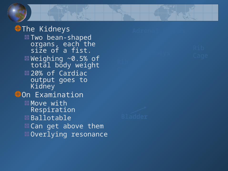
The Kidneys
Two bean-shaped organs, each the size of a fist. Weighing ~0.5% of total body weight20% of Cardiac output goes to Kidney
On ExaminationMove with RespirationBallotableCan get above themOverlying resonance
Rib Cage
Adrenal Glands
Bladder
KidneysRib Cage

Function of the Kidney
Primary balancing organ needed to keep blood in a stable state Remove waste products and toxins
• Urea & Creatinine: • Drugs, toxic substances
Maintain fluid and electrolyte balance• Total body water and fluid distribution
– 50-70% of body weight is water• Sodium Potassium
Maintain normal mineral balance• Calcium Phosphate Magnesium
Regulate acid/base balance

Function of the Kidney
Endocrine Role of the KidneyRegulates blood pressure: Renin-angiotension System and aldosterone
Adjusts final concentration of urine Antidiuretic hormone ADH
Stimulates the production of red blood cells Erythropoietin
Activates Vitamin D (Calcitriol, 1,25(OH)2D3)• Response to Parathyroid hormone
Target Organ Damage Heart
• Left ventricular hypertrophy• Angina or prior myocardial infarction• Prior coronary revascularization• Heart failure
Brain• Stroke or transient ischemic attack
Chronic kidney disease Peripheral arterial disease Retinopathy
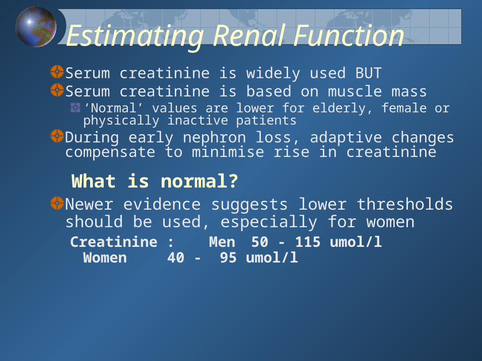
Estimating Renal FunctionSerum creatinine is widely used BUTSerum creatinine is based on muscle mass
‘Normal’ values are lower for elderly, female or physically inactive patients
During early nephron loss, adaptive changes compensate to minimise rise in creatinine
What is normal?Newer evidence suggests lower thresholds should be used, especially for womenCreatinine :Men 50 - 115 umol/l
Women 40 - 95 umol/l

Defining Renal FailureDefining Renal Failure
Cr Clearance = (140 - age) × weight in Kg × SF serum Cr
SF = 1.2 males / 1.05 females
Normal GFR = 90-120mls/minGrade GFR Sr CreatinineMild 60 - 90ml/min 100 - 150umol/l Moderate 30 - 60ml/min 150 - 250µmol/lSevere 15 - 30ml/min 250 - 500µmol/l Endstage < 15ml/min > 500µmol/L
Cockcroft & Gault Formula

Classification of Chronic kidney disease (CKD)
< 15 (or dialysis)End Stage Kidney failure5
15–29Kidney damage Severe GFR4
30–59Kidney damage Moderate GFR3
60–89 Kidney damage mild GFR2
90Kidney damage with normal / GFR1
GFR(mL/min/1.73 m2)
DescriptionStage
www.kidney.org/professionals/kdoqiGFR, glomerular filtration rate
eGFR can be thought of as equivalent to % kidney Function

Key ConceptsKey ConceptsThe importance of early identification
Kidney DiseaseCardiovascular Disease
Focus on quality of care before starting Dialysis
Slowing the progression of Kidney DiseaseSlowing the progression of Co - Morbid Disease
Interplay of pathophysiologyProgressive Kidney DiseaseProgressive Cardiovascular Disease
For persons over age 50, SBP is a more important than DBP as CVD risk factor.
Starting at 115/75 mmHg, CVD risk doubles with each increment of 20/10 mmHg throughout the BP range.
The BP relationship to risk of CVD is continuous, consistent, and independent of other risk factors
JNC 7 Report on Hypertension

New Features and Key Messages
Thiazide-type diuretics should be initial drug therapy for most, either alone or combined with other drug classes.
Certain high-risk conditions are compelling indications for other drug classes.
Most patients will require two or more antihypertensive drugs to achieve goal BP.
If BP is >20/10 mmHg above goal, initiate therapy with two agents, one usually should be a thiazide-type diuretic.
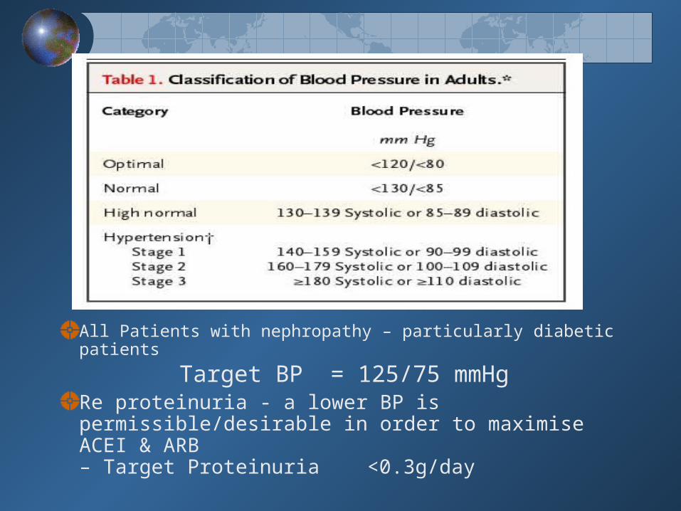
All Patients with nephropathy – particularly diabetic patients
Target BP = 125/75 mmHg
Re proteinuria - a lower BP is permissible/desirable in order to maximise ACEI & ARB – Target Proteinuria <0.3g/day
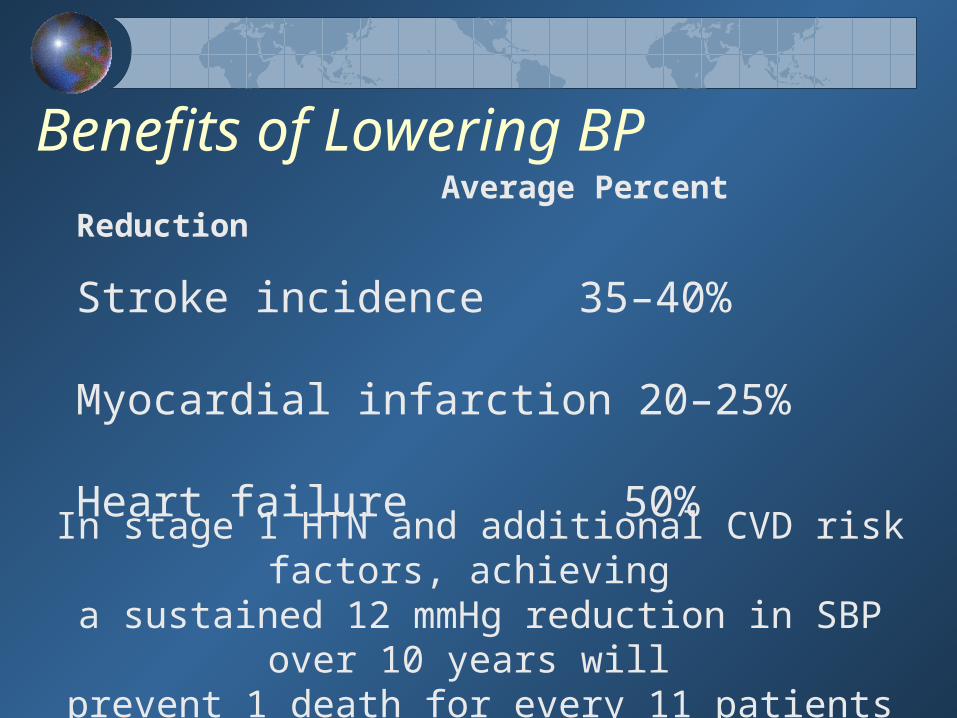
Benefits of Lowering BPAverage Percent Reduction
Stroke incidence 35–40%
Myocardial infarction 20–25%
Heart failure 50%
In stage 1 HTN and additional CVD risk factors, achieving a sustained 12 mmHg reduction in SBP over 10 years will
prevent 1 death for every 11 patients treated.
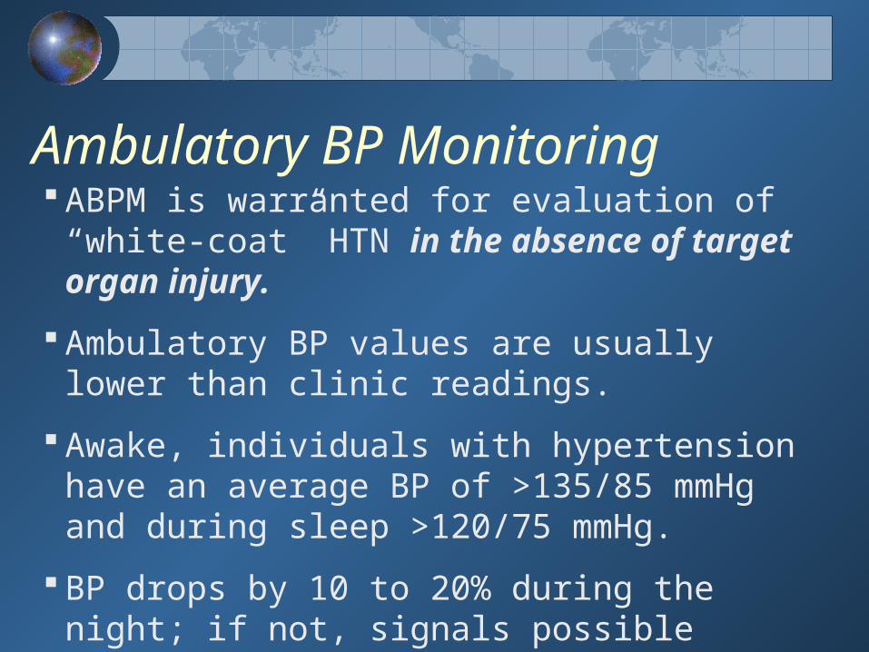
Ambulatory BP Monitoring ABPM is warranted for evaluation of “white-coat” HTN in the
absence of target organ injury.
Ambulatory BP values are usually lower than clinic readings.
Awake, individuals with hypertension have an average BP of >135/85 mmHg and during sleep >120/75 mmHg.
BP drops by 10 to 20% during the night; if not, signals possible increased risk for cardiovascular events.

CVD Risk Factors
Obesity (BMI >30 kg/m2) Physical inactivity Cigarette smoking Hypertension Hyperlipidaemia Diabetes mellitus Microalbuminuria Proteinuria eGFR <60 ml/min Mild Renal failure Creatinine>125 Age (older than 55 for men, 65 for women) Family history of premature CVD
(men under age 55 or women under age 65)*Components of the metabolic syndrome.
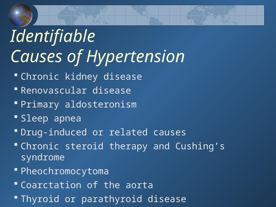
Identifiable Causes of Hypertension Chronic kidney disease Renovascular disease Primary aldosteronism Sleep apnea Drug-induced or related causes Chronic steroid therapy and Cushing’s syndrome Pheochromocytoma Coarctation of the aorta Thyroid or parathyroid disease

Laboratory Tests Routine Tests
• ECG • Urinalysis • Blood glucose, and haemoglobin• Renal profile (potassium, creatinine, calcium)• Lipid profile, Hypokalaemia without diuretics – secondary cause? Hypokalaemia without diuretics – secondary cause?
Optional tests • Microalbuminuria or albumin/creatinine ratio • Renal Ultrasound if renal impairment
More extensive testing for identifiable causes is not generally indicated unless BP control is not achieved
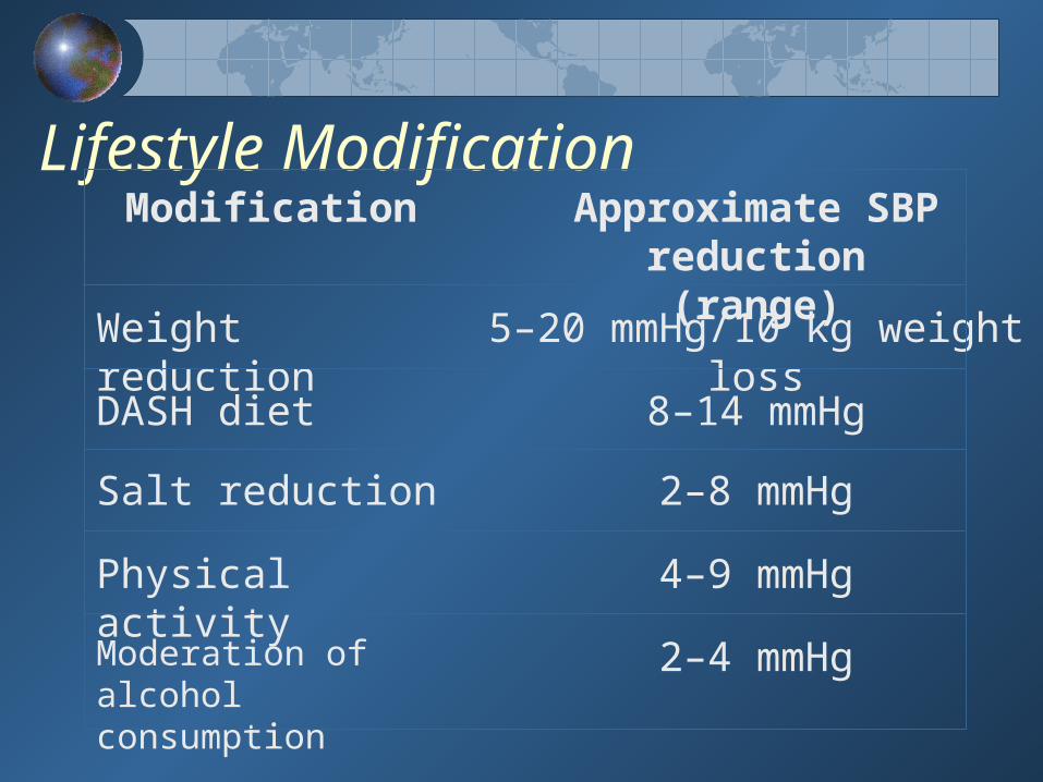
Lifestyle ModificationModification Approximate SBP
reduction(range)Weight reduction 5–20 mmHg/10 kg weight
lossDASH diet 8–14 mmHg
Salt reduction 2–8 mmHg
Physical activity 4–9 mmHg
Moderation of alcohol consumption
2–4 mmHg

Minority Populations In general, treatment similar for all demographic groups. Socioeconomic factors and lifestyle important barriers
to BP control. Prevalence, severity of HTN increased in African
Americans. African Americans demonstrate somewhat reduced BP
responses to monotherapy with BBs, ACEIs, or ARBs compared to diuretics or CCBs.
These differences usually eliminated by adding adequate doses of a diuretic.
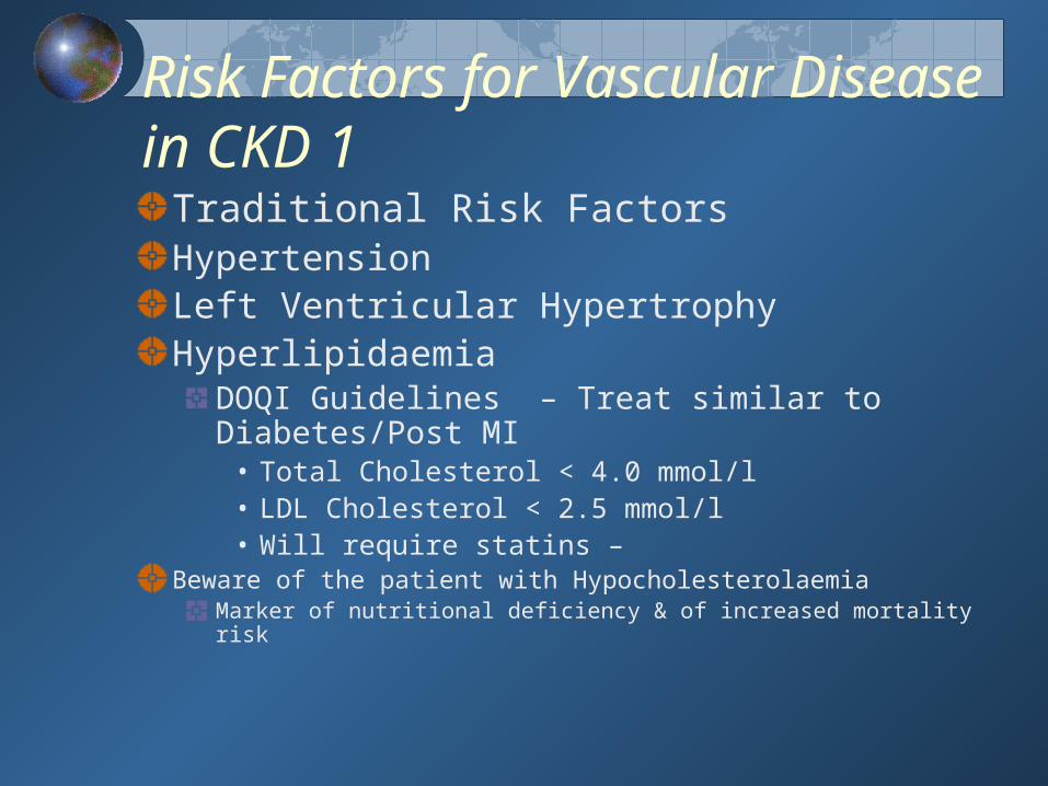
Risk Factors for Vascular Disease in CKD 1
Traditional Risk Factors
Hypertension Left Ventricular HypertrophyHyperlipidaemia
DOQI Guidelines – Treat similar to Diabetes/Post MI
• Total Cholesterol < 4.0 mmol/l• LDL Cholesterol < 2.5 mmol/l • Will require statins –
Beware of the patient with HypocholesterolaemiaMarker of nutritional deficiency & of increased mortality risk
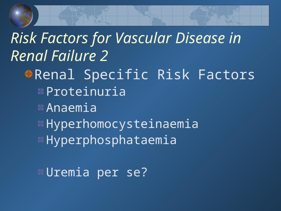
Risk Factors for Vascular Disease in Renal Failure 2
Renal Specific Risk FactorsProteinuriaAnaemiaHyperhomocysteinaemiaHyperphosphataemia
Uremia per se?
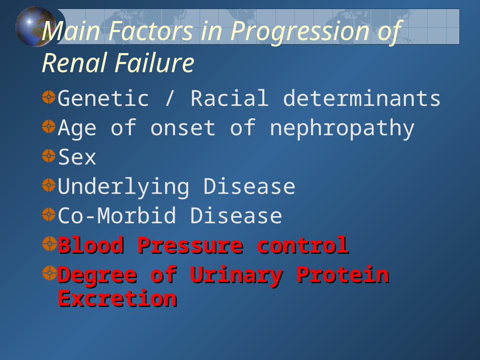
Main Factors in Progression of Renal Failure
Genetic / Racial determinantsAge of onset of nephropathy SexUnderlying DiseaseCo-Morbid DiseaseBlood Pressure controlBlood Pressure controlDegree of Urinary Protein Degree of Urinary Protein ExcretionExcretion
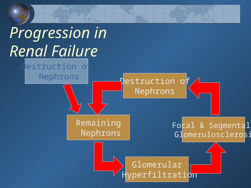
Progression in Renal Failure
Remaining Nephrons
Destruction ofNephrons
Glomerular Hyperfiltration
Focal & Segmental Glomerulosclerosis
Destruction of Nephrons

Renal damage induces hypertension via Plasma volume expansion, Plasma volume expansion, Sodium retention, Sodium retention, Overactivity of both the sympathetic nervous Overactivity of both the sympathetic nervous
system and the renin-angiotensin-aldosterone axis,system and the renin-angiotensin-aldosterone axis, Accumulation of circulating endogenous Accumulation of circulating endogenous
vasoactive substances.vasoactive substances.“Early CRF typically results in a 10-20 mm Hg increase
in diastolic blood pressure until, and unless, renal impairment is identified and treated” Lancet 2000; 356 147-52


Relationship between achieved BP control and declines in GFR in clinical trials of diabetic & non-diabetic renal disease

Blockade of the renin angiotension Blockade of the renin angiotension system is ‘RENOPROTECTIVE’system is ‘RENOPROTECTIVE’
Evidence based medicine suggests preferred initial therapy is either an ACE Inhibitor or Angiotension II Receptor Blocker 3
Most trials note a 24%- 50% risk reduction of overt nephropathy, independent of BP reduction
Recent evidence suggests a synergy between ACE inhibitors and AII Blockers
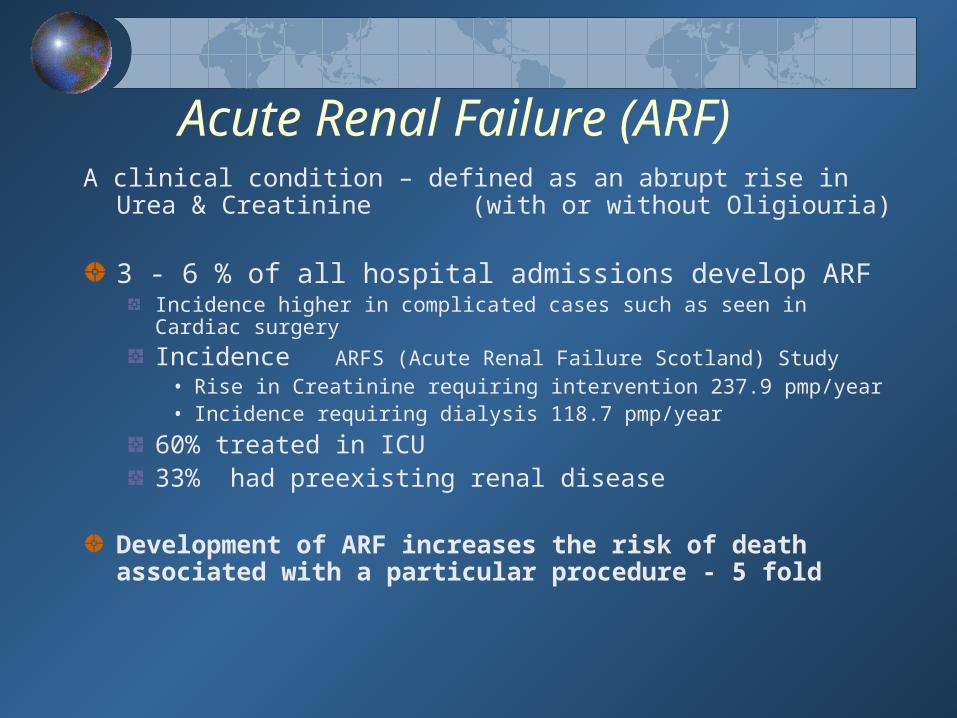
Acute Renal Failure (ARF)A clinical condition – defined as an abrupt rise in Urea &
Creatinine (with or without Oligiouria)
3 - 6 % of all hospital admissions develop ARFIncidence higher in complicated cases such as seen in Cardiac surgery
Incidence ARFS (Acute Renal Failure Scotland) Study• Rise in Creatinine requiring intervention 237.9 pmp/year• Incidence requiring dialysis 118.7 pmp/year
60% treated in ICU33% had preexisting renal disease
Development of ARF increases the risk of death associated with a particular procedure - 5 fold

CASES OF SEVERE ARF (%)Cause Developing Developed
Countries CountriesATNMedical Disease 35 44Obstetrics 14 <1Surgery 8 39
Primary renal disease 10 10
Post renal failure 20 4
Other 13 2
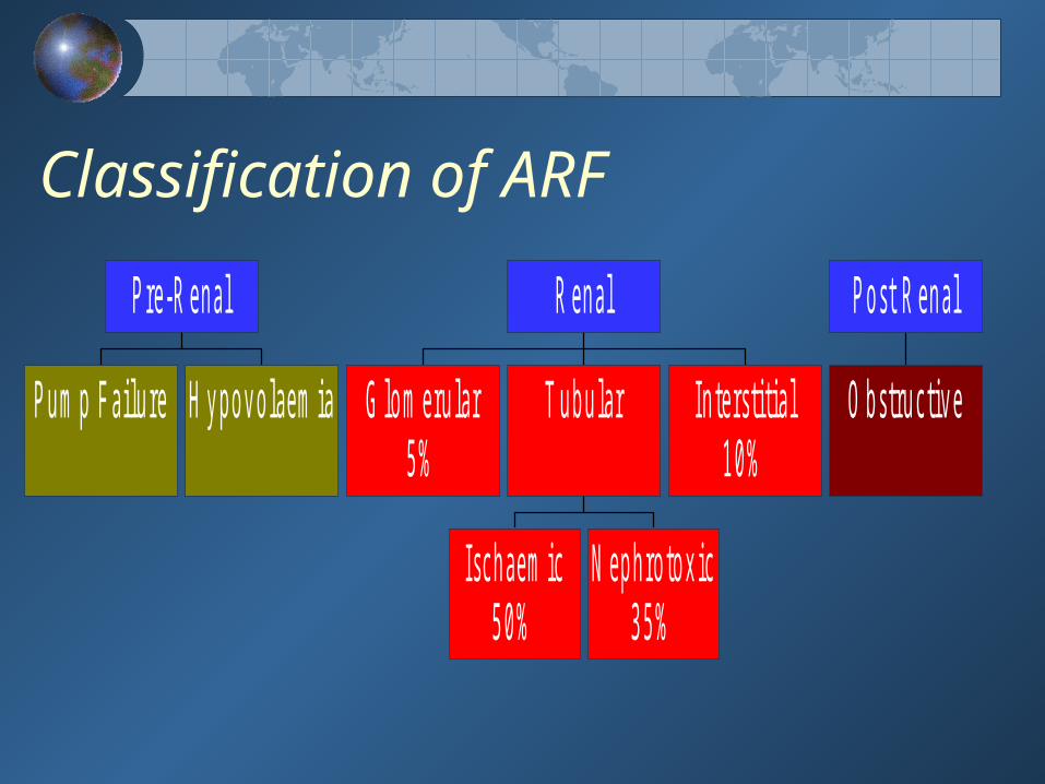
Classification of ARF
P u m p F a ilu re H y p o v o la e m ia
P re -R e n a l
G lo m e ru la r5 %
Isc h a e m ic5 0 %
N e p h ro to x ic3 5 %
T u b u la r In te rs titia l1 0 %
R e n a l
O b stru c tiv e
P o s t R e n a l

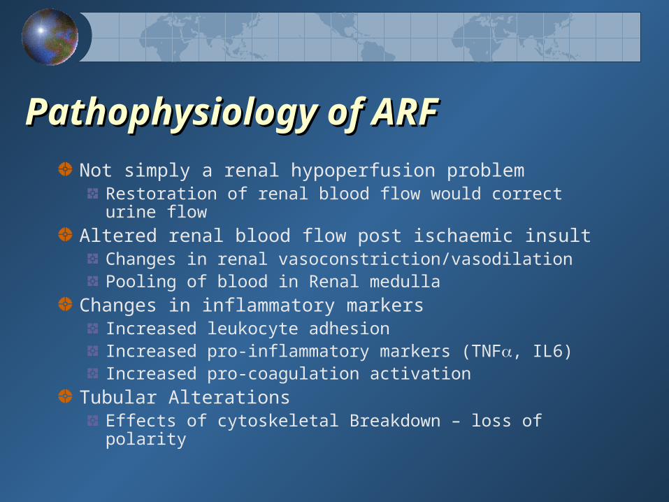
Pathophysiology of ARFPathophysiology of ARF
Not simply a renal hypoperfusion problemRestoration of renal blood flow would correct urine flow
Altered renal blood flow post ischaemic insultChanges in renal vasoconstriction/vasodilationPooling of blood in Renal medulla
Changes in inflammatory markersIncreased leukocyte adhesionIncreased pro-inflammatory markers (TNF, IL6)Increased pro-coagulation activation
Tubular AlterationsEffects of cytoskeletal Breakdown – loss of polarity
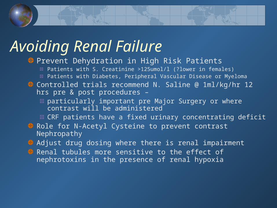
Avoiding Renal FailurePrevent Dehydration in High Risk Patients
Patients with S. Creatinine >125umol/l (?lower in females)Patients with Diabetes, Peripheral Vascular Disease or Myeloma
Controlled trials recommend N. Saline @ 1ml/kg/hr 12 hrs pre & post procedures –
particularly important pre Major Surgery or where contrast will be administeredCRF patients have a fixed urinary concentrating deficit
Role for N-Acetyl Cysteine to prevent contrast NephropathyAdjust drug dosing where there is renal impairmentRenal tubules more sensitive to the effect of nephrotoxins in the presence of renal hypoxia

Guidelines for immediate management of patients with oliguria or anuria
Assess & correct any respiratory or circulatory impairment Manage any life threatening consequences of renal dysfunction (hyperkalaemia, salt and water overload, extreme acidosis)Exclude obstruction of the urinary tract - Get UltrasoundEstablish underlying cause(s) and institute prompt remedial action Get a drug history and alter prescriptions appropriately
Get help from senior appropriately trained specialists
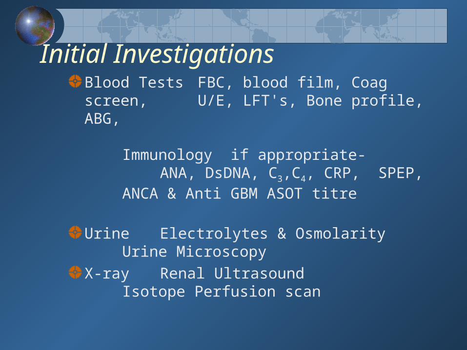
Initial Investigations Blood Tests FBC, blood film, Coag screen, U/E, LFT's, Bone profile, ABG,
Immunology if appropriate- ANA, DsDNA, C3,C4, CRP, SPEP, ANCA & Anti GBM ASOT titre
Urine Electrolytes & OsmolarityUrine Microscopy
X-ray Renal UltrasoundIsotope Perfusion scan

Diagnostic ImagingPlain films (K.U.B.)Screening tool for renal stones. renal calcification
Ultrasoundsafe, high quality images can be obtained on most patientsGood screening tool – esp in ARFUsed to evaluate renal size, renal masses and obstruction
Intravenous Pyelography (I.V.P.)assesses the collecting system and urinary tract Increasingly being replaced by CT-IVP & MRI
Not for patients at risk of contrast nephropathy - Renal failure, Multiple Myeloma, Diabetics Volume depleted patients
Nuclear Medicine – RenogramUsed to asses renal function - DTPA or MAG3MRI
Increasingly being used in general nephrologyMR AngiographyMR Urography

Pre-Renal Vs Established Renal failurePre-Renal Vs Established Renal failure
Pre-renal RF Inadequate renal perfusion
The kidneys concentrating power is normal and the urine produced is highly concentrated
Established RF Failure of tubular function The kidney’s concentrating power is severely
damaged and the urine produced is dilute
Early restoration of effective circulation will avert ATN (Acute Tubular Necrosis)

Renal Hypoperfusion V Established ARF
Measurement Pre Renal ATN
Urinary Na (mmol/l) <20 >40
Fractional excretion <1 >4
of Na (%)
Only appropriate if diuretics not given

Management of Pre Renal FailureManagement of Pre Renal Failure
Restore Renal PerfusionCorrect Hypovolaemia Target CVP = 10 cm Correct HypotensionTarget MAP > 75mmHg
Use fluids in first instanceStart Inotropic support if response insufficient
No Diuresis Frusemide 100 - 250mg IV
Pt must be euvolaemic
Diuresis
Measure hourly urine output & replace losses
Lack of response indicates that ATN has developed
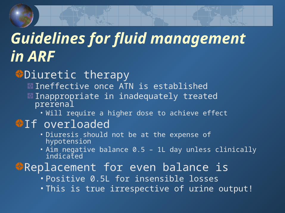
Guidelines for fluid management in ARF
Diuretic therapy Ineffective once ATN is establishedInappropriate in inadequately treated prerenal
• Will require a higher dose to achieve effect
If overloaded • Diuresis should not be at the expense of
hypotension • Aim negative balance 0.5 – 1L day unless clinically
indicated
Replacement for even balance is• Positive 0.5L for insensible losses• This is true irrespective of urine output!

Drugs that induce renal damage
Damage
Decrease in renal perfusion
Impaired intrarenal haemodynamics
Tubular toxicity
Allergic interstitial nephritis
Class of drug Diuretics, ACE inhibitors,
B-Blockers, vasodilators
NSAID’s, radiocontrast
Aminoglycosides, amphotericin, cisplatin
lactams,(penicillins) NSAID’s

ECG changes of HyperkalaemiaPeaked T waves, Flattened P waveProlonged PR interval sinus arrestWide QRS complexes & deep S waves Sine Wave V. Fib asystole

Management of HyperkalaemiaHyperkalaemia is a medical emergency
and must be corrected immediately. V. Fib likely if K+ > 7.0 mmol/l (in ARF)
Rx 1. 10-20 mls of 10% Calcium Gluconate 2. 50 mls of 50% Dextrose with 12 IU Insulin over 30 mins followed by infusion @ 10ml/hour
3. 50 - 100 mls of 8.4% NaHCO3 if acidotic
Indications for renal replacement therapy
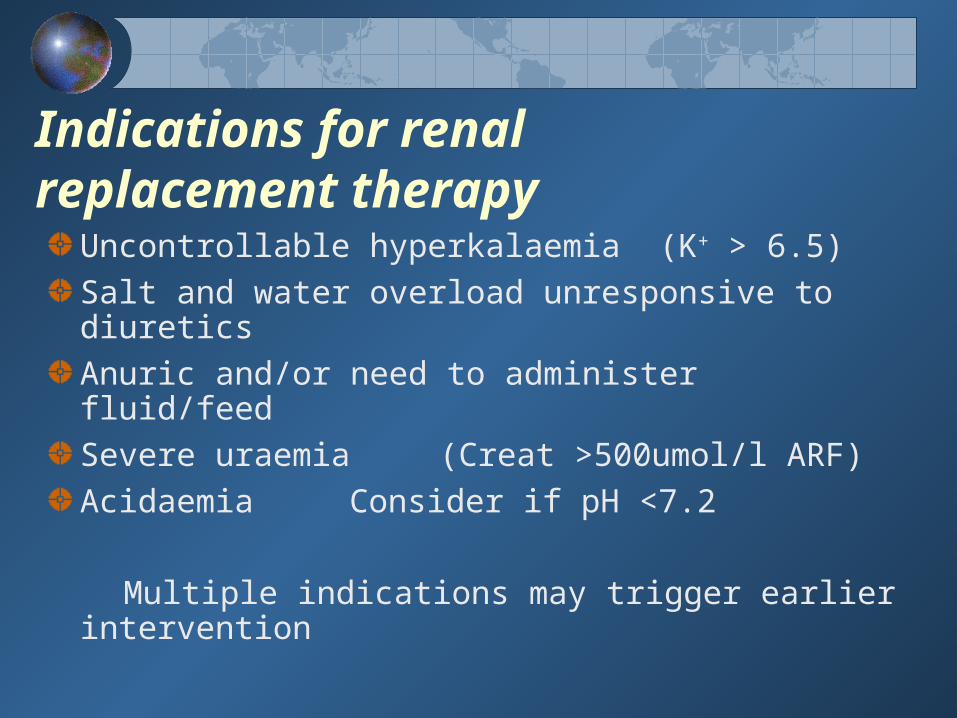
Uncontrollable hyperkalaemia (K+ > 6.5)Salt and water overload unresponsive to diureticsAnuric and/or need to administer fluid/feed Severe uraemia (Creat >500umol/l ARF)Acidaemia Consider if pH <7.2
Multiple indications may trigger earlier intervention

Defined as permanent loss of renal function
Prevalence underestimatedIn USA - while only 0.1% of population require dialysis
5-10 % have renal dsease
Most patients have no symptoms until CRF is advancedAdvanced CRF often termed End Stage Renal Disease (ESRD)Defined as a GFR <15mls/min
Typical symptoms are nausea, anorexia, fatigue, itch and bruisingTypical signs are hypertension, ankle swelling, breathlessness and anaemia
Chronic Renal Failure
Chronic Renal Failure
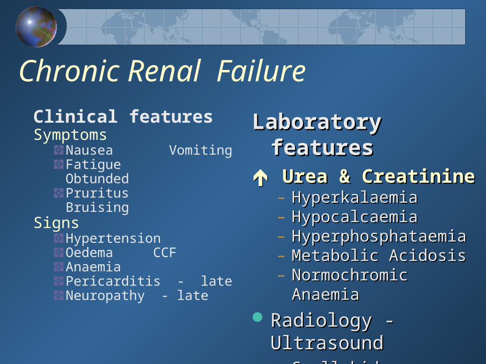
Clinical featuresSymptoms
Nausea VomitingFatigue ObtundedPruritus Bruising
SignsHypertension Oedema CCFAnaemiaPericarditis - lateNeuropathy - late
Laboratory featuresLaboratory features Urea & CreatinineUrea & Creatinine
– HyperkalaemiaHyperkalaemia– HypocalcaemiaHypocalcaemia– HyperphosphataemiaHyperphosphataemia– Metabolic AcidosisMetabolic Acidosis– Normochromic AnaemiaNormochromic Anaemia
Radiology - UltrasoundRadiology - Ultrasound– Small kidneysSmall kidneys - often
scarred
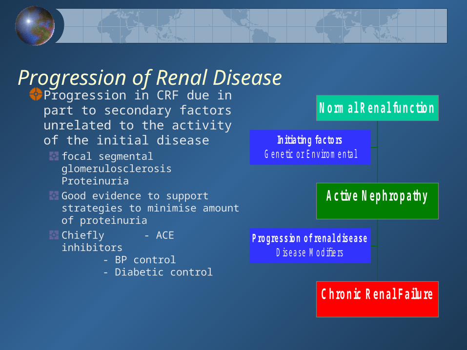
Progression of Renal DiseaseProgression in CRF due in part to secondary factors unrelated to the activity of the initial disease
focal segmental glomerulosclerosis Proteinuria Good evidence to support strategies to minimise amount of proteinuriaChiefly - ACE inhibitors
- BP control- Diabetic
control
Initiating factorsG en e tic o r E n v iro m e nta l
Progression of renal diseaseD isea se M o d if ie rs
C hronic R enal Failure
Active N ephropathy
N orm al R enal function
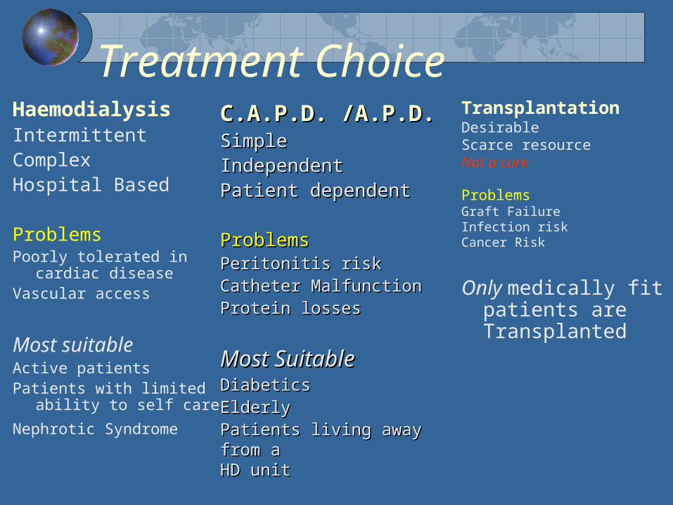
Treatment ChoiceHaemodialysisIntermittentComplex Hospital Based
ProblemsPoorly tolerated in
cardiac diseaseVascular access
Most suitable Active patientsPatients with limited
ability to self care
Nephrotic Syndrome
TransplantationDesirableScarce resourceNot a cure
Problems Graft FailureInfection riskCancer Risk
Only medically fit patients are Transplanted
C.A.P.D. /A.P.D.C.A.P.D. /A.P.D.SimpleSimpleIndependentIndependentPatient dependentPatient dependent
ProblemsProblemsPeritonitis riskPeritonitis riskCatheter MalfunctionCatheter MalfunctionProtein lossesProtein losses
Most SuitableMost SuitableDiabeticsDiabeticsElderlyElderlyPatients living away from aPatients living away from aHD unitHD unit
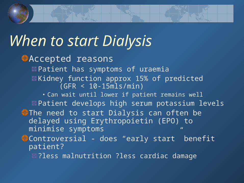
When to start DialysisAccepted reasons
Patient has symptoms of uraemia Kidney function approx 15% of predicted
(GFR < 10-15mls/min)• Can wait until lower if patient remains well
Patient develops high serum potassium levels The need to start Dialysis can often be delayed using Erythropoietin (EPO) to minimise symptomsControversial - does “early start” benefit patient?
?less malnutrition ?less cardiac damage

http://www.kidneypatientguide.org.uk/site/pdanim.html

Peritoneal dialysis
A silastic catheter in the peritoneal cavitySterile dialysis fluid (supplied as 2 - 5L bags)An area for exchange in the homeA pumping device (APD)
The dialysis fluid is infused into the peritoneal cavity (which lies around the bowel) and allowed to dwell for 4-6 hours during which time toxic waste products enter the fluid. The fluid is the drained out and replaced – “An exchange”.
Each exchange lasts 30 - 40 minutesFor CAPD, done 4 times daily, 7 days a weekFor APD, done 4-6 times nightly using a machine “Home Choice”
It is a home based system & requires a committed patient

Automated Peritoneal Dialysis (APD)
Automated Peritoneal Dialysis (APD) uses a machine to perform the fluid exchanges.Dialysis is done at home, at night while pts sleeps.
The APD machine controls the timing of exchanges, drains away the used solution, and fills up the peritoneum with new solution
When patient goes to bed, they connect their catheter to the APD machine's tubing and switch on.The APD machine does exchanges for 8 to 10 hours. In the morning, the patient disconnects from the machine.
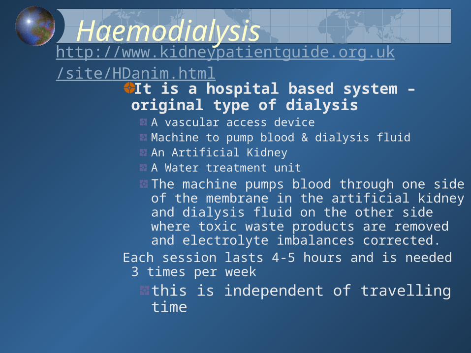
Haemodialysis
It is a hospital based system – original type of dialysis
A vascular access device Machine to pump blood & dialysis fluidAn Artificial KidneyA Water treatment unit
The machine pumps blood through one side of the membrane in the artificial kidney and dialysis fluid on the other side where toxic waste products are removed and electrolyte imbalances corrected.
Each session lasts 4-5 hours and is needed 3 times per week
this is independent of travelling time
http://www.kidneypatientguide.org.uk/site/HDanim.html

Haemodialysis

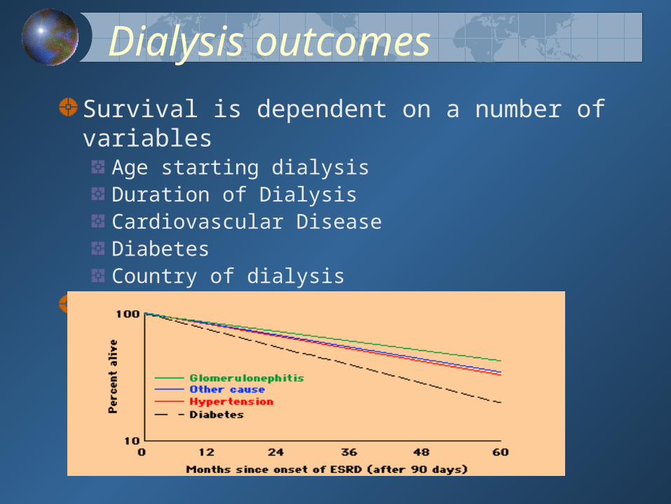
Dialysis outcomes
Survival is dependent on a number of variables
Age starting dialysisDuration of Dialysis Cardiovascular DiseaseDiabetesCountry of dialysis
Expected death rate is 8-40 times that of controls (10 -
15% pa)

TransplantationRecipient Evaluation
Cardiovascular risk, Viral screen, Urological assessment Only 30% patients on dialysis are fit for transplant listAverage waiting time is gone up to 24 monthsAll transplants done in Beaumont
Patients require long term Immunosuppression
Average 1 year graft survival 92% Average Graft survival 12-20 yrsMain cause of graft loss currently• Death of patient! - mainly due to Cardiac disease
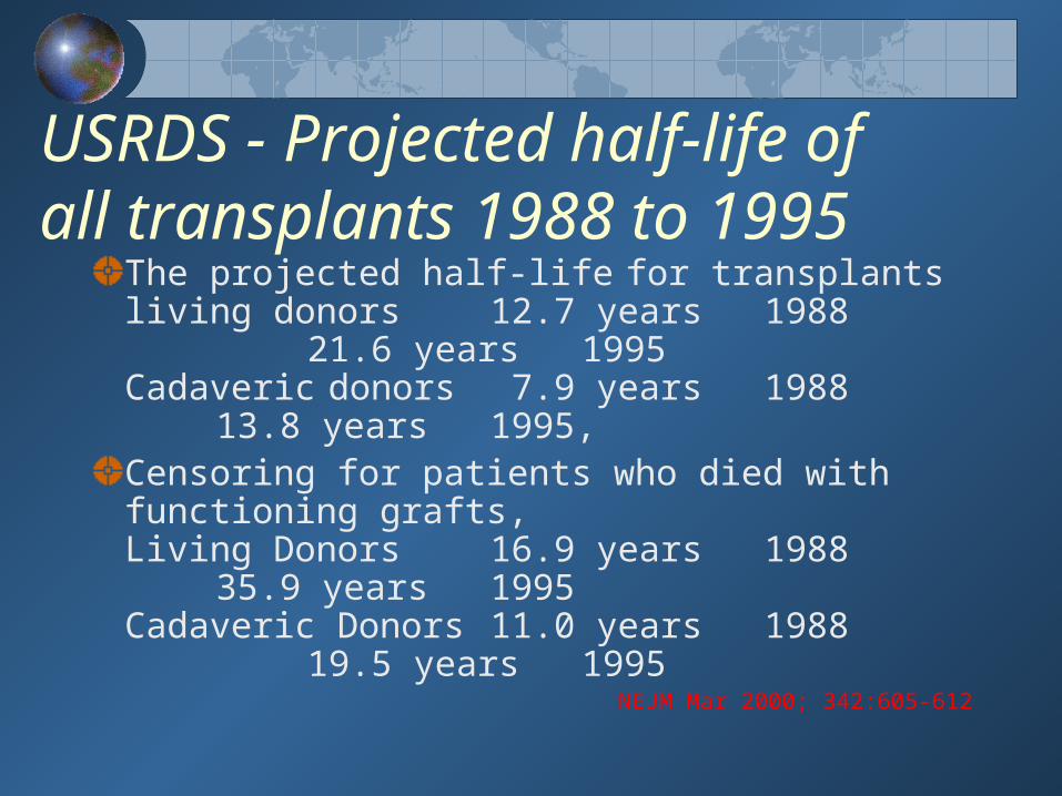
USRDS - Projected half-life of all transplants 1988 to 1995
The projected half-life for transplants living donors 12.7 years 1988
21.6 years 1995 Cadaveric donors 7.9 years 1988
13.8 years 1995, Censoring for patients who died with
functioning grafts, Living Donors 16.9 years 1988
35.9 years 1995Cadaveric Donors 11.0 years 1988
19.5 years 1995NEJM Mar 2000; 342:605-612
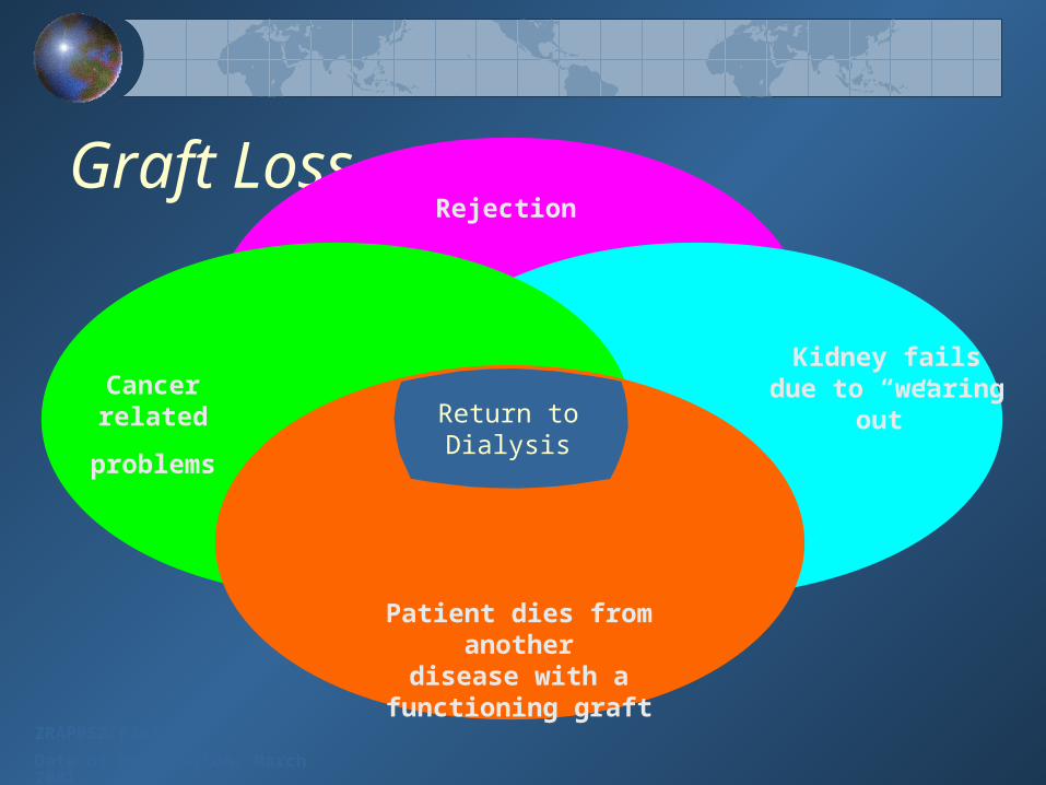
Graft Loss
ZRAP052/0301
Date of Preparation: March 2001
Rejection
Kidney fails due to “wearing out”Cancer related
problems
Patient dies from anotherdisease with a functioning
graft
Return to Dialysis

Dipstick Urinalysis – Haematuria
Dipstick urinalysis detects Haem protein (either red blood cells or haemoglobin)
Highly sensitive but many false positive testsConfirm with urine microscopy. Transient haematuria is relatively common in young subjects and is not indicative of disease.
• Yearly urinalyses in 1000 men between the ages of 18 & 33haematuria – 39% at least once
– 16% two or more occasions BMJ 1984 288:20
Negative tests reliably excludes abnormal haematuria
Coexistent dipstick proteinuria is usually significant and should be investigated further
Dipstick Urinalysis – Protein
Standard dipstick detects albumin >300mg/lhighly specific, but not very sensitiveMeasures urinary protein concentrationThe categories are only a rough guide
Patients with persistent proteinuria should undergo a a 24-hour urine measurement of protein excretion.
Proteinuria > 1g/24 hrs – consider renal biopsy
may biopsy at lower levels

MicroalbuminuriaProtein excretion above normal but below the threshold of “Standard Dipstick”
Albuminuria normally <20mg/24 hrs (15 µg/min); Microalbuminuria = 30-300mg/24 hrs (20-200 µg/min)
Albumin-to-creatinine ratio microalbuminuria = 2.25 - 3.4 mg alb/mmol creatinine
Risk factor in Diabetic NephropathyHigh incidence of false positives

Microalbuminuria Early marker of Diabetic Nephropathy
Usually develops within 10 years of onset of DMDuration of disease before onset of Microalbuminuria correlates with risk of progression to nephropathy
Microalbuminuria < 10 years - Most progress Microalbuminuria > 10 years 30 -50 % progress
Outcome much better than original studies – ?effect of active Rx
Dipstick Urinalysis – other
Pyuria - detects White cells in urineLeukocyte esterase – (75 – 95% sensitivity)
Nitrites - indicates bacterial infectionEnterobacteriaceae convert urinary nitrate to nitriteFalse negative at low colony counts UTIs
Touted as simple and inexpensive screen for UTIMay detect pyuria not associated with infectionNitrite alone insufficient for diagnosis
Abnormalities on dipstick urinalysis seen with UTI should be shown to resolve with clinical cure
Major Clinical Syndromes of Glomerular Disease
Nephrotic Syndrome Nephritic syndromeRapidly Progressive GlomerulonephritisChronic GlomerulonephritisPersistent urinary abnormalities with no symptoms

ProteinuriaIndicative of significant renal
diseaseGlomerular Proteinura» predominantly Albumin» >3.5g/day - classifies as “nephrotic range”» selectivity index useful» amount correlates with long term prognosis Tubular Proteinuria» usually < 2 g/day» due to a failure to reabsorb small molecular weight proteins e.g.. B2 Microglobulin
Light Chain Disease

Glomerular Proteinuria
Primary GN Minimal Change* IgA NephritisFSGS* Membranous*
Hereditary Alport’s*, Infectious SBE, HIV*, Hepatitis, Immunological/Systemic Disease Vasculitis , SLE, PAN, Wegner’s, Goodpastures
Diabetes*, Pregnancy-associated, Drugs Pencillamine, Gold, NSAIDs, Heroin*.Neoplasm's Solid organ CA*, Lymphoma, LeukaemiaOthers Amyloid*, Renal Tx rejection
*Typically nephrotic range

Nephrotic SyndromeHypoalbuminaemia, (<30g/L)Proteinuria (>3.5g/day)Generalised oedema (JVP = N)hypercholesterolaemia
+/- hypertriglyceridaemiaAssociated with - Increased risk of infection
- Increased clotting tendencyPt > 10 years of age should have a renal Biopsy

Acute Poststreptococcal Glomerulonephritis
Principally a disease of children (M>F)Characteristic 10 day latent period between sore throat and renal diseaseNephrotic Urine - ‘Smoky Brown’ haematuria - oliguriaAssociated with oedema and hypertensionDx - rising ASO titre, throat culture - streptococcal A, renal biopsy
Rapidly Progressive Glomerulonephritis
Progression to ESRF within weeks or months of onsetFocal necrotizing GN with crescent formation on renal biopsy
Can form part of a vasculitic processAnti GBM disease Goodpasture’s diseaseANCA positive vasculitisWegners Granulomatosis Churg strauss microscopic polyarteritis SLE any CT disease
Can complicate any primary GN
Outlook for recovery poor unless treated earlySteroids & Cyclophosphamide
Pulse Methyl Prednisolone - plasmapheresis

IgA Nephritis Commonest form of GN worldwide - 30%
typically young males (M:F = 3:1)66% - macroscopic haematuria following (12- 24 hours) onset of sore throat or URTI
may produce ARF - often recurrent 33% -- persistent proteinuria and haematuria
No serological marker 50% raised circulating IgA levels
Long term risk of CRF = 25 - 50%
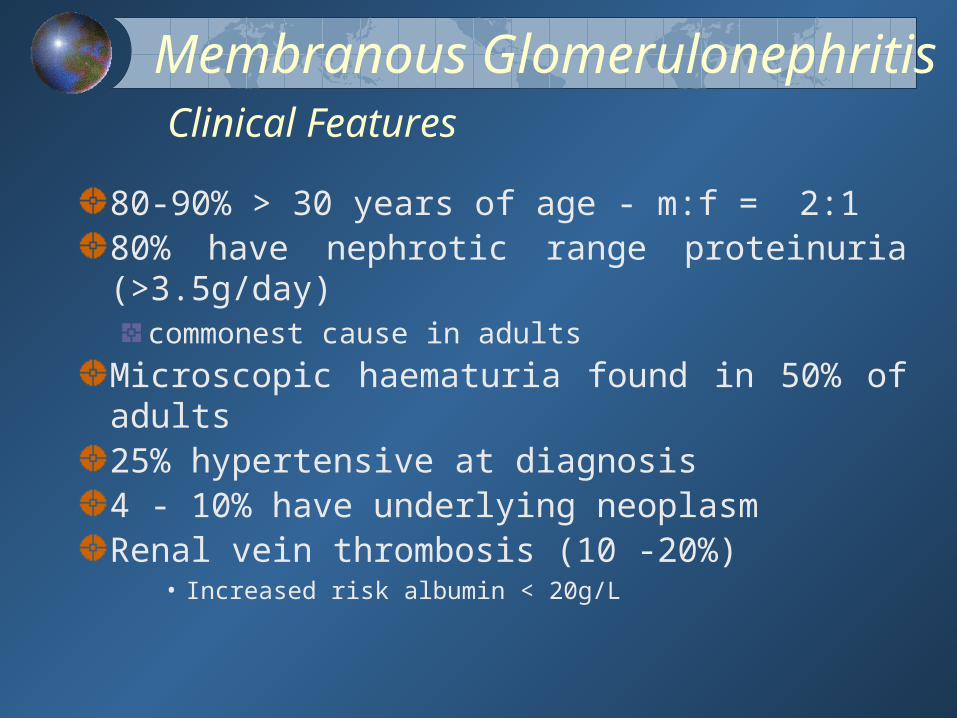
Membranous Glomerulonephritis Clinical Features
80-90% > 30 years of age - m:f = 2:180% have nephrotic range proteinuria (>3.5g/day)
commonest cause in adults
Microscopic haematuria found in 50% of adults25% hypertensive at diagnosis4 - 10% have underlying neoplasm Renal vein thrombosis (10 -20%)
• Increased risk albumin < 20g/L
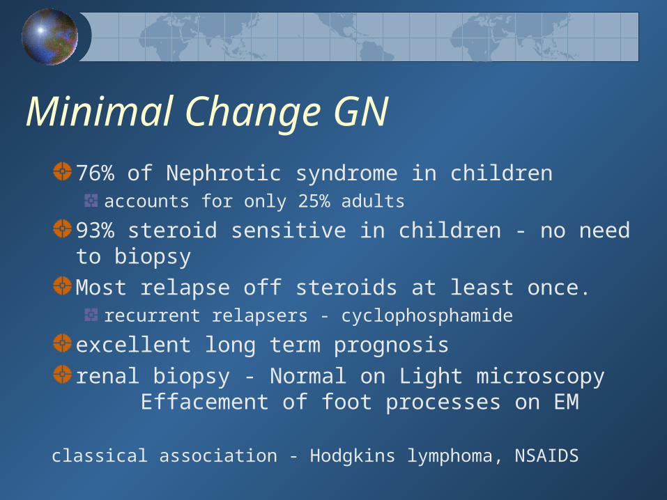
Minimal Change GN76% of Nephrotic syndrome in children
accounts for only 25% adults
93% steroid sensitive in children - no need to biopsyMost relapse off steroids at least once.
recurrent relapsers - cyclophosphamide
excellent long term prognosisrenal biopsy - Normal on Light microscopy
Effacement of foot processes on EM
classical association - Hodgkins lymphoma, NSAIDS
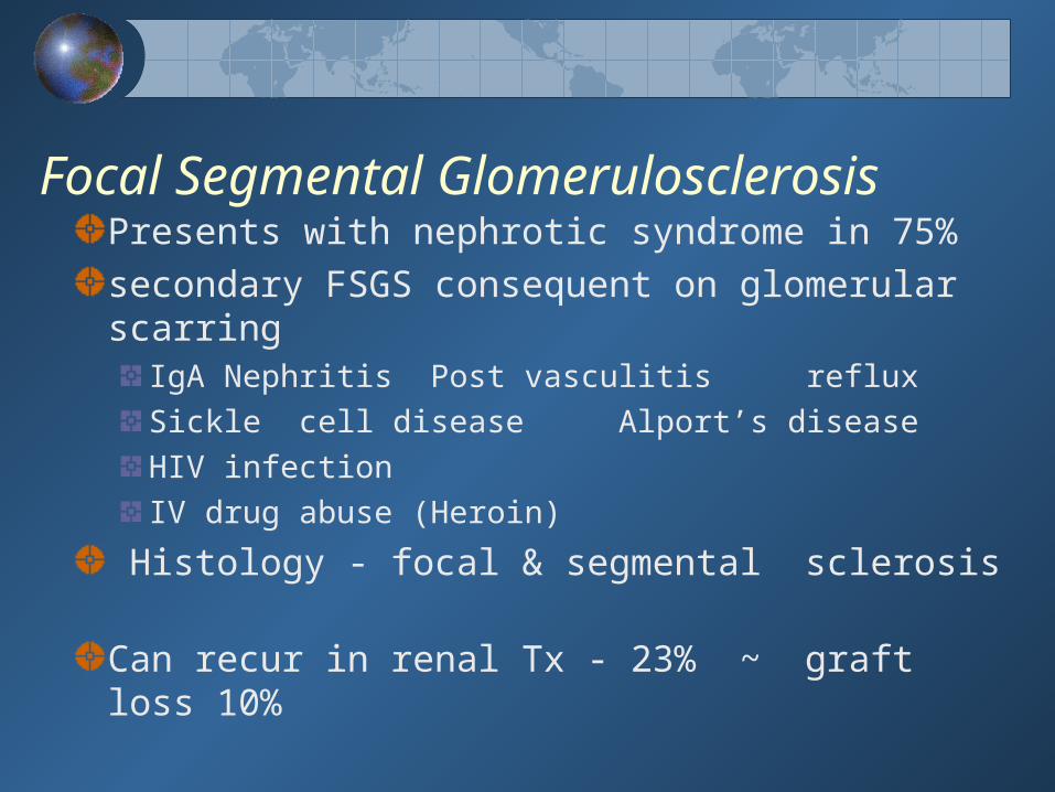
Focal Segmental GlomerulosclerosisPresents with nephrotic syndrome in 75%secondary FSGS consequent on glomerular scarring
IgA Nephritis Post vasculitis reflux Sickle cell disease Alport’s diseaseHIV infectionIV drug abuse (Heroin)
Histology - focal & segmental sclerosis
Can recur in renal Tx - 23% ~ graft loss 10%

MesangioCapillary GN -MCGN(Membranoproliferative GN)
Presentation - Nephrotic (50%) - Nephritic (25%)Histologically Type 1 - Subendothelial deposits
Type 2 - Dense deposit diseaseAssociated with low complenent levels
C3 nephritic factorPartial lipodystrophy
No treatment shown to be effective50 % ESRF at 10 years
Can recur in renal Tx - 15 -35% ~ graft loss 10%


Autosomal Dominant Polycystic Kidney Disease
2 Types PKD 1 85% PKD 2 15%
Prevalence 1 : 500 - 1 : 1000 (Europe)8 - 10% of dialysis patients
Sex Males = Females
Clinical onset Typically 20’s - 50’s

Pathophysiology
Disease begins in uteroMultiple cysts, lined by tubular-type cellsCysts contain uriniferous fluid, blood or pyogenic secretionsCysts can arise anywhere along the nephron
only 1 - 5% of nephrons are involvedIntervening areas show nephrosclerosis and chronic interstitial nephropathy
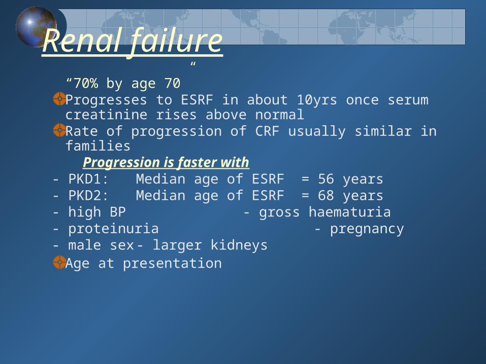
Renal failure“70% by age 70”
Progresses to ESRF in about 10yrs once serum creatinine rises above normalRate of progression of CRF usually similar in families
Progression is faster with- PKD1: Median age of ESRF = 56 years- PKD2: Median age of ESRF = 68 years - high BP - gross haematuria- proteinuria - pregnancy - male sex - larger kidneys
Age at presentation
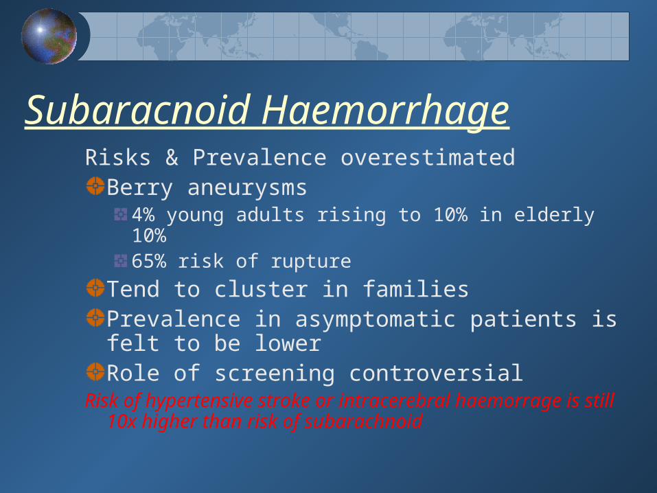
Subaracnoid HaemorrhageRisks & Prevalence overestimated
Berry aneurysms 4% young adults rising to 10% in elderly 10%65% risk of rupture
Tend to cluster in families Prevalence in asymptomatic patients is felt to be lowerRole of screening controversial
Risk of hypertensive stroke or intracerebral haemorrage is still 10x higher than risk of subarachnoid
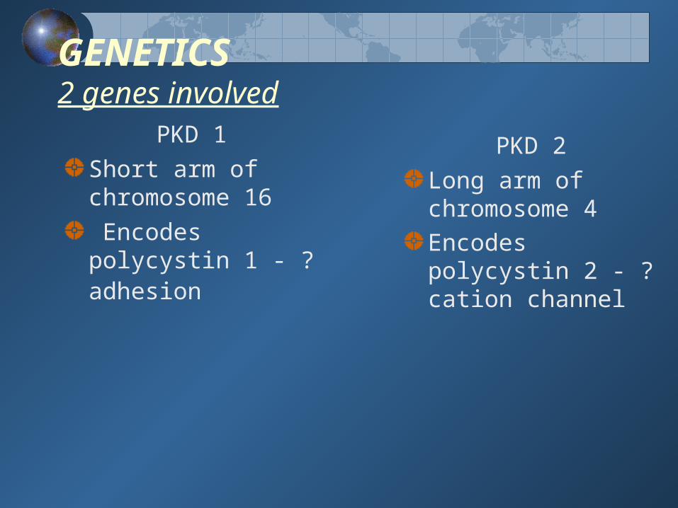
GENETICS 2 genes involved
PKD 1
Short arm of chromosome 16
Encodes polycystin 1 - ? adhesion
PKD 2
Long arm of chromosome 4Encodes polycystin 2 - ? cation channel

GENETICSComplete penetrance & variable expressionOnset of the disease may be earlier if inherited from the motherRate of progression of CRF varies from family to family & within familiesPositive Family history in > 60%
Remainder ? spontaneous mutation

DIAGNOSIS
UltrasoundVery sensitive and specific
Especially in Patient > 30 years of ageDetects cysts as small as 1 - 1.5 cmIncreased false negatives in young patientsCharacteristically multiple cysts in both kidneys which are largeCT (with contrast )More sensitive than USSDetects cysts of 0.5cmDefinitive radiological test

GeneticsOnly 1 - 5% of nephrons develop cysts even
though they all carry a copy of the abnormal gene
2 hit hypothesisAt a cellular level ADPKD seems to be acting as a recessive traitA ‘ second hit ‘ or mutation in the normal copy of the gene seems to have to occur sporadically before the nephron will produce cysts

Renal Ostodystrophy
Develops early in course of CRFFirst signs at GFR~ 40mls/min
Treatment GoalsPTH = 150 - 300ng/l (2-3x normal) in ESRDPhosphate = 0.8 – 1.4 mmol/lCalcium = 2.1 – 2.4 mmol/l
TreatmentRestrict phosphate in dietPhosphate Binders – Ca & non Ca basedActivated Vitamin D (calcitriol / alfacalcidolol/parcalcitriol)New agents being developed - calcimimetics
Increasing concern regarding vascular calcification