remington sample chapter
DESCRIPTION
Remington_sample_chapterTRANSCRIPT
7/18/2019 Remington Sample Chapter
http://slidepdf.com/reader/full/remington-sample-chapter 1/12
Sample chapter from Remington: The Science and Practice of Pharmacy
Chapter 33
Complex FormationThorsteinn Loftsson, MSPharm, PhD and Marcus E. Brewster, Ph
In chemistry and chemical processes the word complex usuallyrefers to molecules or molecular assemblies formed by com-bination of substrates, S, and ligands, L. Most often complex( Sm Ln) formation is a reversible process:
m S n L S L
m n. .+
where m substrate molecules, associate with n ligand moleculesto form a complex of m:n stoichiometry. In this context, com-
plex formation, complexation, binding, association, and che-lation are often synonymous. The substrate and ligand are kepttogether by relatively strong coordinate covalent bonds or byweak non-covalent forces such as hydrogen bonding, van der
Waals forces, electrostatic interactions, dipole forces, chargetransfer, release of conformational strain, or hydrophobic in-
teractions. The complex formation changes the physicochemi-cal properties of its constituents, both of the substrate and theligand, including their aqueous solubility, molar absorptivity,NMR chemical shifts, adsorption to solid surfaces, partitioningbehavior, conductivity, chemical reactivity and/or pKa values.By studying such properties, for example of the substrate as afunction of the ligand concentration, the complex can be iden-tified and quantitatively described. Furthermore, the methodsof chemical kinetics and thermodynamics can be applied to de-scribe the formation and dissociation of a complex. Althoughmost frequently, substrate and ligand molecules are associatedby weak chemical forces, there are complexes where bonds arequite strong and formation of some metal complexes are virtu-ally irreversible. Complexes are usually broadly classified intotwo groups based on the type of S-L bonding involved, namely
coordination complexes and molecular complexes.Coordination complexes consist of ionic substrates, most
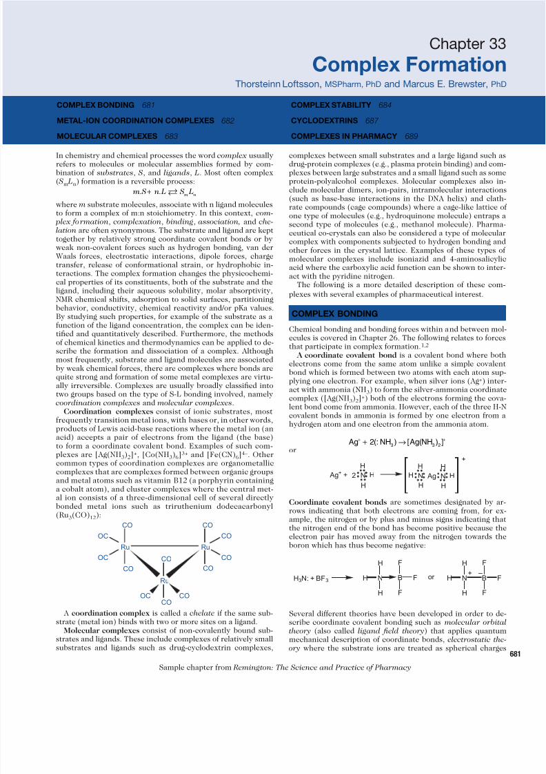
frequently transition metal ions, with bases or, in other words,products of Lewis acid-base reactions where the metal ion (anacid) accepts a pair of electrons from the ligand (the base)to form a coordinate covalent bond. Examples of such com-plexes are [Ag(NH3)2]+, [Co(NH3)6]3+ and [Fe(CN)6]4–. Othercommon types of coordination complexes are organometalliccomplexes that are complexes formed between organic groupsand metal atoms such as vitamin B12 (a porphyrin containinga cobalt atom), and cluster complexes where the central met-al ion consists of a three-dimensional cell of several directlybonded metal ions such as triruthenium dodecacarbonyl(Ru3(CO)12):
CO
CO
CO
CO
CO
CO
CO
CO
CO
OC
OC
OC
Ru Ru
Ru
A coordination complex is called a chelate if the same sub-strate (metal ion) binds with two or more sites on a ligand.
Molecular complexes consist of non-covalently bound sub-strates and ligands. These include complexes of relatively smallsubstrates and ligands such as drug-cyclodextrin complexes,
complexes between small substrates and a large ligand such adrug-protein complexes (e.g., plasma protein binding) and complexes between large substrates and a small ligand such as somprotein-polyalcohol complexes. Molecular complexes also include molecular dimers, ion-pairs, intramolecular interaction(such as base-base interactions in the DNA helix) and clathrate compounds (cage compounds) where a cage-like lattice oone type of molecules (e.g., hydroquinone molecule) entraps second type of molecules (e.g., methanol molecule). Pharmaceutical co-crystals can also be considered a type of moleculacomplex with components subjected to hydrogen bonding another forces in the crystal lattice. Examples of these types omolecular complexes include isoniazid and 4-aminosalicylacid where the carboxylic acid function can be shown to interact with the pyridine nitrogen.
The following is a more detailed description of these complexes with several examples of pharmaceutical interest.
COMPLEX BONDING
Chemical bonding and bonding forces within and between moecules is covered in Chapter 26. The following relates to forcethat participate in complex formation.1,2
A coordinate covalent bond is a covalent bond where botelectrons come from the same atom unlike a simple covalenbond which is formed between two atoms with each atom supplying one electron. For example, when silver ions (Ag+) inteact with ammonia (NH3) to form the silver-ammonia coordinatcomplex ([Ag(NH3)2]+) both of the electrons forming the cova
lent bond come from ammonia. However, each of the three H-covalent bonds in ammonia is formed by one electron from hydrogen atom and one electron from the ammonia atom.
Ag NH Ag NH+ +
+ →23 3 2
(: ) [ ( ) ]
Ag+ + N
H
H
H
N
H
H
H
2 AgN
H
H
H
+
Coordinate covalent bonds are sometimes designated by arows indicating that both electrons are coming from, for example, the nitrogen or by plus and minus signs indicating thathe nitrogen end of the bond has become positive because thelectron pair has moved away from the nitrogen towards th
boron which has thus become negative:
H3N: + BF3 H N B
H
H
F
F
F
or H N B
H
H
F
F
F
+ –
Several different theories have been developed in order to describe coordinate covalent bonding such as molecular orbitatheory (also called ligand field theory) that applies quantummechanical description of coordinate bonds, electrostatic th
ory where the substrate ions are treated as spherical charge
COMPLEX BONDING 681
METAL-ION COORDINATION COMPLEXES 682
MOLECULAR COMPLEXES 683
COMPLEX STABILITY 684
CYCLODEXTRINS 687
COMPLEXES IN PHARMACY 689
or
7/18/2019 Remington Sample Chapter
http://slidepdf.com/reader/full/remington-sample-chapter 2/12
682 PHARMACEUTICS
Sample chapter from Remington: The Science and Practice of Pharmacy
and the ligands as dipoles, and valence bond theory, a quantummechanical theory wherein the ligand donates a pair of elec-trons to a vacant orbital associated with the ion.
Another concept that is used to describe coordination com-plexes is the concept of hard and soft acids and bases (HSAB).
A hard acid is defined as a small Lewis acid (i.e. electron-pairacceptor) atom with high positive charge density and low po-larizability whereas a soft acid is large and polarizable. A hardbase has high electronegativity and low polarizability whereas a
soft base is polarizable (Table 33-1). Polarizability is a measureof the ease with which the electron cloud can be deformed un-der the influence of a charge field. In general, hard acids bindpreferably with hard bases, and soft acids bind preferably withsoft bases.
Non-covalent intermolecular forces (i.e., between molecules)
participating in a complex formation are, in general, relativelyweak forces compared to intramolecular forces (i.e., within amolecule) such as covalent bonds. The following types of non-covalent forces are known to participate in complex formations:
Electrostatic interactions are the consequence of classical at-traction and repulsion effects between charges such as charge-charge, charge-dipole, and dipole-dipole interactions.
Dispersion force is a quantum mechanical effect where syn-chronization of the electronic motion between two moleculesresults in momentary dipole moments and consequently attrac-tion between the molecules. Dispersion force is also called the
van der Waals force. However, some authors use the term vander Waals force for all non-covalent forces.
Hydrogen bonding is an interaction involving formation ofhydrogen bond ( H -bond) between a proton donor ( HA) and aproton acceptor ( B):
A H B A H B− + − The A-H bond has a covalent character but the H ··· B bond ispredominantly electrostatic. The strength of the hydrogen bondis controlled in part by the acid strength of HA and the basestrength of B, but it is also affected by the solvent.
Charge-transfer interaction is a consequence of electrontransfer from an electron donor molecule to an electron accep-tor molecule. Frequently, charge-transfer complexes involveelectron transfer between metal atoms and ligands. However,charge-transfer interactions are also known in other inorganicas well as organic compounds. A well-known charge-transfercomplex is the dark blue or purple iodine/starch complex.
Hydrophobic interaction results from the tendency of liquidwater to exclude non-polar molecules. The water moleculesform a “cage” around two or more non-polar molecules keep-
ing them together in a kind of a complex or loosely associatedmolecules. Hydrophobic interactions are commonly observedduring inclusion complex formations where a non-polar moietyof larger molecule enters a somewhat lipophilic cavity.
Release of conformational strain is known to participate information of cyclodextrin inclusion complexes where the cy-clodextrin molecule frequently undergoes significant confor-mation changes upon complex formation. However, it has beenargued that relief of conformational strain is not a driving forcefor cyclodextrin complex formation.2
Release of high-energy water molecules from, for example,a cyclodextrin cavity does participate in inclusion complexformation. In aqueous solutions the somewhat lipophilic cyclo-
dextrin cavity is occupied by high-energy water molecules (i.e.,water not capable of fulfilling all of its hydrogen-bonding re-quirements) that are expelled during inclusion complex forma-tion. However, based on some thermodynamic observations ithas been argued that the exclusion of water molecules from thecavity is not a driving force of cyclodextrin complex formation.2 This is based on the fact that although the cavity-bound watermolecules are of higher energy (i.e., are enthalpy rich) theyhave more conformational freedom (i.e., form fewer hydrogenbonds). Consequently, although expulsion of water moleculesfrom the cavity is accompanied by a negative enthalpy change,the free energy change of the overall process is not necessarynegative.
METAL-ION COORDINATION COMPLEXES
Metal-ion coordination complexes, sometimes simply calledmetal complexes, consist of a central metal ion bonded to oneor more ligands that are electron-pair donors such as a nitrog-enous base (e.g., ammonia), an ion (e.g., chloride ion), or anaromatic compound (e.g., ferrocene). The number of bondsbetween the metal ion and the ligand or ligands is called the
coordination number of the complex. Metal ions can have morethan one coordination number. The maximum number is de-fined by the size, charge and electronic structure of the metalion. Coordination numbers are normally between 2 and 9 with
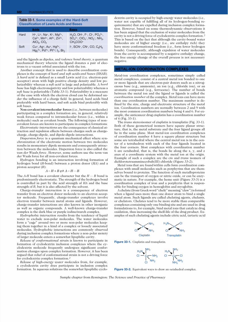
the most common coordination numbers being 4 and 6. For ex-ample, the anticancer drug cisplatin has a coordination numberof 4 (Fig. 33-1).
The trans stereoisomer of cisplatin is transplatin (Fig. 33-1).Both of these geometrical isomers have square planar struc-ture, that is, the metal substrate and the four ligand groups alllie in the same plane. Most metal-ion coordination complexesof coordination number 4 have a square planar structure butsome are tetrahedral where the central metal ion is in the cen-ter of a tetrahedron with each of the four ligands located inthe four corners. Most complexes with coordination number6 are octahedral, that is, the bonds lie along the x, y, and zaxes of a coordinate system with the metal ion at the origin.Example of such a complex are the cis and trans isomers ofdichlorotetraamminecobalt(III) chloride (Figure 33-2).
Metal ions that are found within cells form coordination com-
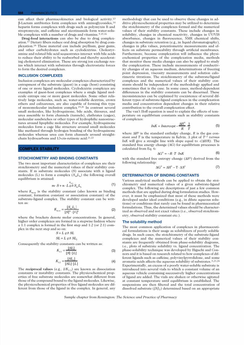
plexes with small molecules such as porphyrins that are them-selves bound to proteins. The function of such metalloproteinscan be the transport of oxygen or nitric oxide, or can be enzy-matic in nature. For example, the heme unit (Figure 33-3) is acoordination complex of iron and a porphyrin that is respon-sible for binding oxygen in hemoglobin and myoglobin.
A chelate (from Greek word “chelè” meaning “claw”) is formedwhen a ligand uses more than one donor atom to bind a singlemetal atom. Such ligands are called chelating agents, chelants,or chelators. Chelates tend to be more stable than comparablecomplexes containing only one binding site and are used in drugformulations to, for example, bind metal ions that catalyze drugoxidation, thus increasing the shelf-life of the drug product. Ex-amples of such chelating agents include citric acid, tartaric acid
Table 33-1. Some examples of the Hard-Soft
Classification of Lewis Acids and Bases
Acids Bases
Hard H+, Li+, Na+, K+, Mg2+,
Ca2+, Mn2+, Al3+, Zn2+,
AlCl3, CO2
H2O, OH–, F–, Cl–, PO43–,
SO42–, ClO4
–, NO3–, NH3,
CH3COO–
Soft Cu+, Ag+, Au+, Hg2+,
Hg22+, Pd2+, Pt2+; I2, Br2
I–, SCN–, CN–, CO, C6H6
Pt
Cl NH3 NH3
NH3Cl
Pt
Cl
H3N Cl
Cisplatin Transplatin
Figure 33-1. Cisplatin and transplatin.
NH2
NH2
NH2
H2N
CoCl
Cl
NH2
NH2
Cl
H2N
CoCl
NH2
Figure 33-2. Equivalent ways to draw an octahedral complex.
7/18/2019 Remington Sample Chapter
http://slidepdf.com/reader/full/remington-sample-chapter 3/12
68COMPLEX FORMATION
Sample chapter from Remington: The Science and Practice of Pharmacy
and EDTA (ethylenediamine tetraacetic acid). Some drugs canalso be chelating agents and bind ions. For instance tetracyclineforms hydrophilic chelates with ions such as calcium (Ca2+),iron (Fe3+, Fe2+), aluminium (Al3+), and magnesium (Mg2+), i.e.complexes that possess poor oral bioavailability (Fig. 33-4).3
Milk and milk products, mineral supplements and antacids con-taining polyvalent cations ingested simultaneously with tetracy-cline antibiotics can reduce their oral bioavailability by as muchas 90%.4 Nalidixic acid, ciprofloxacin and other quinolones doalso form chelates with polyvalent ions that can reduce theiroral bioavailability. The intensity of the effect depends both onthe nature of the drug and the cation, as well as on the dosesused. The drugs that bind metal ions should be taken eithertwo to three hours after or before ingestion of cation containingproducts such as dairy products, antacids and mineral supple-
ments. These types of drug-metal ion interactions are coveredin Chapter 70, Drug Interactions.β-Lactam antibiotics form chelates with metal ions such
as Cu2+ (Fig. 33-5). The β-lactam ring is much more suscep-tible to specific base hydrolysis (i.e., toward OH– attack) whencomplexed than when uncomplexed. Thus, formation of suchchelates can significantly decrease the shelf-life of β-lactamantibiotics.
MOLECULAR COMPLEXES
Molecular complexes can be classified according to 1) the bond-ing or interaction between substrate and ligand (e.g., electro-
static interaction, charge-transfer, hydrogen bonding and hydrophobic interaction), 2) type of substrate and ligand forminthe complex (e.g., small molecule—small molecule, small molecule—macromolecule, enzyme—substrate, drug—receptor anantigen—antibody) or 3) type of structure formed (e.g., self-asembled aggregate, micelle, clathrate, and inclusion complex
Molecular complexes consist of one or more substrates and lgands that are, in general, held together by relatively weak, noncovalent forces. In aqueous solutions, free molecules are mooften in dynamic equilibrium with molecules bound within thcomplex but tightly bond complexes are not unknown. Much othe pioneering work on molecular complexes was published bTakeru Higuchi and coworkers.5
Drug-excipient interactions are quite common in pharmceutical formulations and frequently such interactions are th
result of formation of molecular complexes. Formation of succomplexes will affect the physicochemical and biological properties of the drug bound to the excipient such as its aqueousolubility, release from the drug formulation and bioavailabilit
Non-ionic water-soluble polymers, such as polyvinyl pyrroidone (PVP) and hydroxypropyl methylcellulose (HPMC), arcommonly used to enhance viscosity and to form hydrogelIn aqueous solutions, PVP forms coils and is able to bind drumolecules via non-covalent binding, and PVP is known to formcomplexes with various drugs.6 PVP binds iodine in the preence of iodide ions (povidone-iodine) and as such is used as antibacterial agent for treatment and prevention of skin or wouninfections (Fig. 33-6). Both PVP and HPMC can enhance aqueous solubility of drugs through the formation of water-solubcomplexes and have been used as solubility and dissolutioenhancers.7,8
Ionic water-soluble polymers, such carboxymethylcellulos(CMC), are able to bind drugs. For example, in aqueous solutions CMC forms complexes with some basic drugs such aatenolol, diphenhydramine, lidocaine and propranolol.9 Gum
Arabic and alginates, both of which are anionic polysaccharideare known to interact with drugs affecting their aqueous solubility and chemical stability. Chitosan (a linear polysaccharidcomposed of randomly distributed glucosamine) is positivecharged at acidic pH and forms nano-sized complexes with neatively charged DNA and RNA. Chitosan has been used in gendelivery.10 Alginates form drug complexes and have also beeused to modify drug delivery.11
Drug-drug interactions in the form of complexes are alswell known. Thus, salicylates form complexes with benzocainand the anticancer drugs paclitaxel, doxorubicin, and etoposide have been shown to form dimers and trimers in aqueou
solutions as well as etoposide–paclitaxel complexes.12 Cocainand morphine form a binary complex in aqueous solutions tha
N
C
H
CH
N
H
C
N
HC
N
CH2
CH2
H3C
H3C
COOH
COOH
CH3
CH3
Fe
Figure 33-3. Heme, a coordination complex of iron and a porphyrin.Two of the bonds are coordination covalent bonds indicated by arrows.
O-
NH2
OOOOOH
NCH3HO
CH3H3C
OH
Ca+
Figure 33-4. Calcium (Ca2+) tetracycline complex (chelate).
N
S
CH3
Cu2+
CH3
O
NHC
H2
C
O
COO –
Figure 33-5. Copper (Cu2+) benzylpenicillin complex (chelate).
N
O
C
H
H2
C
N
O
C
H
H+
I3 –
Figure 33-6. The iodine—PVP complex.
7/18/2019 Remington Sample Chapter
http://slidepdf.com/reader/full/remington-sample-chapter 4/12
684 PHARMACEUTICS
Sample chapter from Remington: The Science and Practice of Pharmacy
can affect their pharmacokinetics and biological activity.13 β-Lactam antibiotics form complexes with aminoglycosides,14 heparin forms complexes with drugs such as polymyxin B andstreptomycin, and caffeine and nicotinamide form water-solu-ble complexes with a number of drugs and vitamins.5,15,16
Drug-food interactions can also be due to drug complex-ation.17 Dietary fibers reduce oral drug absorption by drug com-plexation.18 These material can include psyllium, guar gums,and other carbohydrates such as cyclodextrins. Cholestyr-amine and related bile acid sequestrates interact with bile acidsto reduce their ability to be reabsorbed and thereby accelerat-ing cholesterol elimination. These are strong ion exchange res-ins which interact with substrates through electrostatic forcesto form the desired complexes.
INCLUSION COMPLEXES
Inclusion complexes are molecular complexes characterized byentrapment of the substrate (guest) in a cage (host) consistingof one or more ligand molecules. Cyclodextrin complexes areexamples of guest-host complexes where a single ligand mol-ecule entraps one or more guest molecules. Some other rela-tively large molecules, such as, water-soluble dextrins, crownethers and calixarenes, are also capable of forming this typeof monomolecular inclusion complex.19,20 In contrast severalsmall molecules, like hydroquinone, bile acids, thiourea, andurea assemble to form channels (tunnels), clathrates (cages),
molecular sandwiches or other types of hydrophilic nanostruc-tures around lipophilic molecules. For example, hydroquinonemolecules form a cage like structure around small moleculeslike methanol through hydrogen bonding of the hydroquinonemolecules whereas urea can form channels around straight-chain hydrocarbons and 13-cis-retinoic acid.21,22
COMPLEX STABILITY
STOICHIOMETRY AND BINDING CONSTANTS
The two most important characteristics of complexes are theirstoichiometry and the numerical values of their stability con-stants. If m substrate molecules (S) associate with n ligandmolecules (L) to form a complex ( S m L n) the following overallequilibrium is attained:
m S n L S L
K
m n
m n
⋅ + ⋅ :
(1)
where K m:n is the stability constant (also known as bindingconstant, formation constant or association constant) of thesubstrate-ligand complex. The stability constant can be writ-ten as:
K S L
S L m n
m n
m n:
[ ]
[ ] [ ]=
⋅ (2)
where the brackets denote molar concentrations. In general,higher order complexes are formed in a stepwise fashion wherea 1:1 complex is formed in the first step and 1:2 (or 2:1) com-plex in the next step and so on:
S L SL
SL L SL
+
+
2
Consequently the stability constants can be written as:
K SL
S L1 1:
[ ]
[ ] [ ]=
⋅ (3)
K SL
SL L1 2
2:
[ ]
[ ] [ ]=
⋅ (4)
The reciprocal values (e.g., 1/K 1:1) are known as dissociationconstants or instability constants. The physicochemical prop-erties of free substrate molecules are somewhat different fromthose of the compound bound to the ligand molecules. Likewise,the physicochemical properties of free ligand molecules are dif-ferent from those of the ligand in the complex. In general, any
methodology that can be used to observe these changes in ad-ditive physicochemical properties may be utilized to determinethe stoichiometry of the complexes formed and the numericalvalues of their stability constants. These include changes insolubility, changes in chemical reactivity, changes in UV/VISabsorbance, changes in fluorescence, NMR chemical shifts,changes in substrate retention (e.g., in liquid chromatography),changes in pKa values, potentiometric measurements and ef-fects on substrate permeability through artificial membranes.Furthermore, because complexation will influence the physi-cochemical properties of the complexation media, methodsthat monitor these media changes can also be applied to studythe complexation. These include measurements of conductiv-ity changes of an aqueous medium, determinations of freezingpoint depression, viscosity measurements and solution calo-rimetric titrations. The stoichiometry of the substrate/ligandcomplexes and the numerical values of their stability con-stants should be independent of the methodology applied andsometimes that is the case. In some cases, method-dependentdifferences in the stability constants can be discerned. Theseinconsistencies can be explained by coexistence of several dif-ferent types of substrate-ligand complexes in the complexationmedia and concentration dependent changes in their relativecontribution to the overall complexation effect.
The van’t Hoff equation is used to explain the effects of tem-perature on equilibrium constants such as stability constants
of complexes:
InK Intercept
H
R T= −
∆⋅
01
(5)
where ∆ H 0 is the standard enthalpy change, R is the gas con-stant and T is the temperature in Kelvin. A plot of T –1 versuslnK will give a straight line with slope equal to -( ∆ H/R). Thestandard free energy change (∆G) for equilibrium processes iscalculated from Eq. 6:
∆ = − ⋅ ⋅G R T InK 0
(6)
with the standard free entropy change (∆ S0) derived from thefollowing relationship:
∆ = ∆ − ⋅ ∆G H T S0 0 0
(7)
DETERMINATION OF BINDING CONSTANTS
Various analytical methods can be applied to obtain the stoi-chiometry and numerical values of a given substrate-ligandcomplex. The following are descriptions of just a few commonmethods that are applied during drug formulation studies. How-ever, it must be emphasized that most of these methods weredeveloped under ideal conditions (e.g., in dilute aqueous solu-tions) or conditions that rarely can be found in pharmaceuticalformulations. Thus, the determined values should be character-ized as observed and not exact values (i.e., observed stoichiom-etry, observed stability constant etc.).
The solubility method
The most common application of complexes in pharmaceuti-cal formulations is their usage as solubilizers of poorly solubledrugs. In such cases, the stoichiometry of the substrate-ligandcomplexes and the numerical values of their stability con-
stants are frequently obtained from phase-solubility diagrams,i.e., plots of substrate solubility vs. ligand concentration. Thephase-solubility technique was developed by Higuchi and Con-nors and it is based on research related to how complexes of dif-ferent ligands such as caffeine, polyvinylpyrrolidone, and somearomatic acids affects the aqueous solubility of substrates.5,23,24 Experimentally, an excess of a poorly water-soluble substrate isintroduced into several vials to which a constant volume of anaqueous vehicle containing successively higher concentrationsof ligand are added. The vials are shaken or otherwise agitatedat constant temperature until equilibrium is established. Thesuspensions are then filtered and the total concentration ofdissolved substrate ([S]T) determined based on an appropriate
7/18/2019 Remington Sample Chapter
http://slidepdf.com/reader/full/remington-sample-chapter 5/12
68COMPLEX FORMATION
Sample chapter from Remington: The Science and Practice of Pharmacy
analytical technique. The phase-solubility profile is then con-structed by assessing the effect of the ligand concentration onthe apparent solubility of the substrate. The phase-solubilitymethod does not give insight as to how the complexes are
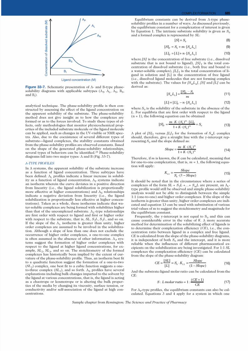
formed or as to the forces involved. To study these types of ef-fects, only methodologies that monitor physicochemical prop-erties of the included substrate molecule or the ligand moleculecan be applied, such as changes in the UV-visible or NMR spec-tra. Also, due to the coexistence of several different types ofsubstrate—ligand complexes, the stability constants obtainedfrom the phase-solubility profiles are observed constants. Basedon the shape of the generated phase-solubility relationships,several types of behaviors can be identified.25 Phase-solubilitydiagrams fall into two major types: A and B (Fig. 33-7).
A-TYPE PROFILES
In A systems, the apparent solubility of the substrate increaseas a function of ligand concentration. Three subtypes havebeen defined: A L profiles indicate a linear increase in solubil-ity as a function of ligand concentration, A P systems indicate
an isotherm wherein the curve deviates in a positive directionfrom linearity (i.e., the ligand solubilization is proportionallymore effective at higher concentrations) and A N relationshipsindicate a negative deviation from linearity (i.e., the ligandsolubilization is proportionally less effective at higher concen-trations). Taken as a whole, these isotherms indicate that wa-ter-soluble complexes are being formed with solubilities higherthan that of the uncomplexed substrate. A L-type relationshipsare first order with respect to ligand and first or higher orderwith respect to the substrate, that is, SL, S2 L, S3 L, and so on.If the slope of the A L isotherm is greater than unity, higherorder complexes are assumed to be involved in the solubiliza-tion. Although a slope of less than one does not exclude theoccurrence of higher order complexes, a one-to-one complexis often assumed in the absence of other information. A P sys-tems suggest the formation of higher order complexes with
respect to the ligand at higher ligand concentrations, for ex-ample, SL2, SL3 , and so on. The stoichiometry of the formedcomplexes has historically been implied by the extent of cur-vature of the phase-solubility profile. Thus, an isotherm best fitto a quadratic function suggest the formation of a one-to-two( SL2) complex, one best fit to a cubic function suggests a one-to-three complex ( SL3), and so forth. A N profiles have severalexplanations including bulk changes imparted to the solvent bythe ligand at various concentrations, that is, the ligand is actingas a chaotrope or kosmotrope or is altering the bulk proper-ties of the media by changing its viscosity, surface tension, orconductivity and/or self-association of the ligand at high con-centrations.
Equilibrium constants can be derived from A-type phasesolubility profiles in a number of ways. As discussed previouslthe equilibrium constant for a complexation of interest is giveby Equation 1. The intrinsic substrate solubility is given as Sand a formed complex is represented by SL:
[ ] S S= 0
(
[ ] [ ] S S m S L
T m n= + ⋅0
(
[ ] [ ] [ ] L L n S L
T m n= + ⋅
(1
where [S] is the concentration of free substrate (i.e., dissolvesubstrate that is not bound to ligand), [S]T is the total concentration of dissolved substrate (i.e., both free and bound ta water-soluble complex), [L]T is the total concentration of lgand in solution and [L] is the concentration of free ligan(i.e., dissolved ligand molecules that are not forming complewith the substrate). The values for [Sm Ln ], [S] and [L] can bderived as:
[ ]
[ ] S L
S S
m m n
T=
− 0
(1
[ ] [ ] [ ] L L n S L
T m n= − ⋅
(1
where S0 is the solubility of the substrate in the absence of th L. For equilibria that are first order with respect to the ligan(n = 1), the following equation can be obtained:
[ ] ( ) [ ]( )
S m K S L K S
ST
m
T
m= ⋅ ⋅ ⋅
+ ⋅
+0
0
01
(1
A plot of [S]T versus [L]T for the formation of Sm L compleshould, therefore, give a straight line with the y-intercept representing S0 and the slope defined as:
Slope m K S
K S
m
m=
⋅ ⋅
+ ⋅
( )
( )0
01
(1
Therefore, if m is known, the K can be calculated, meaning thafor one-to-one complexation, that is, m = 1, the following equation will apply:
K Slope
S Slope1 1
0 1
:( )
=
⋅ −
(1
It should be noted that in the circumstance where a series o
complexes of the form SL + S2 L +…+ S m L are present, an Atype profile would still be observed and simple phase-solubilitanalysis would not be able to distinguish between the simp1:1 (one-to-one) and higher order complexes. If the slope of thisotherm is greater than unity, higher order complexes are indcated and equation 13 can be used with substitution of varioutrial values of m to suggest the stoichiometry and magnitude fothe equilibrium constant.
Frequently, the y-intercept is not equal to S0 and this cacause considerable error in the value of K . A more accuratmethod for determination of the solubilizing effect of ligands to determine their complexation efficiency (CE), i.e., the concentration ratio between ligand in a complex and free ligandCE is calculated from the slope of the phase-solubility diagramit is independent of both S0 and the intercept, and it is morreliable when the influences of different pharmaceutical excipients on the solubilization are being investigated. For 1:1 Scomplexes the complexation efficiency (CE) can be calculatefrom the slope of the phase-solubility diagram:
CE SL
L S K
Slope
Slope= = ⋅ =
−
[ ]
[ ] ( ):0 1 1
1 (1
And the substrate:ligand molar ratio can be calculated from thCE:
S L molar ratio
CE
CE: :
( )=
+1
1
(1
For A P-type profiles, the equilibrium constants can also be caculated. Equations 3 and 4 apply for a system in which on
AL
AN
AP
BS
BI
Ligand concentration (M)
C o n c e n t r a t
i o n o f d i s s o l v e d s u b s t r a t e ( M )
Figure 33-7. Schematic presentation of A- and B-type phase-solubility diagrams with applicable subtypes (A P, A L, A N, BS,and Bi).

7/18/2019 Remington Sample Chapter
http://slidepdf.com/reader/full/remington-sample-chapter 6/12
686 PHARMACEUTICS
Sample chapter from Remington: The Science and Practice of Pharmacy
substrate molecules forms a complex with two ligand moleculesand the following mass balance equations apply:
[ ] [ ] [ ] S S SL SL
T = + +0 2
(18)
[ ] [ ] [ ] [ ] L L SL SL
T = + + ⋅2 2
(19)
Equations 3, 4, 18, and 19 can be combined and converted intothe following quadratic relationship:
[ ] [ ] [ ]: : : S S K S L K K S L
T = + ⋅ ⋅ + ⋅ ⋅ ⋅0 1 1 0 1 1 1 2 0
2
(20)
indicating that a plot of [S]T versus [L]T (assuming that [L] ≈ [L]
T
) fitted to the quadratic relationship will allow for the es-timation of K 1:1 and K 1:2. Note that at low L concentrations,[L]T is sometimes used as an estimate for [L], assuming low CE,meaning that a plot of the ligand concentration versus substratesolubilized can be used to estimate the K values. A linear formof this equation can also be derived:
[ ]
[ ][ ]: : :
S S
L K S K K S L
T −
= ⋅ + ⋅ ⋅ ⋅0
1 1 0 1 1 1 2 0
(21)
Theoretically, higher order complexation can be further exam-ined with higher order curve fitting meaning that the same for-malism can be used when K 1:3, K 1:4,…, K 1:m values are present(the cubic equation is given as an example):
[ ] [ ] [ ]
[ ]
: : :
: : :
S S K S L K K S L
K K K S L
T = + ⋅ ⋅ + ⋅ ⋅ ⋅
+ ⋅ ⋅ ⋅
0 1 1 0 1 1 1 2 0
2
1 1 1 2 1 3 0
33
(22)
It should be emphasized that frequently the K -values obtainedare apparent values that do not describe the actual process on amolecular level. Coexistence of several different types of ligandcomplexes and the nonideality of substrate saturated solutionscomplicate exact determination of the K-values from phase-solubility diagrams. In addition substrate molecules are knownto self-associate in aqueous solution and to interact with otherpharmaceutical excipients and because these equilibria will re-duce the availability of the substrate for complex formation,this may lead to errors in the K -value determinations. Althoughcorrelation is often found between phase-solubility diagramsand the stoichiometry of substrate-ligand complexes deter-mined by other means such as NMR, some discrepancies can befound in the literature.26
The origin of A N-type phase-solubility profiles is uncertain.One possibility is ligand enforced structural changes of theaqueous complexation medium. Another possibility is self-association of ligand molecules at higher concentrations.
B-type profiles indicate formation of complexes with limitedwater solubility. Two subclasses have been described includingBs and BI systems (Fig. 33-7). Bs-type isotherms can be inter-preted in the following manner. As the ligand concentration in-creases, a soluble complex is formed which increases the totalsolubility of the substrate. At a particular point in this solu-bilization process, the maximum solubility of the substrate isachieved which is the sum of S0 plus any substrate solubilizedin the form of the ligand complex ( SL). Additional ligand gener-ates additional complex which precipitates but so long as solidsubstrate remains, dissolution and complexation can occur tomaintain the value of [S]T. During this plateau phase, the fol-lowing equilibrium is assumed to occur:
S S L SL SL
S Aq
K
Aq S + ( ) ( )
(23)
where SS is the solid substrate, S Aq is the dissolved substrate, K is the stability constant of the complex, (SL) Aq is the dis-solved complex, and (SL) S is the solid complex. At some point,all of the solid substrate will have been consumed in the abovedescribed process and further addition of the ligand results inthe formation of additional insoluble inclusion complex whichprecipitates and further depletes the total substrate concentra-tion, [S]T . Finally, the solubility observed in the systems is as-sociated with the solubility of the precipitated complex. If thesame complex which forms in the ascending portion the phase-solubility profile precipitates in the plateau phase, the increase
in the substrate concentration from S0 to the plateau should beequal to the intrinsic solubility of the complex.
The kinetic method
The second most common application of complexes in phar-macy is to influence drug kinetics, usually to decrease drugdegradation in pharmaceutical formulations. Complexes arealways under kinetic equilibrium in aqueous solutions, i.e., themolecules forming a complex are in equilibrium with free mol-ecules in the solution, where the rate of formation and dissocia-tion of the complex is diffusion controlled. In kinetic studiesthe stability constant ( K m:n in Equation 1) can be determinedfrom the stabilizing or destabilizing effects of a ligand on a givensubstrate. If a ligand has for example stabilizing effect on a sub-strate molecule in aqueous solution, then the rate of disappear-ance of the substrate will decrease when the substrate-to-ligandconcentration ratio is decreased (i.e., at increasing ligand con-centration). In the following, we assume that the substratedegradation is first order and that a 1:1 SL complex is beingformed. The first-order rate constant for degradation of the freesubstrate (kf ) is determined in the aqueous complexation me-dia when no ligand is present. The first-order rate constant forthe degradation of the substrate within the ligand complex (kc)and K 1:1 can then be determined from the degradation profile(Fig. 33-8) and the observed first-order rate constant (kobs) forthe rate of disappearance of the substrate:
k k f k f obs f f c c= ⋅ + ⋅ (24)where f f is the fraction of free substrate and f c is the fraction ofsubstrate in complex. If we assume that only a 1:1 substrate-ligand complex ( SL) is being formed and that the total substrateconcentration ([S]T) is the sum of the concentration of freesubstrate ([S]) and the concentration of the complex ([SL]) thefollowing equations are obtained:
[ ] [ ] [ ] S S SL
T = +
(25)
[ ] [ ] [ ] L L SL
T = +
(26)
K SL
S L1 1:
[ ]
[ ] [ ]=
⋅ (27)
f S
S SL K L f
=
+
=
+ ⋅
[ ]
[ ] [ ] [ ]:
1
1 1 1
(28)
f f K L
K L c f = − =
⋅
+ ⋅
11
1 1
1 1
:
:
[ ]
[ ]
(29)
kk k K L
K L obs
f c=
+ ⋅ ⋅
+ ⋅
1 1
1 11
:
:
[ ]
[ ]
(30)
− = ⋅ = + ⋅ ⋅
+ ⋅
⋅ d S
dtk S
k k K L
K L ST
obs T
f c
T
[ ][ ]
[ ]
[ ][ ]
:
:
1 1
1 11
(31)
If the total ligand concentration is much greater than the totalsubstrate concentration ([L]T ≥ 10·[S]T) then it can be assumedthat [L] ≈ [L]T:
kk k K L
K L obs
f c= + ⋅ ⋅
+ ⋅
1 1
1 11
:
:
[ ]
[ ]
(32)
Equation 32 can then be rearranged into several different for-mats including that of the Lineweaver-Burk plot where (kf—kobs )
–1 versus ([L]T )–1 will give a straight line in which kc can be
obtained from the intercept and K 1:1 from the slope:
1 1 1 1
1 1k k K k k L k k
f obs f c T f c−
=
⋅ ⋅
⋅ +
−: ( ) [ ]
(33)
Alternatively, kc and K 1:1 can be obtained by simple non-linearfitting of kobs according to equation 32.
UV/vis spectroscopic method
It is also possible to determine the value of K 1:1 by observingspectrophotometric or spectroscopic changes of the substrate

7/18/2019 Remington Sample Chapter
http://slidepdf.com/reader/full/remington-sample-chapter 7/12
68COMPLEX FORMATION
Sample chapter from Remington: The Science and Practice of Pharmacy
upon binding. Figure 33-9 shows how the ultraviolet spec-trum of p-nitrophenol changes upon complexation withα-cyclodextrin. The spectrometric changes which occur aresimilar to those encountered when a substrate is dissolved insolvents of decreased polarity (e.g., water → dioxane). In thecase of cyclodextrin guest-host complexes, the chromophore ofthe guest (substrate) is transferred from a polar aqueous envi-ronment to a less polar environment within the cyclodextrincavity (ligand). The presence of well-defined isosbestic points is
consistent with the assumption of 1:1 stoichiometry. A wavelength showing significant changes in absorption isselected and the changes in absorption titrated assuming thatBeer’s law is obeyed:
A b S
s T0 = ⋅ ⋅ε [ ]
(34)
where A0 is the absorbance of a substrate solution when no li-gand is present, εS is the molar absorptivity of the substrate, b is the path length and [S]T is the total substrate concentration.In the presence of a ligand the absorbance is:
A b S b L b SL
L s L= ⋅ ⋅ + ⋅ ⋅ + ⋅ ⋅ε ε ε[ ] [ ] [ ]11
(35)
where εL is the absorptivity of the ligand and ε11 is the absorp-tivity of the complex. Combining equation 35 with the mass
balances [S]T = [S] + [SL] and [L]T = [L] +[SL] (i.e., Equation
25 and 26) gives:
A b S b L b SL L s T L T
= ⋅ ⋅ + ⋅ ⋅ + ∆ ⋅ ⋅ε ε ε[ ] [ ] [ ]11 (3
where ∆ε11 = ε11 – ε S – ε L. If the solution absorbance is measured against reference solution containing same concentratioof ligand ([L]T) but no substrate ([S]T = 0) the measured absobance will be:
A b S b SL
S T= ⋅ ⋅ + ∆ ⋅ ⋅ε ε[ ] [ ]11
(3
Combining Equations 3 and 37 gives the Benesi-Hildebranequation:
b
A K S L ST T T
∆=
⋅ ⋅ ∆⋅ +
⋅ ∆
1 1 1
1 1 11 11: [ ] [ ] [ ]ε ε
(3
where ∆ A is the difference in absorbance in the presence anabsence of ligand. Most frequently the [S]T is kept constanwhereas [L]T is varied. Then a plot of b/ ∆ A versus 1/[L]T shou
give a straight line for a 1:1 complex with the ratio intercepslope yielding K 1:1.
NMR spectrometry
Changes is the NMR spectra of the substrate and/or the ligan(i.e., chemical shifts, coupling constants, nuclear Overhauseeffects, and spin-spin and spin-lattice relation times) can bused to probe the solution geometry of complexes as well as tgive kinetic information on their association and dissociationIf the changes in chemical shift of, for example, the substratmolecule is titrated then a modified Benesi-Hildebrand Eqution 38 can be used:
1 1 1 1
1 1∆
=⋅ ∆
⋅ +⋅ ∆δ δ δ K L S
T T: [ ] [ ] max max
where ∆δ is the change in chemical shift at particular liganconcentration and ∆δmax is the limiting change in chemical shiat infinite ligand concentration (i.e., when all the substrate moecules in the solutions are bound to the ligand). A plot of 1/ ∆
versus 1/[L]T should give a straight line for a 1:1 complex witthe ratio intercept/slope yielding K 1:1.
CYCLODEXTRINS
Cyclodextrins are cyclic oligosaccharides consisting of (α-1,4linked D-glucopyranose units, with a hydrophilic outer suface and a lipophilic central cavity. The natural α-, β-, anγ -cyclodextrins consist of 6, 7, and 8 glucopyranose units, r
C
O
OH
O
C CH3
O
+H
C
O
OH
O
C CH3
O
+H
K1:1+
C
OH
OH
O
HO C CH3
O
+
C
OH
OH
O
HO C CH3
O
+
kc
kf
H
H
H
HO O
Figure 33-8. Under acidic conditions unionized aspirin forms 1:1 complex with β-cyclodextrin and its derivatives. When cyclodextrin is addeto the aqueous aspirin solution the lipophilic aromatic ring will be taken into the cyclodextrin cavity where the ester linkage is more stericalhindered against nucleophilic attack by water molecules than outside it (i.e., kf > kc).
2.5
2.0
1.5
1.0
A
0.5
0240 270 300
Wavelength/nm
330 360
Figure 33-9. Ultraviolet absorption spectrum of p-nitrophenol in thepresence of varying concentrations of α-cyclodextrin. The p-nitro-phenol concentration is 1.99·10–4 M, and the cyclodextrin concentra-tion ranges from zero (topmost spectrum) to 0.01 M.
7/18/2019 Remington Sample Chapter
http://slidepdf.com/reader/full/remington-sample-chapter 8/12
688 PHARMACEUTICS
Sample chapter from Remington: The Science and Practice of Pharmacy
spectively (Fig. 33-10 and Table 33-2).25,27,28 The diametersof the central cavities are about 5, 6, and 7 Å for α-, β-, andγ -cyclodextrin, respectively. Larger cyclodextrins containingmore than eight glucopyranose units do exist but are of limitedpharmaceutical interest. Although the natural cyclodextrinsand their complexes are hydrophilic, their aqueous solubil-
ity can be rather limited. This is thought to be due to relativestrong binding of the cyclodextrin molecules in the crystalstate. Random substitution of the hydroxy groups can result indramatic improvements in their aqueous solubility. Cyclodex-trin derivatives of pharmaceutical interest include the hydroxy-propyl derivatives of β- and γ -cyclodextrin, the randomly meth-ylated β-cyclodextrin and sulfobutylether β-cyclodextrin (Table33-2). The molar degree of substitution (MS) is defined as theaverage number of substituents that have reacted with one glu-copyranose repeat unit. In an aqueous environment, cyclodex-trins form inclusion complexes with many lipophilic moleculesthrough a process in which water molecules located inside thecentral cavity are replaced by either a whole molecule, or morefrequently by some lipophilic structure of the molecule (Fig.33-8). Cyclodextrin complexation of a drug molecule changesthe physicochemical properties of the drug, such as its aqueous
solubility, chemical stability and ability to permeate biologicalmembranes. Because the cyclodextrin molecule is hydrophilicon the outside the complex formation usually increases the wa-ter-solubility of lipophilic water-insoluble drugs. Once includedin the cyclodextrin cavity, the drug molecules may be dissoci-ated from the cyclodextrin molecules through complex dilutionor competitive binding by some other suitable molecule (e.g.,lipids) or, if the complex is located in close approximation to alipophilic biological membrane (e.g., mucosa), the drug may betransferred to the matrix for which it has the highest affinity.Importantly, because no covalent bonds are formed or brokenduring the guest-host complex formation, the complexes are
in dynamic equilibrium with free drug and cyclodextrin mol-ecules. In aqueous solutions the rates for formation and dis-sociation of drug-cyclodextrin complexes are very close to thediffusion-controlled limits and the complexes are continuouslybeing formed and dissociated.29 For cyclodextrin complexes thevalue of K 1:1 (Equations 1 and 15) is frequently between 101 and 103 M–1 and K 1:1 greater than 5×10³ M–1 is rarely observed.The effects of cyclodextrins on drug solubility, bioavailability,chemical stability and delivery through biological membraneshave been investigated by a number of research groups.28,30
Cyclodextrins enhance drug delivery through biological mem-branes by increasing drug permeation through the unstirredwater layer that is located adjacent to the membrane surface or,in other words, by increasing the availability of dissolved drugmolecules juxtaposed to the membrane surface. Cyclodextrinsonly enhance drug permeation when a water layer is present atthe membrane exterior. Such water layers can consist of mucusor an aqueous vehicle such as o/w creams or hydrogels. Cyclo-dextrins do not enhance drug permeation from vehicles that donot form an unstirred water layer, such as lipophilic ointmentsand w/o creams. The effect also depends on the physicochemi-cal properties of the drug. Better enhancement is obtained forlipophilic drugs that are poorly soluble in water that form water-soluble complexes with cyclodextrins with stability constants( K 1:1) that are between about 50 and 5000 M–1.30
In aqueous solutions free drug molecules are in equilibrium
with drug molecules bound in a complex and, thus, the releaseof drug molecules from cyclodextrin-containing vehicle will de-pend on the drug/cyclodextrin molar ratio and the K m:n value(Eq. 1). Furthermore, pharmaceutical excipients will affect the
K m:n value. Thus it is of utmost importance to optimize the drugvehicle with regard to the amount of cyclodextrin. Too muchor too little cyclodextrin will result in less than optimal drugbioavailability.
Recently it has been discovered that cyclodextrin moleculesand their complexes self-associate in aqueous solutions to formnanoparticles.31 The general observation is that the aggregateformation increases with increasing cyclodextrin concentration.The anomalously low solubility of β-cyclodextrin is explainedby the intensity of nanoparticle formation (i.e., aggregation).The same explanation is valid for the observed peculiarity ofaqueous γ -cyclodextrin solutions, which are known to become
spontaneously turbid at concentrations of about 1% (w/v) orabove. The fraction of molecules participating in nanoparticleformation is often very low. For example, the mass contributionof the nanoparticles in aqueous 12 mM α-cyclodextrin solutiondoes not exceed 0.8%, that of β-cyclodextrin is only 0.0011% in10 mM in β-cyclodextrin solution, and that of γ -cyclodextrinis only 0.02% in 12 mM γ -cyclodextrin solution.32-34 However,formation of cyclodextrin complexes enhances the cyclodex-trin aggregation.35 In some cases the mass contribution of thenanoparticles in aqueous complexation media can be well above50%. Another interesting feature of cyclodextrin nanoparticlesis their shape, such as disks, rods and fibers. Thus, cyclodextrins
O
O
O
O
O
O
O
O O
O
O
O
O
OH
OH
OH
OH
OH
OH
OH
HO
HO
HO
HO
HO
HO OH
OHOH
OH
OHHO
HO
HO
Figure 33-10. The structure of β-cyclodextrin.
Table 33-2. The natural cyclodextrins and some of their derivatives.
Cyclodextrin Synonyms MS MW* (Da) Solubility (mg/ml)
α-Cyclodextrin alfadex 972.8 130
β-Cyclodextrin betadex 1135 18.4
2-Hydroxypropyl-β-cyclodextrin hydroxypropyl betadex 0.65 1400 > 600
Sulfobutylether β-cyclodextrin sodium salt 0.9 2163 > 500
Methylated β-cyclodextrin 1.8 1312 > 600
0.57 1191 200
γ -Cyclodextrin gammadex 1297 249
2-Hydroxypropyl-γ -cyclodextrin hydroxypropylgammadex 0.6 1576 > 600
*The molecular weights (MW) of the cyclodextrin derivatives will depend on their molar degree of substitution (MS), i.e., the number of
substituents per glucopyranose repeat unit.
7/18/2019 Remington Sample Chapter
http://slidepdf.com/reader/full/remington-sample-chapter 9/12
68COMPLEX FORMATION
Sample chapter from Remington: The Science and Practice of Pharmacy
and cyclodextrin complexes form supramolecular complexes atelevated cyclodextrin concentrations.
Due to their favorable toxicological profile cyclodextrins arefrequently preferred to organic solvents during in vitro / in vivo evaluation of new chemical entities.
Worldwide there are between 30 and 40 different cyclo-dextrin-containing drug products on the market and few ex-amples are shown in Table 33-3. In most cases cyclodextrinsare used as solubilizers, either to enhance dissolution and oralbioavailability of poorly-soluble drugs in solid dosage forms orto replace organic solvents in parenteral dosage forms. Cyclo-dextrins are also used to increase both chemical and physicalstability of drugs, including both peptide and protein drugs, toreduce drug-drug and drug-excipient interactions and to con-vert liquids to solid powders. Cyclodextrins can also have someadverse effects. For example, excess cyclodextrin can hamperdrug absorption from the gastro-intestinal tract and permeationof drugs through skin and other biological membranes. In fact,cyclodextrins have been used to prevent permeation of topi-cally applied sunscreen agents into skin and to reduce absorp-tion of fat from the gastrointestinal tract.
COMPLEXES IN PHARMACY
APPLICATION TO DRUG DELIVERY
Complexation may affect physicochemical and biopharmaceu-tical properties of drugs, such as aqueous solubility, chemicalstability, dissolution rate, partition coefficient, permeability,rate of absorption, bioavailability, biological activity, volatil-ity and physical state (e.g., converting liquid drug into soliddrug complex). Pharmaceutical formulators sometimes apply
complexation agents in their formulation design. For examplcyclodextrins and their derivatives are commonly appliefunctional pharmaceutical excipients with products containing these materials associated with numerous therapeutcategories and administration routes (Table 33-3). These materials are present in formulations intended for parenteral (intravenous, intramuscular, intracavernosal), oral, sublinguabuccal, nasal, ophthalmic, and dermal use. Parenteral use cyclodextrins include drug products intended to deliver prostglandins with examples including prostaglandin E1 formulatewith α-cyclodextrin (αCD) (alprostadil alfadex). This formulation is used in the treatment of various vascular complications including Buerger’s disease. More recently, the materihas been shown to be of benefit in male erectile dysfunctiowhen administered intracavernosally. Studies have found thait is useful in patients who do not respond to oral sildenafi(Viagra®, Pfizer) treatment. Although β-cyclodextrin (βCD) contraindicated parenterally, its derivatives can be safely administered using the oral route. 2-Hydroxypropyl-β-cyclextri(HPβCD) is available in several intravenous products includinin the Sporanox® IV solution (itraconazole) product. Itraconazole is a triazole-type drug which exerts its effect by inhibitinfungal P450 and inhibiting the biosynthesis of ergosterol, an esential component of the fungal membrane. The compound noteworthy in that it was the first approved orally bioavailablagent with significant clinical activity against both candidia
sis and Aspergillus spp
., the two most common human fungpathogens. Intravenous use of itraconazole is indicated for empiric therapy of idiopathic fever as well as for blastomycos(pulmonary and extra-pulmonary), histomycosis (pulmonarand disseminated, non-meningeal) and aspergillosis (pulmonary and non-pulmonary). Cyclodextrins were enabling in th
Table 33-3. Examples of cyclodextrin-containing drug products.
Drug/Cyclodextrin Therapeutic usage Formulation Trade Name
α -Cyclodextrin ( α CD)
Alprostadil To treat erectile dysfunction IV solution Caverject Dual
β -Cyclodextrin ( β CD)
Cetirzine Antihistamine drug Chewing tablets Cetrizin
Dexamethasone Anti-inflammatory steroid Ointment, tablets Glymesason
Ethinylestradiol and drospirenone Birth control Tablets Yaz
Iodine Throat disinfection Solution Mena-Gargle
Nicotine Nicotine replacement product Sublingual tablets Nicorette
Nimesulide Non-steroidal anti-inflammatory drug Tablets Nimedex
Omeprazole To treat gastroesophageal reflux Tablets Omebeta
Piroxicam Non-steroidal anti-inflammatory drug Tablets, suppository Brexin
Tiaprofenic acid Non-steroidal anti-inflammatory drug Tablets Surgamyl
2-Hydroxypropyl-β -cyclodextrin (HPβ CD)
Indomethacin Non-steroidal anti-inflammatory drug Eye drop solution Indocid
Itraconazole Antifungal agent Oral and i.v. solutions Sporanox
Mitomycin Anticancer agent IV infusion MitoExtra
Sulfobutylether β -cyclodextrin sodium salt (SBE β CD)
Aripiprazole Antipsychotic drug IM solution AbilifyMaropitant Anti-emetic drug (motion sickness in dogs) Parenteral solution Cerenia
Voriconazole Antifungal agent IV solution Vfend
Ziprasidone mesylate Antipsychotic drug IM solution Geodon
Randomly methylated β -cyclodextrin (RMβ CD)
Cloramphenicol Antibacterial agent Eye drop solution Clorocil
γ -Cyclodextrin ( γ CD)
Tc-99 Teoboroxime Diagnostic aid, cardiac imaging IV solution CardioTec
2-Hydroxypropyl-γ -cyclodextrin (HPγ CD)
Diclofenac sodium salt Non-steroidal anti-inflammatory drug Eye drop solution Voltaren Ophtha
Tc-99 Teoboroxime Diagnostic aid, cardiac imaging IV solution CardioTec

7/18/2019 Remington Sample Chapter
http://slidepdf.com/reader/full/remington-sample-chapter 10/12
690 PHARMACEUTICS
Sample chapter from Remington: The Science and Practice of Pharmacy
product due to their solubilizing effect on itraconazole whichhas an estimated aqueous solubility at neutral pH of about 1 ng/ mL. The formulation contains 40% w/v HPβCD and increasesthe solubility of itraconazole to 10 mg/mL (or approximately100,000-fold). Based on i.v. doses of itraconazole between 200and 400 mg and the formulation containing 10 mg/mL itra-conazole in a 40% HPβCD solution, the i.v. dose of HPβCD isbetween 8 and 16 g/day. Another widely used β-cyclodextrinderivative is the sulfobutylether β-cyclodextrin sodium salt(SBEβCD). This anionic excipient is found in a number ofparenteral products including the intravenous formulation forvoriconazole (Vfend, Pfizer) as well as intramuscular prepara-tions for ziprasidone (Geodon, Zeldox, Pfizer) and aripiprazole(Abilify, BMS). In the case of voriconazole, the cyclodextrinsolubilizes the antifungal such that its solubility increases from0.2 mg/mL at pH 3 in the absence of cyclodextrin to 10 mg/ mL using 15% w/v SBEβCD. Based on standard injection dos-es of the API, 3.5 to 7 g of the cyclodextrin are administered.2-Hydroxypropyl-γ -cyclodextrin (HPγ CD) is also available in anapproved intravenous product.
Oral use of cyclodextrins includes products containing αCD,βCD, and HPβCD. An oral formulation has been developedand marketed for itraconazole. This dosage form provides forincreased and more consistent oral bioavailability of itracon-azole relative to other solid formulation in various subpopula-tions with a fraction absorbed of 85% and oral bioavailability of
55%. Based on oral doses of 200 mg itraconazole, the dose ofHPβCD is 8 g/day. This formulation also allowed the effectivetreatment of esophageal candidiasis. Solid oral dosage formscontaining βCD include tablets containing ethinylestradiol anddrospirenone (Yaz®, Bayer), piroxicam (Brexin®, Chiesi) andprostaglandin E2 (Prostarmon E®, Ono). Cyclodextrins are alsoused to enhance drug absorption from suppositories as well asto increase the bioavailability of drug in eye drops and nasalsprays.28,30
Complexation does not always improve the physicochemicaland biopharmaceutical properties of drugs and, as previouslymentioned, some incompatibilities (i.e., drug-drug and drug-excipient interactions) as well as drug-food interactions are theresults of unwanted drug complexation.
Other complexing agents commonly used in drug formula-tions are, for example, EDTA and citric acid for complexation of
metal ions. Metal ions catalyze various drug degradation path-ways such as oxidation. However, the complex bound metalions are inactive. Polyalcohols, such as sorbitol, are added asstabilizers to lyophilized protein drugs. The polyalcohols formcomplexes with proteins and prevent their self-aggregation.
COMPLEXES IN PHARMACEUTICAL ANALYSIS
Complexation is an essential aspect of many types of bioanaly-sis. Determination of metal ions is frequently based on metalion coordination complexes. Complexometric (chelatometric)titration is often based on EDTA complexation of metal ions.Spectrophotometric (i.e., colorimetric) determinations of met-al ions are sometimes based on complex formations. Recently,complexes have been applied in chiral chromatography. Cy-clodextrins have found numerous applications in this regard inpart due to their ability to interact with compounds by com-
plexation as well as their molecular recognition abilities. Theseproperties have allowed cyclodextrin to aid in chiral analysisin gas and high performance liquid chromatography as well ascapillary electrophoresis and NMR assessments. Chromato-graphically, cyclodextrins can be added to the mobile phaseor bound to the stationary phase with both approaches beingwidely exploited. Cyclodextrins are chiral sugars and can beuseful adjuncts in circular dichroism measurements due totheir ability to induce chirality in non-chiral substrates. Fur-thermore, cyclodextrins can increase fluorescence quantumyield by shielding the excited singlet state in the central cav-ity thus protecting it from quenching and related effects. Phos-
phorescence can likewise be allowed in solution by providing
pseudo-ordered stated within the cyclodextrin cavity in whichthe triplet state can relax.
DRUG-PROTEIN BINDING
After a drug has been absorbed into plasma, or after it has beeninjected into the blood circulation, the drug molecules are car-ried by the blood circulation to the target tissue within the bodywhere they bind to receptors. The drug molecules are also car-ried to the eliminating organs such as the liver and kidneys.Blood plasma and the various body tissues contain proteins thatare able to form complexes with drugs. Formation of such com-plexes is reversible and is normally referred to as drug-proteinbinding. Irreversible drug-protein binding is usually a conse-quence of covalent binding of drug molecules to proteins, notcomplexation, and may account for certain types of adverse ef-fects such as allergy, carcinogenicity, teratogenicity or tissuetoxicity. Drug-protein binding does affect the ability of drugmolecules to permeate biological membranes and their abilityto interact with enzymes and receptors. Only unbound drugmolecules permeate membrane barriers and interact with drugreceptors or undergo metabolism and glomerular filtration, and,thus, drug-protein binding will affect the drug pharmacokinet-ics (see Chapter 55).
Plasma protein binding is the result of complex formationbetween drug molecules and plasma proteins, such as humanserum albumin (HSA), lipoprotein, glycoprotein, and α-, β-‚ and
γ -globulin, of which HSA is the most important and most stud-ied. HSA consists of 585 amino acid residues having calculatedmolecular weight of 66.47 kDa. Normal plasma concentrationof HSA is between 35 and 55 mg/ml but it varies with age, ex-ercise, stress and disease. One of the functions of HSA is to actas a transporter for poorly soluble drugs. However, HSA is verynon-specific complexing agent that besides drugs binds water,various cations, fatty acids and thyroid hormones. Also plasmaα1-acid-glycoprotein (α-AGP) has been shown to bind a varietyof drugs. Other plasma proteins play a smaller role in drug-protein binding.36,37 In general, weak bonds such as hydropho-bic bonds, van der Waals dispersion forces, hydrogen bonds, andionic interactions are involved in the protein binding of drugs.Each protein molecule may possess several binding sites (i.e.bind several drug molecules) and, thus Equation 1 is not precise-ly applicable but it can be used to explain the basis of binding of
drug ( D) to a protein ( P) to form a drug-protein complex ( PD): P D PD
K
+ → ←
(40)
If one drug molecule is bound to one protein molecule then (seeEquations 28 and 29 for comparison):
r PD
= =concentration of bound drug
total concentration of protein
[ ]
[[ ] [ ]
[ ]
[ ]
[ ]
[ ]
PD P
PD
P
K D
K DT
+
= =⋅
+ ⋅1
(41)
where K is the binding constant or the equilibrium constantof the complex formation and [D] is the concentration of freedrug. However, because each protein has n number of indepen-dent binding sites we get:
r
n K D
K D
=⋅ ⋅
+ ⋅
[ ]
[ ]1 (42)
Then the large protein molecule may contain more than onetype of binding sites:
r n K D
K D
n K D
K D=
⋅ ⋅
+ ⋅
+⋅ ⋅
⋅
+1 1
1
2 2
21 1
[ ]
[ ]
[ ]
[ ]...
+
(43)
where n1 is the number of binding sites of type 1 with bindingconstant K 1, n2 is the number of binding sites of type 2 withbinding constant K 2, and so on. If only one type of binding siteexists then Equation 42 can be converted to:
1 1 1 1
r n K D n=
⋅
⋅ +
[ ] (44)

7/18/2019 Remington Sample Chapter
http://slidepdf.com/reader/full/remington-sample-chapter 11/12
69COMPLEX FORMATION
Sample chapter from Remington: The Science and Practice of Pharmacy
Plotting 1/r against 1/[D] will give a straight line where the valueof K is obtained from the slope and the number of binding sites( n) from the intercept. The concentration of free drug can bedetermined by dialysis. The fraction of bound drug (β) is ex-presses as:
β = =concentration of bound drug
total concentration of drug
[ ]
[
PD
PD]] [ ]
[ ]
[ ] [ ]
+
= ⋅ ⋅
+ ⋅ + ⋅ ⋅
D
n K P
K D n K P
T
T1
(45)
The plasma protein binding is non-linear and, thus, the frac-tion of drug bound is dependent on the concentration of bothdrug and protein. However, at very low free drug concentrations([D]) β becomes essentially independent of drug concentrationbut β increases with increasing protein concentration ([P]T).Drugs with high K value may saturate the protein resulting ina decrease in β with increasing drug concentration. Drugs withrelatively high β values are susceptible to drug-drug interac-tions due to competitive drug protein binding.
Enzymes are proteins that catalyze reactions and like all pro-teins, enzymes are long, linear chains of amino acids that foldto produce a three-dimensional structure. Enzymes ( E) forman intermediate complex ( ES) with the substrate which is thenconverted to product upon release of the enzyme:
E S ES E P K k
+ → ← → +2
(46)
K is the binding constant or the equilibrium constant of thecomplex formation and k2 is the rate constant for the catalyzedreaction. Enzymes can be highly specific catalyzing only cer-tain class of chemical reactions or even only one particular re-action. Enzyme catalysis is covered in Chapter 32.
Drug receptors ( R) are most often, but not always, proteinsthat interact with the drug molecules ( D) to form a drug-recep-tor complex ( DR) (see Chapter 53):
D R DR K
+ → ←
(47)
The complex formation changes the receptor that consequentlyleads to a biological activity and pharmacological response and/ or adverse toxic effects. The forces involved in drug-receptorbinding are the same as responsible for drug-protein bindingsuch as ionic interactions, hydrogen bonds van der Waals dis-persion forces and hydrophobic bonds. Formation of irrevers-
ible covalent bonds between drug and receptor leads to long-lasting effects.
COMPLEXES IN THERAPEUTICS
Complexes are very common in biological systems. As men-tioned above drugs interact with proteins, including enzymesand receptors, through complex formations. For example, bio-logical activity of some antimicrobial and antineoplastic agentsis based on complex formation with DNA base-pairs. Molecu-lar complexes in biological systems include DNA base-pairingand folding of proteins. Charge-transfer interactions play animportant role in some membrane-transport processes. Metalion coordination complexes are important parts of many bio-logically active compounds.38 Examples of such compoundsare hemoglobin (iron), cytochrome (iron), carboxypeptidase A(zinc), carbonic anhydrase (zinc), superoxide dismutase (zinc
and copper), vitamin B12 (cobalt), chlorophyll (magnesium),and urease (nickel).
ACKNOWLEDGMENTS
Kenneth A. Connors, PhD is acknowledged for his contributionto previous editions of this work.
REFERENCES
1. Müller-Dethlefs K, Hobza P. Noncovalent interactions: a chal-lenge for experiment and theory. Chem Rev 2000; 100:143–167.
2. Liu L, Guo Q-X. The driving forces in the inclusion complexationof cyclodextrins. J Incl Phenom Macroc Chem 2002; 42:1–14.
3. Palm GJ et al. Specific binding of divalent metal ions to tetracycline and to the Tet repressor/tetracycline complex. J Biol Inorg
Chem 2008; 13:1097–1110.4. Leyden JJ. Absorption of minocycline hydrochloride and tetra-
cycline hydrochloride. Effect of food, milk, and iron. J Am Acad
Dermatol 1985; 12:308–312.5. Riley CM et al. eds. Takeru Higuchi, a Memorial Tribute.
Volume 3 - Equilibria and Thermodynamics. Lawrence: AllenPress, 1991.
6. Plaizier-Vercammen JA, de Nève RE. Interaction of povidonewith aromatic compounds II: Evaluation of ionic strength, buf-
fer concentration, temperature, and pH by factorial analysis. J Pharm Sci 1981; 70:1252–1256.
7. Rácz I. Drug Formulations. Budapest: John Wiley and Sons, 1988. Loftsson T et al. The effect of water-soluble polymers on aque-
ous solubility of drugs. Int J Pharm 1996; 127:293–296.9. Ramírez Rigo MV et al. A linear free energy relationship treat-
ment of the affinity between carboxymethylcellulose and basicdrugs. Mol Pharm 2004; 1:383–386.
10. Mao S et al. Chitosan-based formulations for delivery of DNAand siRNA. Adv Drug Deliv Rev 2010; 62:12–27.
11. Cafaggi S et al. Preparation and evaluation of nanoparticlesmade of chitosan or N-trimethyl chitosan and a cisplatin-alginate complex. J Control Rel 2007; 121:110–123.
12. Lorenz SA et al. Using solution phase hydrogen/deuterium (H/Dexchange to determine the origin of non-covalent complexes observed by electrospray ionization mass spectrometry: in solutioor in vacuo? J Am Soc Mass Spectrom 2001; 12:795–804.
13. Garrido JMPJ et al. Spectroscopic and electrochemical stud-ies of cocaine-opioid interactions. Anal Bioanal Chem 2007;388:1799–1808.
14. Greenslade MD et al. Interactions between β-lactam and aminoglycoside antibiotics. Int J Pharm 1985; 26:133–144.
15. Lim L-Y, Go M-L. Caffeine and nicotinamide enhances theaqueous solubility of the antimalarial agent halofantrine. Eur J
Pharm Sci 2000; 10:17–28.16. Evstigneev MP et al. Effect of a mixture of caffeine and nicotin-
amide on the solubility of vitamin (B2) in aqueous solution. Eu
J Pharm Sci 2006; 28:59–66.17. Singh BN. Effects of food on clinical pharmacokinetics. Clin
Pharmacokinet 1999; 37:213–255.18. Dongowsk G et al. Interactions between food components and
drugs. Part 4: Influence of pectins and bile salts on propranololabsorption. Int J Pharm 1996; 144:233–239.19. Kralj M et al. Biomedical potentials of crown ethers: prospective
antitumor agents. Chem Med Chem 2008; 3:1478–1492.20. Rodik RV et al. Calixarenes in bio-medical researches. Curr Me
Chem 2009; 16:1630–1655.21. Brown ME, Hollingsworth MD. Stress-induced domain
reorientation in urea inclusion compounds. Nature 1995;376:323–327.
22. Thakral S, Madan AK. Urea co-inclusion compounds of 13cis-retinoic acid for simultaneous improvement of dissolutionprofile, photostability and safe handling characteristics. J Pharm
Pharmacol 2008; 60:823–832.23. Higuchi T, Connors KA. Phase-solubility techniques. Adv Anal
Chem Instrum 1965; 4:117–212.24. Repta AJ. Alteration of apparent solubility through complex-
ation. In: Yalkowski SH, ed. Techniques of solubilization of drugs. New York: Marcel Dekker, 1985: 135–157.
25. Brewster ME, Loftsson T. Cyclodextrins as pharmaceutical solubilizers. Adv Drug Deliv Rev 2007; 59:645–666.
26. Loftsson T et al. Self-association of cyclodextrins and cyclodex-trin complexes. J Pharm Sci 2004; 93:1091–1099.
27. Loftsson T, Duchêne D. Cyclodextrins and their pharmaceuticaapplications. Int J Pharm 2007; 329:1–11.
28. Loftsson T, Brewster ME. Pharmaceutical applications of cy-clodextrins: basic science and product development. J Pharm
Pharmacol 2010; 62:1607–1621.29. Stella VJ et al. Mechanisms of drug release from cyclodextrin
complexes. Adv Drug Deliv Rev 1999; 36:3–16.

7/18/2019 Remington Sample Chapter
http://slidepdf.com/reader/full/remington-sample-chapter 12/12
Sample chapter from Remington The Science and Practice of Pharmacy
692 PHARMACEUTICS
30. Loftsson T, Brewster ME. Pharmaceutical applications of cyclo-dextrins: effects on drug permeation through biological mem-branes. J Pharm Pharmacol 2011; 63:1119–1135.
31. Messner M et al. Self-assembled cyclodextrin aggregates andnanoparticles. Int J Pharm 2010; 387:199–208.
32. Bonini M et al. Self-assembly of beta-cyclodextrin in water.Part 1: Cryo-TEM and dynamic and static light scattering. Lang-
muir 2006; 22:1478–1484.33. Wu A et al. Micrometer-sized rodlike structure formed by the
secondary assembly of cyclodextrin nanotube. J Coll Interf Sci
2006; 302:87–94.
34. He W et al. Cyclodextrin-based aggregates and characterizationby microscopy. Micron 2008; 39:495–516.
35. Messner M et al. Self-assembly of cyclodextrins: the effect of theguest molecule. Int J Pharm 2011; 408:235–247.
36. Bertucci C, Domenici E. Reversible and covalent binding ofdrugs to human serum albumin: methodological approaches andphysiological relevance. Curr Med Chem 2002; 9:1463–1481.
37. Vuignier K et al. Drug-protein binding: a critical review of ana-lytical tools. Anal Bioanal Chem 2010; 398:53–66.
38. Frezza M et al. Novel metals and metal complexes as platformsfor cancer therapy. Curr Pharm Des 2010; 16:1813–1825.