relevance of nck–cd3 interaction for t cell activation in … · have studied the relevance of...
TRANSCRIPT

of June 20, 2018.This information is current as
Cell Activation In Vivo Interaction for TεCD3−Relevance of Nck
AlarcónMiloslav Suchànek, Wolfgang W. Schamel and BalbinoFuentes, Alberto Orfao, Elaine P. Dopfer, Marek Prouza, Aldo Borroto, Irene Arellano, Raquel Blanco, Manuel
http://www.jimmunol.org/content/192/5/2042doi: 10.4049/jimmunol.1203414January 2014;
2014; 192:2042-2053; Prepublished online 27J Immunol
MaterialSupplementary
4.DCSupplementalhttp://www.jimmunol.org/content/suppl/2014/01/25/jimmunol.120341
Referenceshttp://www.jimmunol.org/content/192/5/2042.full#ref-list-1
, 9 of which you can access for free at: cites 24 articlesThis article
average*
4 weeks from acceptance to publicationFast Publication! •
Every submission reviewed by practicing scientistsNo Triage! •
from submission to initial decisionRapid Reviews! 30 days* •
Submit online. ?The JIWhy
Subscriptionhttp://jimmunol.org/subscription
is online at: The Journal of ImmunologyInformation about subscribing to
Permissionshttp://www.aai.org/About/Publications/JI/copyright.htmlSubmit copyright permission requests at:
Email Alertshttp://jimmunol.org/alertsReceive free email-alerts when new articles cite this article. Sign up at:
Print ISSN: 0022-1767 Online ISSN: 1550-6606. Immunologists, Inc. All rights reserved.Copyright © 2014 by The American Association of1451 Rockville Pike, Suite 650, Rockville, MD 20852The American Association of Immunologists, Inc.,
is published twice each month byThe Journal of Immunology
by guest on June 20, 2018http://w
ww
.jimm
unol.org/D
ownloaded from
by guest on June 20, 2018
http://ww
w.jim
munol.org/
Dow
nloaded from

The Journal of Immunology
Relevance of Nck–CD3« Interaction for T Cell ActivationIn Vivo
Aldo Borroto,* Irene Arellano,* Raquel Blanco,* Manuel Fuentes,† Alberto Orfao,†
Elaine P. Dopfer,‡ Marek Prouza,x Miloslav Suchanek,x Wolfgang W. Schamel,‡,{ and
Balbino Alarcon*
On TCR ligation, the adaptor Nck is recruited through its src homology 3.1 domain to a proline-rich sequence (PRS) in CD3«. We
have studied the relevance of this interaction for T cell activation in vitro and in vivo by targeting the interaction sites in both
partners. The first approach consisted of studying a knockin (KI) mouse line (KI-PRS) bearing a conservative mutation in the PRS
that makes the TCR incompetent to recruit Nck. This deficiency prevents T cell activation by Ag in vitro and inhibited very early
TCR signaling events including the tyrosine phosphorylation of CD3z. Most important, KI-PRS mice are partly protected against
the development of neurological symptoms in an experimental autoimmune encephalitis model, and show a deficient antitumoral
response after vaccination. The second approach consisted of using a high-affinity peptide that specifically binds the src homology
3.1 domain and prevents the interaction of Nck with CD3«. This peptide inhibits T cell proliferation in vitro and in vivo. These
data suggest that Nck recruitment to the TCR is fundamental to mount an efficient T cell response in vivo, and that the Nck–CD3«
interaction may represent a target for pharmacological modulation of the immune response. The Journal of Immunology, 2014,
192: 2042–2053.
Inmost T cells, the TCR is composed of the sequence-variableTCRa- and TCRb-chains, which are responsible for pMHCrecognition, and the CD3 subunits responsible for signal
transduction (CD3g, CD3d, CD3ε, and CD3z [also named asCD247]) (1). The TCR has to be able to interpret small differencesin the chemical composition of the peptide Ag bound to MHC asquantitatively and qualitatively different signaling outcomes, al-though the mechanism underlying this process remains poorlyunderstood. The most prevalent, simplistic model proposes twotypes of cytoplasmic tyrosine kinases as the sole direct effectorsof the TCR: Lck and ZAP70 (2). However, direct recruitment ofother proteins to the CD3 subunits of the TCR has also been de-
scribed (2–6), suggesting that the diversity of signaling outcomesemanating from the TCR may be modulated by the composition ofthe “TCR signalosome.” Thus, these mechanisms may involve therecruitment and activation of different cytoplasmic and membraneeffectors to the TCR.Nck is an src homology 2 (SH2)/SH3 adaptor protein that plays
a universal role in coordinating the signaling networks critical fororganizing the actin cytoskeleton, cell movement or axon guidance,connecting transmembrane receptors to multiple intracellular sig-naling pathways (7, 8). Nck effectors include proteins that havea pivotal role in the nucleation and polymerization of the actincytoskeleton such as the SCAR/WAVE proteins and the serine/threonine kinase Pak1. Another important Nck-binding partnerin T cells is the cytosolic scaffolding protein SLP76, which in-teracts with the SH2 domain (9, 10). Typically, Nck is recruitedvia its SH2 domain to phosphotyrosine residues in the tail oftransmembrane receptors, including the BCR (11). On TCR liga-tion, Nck is, however, recruited to a proline-rich sequence (PRS) inthe cytoplasmic tail of CD3ε via its N-terminal SH3 (SH3.1) do-main (8, 12, 13).Bone marrow reconstitution with CD3ε PRS mutants in CD3ε-
deficient mice, Nck overexpression in primary T cells, Nckknockout mice, and PRS knockin (KI) mice have all been used tostudy the role of the PRS and Nck in T cell development andmature T cell activation (14–20). Some experiments have sug-gested that the PRS is important for thymic maturation, but not formature T cell activation in vitro (15), although experiments withbone marrow chimeras indicate that the PRS is important to ac-tivate mature T cells by weak, but not by strong, agonists in vitro(19). Furthermore, in KI mice bearing an 8-aa replacement of thePRS, Nck recruitment to the PRS was shown to regulate TCRlevels at the plasma membrane in preselection double-positiveCD4+CD8+ thymocytes, but not at later stages, promoting thedegradation of the TCR (15, 21). However, a second KI linebearing a more conservative mutation in the PRS did not revealsuch TCR degradation defect but rather a partial arrest of thy-
*Centro de Biologıa Molecular Severo Ochoa, Consejo Superior de InvestigacionesCientıficas, Universidad Autonoma de Madrid, Cantoblanco, Madrid 28049, Spain;†Cancer Research Center, Spanish National Research Council, University of Sala-manca, Campus Unamuno, 37007 Salamanca, Spain; ‡Centre for Biological Signal-ling Studies, Faculty of Biology, University Freiburg and Max Planck Institute forImmunobiology and Epigenetics, 79108 Freiburg, Germany; xExbio Praha, a.s., 25242 Vestec, Czech Republic; and {Centre of Chronic Immunodeficiency, UniversityClinics Freiburg, 79106 Freiburg, Germany
Received for publication December 14, 2012. Accepted for publication December 20,2013.
This work was supported by Comision Interministerial de Ciencia y Tecnologia GrantSAF2010-14912, Redes Tematicas de Investigacion Cooperativa en Salud GrantRD06/0020/1002, the ‘Fundacion Cientıfica’ of the ‘Asociacion Espanola Contra elCancer,’ European Union Grant FP7/2007-2013 “SYBILLA,” Deutsche Forschungs-gemeinschaft Grant SFB620, Bundesministerium f€ur Bildung, Wissenschaft, For-schung und Technologie Grant 01 EO 0803, and the Fundacion Ramon Areces (tothe Centro de Biologıa Molecular Severo Ochoa).
Address correspondence and reprint requests to Prof. Balbino Alarcon, Centro deBiologıa Molecular Severo Ochoa, Consejo Superior de Investigaciones Cientıficas,Universidad Autonoma de Madrid, Nicolas Cabrera 1, Cantoblanco, Madrid 28049,Spain. E-mail address: [email protected]
The online version of this article contains supplemental material.
Abbreviations used in this article: EAE, experimental autoimmune encephalitis; IS,immunological synapse; KI, knock-in; MOG, myelin oligodendrocyte glycoprotein;MVA, modified vaccinia Ankara; PRS, proline-rich sequence; SH3, src homologydomain 3; SPR, surface plasmon resonance; WT, wild type.
Copyright� 2014 by TheAmericanAssociation of Immunologists, Inc. 0022-1767/14/$16.00
www.jimmunol.org/cgi/doi/10.4049/jimmunol.1203414
by guest on June 20, 2018http://w
ww
.jimm
unol.org/D
ownloaded from
mocyte differentiation at a double-positive stage with high TCRexpression (22).To evaluate the functional relevance of the Nck–CD3ε interac-
tion in vivo, whereas trying to solve the conflicting data regardingthe role of PRS in mature T cell function, we have studied thenovel KI mouse line (KI-PRS). Our results demonstrate that Ag-dependent activation of mature T cells is impaired both in vitroand in vivo. The importance of the Nck–CD3ε interaction forT cell activation was also studied using a high-affinity peptideinhibitor of the Nck(SH3.1)–CD3ε interaction. The use of thisinhibitor reproduced the effects of the PRS mutation and inhibitedT cell activation by Ag in vitro and in vivo. These results suggestthat Nck recruitment to the PRS of CD3ε is essential for full T cellactivation.
Materials and MethodsEthics statement
All mice were maintained under specific pathogen-free conditions at theanimal facility of the ‘Centro de Biologıa Molecular Severo Ochoa’ in ac-cordance with current national and European guidelines. All animal proce-dures were approved by the ethical committee of the ‘Centro de BiologıaMolecular Severo Ochoa.’
Cells and mice
KI mice bearing the PxxP to AxxA double mutation in the PRS of CD3εwere generated by Genoway (Lyon, France) (22). The human Jurkat T celllymphoma, the human lymphoblastoid B cell line Raji, and the C57BL/6mouse melanoma B16-OVA cell line (a generous gift from Dr. I. Melero,Center for Applied Medical Research, Pamplona, Spain) were grown inRPMI 1640 plus 5% FBS. The DCEK fibroblast cell line stably transfectedwith plasmids encoding I-Ek and CD80, and African green monkey COS7cells, were grown in DMEM plus 10% FBS. Lymph node T cells weremaintained in RPMI 1640 10% FBS supplemented with 20 mM 2-ME and10 mM sodium pyruvate. KI-PRS mice were crossed with OT-I TCRtransgenic (Tg) mice (OVAp specific, H-2Kb restricted) (23) for the ANDTCR (MCC specific, I-Ek restricted) (24), and for the HY TCR (HY Agspecific, H-2Db restricted) (25). The resulting heterozygous mice werecrossed again to generate TCR Tg wild type (WT) and KI homozygousmice. All experiments involved the use of littermates homozygous for theWT or the KI alleles.
Abs and other reagents
The following Abs were obtained from BD Pharmingen: FITC-CD4, AlexaFluor 647–anti-CD4, FITC–anti-CD8a, biotinylated-anti-CD8a, PerCP–anti-CD3 (145-2C11), FITC–anti-CD3 (17A2), FITC–H57-597 Ab against mouseCb, biotinylated-H57-597, PE-Va2 Ab against TCRa, FITC–anti-CD69, andanti-hamster IgG (G70-204). The fluorochrome-conjugated streptavidinsused to develop staining with biotinylated Abs and the CD16/32 FcBlockwere also purchased from BD Pharmingen. The PerCP–anti-CD25 was fromBiolegend. The rabbit anti-mouse AlexaFluor 488 secondary antiserum wasfrom Invitrogen, and the AffiniPure F(ab9)2 fragment Donkey anti-RabbitAlexa Fluor 488 was from Jackson Immunoresearch. Abs against phos-phorylated ZAP70-Y319, Akt-S473, and ERK-T202/T204 were obtainedfrom Cell Signaling. The hybridoma producing the 145-2C11 mAb againstmouse CD3 was a generous gift from Dr. Jeffrey Bluestone (University ofCalifornia San Francisco). The OT1 TCR agonist (OVAp, SIINFEKL) andall peptides used in this article were synthesized at the ‘Centro de BiologıaMolecular Severo Ochoa’ facility by the fmoc method, verified by massspectrometry, and purified by HPLC to .90% purity.
Confocal microscopy
For confocal microscopy, cells were first adhered to poly-L-lysine–coatedcoverslips, and they were then fixed and permeabilized as described previ-ously (5), before staining with the appropriate Abs. An inverted Axio-vert200M microscopy system coupled to a Confocal LSM510 was used with633 PlanApo oil immersion objective lens (1.4 numerical aperture) anda 1003 Plan-Neofluar oil immersion objective lens (1.3 numerical aperture).
Flow cytometry
Cells were preincubated with the anti-CD16/32–specific mAb 2.4G2 inPBS, 1% BSA, 0.02% sodium azide before labeling with saturating
amounts of the indicated fluorochrome-labeled or biotinylated mAbs and,where applicable, fluorochrome-labeled streptavidin (reagents purchasedfrom BD Pharmingen, eBioscience, Immunotools, Santa Cruz, and Mil-tenyi). Labeled cells were analyzed on a FACSCalibur or FACSCanto IIflow cytometer (Becton-Dickinson), and the data were analyzed withFlowJo software (TreeStar).
In vivo cytotoxicity assay
WT and KI-PRS OT-I mice were sensitized by i.p. inoculation of 1 3 106
PFU modified vaccinia Ankara (MVA)-OVA (26), and 2 d later a mixtureof 1 3 106 C57BL/6 spleen cells, labeled with 4, 0.8, 0.016, or 0.0032 mMCFDA-SE (CFSE) and loaded with 0, 100, 10 or 1 nM OVAp, respectively,was injected i.v. into these mice. Twenty-four hours later, lymph nodes andspleens were isolated and analyzed by flow cytometry. Specific killing wascalculated using the formula:
100-ðð%CFSE_OVAp½x�exp=%CFSE_OVAp½0�expÞ3 ð%CFSE_OVAp½0�ctrl=%CFSE_OVAp½x�ctrlÞ3 100Þ
Tumors and vaccination
Mice were injected s.c. in the middle of the right flank with 5 3 106 B16-OVA melanoma cells. Cells were harvested and injected in 100 ml PBS.Tumor growth was monitored every 2–3 d. Tumors were measured witha dial-caliper, and areas were determined by multiplying the length andwidth. Tumors grew quite flat, and measurement of the tumor volume wastherefore unreliable.
For vaccination, we used a high-titer MVA-OVA preparation kindlyprovided by Drs. Astrid Schwantes and Gerd Sutter (Paul-Ehrlich-Institut,Langen, Germany). The preparation was diluted in saline immediatelybefore administration. A total of 200 ml of the viral suspension containing107 PFU was administered i.p. per mouse a few minutes after injection ofthe tumor.
Experimental autoimmune encephalomyelitis
Chronic experimental autoimmune encephalitis (EAE) was induced infemale C57BL/6 mice (6–8 wk old; 20 g body weight: Janvier), s.c.injecting a total of 150 mg myelin oligodendrocyte glycoprotein peptide(MOG35–55) emulsified in CFA (Sigma-Aldrich) and supplemented with5 mg/ml Mycobacterium tuberculosis (H37Ra strain from Difco) into bothfemoral regions. The mice were immediately i.p. injected with 150 ngpertussis toxin (Sigma-Aldrich) and again 48 h after the immunization.The animals were weighed and inspected for clinical signs of disease ona daily basis by an observer blinded to the genotype. Disease severity ofEAE was assessed according to the following scale: 0 = normal; 1 = limptail or mild hind-limb weakness; 2 = moderate hind-limb weakness or mildataxia; 3 = moderately severe hind-limb weakness; 4 = severe hind-limbweakness or mild forelimb weakness or moderate ataxia; 5 = paraplegiawith no more than moderate forelimb weakness; and 6 = paraplegia withsevere forelimb weakness or severe ataxia or moribund condition.
T cell stimulation
The expression of CD69 at the membrane of AND CD4+ cells was analyzed24 h after stimulation with 5 3 104 DCEK cells preloaded with differentconcentrations of MCC peptide (ANERADLIAYLKQATK). Cells wereincubated with an Ab against CD69 and analyzed in a FACSCalibur(Becton Dickinson) flow cytometer. Likewise, CD25 expression by ANDCD4+ T cells was analyzed 48 h after stimulation with MCC-loadedDCEK. AND T cell proliferation in response to Ag was measured byCFSE dye dilution after 68 h at 37˚C. The proliferation index was cal-culated according to the number of cell divisions and the percentage ofcells in each CFSE peak of the CD4+ population. The intracellular ex-pression of IFN-g by OT-I T cells was analyzed 48 h after stimulation with5 3 104 T2Kb cells preloaded with different concentrations of OVAppeptide (SIINFEKL). Cells were permeabilized and intracellular stainingperformed following the manufacturer’s indications (Miltenyi). Likewise,intracellular IFN-g expression by female HY T cells was analyzed 48 hafter stimulation with 105 irradiated spleen cells from female C57BL/6mice, preloaded with different concentrations of the HY peptide Ag(KCSRNRQYL).
Immunoblot analysis of T cell activation
A total of 33 107 naive T cells of each genotype was activated at differenttimes with APCs preloaded with Ag. After different incubation times, thecells were lysed in 1 ml Brij96 lysis buffer containing protease and
The Journal of Immunology 2043
by guest on June 20, 2018http://w
ww
.jimm
unol.org/D
ownloaded from

phosphatase inhibitors (0.3% Brij96, 140 mM NaCl, 20 mM Tris-HCl [pH7.8], 10 mM iodoacetamide, 1 mM PMSF, 1 mg/ml leupeptin, 1 mg/mlaprotinin, 1 mM sodium orthovanadate, and 20 mM sodium fluoride).Immunoprecipitation was performed with anti-CD3z serum 448 (27) oranti-CD3 mAb (145-2C11) and protein A-Sepharose beads. SDS-PAGEand immunoblotting were performed according to standard protocols,and the membranes were probed with the anti-CD3z serum and the anti-CD3ε mAb M-20 (Santa Cruz), which were visualized by ECL. Quanti-fication was performed on ECL autoradiography films using ImageJsoftware.
Flow cytometry analysis of T cell activation
To study protein phosphorylation, we loaded 5 3 104 T2kb cells overnightwith different concentrations of OVAp and discarded the supernatant, and2 3 105 lymph node cells from OT1 mice were at 37˚C for 5 min. Stim-ulation was stopped by adding paraformaldehyde to a final concentrationof 2% for 15 min at room temperature, and the fixed cells were thenpermeabilized for 3 min with 0.1% Nonidet P-40 on ice. Cells werewashed and labeled intracellularly overnight with specific phospho-Abs.
Peptide entry assays
FITC-labeled 11Rwt and 11R085 peptides were used to measure theconcentration dependence of internalization in Jurkat cells. Cells wereattached to poly-L-lysine–coated coverslips and incubated for 30 min withdifferent concentrations of the FITC-labeled peptides before they wereexamined by confocal microscopy. Alternatively, Jurkat cells were incu-bated with different concentrations of the FITC-labeled peptides for 30min and then washed twice for 1 min with acidic medium (50 mM glycine,150 mM NaCl, 100 mM acetic acid, pH 2.5) to remove all noninternalizedpeptide. Once washed, the cells were analyzed by flow cytometry.
Surface plasmon resonance
Surface plasmon resonance (SPR) analysis was performed on a Biacore XInstrument (General Electric). Instrument setup was performed according tothe manufacturer’s instructions, using CM5 as sensor chip and HBS-EP (10mM HEPES pH 7.4, 150 mM NaCl, 3 mM EDTA, 0.005% [v/v] SurfactantP20) as running buffer. The immobilization of the SH3.1 domain (at a finalconcentration of 30 mg/ml) was performed by the amino coupling strategyusing sodium acetate (10 mM) at pH 4.5 and 150 ml solution (v/v) of 1-ethyl-3-(3-dimethylaminopropyl)carbodiimide/N-hydroxysuccinimide, being an op-timum immobilization level of approximately 5000 resonance units for bothpeptides. For interaction experiments, different concentrations of 11R058 and11Rwt were used onto the same chip. In both cases, the flow rate was 10 ml/s.After each interaction, glycine 2.5 M pH 2 was used to regenerate the surface.In all experiments after the regeneration, the analyte response was constantafter repeated injections and within 610% of the level reached in the firstinjection. SPR raw data were prepared for global analysis by BIAEvaluation3.2 (General Electric). The corrected binding data were then analyzed by directcurve fitting to a simple bimolecular interaction mechanism (1:1) and masstransport effect. These were fit to the association and dissociation phase sensordata in all the experiments. The error space for each of the parameters wasassessed using statistical profiling (Rmax , 0.001). Statistical analysis wasperformed by MathLab versus7.8 (2009) software considering a normal dis-tribution of each data set (kon, koff) per feature and per assay, using medianabsolute deviation for a robust measure of statistical dispersion of the obtaineddata. The values (kon, koff) outside of the criteria (average 6 3 SD) were re-moved. KD was established as koff/kon values.
Statistical analysis
Quantitative data are shown as the mean6 SD. A two-tailed unpaired t testwas used to assess the confidence intervals.
ResultsInhibition of T cell activation in mice bearing a germlinemutation in the PRS
The PRS of CD3ε interacts in an unusual manner with the SH3.1domain of Nck. The canonical PxxP sequence for PRS–SH3 do-main interaction is followed by 2 aa at the position +3 that es-tablish interactions with a third hydrophobic pocket of the SH3.1domain (pocket magenta in Fig. 1A) (28–30). Thus, the interactionsequence in the PRS of CD3ε is actually defined by the motifPxxPxxDY. This motif overlaps with the N-terminal part ofCD3ε’s ITAM and with a consensus binding site (NPxY) for
proteins containing PTB domains (Fig. 1A). A first KI mouse linebearing an 8-aa replacement of the PRS affected both the SH3 andthe PTB domain interaction motifs. To fully prevent the interac-tion with Nck, whereas minimizing possible interferences withother proteins, we generated a second KI mouse bearing a germ-line substitution of the two central prolines in the PxxPxxDYmotif with alanine. T cell development in these mutant KI (KI-PRS) mice is partly arrested at each step in which pre-TCR orTCR signaling are required (22). Despite this partial defect duringdevelopment, peripheral lymphoid organs of KI-PRS mice, eithernon-Tg or Tg for two different TCRs, were normally populatedwith mature T cells expressing normal levels of TCR and the CD4and CD8 coreceptors (Supplemental Fig. 1). Notwithstanding,recruitment of endogenous Nck to the TCR upon stimulation oflymph node T cells from OT-I TCR Tg (OT-ITg) mice with Ag wasblocked: a fast and transient recruitment of Nck to the TCR wasobserved in WT Ag-stimulated OT-ITg T cells, but not in KI-PRScells (Fig. 1B).To study how blocking the TCR–Nck interaction affected T cell
activation in vitro, we first tested the response of KI-PRS T cells toAb, anti-CD3 plus anti-CD28, stimulation. The proliferative re-sponse of KI-PRS T cells was impaired, but not abrogated, whencompared with WT T cells (Fig. 1C). Likewise, in a mixed-lymphocyte reaction assay, KI-PRS T cells were less sensitive tothe dose of alloantigen than WT T cells (Fig. 1D). The effect ofthe PRS mutation on the T cell proliferative response was muchstronger when cells were stimulated with Ag-loaded APCs. Thus,the sensitivity of KI-PRS AND TCR Tg (ANDTg) T cells to Agstimulation was strongly inhibited (Fig. 1E). In addition to Ag-induced T cell proliferation, Nck recruitment was also importantfor IFN-g production by CD8 T cells bearing either a weak(Fig. 1F) or a strong (Fig. 1G) TCR. By contrast, upregulation ofCD69 was stronger in ANDTg KI-PRS T cells than in WT controls(Fig. 1H). Because CD69 expression is less dependent on thequality or strength of the TCR signal (31), these data suggest thatNck binding to CD3ε is necessary for full T cell activation.To study how blocking the TCR–Nck interaction affected TCR
signaling, we first examined the capacity of T cells to spread oncoverslips coated with anti-CD3. TCR triggering promoted thespreading of the WT cells on the coverslip (Fig. 2A) after therearrangement of the actin cytoskeleton and the formation of anexpanding actin ring (32, 33). Cell spreading and actin polymer-ization were strongly inhibited in KI-PRS T cells (Fig. 2A). Whenthe total F-actin content was measured by flow cytometry afterstimulating T cells with soluble anti-CD3, actin polymerizationwas less evident in KI-PRS than in WT T cells (Fig. 2B). Theseresults indicated that TCR-triggered polymerization of the actincytoskeleton requires Nck binding to the PRS. As expected, therecruitment of Nck to the immunological synapse (IS) formed byANDTg WT cells was strongly inhibited in KI-PRS mice, indi-cating that the PRS is required for Nck recruitment to the TCR inthe IS (Fig. 2C). Interestingly, in AND KI-PRS T cells, CD3zaccumulation in the IS was clearly inhibited, suggesting that Nckrecruitment to the TCR is necessary for the formation or thestabilization of the IS. Furthermore, staining with a mAb specificfor the phosphorylated form of the N-terminal tyrosine of the firstITAM of CD3z (phospho-zY1) or with a polyclonal Ab specificfor the N-terminal tyrosine of the ITAM of CD3ε (phoshpho-εY1)(34) demonstrated deficient phosphorylation of CD3z and CD3ε atthe IS in KI-PRS T cells (Fig. 2C). These data indicate that thePRS is required for the formation of the IS.Likewise, the defective phosphorylation of CD3z at the IS was
paralleled by defective phosphorylation of CD3z immunopreci-pitated with anti-CD3 from KI-PRS T cells (Fig. 3A). The inhi-
2044 REGULATION OF TCR SIGNALING BY Nck
by guest on June 20, 2018http://w
ww
.jimm
unol.org/D
ownloaded from

bition in total CD3z tyrosine phosphorylation was accompaniedby a general reduction of tyrosine phosphorylated proteins of 37,50, 75, and 100 kDa that copurified with the TCR (Fig. 3A).Immunoblotting with anti–phospho-zY1 showed that at least partof the defective phosphorylation of CD3z in KI-PRS T cells isdue to a deficient phosphorylation of the first tyrosine of themembrane-proximal ITAM (Fig. 3B). Likewise, immunoblotting
with an Ab specific for ZAP70 phosphorylated on residue Tyr319
showed less active ZAP70 copurified with the TCR in KI-PRSthan in WT control T cells (Fig. 3C). The reduction in CD3zand ZAP70 phosphorylation was paralleled by an inhibition,downstream, of ERK phosphorylation (Fig. 3C). The resultsgenerated by immunoblotting from Ab-stimulated T cells wereconfirmed by flow cytometry after intracellular staining of OT-I
FIGURE 1. Mutation of the two central proline residues in CD3ε abolishes Nck recruitment to the TCR. (A) Surface model of the SH3.1 domain of Nck1
(PDB 2jw4) and amino acid sequence of the cytoplasmic tail of CD3ε. The three shallow hydrophobic pockets of Nck(SH3.1) that bind CD3ε are shown in
cyan, green, and magenta. The two prolines of the PxxPxxDY SH3.1-binding motif of CD3ε are shown in cyan and green, and the Asp-Tyr residues in magenta.
Boxes indicate amino acids comprising the PRS, the PTB, and the ITAM motifs. (B) OT-ITg WT and KI-PRS lymph node T cells were stimulated with T2-Kb
APCs loaded with OVAp. TCR–Nck copurification was assessed in anti-Nck immunoblots after immunoprecipitation with anti-CD3. Reprobing with anti-CD3z
served as a loading control. Data are representative of three experiments. (C) Proliferation of non-Tg T cells 96 h after stimulation with plastic-bound anti-CD3
and 1 mg/ml soluble anti-CD28. Data represent the mean 6 SD of triplicates and are representative of four experiments. (D) MLR of 5 3 105 spleen WT and
KI-PRS T cells in response to different doses (1.2 3 105 to 5 3 105) of irradiated BALB/c spleen cells. Data represent the mean 6 SD of triplicates and are
representative of three experiments. (E) Proliferation of ANDTg T cells 68 h after stimulation with MCC peptide Ag-loaded DCEK APCs. Data represent the
mean6 SD of triplicates and are representative of four experiments. (F) Intracellular IFN-g expression by T cells from female HYTg mice after stimulation for
48 h with HY peptide Ag-loaded irradiated spleen cells from female C57BL/6 mice. Data represent the mean6 SD of triplicates and are representative of three
experiments. (G) Intracellular IFN-g expression by OT-ITg T cells after stimulation for 48 h with OVAp-loaded T2-Kb APCs. Data represent the mean6 SD of
triplicates and are representative of four experiments. (H) CD69 induction in ANDTg T cells measured 24 h after stimulation with DCEK APCs loaded with
MCC peptide Ag. Data represent the mean 6 SD of triplicates and are representative of four experiments. *p , 0.05, **p , 0.005.
The Journal of Immunology 2045
by guest on June 20, 2018http://w
ww
.jimm
unol.org/D
ownloaded from

TCR Tg T cells stimulated with Ag. Compared with WT controls,KI-PRS cells responded with weaker phosphorylation of ZAP70,ERK, and Akt (Fig. 3D), indicating that mutation of the PRSinhibited, but not blocked, immediate TCR-proximal and down-stream activation events.These results suggest that Nck recruitment to the TCR in mature
T cells requires an intact CD3ε PRS, and that disruption of this
interaction results in decreased tyrosine phosphorylation of CD3z,impaired formation of the IS, and the failure to trigger TCR-dependent actin cytoskeleton remodeling. Furthermore, the ef-fect of the KI-PRS mutant on CD3z tyrosine phosphorylationsuggests that Nck recruitment to the PRS of CD3ε affects eitherthe recruitment of priming tyrosine kinases or the accessibility ofCD3z tyrosine residues.
FIGURE 2. Blocking Nck–CD3ε interaction impairs TCR-triggered actin polymerization and IS formation. (A) Cell spreading and polymerization of the
actin cytoskeleton of lymph node WT and KI-PRS cells incubated for 15 min on coverslips coated with either anti-TCRb (H57) or anti-CD3 (2C11) Abs, or
uncoated (polylysine). Photographs were taken at 325 and 363 magnification of cells stained with Texas Red–conjugated phalloidin. Scale bars: 50 mm
(original magnification 325), 20 mm (original magnification 363). Quantification of cell spreading was performed with cells incubated on anti-TCRb–
coated coverslips by counting cells with a diameter.30 mm in the major axis in 10 photographs per cell type each containing ∼40 cells. Data represent the
mean6 SD of triplicates and are representative of three experiments. (B) Flow cytometry analysis of F-actin content in CD4 and CD8 T cells stimulated for
5 min with anti-CD3 (filled bars) or unstimulated (open bars). Values represent the mean fluorescence intensity of fluorescein-conjugated phalloidin staining
after permeabilization. Data represent the mean 6 SD of triplicates and are representative of three experiments. (C) Formation of the IS by ANDTg T cells
stimulated for 15 min with MCC-loaded DCEK APCs was evaluated in fixed cells after staining with anti-CD3z. Nck recruitment to the IS, and phospho-
CD3z(Y1) and phospho-CD3ε(Y1) localization were visualized with specific Abs; actin polymerization with Texas Red–conjugated phalloidin. Arrows
indicate the presence of polymerized actin in the IS of WT cells. Staining with anti–phospho-ε was carried out separately. Scale bars, 5 mm. Quantification
of labeled ISs was performed by counting 5 photographs per cell type and condition, each containing ∼20 cells. Data represent the mean 6 SD and are
representative of three experiments. *p , 0.05, **p , 0.005.
2046 REGULATION OF TCR SIGNALING BY Nck
by guest on June 20, 2018http://w
ww
.jimm
unol.org/D
ownloaded from

Impairment of T cell–dependent tumor rejection in KI-PRSmice
Given the impaired response to Ag observed in vitro, we studiedwhether KI-PRS mice could mount an efficient immune responsein vivo. To this end, we analyzed s.c. growth of the B16 mouse
melanoma cell line expressing OVA as a model Ag, and the an-titumor response elicited after vaccination (Fig. 4A). After vac-cination with a recombinant nonreplicative vaccinia virus en-coding the OVA protein (MVA-OVA) (26), tumors progressedmuch slower in WT mice. However, KI-PRS mice responded
FIGURE 3. Blocking Nck–CD3ε interaction impairs early activation events in mature T cells. (A) Tyrosine phosphorylation of the TCR and TCR-associated
proteins was analyzed after T cell stimulation with anti-CD3 and probing with anti-phosphotyrosine Ab 4G10 after immunoprecipitation with anti-CD3. The
position of tyrosine phosphorylated CD3z in its different m.w. forms is indicated. Data are representative of four experiments. (B) Tyrosine phosphorylation of
CD3z was analyzed as in (A) after probing with anti-phosphoz(Y1). Reprobing with anti-CD3z served as loading control. Data are representative of three
experiments. (C) ZAP70 phosphorylation was analyzed after T cell stimulation with anti-CD3 and probing with anti–phospho-ZAP70 (Y319) Ab after im-
munoprecipitation with anti-CD3. Anti-ERK phosphorylation triggered after stimulation with anti-CD3 evaluated by immunoblotting with anti–phospho-ERK.
Data are representative of three experiments. (D) Defective ZAP70 and ERK, and Akt phosphorylation, in CD8+ T cells from lymph nodes of OT-I Tg mice
stimulated with OVAp-loaded APCs as analyzed by flow cytometry. Data represent the mean6 SD mean fluorescence intensity (n = 3 mice/group). *p, 0.05.
The Journal of Immunology 2047
by guest on June 20, 2018http://w
ww
.jimm
unol.org/D
ownloaded from

FIGURE 4. Blocking Nck–CD3ε interaction abrogates T cell–mediated immunity in vivo. (A) Protective response after vaccination with MVA-OVA (107
PFU, i.p.) against OVA-expressing B16 melanoma cells (53 106) injected s.c. just after vaccination (day 0). Data represent the mean 6 SD for n = 4 mice/
group and are representative of three experiments. (B) Protective response after adoptive transfer on day 0 of OT-ITg T cells (103, i.v.) from either WTor KI-
PRS mice to non-Tg WT recipient mice that were subsequently immunized and challenged with B16-OVA cells as described in (A). Data represent the
mean 6 SD for n = 5 mice/group and are representative of three experiments. (C) Proliferative response to MVA-OVA vaccination of CFSE-labeled OT-ITg
T cells (107) inoculated i.v. into C57BL/6 mice immunized with MVA-OVA. Sixty hours after transfer, mice were sacrificed and CFSE fluorescence in-
tensity within the CD8+ Va2+(OT-I TCR) population in spleen cells was analyzed by flow cytometry. The proliferation index was calculated as indicated in
Materials and Methods. Data represent the mean 6 SD for n = 4 mice/group and are representative of two experiments. (D) In vivo cytotoxicity. Spleen
cells from C57BL/6 mice were labeled with CFSE at four different concentrations. Each CFSE-labeled population was preincubated with a dose of OVAp
and then mixed in approximately equal proportions before i.v. injection into OT-I WT or OT-I KI-PRS mice. Recipient mice were sacrificed 24 h later, and
the percentage of CFSE-labeled cells corresponding to each intensity peak was calculated after flow cytometry analysis. To the left are examples of cells
analyzed according to CFSE intensity and size (forward scatter [FS]). OVAp dose-dependent killing was calculated according to the ratio of cells under
each CFSE peak divided by the percentage of cells nonloaded with OVAp. non-Tg, mice non-Tg for OT-I. Data represent the mean 6 SD for n = 5 mice/
group and are representative of two experiments. *p , 0.05, ***p , 0.0005, ****p , 0.00005. n.i., WT mice injected with B16-OVA melanoma cells but
not immunized with MVA-OVA; N.T., WT vaccinated mice that did not receive OT-ITg T cells; WT n.i., proliferation of OT-I WT CD8 T cells in non-
immunized C57BL/6 recipient mice.
2048 REGULATION OF TCR SIGNALING BY Nck
by guest on June 20, 2018http://w
ww
.jimm
unol.org/D
ownloaded from

poorly to vaccination and developed large tumors (Fig. 4A). Theseresults indicate that KI-PRS mice were unable to mount an effi-cient adaptive immune response to a model tumor Ag after vac-cination.Control of the B16-OVA melanoma requires an efficient CD8
T cell cytotoxic response (35). To determine whether the antitumorimmunity conferred by CD8 T cells was defective in the absenceof a functional PRS, purified OVA-responsive CD8 T cells fromeither WT or KI-PRS OT-ITg mice were adoptively transferred toWT mice on the day of immunization (Fig. 4B). Compared withWT mice just vaccinated with MVA-OVA, tumor progression wassignificantly reduced when mice received OT-I WT CD8 T cells.Conversely, no protection other than that afforded by vaccinationwas observed when immunized WT mice received OT-I CD8T cells from KI-PRS mice (Fig. 4B). Overall, these data indicatethat Nck recruitment to the TCR is required to elicit a protectiveadaptive immune response to tumor Ags in vivo.To understand the inability of KI-PRS mice to mount an efficient
antitumor response after vaccination, we first tested the prolifer-ative response of OT-I T cells to MVA-OVA in vivo. WT and KI-PRS OT-I T CD8 T cells were CFSE-labeled and adoptivelytransferred into MVA-OVA–vaccinated WT mice. The prolifera-tion of OT-I cells was calculated 60 h after inoculation accordingto CFSE dilution. KI-PRS OT-ITg cells proliferated poorly inresponse to MVA-OVA vaccination, compared with WT cells(Fig. 4C), suggesting that Nck binding to the PRS of CD3ε isrequired for an efficient proliferative response of CD8 T cells tothe MVA-OVAvaccine. In addition, we tested the in vivo cytotoxicresponse of KI-PRS T cells by inoculating WT and KI-PRS OT-ITg
mice with CFSE-labeled target spleen cells preloaded with dif-ferent amounts of OVAp. Twenty-four hours after inoculation,target cells preloaded with the lowest concentration of OVAp (1nM) had been efficiently killed in WT mice, whereas KI-PRS micerequired target cells to be preloaded with a 100-fold higher con-centration of OVAp to achieve a similar response (Fig. 4D). Theseresults indicated that the PRS is required for an efficient cytotoxicT cell response in vivo. Therefore, the inefficient proliferative re-sponse to the vaccine and the inefficient cytotoxic response ofT cells bearing the PRS mutation could be behind their inability toreject a model tumor in vivo.
Delayed onset of neurological symptoms in an EAE model inthe absence of a functional PRS
EAE is the most commonly used experimental model for multiplesclerosis, which is frequently induced by immunization withmyelin-derived Ags in adjuvant, apparently reproducing the keypathological features of multiple sclerosis: inflammation, demy-elination, axonal loss, and gliosis (36). To assess the effect of thePRS mutation on the development of EAE symptoms, WT and KI-PRS mice were immunized with MOG35–55 and scored according
to the acquisition of neurological symptoms and weight loss. Bothsymptoms were significantly delayed in KI-PRS compared withWT controls (Fig. 5), suggesting that in the absence of a func-tional PRS, T cell response against an autoantigen is impaired.
A high-affinity peptide for the SH3.1 domain of Nck inhibitsT cell activation in vitro and in vivo
The effect of the KI-PRS mutation indicated that the PRS sequenceresponsible for Nck binding is important both for a functionalimmune response of mature T cells in vitro and in vivo. To preventthe CD3ε PRS–Nck interaction by blocking the SH3.1 domain ofNck, that is, without affecting the other domains of Nck, we useda synthetic peptide derived from the CD3ε PRS, identified aftertwo sequential rounds of PEPSCAN to ensure high-affinity bind-ing to the SH3.1 domain of Nck1 (29). This peptide (peptide 085)was synthesized with an N-terminal polyarginine sequence thatfavored cell entry (37) (Fig. 6A). For comparison, we synthesizedthe corresponding peptide derived from the WT sequence (11Rwt)and another with the same amino acid composition as peptide 085but with a scrambled sequence (11Rscr). Using a pull-down assaywith GST fused to a truncated Nck2 construct containing the threeSH3 domains, binding of anti-CD3–triggered TCR to GST-Nckwas completely abolished by the 11Rwt peptide at 100 mM, butonly slightly affected at a dose of 30 mM (Fig. 6B). Peptide11R085 was more potent, blocking TCR binding at 30 mM andwith only residual binding observed at 10 mM. Peptide 085 wasselected by its high affinity for the SH3.1 domain and unaffectedbinding to the SH3.2 and SH3.3 domains (29). Therefore, it wasnot surprising that neither 11R085 nor 11Rwt affected the bindingof Pak1 to the SH3.2 domain of Nck (Fig. 6B), suggesting that11R085 is selective for the SH3.1 domain.Because the pull-down assay is not very sensitive for inhibition
studies because of the large amounts of GST-Nck fusion proteinrequired, we analyzed the inhibitory capacity of these peptidesin vitro using a more sensitive immunoprecipitation measured byflow cytometry method, as previously described (38). Peptide11R085 inhibited TCR binding to the SH3.1 domain of Nck1 withan IC50 of 0.3 nM, whereas peptide 11Rwt was 300-fold lesspotent (Fig. 6C). These data are consistent with the affinity of11R085 and 11Rwt for SH3.1 of Nck1 observed by SPR (0.28 and580 nM, respectively; Fig. 6D).In spite of the presence of the poly-basic amino acid stretch, the
permeability of T cells to these peptides was low, and peptide entrywas barely detectable at concentrations ,1 mM (Fig. 6E, 6F).Peptide 11R085 was nevertheless useful to test the effect of in-hibition of Nck recruitment to the TCR on T cell activation.Stimulation of Jurkat T cells with superantigen-loaded APCsproduced a fast and transient recruitment of Nck to the TCR(Fig. 6G), such as it had been observed for OT-I WT T cells(Fig. 1B). The recruitment of endogenous Nck was prevented in
FIGURE 5. Delayed onset of neurological symptoms in
an EAE model in the KI-PRS mice. Weight loss (A) and
neurological score (B) in MOG immunized WT and KI-PRS
mice. Neurological scores were as follows: 0, normal be-
havior; 1, distal limp tail; 1.5, complete limp tail; 2, righting
reflex; 3, ataxia; 4, early paralysis; 5, full paralysis; 6,
moribund/dead. Data represent the mean 6 SD (n = 5)
where the p values were calculated with the Student t test.
*****p , 0.000005.
The Journal of Immunology 2049
by guest on June 20, 2018http://w
ww
.jimm
unol.org/D
ownloaded from
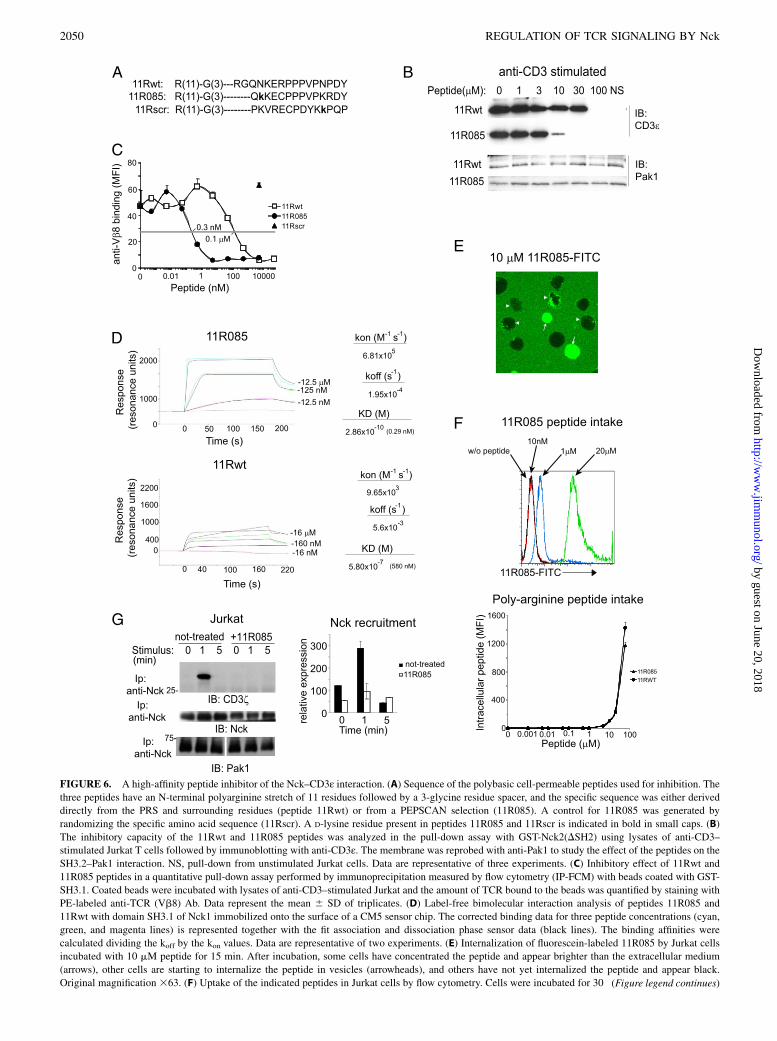
FIGURE 6. A high-affinity peptide inhibitor of the Nck–CD3ε interaction. (A) Sequence of the polybasic cell-permeable peptides used for inhibition. The
three peptides have an N-terminal polyarginine stretch of 11 residues followed by a 3-glycine residue spacer, and the specific sequence was either derived
directly from the PRS and surrounding residues (peptide 11Rwt) or from a PEPSCAN selection (11R085). A control for 11R085 was generated by
randomizing the specific amino acid sequence (11Rscr). A D-lysine residue present in peptides 11R085 and 11Rscr is indicated in bold in small caps. (B)
The inhibitory capacity of the 11Rwt and 11R085 peptides was analyzed in the pull-down assay with GST-Nck2(DSH2) using lysates of anti-CD3–
stimulated Jurkat T cells followed by immunoblotting with anti-CD3ε. The membrane was reprobed with anti-Pak1 to study the effect of the peptides on the
SH3.2–Pak1 interaction. NS, pull-down from unstimulated Jurkat cells. Data are representative of three experiments. (C) Inhibitory effect of 11Rwt and
11R085 peptides in a quantitative pull-down assay performed by immunoprecipitation measured by flow cytometry (IP-FCM) with beads coated with GST-
SH3.1. Coated beads were incubated with lysates of anti-CD3–stimulated Jurkat and the amount of TCR bound to the beads was quantified by staining with
PE-labeled anti-TCR (Vb8) Ab. Data represent the mean 6 SD of triplicates. (D) Label-free bimolecular interaction analysis of peptides 11R085 and
11Rwt with domain SH3.1 of Nck1 immobilized onto the surface of a CM5 sensor chip. The corrected binding data for three peptide concentrations (cyan,
green, and magenta lines) is represented together with the fit association and dissociation phase sensor data (black lines). The binding affinities were
calculated dividing the koff by the kon values. Data are representative of two experiments. (E) Internalization of fluorescein-labeled 11R085 by Jurkat cells
incubated with 10 mM peptide for 15 min. After incubation, some cells have concentrated the peptide and appear brighter than the extracellular medium
(arrows), other cells are starting to internalize the peptide in vesicles (arrowheads), and others have not yet internalized the peptide and appear black.
Original magnification 363. (F) Uptake of the indicated peptides in Jurkat cells by flow cytometry. Cells were incubated for 30 (Figure legend continues)
2050 REGULATION OF TCR SIGNALING BY Nck
by guest on June 20, 2018http://w
ww
.jimm
unol.org/D
ownloaded from

min with fluorescein-labeled 11R085 or 11Rwt peptides and intake calculated according to the mean fluorescence intensity of the cells after washing out the
bound, but not internalized peptide, with acidic buffer. Autofluorescence of cells is represented by a black line, green fluorescence of cells incubated with
10 nM peptide by a red line, with 1 mM peptide by a blue line, and with 20 mM peptide by a green line. The panel on the bottom shows a plot of mean
fluorescence intensity data for all concentrations of peptides 11R085-FITC and 11Rwt-FITC. Data represent the mean 6 SD of triplicates and are rep-
resentative of three experiments. (G) Recruitment of endogenous Nck to the TCR was assessed by precipitation with anti-Nck of lysates from Jurkat cells
stimulated with SEE-loaded Raji APCs for the times indicated. Immunoblots were subsequently probed with anti-CD3z. Jurkat cells were either incubated
with 20 mM 11R085 or mock treated. The membrane was reprobed with anti-Nck as a loading control. Data are representative of two experiments.
FIGURE 7. The inhibitor of the Nck–CD3ε interaction blocks T cell activation in vitro and in vivo. (A) Proliferation of T cells from lymph nodes of OT-I
TCR Tg WT mice was examined 72 h after stimulation with T2-Kb APCs loaded with 1 nM OVAp in the presence of the indicated concentrations of the
inhibitory peptides. Data represent the mean 6 SD of triplicates and are representative of five experiments. ***p , 0.0005. (B) Proliferation of human
blood CD8+ T cells in response to anti-CD3 plus anti-CD28 for 96 h in the presence of the concentrations of peptide inhibitors indicated. The effect of the
peptide inhibitors on IL-2–dependent T cell proliferation was analyzed on T lymphoblasts stimulated for 72 h with IL-2. Data represent the mean 6 SD of
triplicates and are representative of five experiments. (C) Intracellular IFN-g expression by lymph node T cells from OT-I TCR Tg WT and KI-PRS mice
after stimulation for 48 h with T2-Kb APCs loaded with OVAp in the presence of the indicated concentrations of peptides 11R085 or 11Rscr. Data represent
the mean 6 SD of triplicates and are representative of three experiments. *p , 0.05. (D) Proliferation of T cells from lymph nodes of AND TCR Tg WT
and KI-PRS mice was examined 68 h after stimulation with MCC peptide Ag-loaded DCEK APCs. Data represent the mean 6 SD of triplicates and are
representative of three experiments. ***p , 0.0005. (E) Stability of intracellular 11R085 in T cells in vivo. Lymph node cells from OT-I WT mice were
preincubated ex vivo with 10 mM FITC-labeled 11R085 for 1 h. Treated cells were subsequently inoculated (107, i.v.) into C57BL/6 mice previously
immunized with MVA-OVA. Two mice were killed at each indicated time point after inoculation and FITC-11R085 fluorescence intensity within the CD81
Va21(OT-I TCR) population in lymph node cells was analyzed by flow cytometry. Data represent the mean 6 SD for n = 2 mice per group analyzed in
triplicates. Mean fluorescence values (MFI) were normalized to those of the cells before inoculation. (F) Effect of the Nck–CD3ε interaction inhibitor on theT cell proliferative response to MVA-OVA vaccination. Lymph node cells from OT-I WT mice were labeled with CFSE and preincubated ex vivo with the
indicated concentrations of peptides 11R085 or 11Rscr for 1 h. Treated cells were subsequently inoculated (107, i.v.) into C57BL/6 mice previously
immunized with MVA-OVA. Sixty hours after transfer, mice were killed and the CFSE fluorescence intensity within the CD8+ Va2+(OT-I TCR) population
in lymph node and spleen cells was analyzed by flow cytometry. The proliferation index was calculated as indicated in Materials and Methods. Data
represent the mean 6 SD for n = 4 mice/group and are representative of two experiments. *p , 0.05. n.i., Proliferation of OT-I WT CD8 T cells in
nonimmunized C57BL/6 recipient mice; n.s., not significant, p . 0.05.
The Journal of Immunology 2051
by guest on June 20, 2018http://w
ww
.jimm
unol.org/D
ownloaded from

the presence of the 11R085 peptide (Fig. 6G), demonstrating thatthe peptide is capable of entering cells and inhibiting the Nck–CD3ε interaction.We then examined the effect of the 11R085 peptide on T cell
activation. This peptide blocked Ag-triggered proliferation ofOT-I WT T cells at a concentration of 10 mM, whereas peptide11Rwt was less potent (Fig. 7A). The scrambled peptide did notaffect OT-I T cell proliferation even at 60 mM, demonstrating thespecificity of the inhibitory effect. To determine whether the in-hibitory effect of the 11R085 peptide was restricted to TCR-dependent stimuli, its effect on cell proliferation was studied infreshly isolated human T cells stimulated with anti-CD3 and inhuman T lymphoblasts stimulated with exogenous IL-2. Thepeptide only inhibited TCR-dependent proliferation, indicatingthat no nonspecific toxic effects on non–TCR-dependent pathwaysoccurred (Fig. 7B). Further proof of the specificity of 11R085peptide inhibition came from its dependence on an intact PRS.Thus, both IFN-g production (Fig. 7C) and Ag-triggered prolif-eration (Fig. 7D) of WT OT-I T cells were inhibited by peptide11R085. However, this peptide did not inhibit Ag-triggered acti-vation of KI-PRS OT-I T cells (Fig. 7C, 7D), indicating that itdoes not have targets other than the Nck–CD3ε interaction duringTCR-triggered T cell activation.Because of the presence of the polyarginine stretch, both the
11R085 and the control 11Rscr peptides were too toxic when directlyadministered to mice as to perform experiments in vivo (data notshown). Notwithstanding, to test the inhibitory capacity of the11R085 on T cell activation in vivo, we performed an experimentsimilar to this of Fig. 4C. To this aim, we first tested the intracellularstability of the 11R085 peptide in vivo. OT-I CD8 WT T cells werepreincubated with FITC-labeled peptide 11R085 ex vivo and wereadoptively transferred into MVA-OVA–vaccinated WT mice. Fortyand twenty-five percent of the initial fluorescence intensity was stilldetected in OT-I T cells taken from draining lymph nodes 4 and16 h after inoculation, respectively (Fig. 7E). These data suggestedthat after preincubation ex vivo, peptide 11R085 may be sufficientlystable as to have an impact on initial TCR triggering events in vivo.To study the effect of peptide 11R085 on T cell proliferationin vivo, CD8 T cells from OT-I WT mice were labeled with CFSEand preincubated ex vivo with peptides 11R085 or 11Rscr. Sixtyhours after adoptive transfer into MVA-OVA–vaccinated WT mice,T cell proliferation was calculated according to CFSE dilution onCD8 OT-I T cells removed from draining lymph nodes and spleen.A pretreatment with 11R085 inhibited OT-I cell proliferation inresponse to MVA-OVA vaccination, compared with 11Rscr-treatedcells (Fig. 7F). These results suggest that the Nck–CD3ε interactioncould be an interesting target for immunosuppressive or immuno-modulatory drugs.
DiscussionIn this study, we used a dual approach to study the relevance ofthe Nck–CD3ε interaction in T cell responses to Ag in vivo.We generated a genetically modified mouse line bearing alaninereplacements for the two central prolines of the canonical PxxPmotif of CD3ε. This mutation severely impaired T cell response toAg in vitro in all the TCR systems used. More importantly, thecapacity of mutant T cells to kill target cells in vivo and of mutantmice to acquire immunity to a model tumor Ag after vaccinationwas compromised. This incapacity to respond in vivo was prob-ably caused by deficient T cell proliferation in response to theviral vaccine and by inefficient cytotoxicity. The second approachwas to inhibit the Nck–CD3ε interaction by specifically targetingthe SH3.1 domain of Nck. This approach confirmed the impor-
tance of this interaction for T cell activation. Accordingly, wegenerated a peptide inhibitor with an extremely high affinity forthe SH3.1 domain in cell-free systems (KD ,1 nM), although withconsiderably reduced potency in intact cells, probably because ofpoor entry.An important role for Nck during mature T cell function has been
demonstrated in double knockout mice lacking Nck1 in all tissuesand conditionally lacking Nck2 in T cells only (16, 17). Thephenotype of mice lacking Nck in T cells resembles that of ourKI-PRS mice, although in some aspects the phenotype of thesemice is stronger or milder than ours. This may be explained by theparticipation of Nck in T cell activation in both TCR recruitment–dependent and –independent pathways, and by the incompleteelimination of Nck2 through the conditional approach. Therefore,unlike previous articles on Nck-deficient mice (16, 17), this articlehighlights the importance of Nck in T cell activation by binding toCD3ε, and therefore being directly recruited to the TCR. Fur-thermore, our data with the 11R085 peptide pinpoints the SH3.1domain of Nck, the one binding CD3ε, as a fundamental domainfor T cell activation.In summary, our results demonstrate that the Nck–CD3ε inter-
action plays an important role in TCR signaling, rearrangement ofthe actin cytoskeleton, and T cell activation. Disruption of Nckrecruitment to the TCR results in an impaired T cell Ag responsein vitro and prevents the T cell response to a model tumor Agin vivo. Furthermore, pretreatment with a peptide that preventsNck–CD3ε interaction inhibits T cell activation in vitro and in vivo,suggesting that this interaction might be an interesting target for thedevelopment of immunosuppressive or immunomodulatory drugs.Indeed, KI-PRS mice present a delayed onset of neurologicalsymptoms after vaccination with a self MOG peptide. Of note, thePRS mutation and the peptide inhibitor 11R085 inhibited but didnot block T cell responses to Ag, suggesting that the Nck–CD3εinteraction modulates the TCR signal rather than being absolutelyrequired. It will be interesting to determine whether such a modu-lating role could make it more attractive to selectively inhibit some,but not all, T cell responses in models of autoimmunity.
AcknowledgmentsWe thank Fabien Bertaux and Kader Thiam (Genoway) for generation of the
KI-PRS mice and Ignacio Melero, Astrid Schwantes, and Gerd Sutter for
providing reagents. We also thank Manuel Fresno, Francisco Sanchez-
Madrid, and Mark Sefton for critical reading of the manuscript. We are
indebted to Cristina Prieto, Valentina Blanco, Tania Gomez, and Fernando
Barahona for expert technical assistance.
DisclosuresThe authors have no financial conflicts of interest.
References1. Wucherpfennig, K. W., E. Gagnon, M. J. Call, E. S. Huseby, and M. E. Call.
2011. Structural biology of the T-cell receptor: insights into receptor assembly,
ligand recognition, and initiation of signaling. Cold Spring Harb. Perspect. Biol.2: a005140.
2. Love, P. E., and S. M. Hayes. 2010. ITAM-mediated signaling by the T-cellantigen receptor. Cold Spring Harb. Perspect. Biol. 2: a002485.
3. de Aos, I., M. H. Metzger, M. Exley, C. E. Dahl, S. Misra, D. Zheng,L. Varticovski, C. Terhorst, and J. Sancho. 1997. Tyrosine phosphorylation of theCD3-epsilon subunit of the T cell antigen receptor mediates enhanced associa-
tion with phosphatidylinositol 3-kinase in Jurkat T cells. J. Biol. Chem. 272:25310–25318.
4. DeFord-Watts, L. M., J. A. Young, L. A. Pitcher, and N. S. van Oers. 2007. The
membrane-proximal portion of CD3 epsilon associates with the serine/threoninekinase GRK2. J. Biol. Chem. 282: 16126–16134.
5. Delgado, P., B. Cubelos, E. Calleja, N. Martinez-Martin, A. Cipres, I. Merida,C. Bellas, X. R. Bustelo, and B. Alarcon. 2009. Essential function for the GTPaseTC21 in homeostatic antigen receptor signaling. Nat. Immunol. 10: 880–888.
2052 REGULATION OF TCR SIGNALING BY Nck
by guest on June 20, 2018http://w
ww
.jimm
unol.org/D
ownloaded from

6. Martın-Cofreces, N. B., F. Baixauli, M. J. Lopez, D. Gil, A. Monjas, B. Alarcon,and F. Sanchez-Madrid. 2012. End-binding protein 1 controls signal propagationfrom the T cell receptor. EMBO J. 31: 4140–4152.
7. Buday, L., L. Wunderlich, and P. Tamas. 2002. The Nck family of adapterproteins: regulators of actin cytoskeleton. Cell. Signal. 14: 723–731.
8. Lettau, M., J. Pieper, A. Gerneth, B. Lengl-Janssen, M. Voss, A. Linkermann,H. Schmidt, C. Gelhaus, M. Leippe, D. Kabelitz, and O. Janssen. 2010. Theadapter protein Nck: role of individual SH3 and SH2 binding modules for proteininteractions in T lymphocytes. Protein Sci. 19: 658–669.
9. Jordan, M. S., and G. A. Koretzky. 2010. Coordination of receptor signaling inmultiple hematopoietic cell lineages by the adaptor protein SLP-76. Cold SpringHarb. Perspect. Biol. 2: a002501.
10. Simeoni, L., J. A. Lindquist, M. Smida, V. Witte, B. Arndt, and B. Schraven.2008. Control of lymphocyte development and activation by negative regulatorytransmembrane adapter proteins. Immunol. Rev. 224: 215–228.
11. Castello, A., M. Gaya, J. Tucholski, T. Oellerich, K. H. Lu, A. Tafuri, T. Pawson,J. Wienands, M. Engelke, and F. D. Batista. 2013. Nck-mediated recruitment ofBCAP to the BCR regulates the PI(3)K-Akt pathway in B cells. Nat. Immunol.14: 966–975.
12. Gil, D., W. W. Schamel, M. Montoya, F. Sanchez-Madrid, and B. Alarcon. 2002.Recruitment of Nck by CD3 epsilon reveals a ligand-induced conformationalchange essential for T cell receptor signaling and synapse formation. Cell 109:901–912.
13. Kesti, T., A. Ruppelt, J. H. Wang, M. Liss, R. Wagner, K. Tasken, andK. Saksela. 2007. Reciprocal regulation of SH3 and SH2 domain binding viatyrosine phosphorylation of a common site in CD3epsilon. J. Immunol. 179:878–885.
14. Brodeur, J. F., S. Li, M. da Silva Martins, L. Larose, and V. P. Dave. 2009.Critical and multiple roles for the CD3epsilon intracytoplasmic tail in doublenegative to double positive thymocyte differentiation. J. Immunol. 182: 4844–4853.
15. Mingueneau, M., A. Sansoni, C. Gregoire, R. Roncagalli, E. Aguado, A. Weiss,M. Malissen, and B. Malissen. 2008. The proline-rich sequence of CD3epsiloncontrols T cell antigen receptor expression on and signaling potency in prese-lection CD4+CD8+ thymocytes. Nat. Immunol. 9: 522–532.
16. Roy, E., D. Togbe, A. Holdorf, D. Trubetskoy, S. Nabti, G. Kublbeck, S. Schmitt,A. Kopp-Schneider, F. Leithauser, P. Moller, et al. 2010. Fine tuning of thethreshold of T cell selection by the Nck adapters. J. Immunol. 185: 7518–7526.
17. Roy, E., D. Togbe, A. D. Holdorf, D. Trubetskoy, S. Nabti, G. Kublbeck,A. Klevenz, A. Kopp-Schneider, F. Leithauser, P. Moller, et al. 2010. Nckadaptors are positive regulators of the size and sensitivity of the T-cell repertoire.Proc. Natl. Acad. Sci. USA 107: 15529–15534.
18. Szymczak, A. L., C. J. Workman, D. Gil, S. Dilioglou, K. M. Vignali, E. Palmer,and D. A. Vignali. 2005. The CD3epsilon proline-rich sequence, and its inter-action with Nck, is not required for T cell development and function. J. Immunol.175: 270–275.
19. Tailor, P., S. Tsai, A. Shameli, P. Serra, J. Wang, S. Robbins, M. Nagata,A. L. Szymczak-Workman, D. A. Vignali, and P. Santamaria. 2008. The proline-rich sequence of CD3epsilon as an amplifier of low-avidity TCR signaling. J.Immunol. 181: 243–255.
20. Yiemwattana, I., J. Ngoenkam, P. Paensuwan, R. Kriangkrai, B. Chuenjitkun-taworn, and S. Pongcharoen. 2012. Essential role of the adaptor protein Nck1 inJurkat T cell activation and function. Clin. Exp. Immunol. 167: 99–107.
21. Wang, H., J. Holst, S. R. Woo, C. Guy, M. Bettini, Y. Wang, A. Shafer,M. Naramura, M. Mingueneau, L. L. Dragone, et al. 2010. Tonic ubiquitylationcontrols T-cell receptor:CD3 complex expression during T-cell development.EMBO J. 29: 1285–1298.
22. Borroto, A., I. Arellano, E. P. Dopfer, M. Prouza, M. Suchanek, M. Fuentes,A. Orfao, W. W. Schamel, and B. Alarcon. 2013. Nck recruitment to the TCR
required for ZAP70 activation during thymic development. J. Immunol. 190:1103–1112.
23. Hogquist, K. A., S. C. Jameson, W. R. Heath, J. L. Howard, M. J. Bevan, andF. R. Carbone. 1994. T cell receptor antagonist peptides induce positive selec-tion. Cell 76: 17–27.
24. Kaye, J., M. L. Hsu, M. E. Sauron, S. C. Jameson, N. R. Gascoigne, andS. M. Hedrick. 1989. Selective development of CD4+ T cells in transgenic miceexpressing a class II MHC-restricted antigen receptor. Nature 341: 746–749.
25. Kisielow, P., H. Bl€uthmann, U. D. Staerz, M. Steinmetz, and H. von Boehmer.1988. Tolerance in T-cell-receptor transgenic mice involves deletion of non-mature CD4+8+ thymocytes. Nature 333: 742–746.
26. Albrecht, M., Y. Suezer, C. Staib, G. Sutter, S. Vieths, and G. Reese. 2008.Vaccination with a Modified Vaccinia Virus Ankara-based vaccine protects micefrom allergic sensitization. J. Gene Med. 10: 1324–1333.
27. San Jose, E., A. G. Sahuquillo, R. Bragado, and B. Alarcon. 1998. Assembly ofthe TCR/CD3 complex: CD3 epsilon/delta and CD3 epsilon/gamma dimersassociate indistinctly with both TCR alpha and TCR beta chains. Evidence fora double TCR heterodimer model. Eur. J. Immunol. 28: 12–21.
28. Aitio, O., M. Hellman, T. Kesti, I. Kleino, O. Samuilova, K. Paakkonen,H. Tossavainen, K. Saksela, and P. Permi. 2008. Structural basis of PxxDY motifrecognition in SH3 binding. J. Mol. Biol. 382: 167–178.
29. Santiveri, C. M., A. Borroto, L. Simon, M. Rico, B. Alarcon, and M. A. Jimenez.2009. Interaction between the N-terminal SH3 domain of Nck-alpha and CD3-epsilon-derived peptides: non-canonical and canonical recognition motifs. Bio-chim. Biophys. Acta 1794: 110–117.
30. Takeuchi, K., H. Yang, E. Ng, S. Y. Park, Z. Y. Sun, E. L. Reinherz, andG. Wagner. 2008. Structural and functional evidence that Nck interaction withCD3epsilon regulates T-cell receptor activity. J. Mol. Biol. 380: 704–716.
31. Perez-Martınez, M., M. Gordon-Alonso, J. R. Cabrero, M. Barrero-Villar,M. Rey, M. Mittelbrunn, A. Lamana, G. Morlino, C. Calabia, H. Yamazaki, et al.2010. F-actin-binding protein drebrin regulates CXCR4 recruitment to the im-mune synapse. J. Cell Sci. 123: 1160–1170.
32. Borroto, A., D. Gil, P. Delgado, M. Vicente-Manzanares, A. Alcover, F. Sanchez-Madrid, and B. Alarcon. 2000. Rho regulates T cell receptor ITAM-inducedlymphocyte spreading in an integrin-independent manner. Eur. J. Immunol.30: 3403–3410.
33. Bunnell, S. C., V. Kapoor, R. P. Trible, W. Zhang, and L. E. Samelson. 2001.Dynamic actin polymerization drives T cell receptor-induced spreading: a rolefor the signal transduction adaptor LAT. Immunity 14: 315–329.
34. Dopfer, E. P., B. Schopf, C. Louis-Dit-Sully, E. Dengler, K. Hohne, A. Klescova,M. Prouza, M. Suchanek, M. Reth, and W. W. Schamel. 2010. Analysis of novelphospho-ITAM specific antibodies in a S2 reconstitution system for TCR-CD3signalling. Immunol. Lett. 130: 43–50.
35. Aranda, F., D. Llopiz, N. Diaz-Valdes, J. I. Riezu-Boj, J. Bezunartea, M. Ruiz,M. Martinez, M. Durantez, C. Mansilla, J. Prieto, et al. 2011. Adjuvant com-bination and antigen targeting as a strategy to induce polyfunctional and high-avidity T-cell responses against poorly immunogenic tumors. Cancer Res. 71:3214–3224.
36. Mix, E., H. Meyer-Rienecker, H. P. Hartung, and U. K. Zettl. 2010. Animalmodels of multiple sclerosis–potentials and limitations. Prog. Neurobiol. 92:386–404.
37. Noguchi, H., M. Matsushita, T. Okitsu, A. Moriwaki, K. Tomizawa, S. Kang,S. T. Li, N. Kobayashi, S. Matsumoto, K. Tanaka, et al. 2004. A new cell-permeable peptide allows successful allogeneic islet transplantation in mice.Nat. Med. 10: 305–309.
38. Schrum, A. G., D. Gil, E. P. Dopfer, D. L. Wiest, L. A. Turka, W. W. Schamel,and E. Palmer. 2007. High-sensitivity detection and quantitative analysis ofnative protein-protein interactions and multiprotein complexes by flow cytom-etry. Sci. STKE 2007: pl2.
The Journal of Immunology 2053
by guest on June 20, 2018http://w
ww
.jimm
unol.org/D
ownloaded from