rehabilitation of a patient with non-syndromic partial ... · rehabilitation of a patient with...
TRANSCRIPT

The Journal of Advanced Prosthodontics 241
Rehabilitation of a patient with non-syndromic partial oligodontia
Hyeon-Goo Kang, Yoon-Hyuk Huh, Chan-Jin Park, Lee-Ra Cho*Department of Prosthodontics and Research Institute of Oral Science, College of Dentistry, Gangneung-Wonju National University, Gangneung, Republic of Korea
Oligodontia is defined as a congenital tooth agenesis with the absence of six or more permanent teeth. This clinical report describes a patient with non-syndromic partial oligodontia, with retained deciduous teeth and the absence of 16 permanent teeth. Anterior esthetic problems were caused by interarch tooth size discrepancy, interdental space, aberrant tooth dimensions, and the absence of centric contacts of the anterior teeth. Prosthetic restoration after orthodontic and implant treatment was performed with a multi-disciplinary team approach. Favorable functional and esthetic results were obtained using a definitive prosthesis. [ J Adv Prosthodont 2016;8: 241-50]
KEY WORDS: Anterior guidance; Congenital tooth agenesis; Interarch tooth size discrepancy; Oligodontia
http://dx.doi.org/10.4047/jap.2016.8.3.241http://jap.or.kr J Adv Prosthodont 2016;8:241-50
IntRODuCtIOn
Anomalies of tooth number, such as tooth agenesis or supernumerary teeth, result from developmental distur-bances in the initiation and proliferation stages of tooth formation.1 Congenital tooth agenesis is defined as a state in which one or more teeth, excluding the third molars, are missing.2 Although various classification methods are avail-able, tooth agenesis is usually classified according to the number of missing teeth; hypodontia, oligodontia, and ano-dontia refer to the states in which less than six teeth, more than six teeth, or all teeth, respectively, are missing.3 Among these, oligodontia has a particularly low prevalence (0.08-1.1%) and has been reported to be one of the symptoms of systemic diseases such as ectodermal dysplasia, Down syndrome, Nance-Horan syndrome, Rieger syndrome, and cleft lip and palate.4 Unlike syndromic oligodontia, muta-tions in PAX9, AXIN2, EDA, and MSX1 or environmen-
tal causes, such as drug-induced disturbances of the tooth germ and nutritional imbalances, are etiological factors in non-syndromic oligodontia.5,6
In most oligodontia patients, congenitally delayed tooth genesis and prolonged retention of primary teeth are observed, and unstable occlusion, such as traumatic occlu-sion and hypo-occlusion, occurs as a result of root resorp-tion and ankylosis.7 Teeth in such cases are narrow com-pared with normal tooth dimensions, leading to dental anomalies such as microdontia or conical teeth. Excessive or insufficient restoration space can occur depending on the pattern and severity of tooth absence. Additionally, the absence of permanent tooth germ can result in severe atro-phy of the alveolar ridge, which can pose problems in prosthodontic treatment involving implants.8 Patients with oligodontia often suffer from psychosocial impact in their childhood and adolescence due to their appearance and the inarticulate pronunciation that is caused by congenital tooth agenesis.9
Since any tooth on the dental arch can be missing, it is essential to evaluate the number and position of the miss-ing teeth and residual teeth, and the state of the alveolar ridge during treatment planning. Malocclusion caused by retained deciduous teeth and congenital permanent tooth agenesis should be corrected, and space redistribution should be performed to make prosthetic restoration possi-ble. Prosthetic restoration can be achieved by various meth-ods, such as resin adhesion, fixed dental prosthesis (FDP), removable dental prosthesis, and implants; treatment options can be limited by the number and position of miss-
Corresponding author: Lee-Ra ChoDepartment of Prosthodontics and Research Institute of Oral Science, College of Dentistry, Gangneung-Wonju National University, 7 Jukheon-gil, Gangneung 25457, Republic of KoreaTel. 82336403153: e-mail, [email protected] September 22, 2015 / Last Revision December 28, 2015 / Accepted January 12, 2016
© 2016 The Korean Academy of ProsthodonticsThis is an Open Access article distributed under the terms of the Creative Commons Attribution Non-Commercial License (http://creativecommons.org/licenses/by-nc/3.0) which permits unrestricted non-commercial use, distribution, and reproduction in any medium, provided the original work is properly cited.
pISSN 2005-7806, eISSN 2005-7814

242
ing teeth and the state of the residual teeth and alveolar ridge. Given that conventional prosthetic treatment alone cannot achieve esthetic and functional rehabilitation, a multi-disciplinary team approach combining orthodontics, oral and maxillofacial surgery, and prosthodontics is indis-pensable. Early diagnosis using clinical as well as radio-graphic examination is essential and lifetime maintenance is also required. Comprehensive treatment goals should include restoration of stable occlusion and support for psy-chosocial adjustment by improving not only functionality, but also harmonious esthetics.
This case report writes about a patient diagnosed with non-syndromic oligodontia who received an FDP after orthodontic and implant treatment with a multi-disciplinary team approach.
CASE REPORt
A 17-year-old patient visited our clinic for prosthetic resto-ration of her congenital tooth agenesis. She had no medical history of systemic diseases, but there was a familial history of partial oligodontia in her father and her sister. Extraoral examination revealed no abnormalities in the frontal and lateral facial profile, nor did lateral cephalometric radio-
graphic examination reveal any skeletal anomalies. However, the panoramic radiograph revealed a total of 16 missing permanent teeth and external root resorption in some of the retained deciduous teeth (Fig. 1). A transcrani-al radiograph was taken to evaluate the temporomandibular joint. The joint function was normal and no clinical symp-toms were observed, indicating that the condylar head was in a physiologically stable position. Intraoral examination revealed multiple deciduous teeth retained with spacing, an anterior open occlusal relationship (open bite), and an unesthetic smile due to diastema (Fig. 2). The retained
Fig. 1. Panoramic radiograph.
Fig. 2. Pre-operative intraoral photographs. (A) Maxillary occlusal view, (B) Right lateral view, (C) Frontal view, (D) Left lateral view, (E) Mandibular occlusal view.
A
B C D
E
J Adv Prosthodont 2016;8:241-50

The Journal of Advanced Prosthodontics 243
deciduous teeth and permanent teeth resulted in mixed dentition. However, stable occlusion was present, with proper functioning without hypo-occlusion from ankylosis, despite the slight root resorption. Due to a poor tooth axis and loss of occlusal contacts, the anterior teeth could not properly guide mandibular protrusive movement. A diag-nostic cast analysis showed a steep curve of Spee and an extruded mandibular left second molar, resulting in insuffi-cient antagonist restoration space. Additionally, narrow maxillary central incisors led to a 5-mm interarch tooth size discrepancy (disproportion in the mesiodistal dimensions of teeth of opposing dental arches; Fig. 3A). Esthetic and functional problems, such as malocclusion, were local and
limited to the anterior teeth. These findings led to a diagnosis of non-syndromic oli-
godontia. Accordingly, the treatment goal was establishing anterior guidance and esthetics from solving the interarch tooth size discrepancy by restoring the missing teeth. With the involvement of an orthodontist, the initial treatment plan was decided to improve tooth axis and induce redistri-bution of the restoration space. A model of tooth arrange-ment, showing ideal tooth positions, was fabricated, and a diagnostic wax-up was performed on this model (Fig. 3B). After 11 months of the orthodontic treatment, teeth were re-aligned and diastema was closed (Fig. 4). After the orth-odontic treatment and re-evaluation of the space and abut-
Fig. 3. (A) Anterior tooth size discrepancy. Bolton anterior ratio = 85.3%, (B) Kesling setup.
A B
Fig. 4. Orthodontic treatment. (A) Pre-orthodontic treatment, (B) Post-orthodontic treatment.
A B
Rehabilitation of a patient with non-syndromic partial oligodontia

244
ment, maxillary deciduous lateral incisors, canines, and mandibular deciduous central incisors were extracted because these teeth were risk factors for a poor prognosis. To minimize invasiveness, implants and FDPs were selected for treatment.
A face-bow transfer was performed to mount the diag-nostic cast on a semi-adjustable ar t iculator (KaVo PROTAR Evo 7, KaVo, Biberach, Germany), and the posi-tion of the incisal edge and gingival margin was designed, taking into account the shapes of the central incisors. The diagnostic cast was then reduced in accordance with the shape of the gingival margin, and a diagnostic wax-up was again performed (Fig. 5). Consequently, the initial interarch tooth size discrepancy of 5 mm was reduced to 2 mm.
Irreversible hydrocolloid impression material (Aroma Fine Plus; GC Corp., Tokyo, Japan) was used to obtain an impression for fabricating a duplicate cast. On the duplicate cast, the lead embedded in the periapical radiographic film was cut to a 1-mm width and was positioned in the bucco-lingual cervical area of the diagnostic wax-up cast. A ther-moplastic template (Duran 0.5 mm, Scheu Dental, Iserlohn, Germany) was used to fabricate a radiographic stent, and a cone beam computed tomography (CBCT) image was taken to evaluate the positional relationships between the bone, definitive prosthesis, and residual ridge. An additional bone graft was necessary at the maxillary deciduous lateral incisor and mandibular left deciduous canine areas due to the insufficient bone volume for implant installation (Fig. 6). In
Fig. 6. CBCT images. Narrow ridge and inadequate bone width of both maxillary deciduous lateral incisor and mandibular left deciduous canine areas.
Fig. 5. Diagnostic wax-up. (A) Resting, (B) Smile, (C) Esthetic design of the planned crown. Red lines represent the ideal cervical margin. Blue lines represent the incisal edge of maxillary incisors, (D) Soft tissue contour design. Red lines represent the ideal cervical margin. Blue “T” represents the zenith of the planned anterior crowns. White lines represent the natural balance of gingival levels, (E) Diagnostic waxing after soft tissue contour design, (F) Interarch tooth size discrepany (2 mm) indicated by the Bolton anterior ratio.
A
B
C
D
E
F
J Adv Prosthodont 2016;8:241-50

The Journal of Advanced Prosthodontics 245
consideration of the patient’s age and concerns about the surgical intervention required for the bone graft, the defini-tive treatment was changed to installing implants in the maxillary canine areas without bone graft, restoring the maxillary lateral incisors with a cantilever, and installing a mini-implant in the mandibular left canine area.
Prior to implant installation, mandibular teeth were pre-pared on the basis of the diagnostic wax pattern, and a pro-visional prosthesis was placed after extracting the decidu-ous anterior teeth. The position of the implant was chosen in accordance with the biological width and estimated gingi-val margin (Fig. 7). After extracting the deciduous canine
without alveolar bone trauma, immediate implant installa-tion was performed without invasion to the nasal floor, adjacent teeth, or nerve (Fig. 8). Tapered internal connec-tion type implants (TSIV; Osstem, Busan, Korea) with a diameter of 4.0 mm and a length of 10 mm were installed in both maxillary canines, and a mini-implant (MS SA; Osstem) with a diameter of 3.0 mm and a length of 10 mm was installed in mandibular left canine. Immediate provi-sional restoration was placed after affirming the primary stability of the implant. All contact points during maximal intercuspation and lateral excursion were removed to mini-mize the stress exerted on the implant.
Fig. 8. Implant surgery procedures. (A) Bucco-lingual bone thickness of mandibular left canine region, (B) Implant placement of mandibular left canine, (C) Extraction of both maxillary deciduous canines, (D) Implant placement of both maxillary canines, (E) Provisional restorations. Note the concave emergence profile.
A B
C D E
Fig. 7. Distance between the cervical margin of the crown and the planned implant position.
Rehabilitation of a patient with non-syndromic partial oligodontia
246
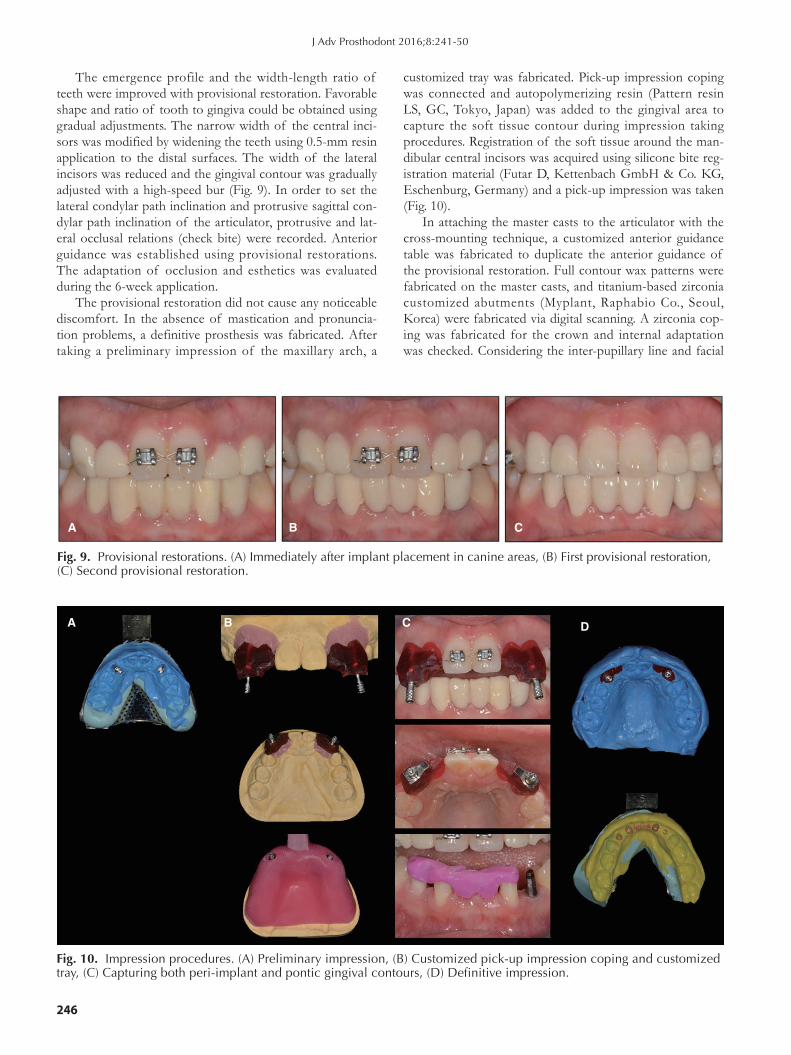
The emergence profile and the width-length ratio of teeth were improved with provisional restoration. Favorable shape and ratio of tooth to gingiva could be obtained using gradual adjustments. The narrow width of the central inci-sors was modified by widening the teeth using 0.5-mm resin application to the distal surfaces. The width of the lateral incisors was reduced and the gingival contour was gradually adjusted with a high-speed bur (Fig. 9). In order to set the lateral condylar path inclination and protrusive sagittal con-dylar path inclination of the articulator, protrusive and lat-eral occlusal relations (check bite) were recorded. Anterior guidance was established using provisional restorations. The adaptation of occlusion and esthetics was evaluated during the 6-week application.
The provisional restoration did not cause any noticeable discomfort. In the absence of mastication and pronuncia-tion problems, a definitive prosthesis was fabricated. After taking a preliminary impression of the maxillary arch, a
customized tray was fabricated. Pick-up impression coping was connected and autopolymerizing resin (Pattern resin LS, GC, Tokyo, Japan) was added to the gingival area to capture the soft tissue contour during impression taking procedures. Registration of the soft tissue around the man-dibular central incisors was acquired using silicone bite reg-istration material (Futar D, Kettenbach GmbH & Co. KG, Eschenburg, Germany) and a pick-up impression was taken (Fig. 10).
In attaching the master casts to the articulator with the cross-mounting technique, a customized anterior guidance table was fabricated to duplicate the anterior guidance of the provisional restoration. Full contour wax patterns were fabricated on the master casts, and titanium-based zirconia customized abutments (Myplant, Raphabio Co., Seoul, Korea) were fabricated via digital scanning. A zirconia cop-ing was fabricated for the crown and internal adaptation was checked. Considering the inter-pupillary line and facial
Fig. 9. Provisional restorations. (A) Immediately after implant placement in canine areas, (B) First provisional restoration, (C) Second provisional restoration.
A B C
Fig. 10. Impression procedures. (A) Preliminary impression, (B) Customized pick-up impression coping and customized tray, (C) Capturing both peri-implant and pontic gingival contours, (D) Definitive impression.
A B C D
J Adv Prosthodont 2016;8:241-50

The Journal of Advanced Prosthodontics 247
midline, the tooth axis was evaluated with the zirconia cop-ing (Fig. 11). During an up-close examination while per-forming shade selection, a demineralized lesion was found on the buccal surfaces of the maxillary anterior teeth. In order to restore the shade of the teeth, resin infiltration
Fig. 12. Resin infiltration. (A) Pre-orthodontic treatment, (B) Post-orthodontic treatment, (C) After resin infiltration.
A B C
Fig. 13. Definitive prosthesis. (A) Working side during right lateral excursion, (B) Maxillary occlusal view, (C) Non-working side during right lateral excursion, (D) Left lateral view at centric occlusion, (E) Frontal view at centric occlusion, (F) Right lateral view at centric occlusion, (G) Non-working side during left lateral excursion, (H) Mandibular occlusal view, (I) Working side during left lateral excursion.
A
D
G
B
E
H
C
F
I
was performed using unfilled resin (Icon, DMG, Hamburg, Germany) with an enamel-like refractive index. Then, a shade was selected and porcelain build-up was performed (Fig. 12). The definitive prosthesis was placed and adjusted to achieve mutually protected occlusion (Fig. 13).
Fig. 11. (A) Fabrication of zirconia coping, (B) Trial placement.
A B
Rehabilitation of a patient with non-syndromic partial oligodontia

248
In the follow-up examination after 18 months, oral hygiene was verified to be excellent, and there were no complaints in terms of occlusion, mastication, phonetics, esthetics, or the temporomandibular joint. The peri-implant marginal bone level was maintained without noticeable changes, and no abutment screw loosening or dislodgement of the cement-retained prosthesis was detected (Fig. 14).
DISCuSSIOn
Prosthodontic treatment alone is not sufficient to achieve ideal results in the treatment of congenital tooth agenesis in oligodontia patients; thus, a multi-disciplinary approach, including orthodontics or implants, is necessary.9 A defini-tive treatment plan for replacing the missing teeth should be decided after analyzing the state of the remaining teeth and the interarch relationship. Orthodontic treatment can improve the space redistribution of missing area, rearrange-ment of teeth, and correction of the tooth axis. Factors such as residual ridge configuration and the remaining bone amount can pose great challenges for prosthodontic treat-ment. In this report, the case was complicated due to migration of the adjacent teeth following multiple perma-
Fig. 14. Periapical radiographs. (A) Implant placement, (B) Provisional restoration, (C) Recall check after 18 months.
A
B
C
nent teeth missing, limited restoration space, and abnormal-ities of the remaining teeth. Therefore, prosthetic restora-tion alone was considered insufficient for obtaining good esthetics and improving masticatory function, and thus the plan was decided to proceed to orthodontic treatment with prosthodontic treatment involving an implant.
It has been reported that missing permanent teeth make it difficult to provide good anchorage for orthodontic treat-ment.10 When 4-16 permanent teeth are missing, root resorption is likely to happen after orthodontic treatment.10
Another report has stated that deciduous canines and man-dibular deciduous second molars could remain in the oral space longer than any other retained deciduous teeth. It has also reported that if a deciduous second molar still remained at the age of 20, good prognosis could be expect-ed.11 However, if a deciduous molar was involved in hypo-occlusion and was expected to hamper the growth of the alveolar bone through root ankylosis or resorption, it was necessary to extract it and replace it with an implant or FDP. Without the succeeding permanent teeth, the maxil-lary deciduous lateral incisors tend to be prematurely exfoli-ated and affect anterior esthetics; therefore, restoration after extraction should be considered.11,12 After the diagnos-tic procedures in the case reported here, it was planned to extract the maxillary deciduous lateral incisors and canines and mandibular deciduous central incisors, because of their short root length and poor prognosis. During the orth-odontic treatment, root resorption and pathologic tooth mobility of the mandibular left deciduous canine occurred, and thus an additional extraction was performed.
Congenital tooth agenesis is characterized by a narrower mesio-distal tooth width and a smaller dental arch size, and a smaller width of the remaining teeth is known to be relat-ed to a higher number of missing teeth.13,14 Interarch tooth size discrepancy is defined as the difference in the sum of the mesio-distal dimensions of teeth of opposing dental arches; such discrepancy poses an esthetic problem and dis-turbs proper occlusal relations. In the anterior teeth, a dis-crepancy of about 2 mm is reported to be clinically prob-lematic.15 In our case, the patient had a total of 16 missing permanent teeth; however, the esthetic and functional problems were local and limited to the anterior teeth. Additionally, the remaining teeth had were narrow and there was a discrepancy in the interarch tooth size. When comparing the patient’s case with the ideal anterior Bolton ratio16 of the summed mesio-distal dimensions of the man-dibular to those of the maxillary anterior teeth, the mandib-ular teeth width was found to exceed that of the normal range by about 5 mm. To resolve the interarch tooth size discrepancy and achieve an adequate arrangement of teeth for prosthetic restoration, the orthodontic treatment involved space evaluation through a diagnostic wax-up in a setup model.17 Orthodontic treatment resulted in tooth axis improvement and space redistribution, and the interarch tooth size discrepancy was reduced to 2 mm. In the defini-tive prosthesis, this discrepancy was further reduced to 1 mm by adjusting the provisional restoration.
J Adv Prosthodont 2016;8:241-50

The Journal of Advanced Prosthodontics 249
In FDP, adequate crown width/length ratio and symme-try, as well as the gingival emergence profile, are essential for optimal esthetics. Rehabilitation of missing teeth was performed to improve the patient’s smile using a diagnostic wax pattern after setting the position of the incisal edge and the gingival margin, taking into account the incisal edge exposure in the resting position and the curvature of the lower lip in a smile.
Implants were installed into the maxillary canine area about 2-mm palatally and 3-mm inferiorly to the gingival margin of the expected definitive prosthesis so that 1.8-2 mm of the buccal bone could be preserved.18,19 The biolog-ical width was also considered. The provisional restoration was placed immediately after implant installation to sculp-ture the emergence profile. Excellent esthetics could be restored by gradually adjusting the gingival contour.
Maxillary and mandibular dental and skeletal growth leads to dramatic 3-dimensional changes in younger patients. Remodeling in the region of the implant installa-tion site could cause several problems such as implants unsupported by bone or submerged within it, and loss of implants.20 However, implants placed during late puberty or early adulthood, like in our patient’s case, have a good long-term prognosis.21
Dawson22 insisted that stable occlusion can be achieved by having stable occlusal stops in all teeth when the con-dyles are in a centric relation, avoiding interference of all posterior teeth, and having anterior guidance in harmony with the border movement of the envelope of function. The patient in the present report did not show any skeletal problems that might induce anomalies in the temporoman-dibular joint; it demonstrated normal function without any clinical symptoms, and the condyle was considered to be in a physiologically stable position.
With the missing permanent teeth and retained decidu-ous teeth, this patient developed an occlusion of mixed dentition that was functioning properly. However, the max-illary central incisors and mandibular lateral incisors were the only permanent teeth present on the anterior arch, exhibiting a poor tooth axis and loss of occlusal contacts. The anterior teeth could not guide mandibular movement, which resulted in occlusal interference of the posterior teeth. Therefore, a new anterior guidance had to be estab-lished using provisional restoration. The anterior guidance determined by the envelope of function was evaluated based on the patient’s adaptation.22 Orthodontic treatment, implant installation, and gradual occlusal adjustment were used to establish stable, functionally harmonious anterior guidance (Fig. 14).
In this case, functional and esthetic occlusion was attained in a patient with non-syndromic oligodontia con-genitally missing 16 permanent teeth through orthodontic treatment, followed by prosthetic restoration including implant. The treatment also brought about psychosocial adjustment in the adolescent patient. Although the patient demonstrate a stable function, lifetime maintenance is con-sidered necessary due to some clinical factors specific to
oligodontia.
COnCLuSIOn
This case report concerns a patient with non-syndromic oligodontia without any specific systemic disease. The patient was rehabilitated by means of space redistribution and prosthetic restoration involving an implant, through multi-disciplinary treatments. Optimal esthetics could be achieved by resolving the interarch tooth size discrepancy, gradually adjusting the gingival contour, and improving the crown width/length ratio. A new anterior guidance was established in accordance with function. In patients with oligodontia, lifetime maintenance care is essential for pres-ervation of esthetics and function.
ORCID
Hyeon-Goo Kang http://orcid.org/0000-0001-6293-0121Yoon-Hyuk Huh http://orcid.org/0000-0003-4072-5199Chan-Jin Park http://orcid.org/0000-0003-4734-214XLee-Ra Cho http://orcid.org/0000-0003-3989-2870
REFEREnCES
1. Hobkirk JA, Brook AH. The management of patients with severe hypodontia. J Oral Rehabil 1980;7:289-98.
2. AlShahrani I, Togoo RA, AlQarni MA. A review of hy-podontia: classification, prevalence, etiology, associated anomalies, clinical implications and treatment options. World J Dent 2013;4:117-25.
3. Fekonja A. Hypodontia in orthodontically treated children. Eur J Orthod 2005;27:457-60.
4. Bergendal B, Norderyd J, Bågesund M, Holst A. Signs and symptoms from ectodermal organs in young Swedish individ-uals with oligodontia. Int J Paediatr Dent 2006;16:320-6.
5. Ruf S, Klimas D, Hönemann M, Jabir S. Genetic background of nonsyndromic oligodontia: a systematic review and meta-analysis. J Orofac Orthop 2013;74:295-308.
6. Rakhshan V. Congenitally missing teeth (hypodontia): A re-view of the literature concerning the etiology, prevalence, risk factors, patterns and treatment. Dent Res J (Isfahan) 2015;12:1-13.
7. Becelli R, Morello R, Renzi G, Dominici C. Treatment of oli-godontia with endo-osseous fixtures: experience in eight con-secutive patients at the end of dental growth. J Craniofac Surg 2007;18:1327-30.
8. Carvalho JC, Vinker F, Declerck D. Malocclusion, dental inju-ries and dental anomalies in the primary dentition of Belgian children. Int J Paediatr Dent 1998;8:137-41.
9. Durey K, Cook P, Chan M. The management of severe hy-podontia. Part 1: Considerations and conventional restorative options. Br Dent J 2014;216:25-9.
10. Levander E, Malmgren O, Stenback K. Apical root resorp-tion during orthodontic treatment of patients with multiple aplasia: a study of maxillary incisors. Eur J Orthod 1998;20: 427-34.
Rehabilitation of a patient with non-syndromic partial oligodontia

250
11. Bjerklin K, Bennett J. The long-term survival of lower sec-ond primary molars in subjects with agenesis of the premo-lars. Eur J Orthod 2000;22:245-55.
12. Savarrio L, McIntyre GT. To open or to close space--that is the missing lateral incisor question. Dent Update 2005;32:16-8, 20-2, 24-5.
13. Fekonja A. Comparison of mesiodistal crown dimension and arch width in subjects with and without hypodontia. J Esthet Restor Dent 2013;25:203-10.
14. Brook AH, Griffin RC, Smith RN, Townsend GC, Kaur G, Davis GR, Fearne J. Tooth size patterns in patients with hy-podontia and supernumerary teeth. Arch Oral Biol 2009;54: S63-70.
15. Profit WR. Contemporary orthodontics. 4th ed. St. Louis, (MO): Mosby Elsevier; 2007.
16. Prasanna AL, Venkatramana V, Aryasri AS, Katta AK, Santhanakrishnan K, Maheshwari U. Evaluation and compar-ison of intermaxillary tooth size discrepancy among class I, class II division 1, and class III subjects using bolton’s analy-sis: an in vitro study. J Int Oral Health 2015;7:58-64.
17. Araújo TM, Fonseca LM, Caldas LD, Costa-Pinto RA. Preparation and evaluation of orthodontic setup. Dent Press J Orthod 2012;17:146-65.
18. Hermann JS, Buser D, Schenk RK, Higginbottom FL, Cochran DL. Biologic width around titanium implants. A physiologically formed and stable dimension over time. Clin Oral Implants Res 2000;11:1-11.
19. Evans CD, Chen ST. Esthetic outcomes of immediate im-plant placements. Clin Oral Implants Res 2008;19:73-80.
20. Oesterle LJ, Cronin RJ Jr, Ranly DM. Maxillary implants and the growing patient. Int J Oral Maxillofac Implants 1993;8: 377-87.
21. Cronin RJ Jr, Oesterle LJ, Ranly DM. Mandibular implants and the growing patient. Int J Oral Maxillofac Implants 1994;9:55-62.
22. Dawson PE. Functional occlusion: From TMJ to smile de-sign. Mosby; Elsevier Health Sciences, 2006.
J Adv Prosthodont 2016;8:241-50