rehabilitation after hip arthroscopy - andrew wolff, md · much of the evidence on rehabilitation...
TRANSCRIPT

Rehabil itation After HipArthroscopy
A Movement Control–Based PerspectivePhilip Malloy, MS, PT, SCSa,*, Kim Gray, DPTb, Andrew B. Wolff, MDc
KEYWORDS
� Rehabilitation � Hip arthroscopy � Movement control � Hip rehabilitation
KEY POINTS
� Joint protection immediately after hip arthroscopy is essential and must be tailored spe-cifically to the severity of hip injury and surgical procedures performed.
� Mobility, muscle performance and stability, and neuromuscular control are vital aspects tomovement control that are commonly addressed in rehabilitation programs after hiparthroscopy.
� Each phase of hip arthroscopy should be adapted to the specific functional demands ofthe patient. Exercise progressions should be monitored closely and patients should beprogressed slowly to prevent complications, such as persistent soft tissue irritation.
BACKGROUND
Diagnosis and management of nonarthritic hip pathology have evolved significantlyover the years through the advancements in arthroscopic surgical interventions forintra-articular and extra-articular hip injury.1 The rapid growth of hip arthroscopic sur-gery necessitates parallel advancement in rehabilitation after surgery.2–4 Currently,much of the evidence on rehabilitation after hip arthroscopy is limited to case-control studies.5–9 In part, this population is difficult to study because hip arthroscopyoften requires patient-specific postoperative restrictions. Additionally, the diversebackgrounds of rehabilitation specialists creates a situation where different treatmenttechniques may be equally effective based on a patient’s specific need.In recent years, many investigators have presented guidelines for hip arthroscopy
rehabilitation with much of the evidence based on empirical experience and rehabili-tation literature from similar patient populations.2–4,10,11 Most presented guidelines
andrewwolffmd.com
Disclosures: Dr A.B. Wolff is a paid consultant for Strkyer.a Department of Physical Therapy, Marquette University, 604 North 16th Street, Milwaukee, WI 53233, USA; b SMARTherapy, Washington Orthopaedics and Sports Medicine, 2021 K Street Northwest, Washington, DC 20006, USA; c Department of Orthopedic Surgery, Washington Orthopaedics and Sports Medicine, 2021 K Street Northwest, Washington, DC 20006, USA
E-mail address: [email protected]

Malloy et al2
break down hip arthroscopy rehabilitation into 4 or 5 phases. The focus of each phaseis related to pivotal aspects of rehabilitation after surgery, which includes joint protec-tion, range of motion and mobility, restoration of normal gait, muscle strength andneuromuscular control, and sport-specific or functional training.2–4,11 Objective mile-stones for progression from one phase to the next provide clinicians and patients withtangible goals while respecting healing time frames for surgically repaired tissues.12 Inaddition, many of these published guidelines provide surgery-specific limitations, forexample, weight-bearing restriction after microfracture or range-of-motion limitationafter soft tissue repair.2,4,13 Other guidelines have presented pitfalls potentiallyencountered at each rehabilitative phase as well as prevention strategies to mitigatethe occurrence of these setbacks.3
The understanding of human movement control is essential to the development ofan effective rehabilitation program for patients after hip arthroscopy. Vital aspectsof movement control include mobility, muscle performance and stability, and neuro-muscular control, which serve as common rehabilitation targets for clinicians. Eachof these aspects of movement control is essential for safe return to functional activitiesafter hip arthroscopy. For that reason, it is the purpose of this article to present hiparthroscopy rehabilitation guidelines based on the important aspects of movementcontrol. Initial joint protection techniques are discussed and rehabilitation is brokendown into 4 phases in the context of mobility, muscle performance and stability,and neuromuscular control exercises.
JOINT PROTECTION
It is known that cellular repair and remodeling mechanisms begin immediately afterjoint injury or surgery.14 To promote an optimal environment for healing, joint protec-tion aimed at the restoration of joint homeostasis is the initial primary goal after hiparthroscopy. For practical purposes, joint homeostasis can be defined as the elimina-tion of outward signs of acute or subacute inflammation, which may include edema,ecchymosis, pain at rest, and/or pain at the end of the day. Healing of the incision por-tal sites and a reduction in ecchymosis provides a good indication of when acuteinflammation is no longer present. The rehabilitation during this phase is crucial toset the foundation for progression to the next phases. Emphasis should be placedon significant activity limitation and rest during this phase to allow the natural healingprocess to take place. Pharmacologic treatments during this phase include the use ofpain medication and nonsteroidal anti-inflammatory drugs to reduce pain and inflam-mation and for prevention of heterotopic ossification after surgery.Patient education on joint protection strategies is essential to prevent both intra-
articular and extra-articular soft tissue irritation. Restricted weight bearing iscommonly recommended to reduce joint reaction forces to protect healing tis-sues, such as the femoral neck, acetabular labrum, joint capsule, and contractiletissues of the hip joint.15 Patients should be instructed in a foot flat or normal heel-toe weight-bearing pattern using an assistive device. The use of a non–weight-bearing or toe-touch weight-bearing pattern is contraindicated because this leadsto recurrent activation of the hip flexor muscle group. Persistent activation of thehip flexors after surgery can result in persistent anterior pain secondary to muscleoveruse. To prevent stiffness in the anterior hip during this initial phase, patientsshould be instructed to limit sitting time and encouraged to change positionsfrequently. Prone lying should be emphasized to position the hip in neutral; how-ever, caution should be exercised for prone positioning in patients with low backpain.13

Rehabilitation After Hip Arthroscopy 3
A continuous passive motion machine may be prescribed after surgery to begincontrolled passive movement of the hip. Rehabilitation specialists must provide in-struction on how to get in and out of the continuous passive motion machine to avoidactively lifting the leg immediately after surgery. In the first few postoperative days,caregiver assistance or the use of a leg-lifting device is helpful. Patients can beginriding an upright stationary bicycle the first week after surgery. Recumbent bicyclesshould be discouraged. Instructing patients on safe techniques for getting on andoff a bike and setting the proper seat height to avoid pinching in the anterior hip isimperative.
MOBILITYPhase 1 Mobility Exercises
Passive range-of-motion exercises can be initiated during the first week after hiparthroscopy. Although range-of-motion restrictions are procedure dependent, smalloscillatory motions in the midranges of all planes are recommended.16 Although casesof intra-articular adhesions have been reported, these complications are uncom-mon.17,18 Therefore, during this phase, range-of-motion exercises should always beperformed in pain-free ranges. In addition, limitation in specific motion, such as exten-sion and external rotation, may be prescribed in patients where capsular modificationor labral reconstruction procedures were performed.2,12 The rehabilitation specialistmust communicate with the surgeon to determine the extent of postoperativerange-of-motion limitations on the tissues addressed.Gentle soft tissue mobilization should be initiated in the first week postopera-
tively to assist with scar and edema management. Soft tissue mobilization mayalso be useful for pain reduction and increased tone in muscles surrounding thehip and trunk.9
Phase 2 Mobility Exercises
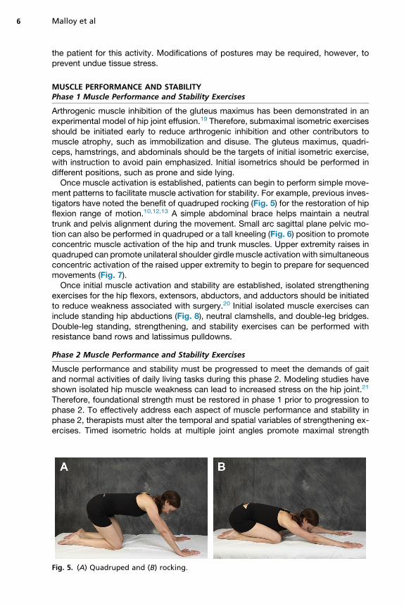
Passive range-of-motion exercises should be continued with gentle end-rangestretching as needed. Active movements should be performed through larger arcsof motion and into terminal ranges while continuing to respect the surgery-specificrange-of-motion restrictions. It may be useful for clinicians to use functional tests toassess range of motion into terminal ranges, such as a double-leg squat (Fig. 1) orstanding hip hike (Fig. 2). Frontal plane mobility may be assessed with crossover step-ping or a lateral slide side lunge (Fig. 3). A standing stool rotation exercise can be usedto assess transverse plane hip mobility (Fig. 4). In patients with persistent mobility is-sues, soft tissue and joint mobilization may be beneficial. Prescribing a dynamic self-stretching program assists with maintaining the mobility gained with manual tech-niques. Also, performing active movements into the range of motion can assist withre-establishing movement control into the range.
Phase 3 Mobility Exercises
At this point, patients should have the required hip joint mobility for the desired func-tional activity. Using exercises to maintain this range of motion, however, especially asmuscle size increases with training, is important. In addition, the ankle, knee, lumbar,and thoracic spine should be evaluated early in this phase to ensure adequatesegmental mobility is present for advanced function and dynamic exercise. A dynamicwarm-up or movement preparation exercises are recommended prior to a trainingsession to ensure adequate warm-up and mobility before beginning exercise. Patientsmay return to yoga for stretching as long as the physician and therapist have released
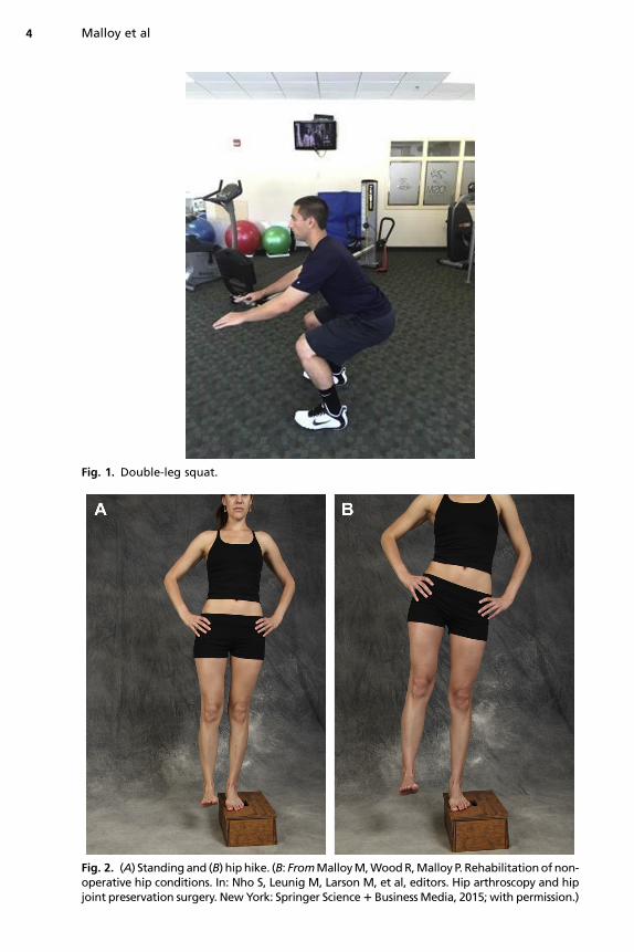
Fig. 1. Double-leg squat.
Fig. 2. (A) Standingand (B) hiphike. (B: FromMalloyM,WoodR,Malloy P. Rehabilitationofnon-operative hip conditions. In: Nho S, Leunig M, Larson M, et al, editors. Hip arthroscopy and hipjoint preservation surgery. NewYork: Springer Science1 BusinessMedia, 2015; with permission.)
Malloy et al4
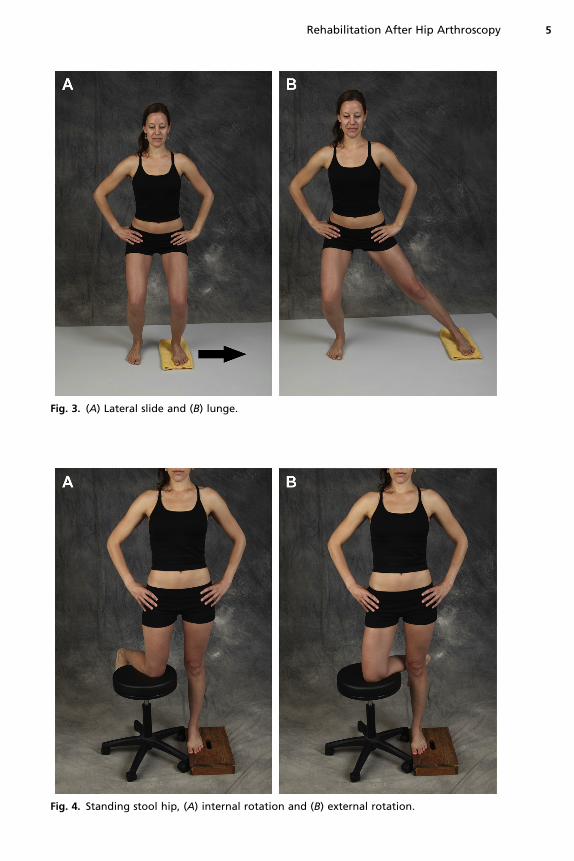
Fig. 3. (A) Lateral slide and (B) lunge.
Fig. 4. Standing stool hip, (A) internal rotation and (B) external rotation.
Rehabilitation After Hip Arthroscopy 5
Malloy et al6
the patient for this activity. Modifications of postures may be required, however, toprevent undue tissue stress.
MUSCLE PERFORMANCE AND STABILITYPhase 1 Muscle Performance and Stability Exercises
Arthrogenic muscle inhibition of the gluteus maximus has been demonstrated in anexperimental model of hip joint effusion.19 Therefore, submaximal isometric exercisesshould be initiated early to reduce arthrogenic inhibition and other contributors tomuscle atrophy, such as immobilization and disuse. The gluteus maximus, quadri-ceps, hamstrings, and abdominals should be the targets of initial isometric exercise,with instruction to avoid pain emphasized. Initial isometrics should be performed indifferent positions, such as prone and side lying.Once muscle activation is established, patients can begin to perform simple move-
ment patterns to facilitate muscle activation for stability. For example, previous inves-tigators have noted the benefit of quadruped rocking (Fig. 5) for the restoration of hipflexion range of motion.10,12,13 A simple abdominal brace helps maintain a neutraltrunk and pelvis alignment during the movement. Small arc sagittal plane pelvic mo-tion can also be performed in quadruped or a tall kneeling (Fig. 6) position to promoteconcentric muscle activation of the hip and trunk muscles. Upper extremity raises inquadruped can promote unilateral shoulder girdle muscle activation with simultaneousconcentric activation of the raised upper extremity to begin to prepare for sequencedmovements (Fig. 7).Once initial muscle activation and stability are established, isolated strengthening
exercises for the hip flexors, extensors, abductors, and adductors should be initiatedto reduce weakness associated with surgery.20 Initial isolated muscle exercises caninclude standing hip abductions (Fig. 8), neutral clamshells, and double-leg bridges.Double-leg standing, strengthening, and stability exercises can be performed withresistance band rows and latissimus pulldowns.
Phase 2 Muscle Performance and Stability Exercises
Muscle performance and stability must be progressed to meet the demands of gaitand normal activities of daily living tasks during this phase 2. Modeling studies haveshown isolated hip muscle weakness can lead to increased stress on the hip joint.21
Therefore, foundational strength must be restored in phase 1 prior to progression tophase 2. To effectively address each aspect of muscle performance and stability inphase 2, therapists must alter the temporal and spatial variables of strengthening ex-ercises. Timed isometric holds at multiple joint angles promote maximal strength
Fig. 5. (A) Quadruped and (B) rocking.
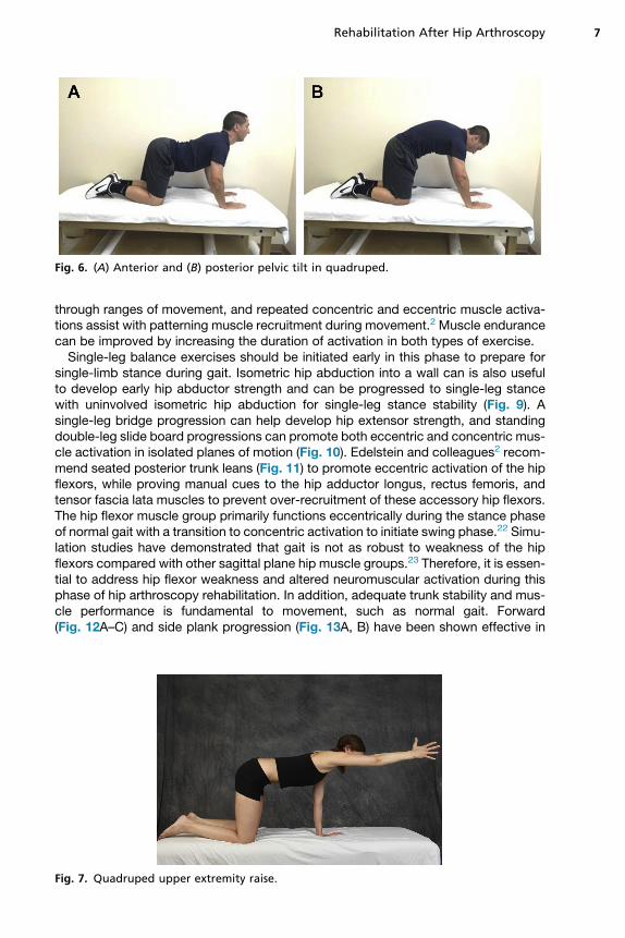
Fig. 6. (A) Anterior and (B) posterior pelvic tilt in quadruped.
Rehabilitation After Hip Arthroscopy 7
through ranges of movement, and repeated concentric and eccentric muscle activa-tions assist with patterning muscle recruitment during movement.2 Muscle endurancecan be improved by increasing the duration of activation in both types of exercise.Single-leg balance exercises should be initiated early in this phase to prepare for
single-limb stance during gait. Isometric hip abduction into a wall can is also usefulto develop early hip abductor strength and can be progressed to single-leg stancewith uninvolved isometric hip abduction for single-leg stance stability (Fig. 9). Asingle-leg bridge progression can help develop hip extensor strength, and standingdouble-leg slide board progressions can promote both eccentric and concentric mus-cle activation in isolated planes of motion (Fig. 10). Edelstein and colleagues2 recom-mend seated posterior trunk leans (Fig. 11) to promote eccentric activation of the hipflexors, while proving manual cues to the hip adductor longus, rectus femoris, andtensor fascia lata muscles to prevent over-recruitment of these accessory hip flexors.The hip flexor muscle group primarily functions eccentrically during the stance phaseof normal gait with a transition to concentric activation to initiate swing phase.22 Simu-lation studies have demonstrated that gait is not as robust to weakness of the hipflexors compared with other sagittal plane hip muscle groups.23 Therefore, it is essen-tial to address hip flexor weakness and altered neuromuscular activation during thisphase of hip arthroscopy rehabilitation. In addition, adequate trunk stability and mus-cle performance is fundamental to movement, such as normal gait. Forward(Fig. 12A–C) and side plank progression (Fig. 13A, B) have been shown effective in
Fig. 7. Quadruped upper extremity raise.
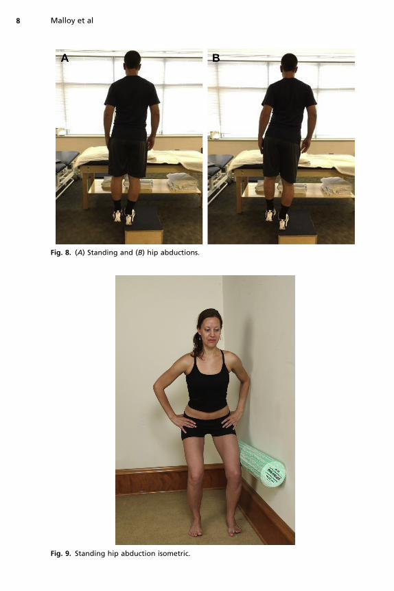
Fig. 8. (A) Standing and (B) hip abductions.
Fig. 9. Standing hip abduction isometric.
Malloy et al8

Fig. 10. Supine single-leg bridge.
Rehabilitation After Hip Arthroscopy 9
achieving high levels of muscle activation of the core and trunk muscles; therefore,these common exercises should be initiated during this phase of hip arthroscopyrehabilitation.24,25
Phase 3 Muscle Performance and Stability Exercises
The ultimate goal of phase 3 hip arthroscopy rehabilitation is to return patients to un-restricted functional activities. The muscle performance and stability exercises duringthis phase are individual, based on a patient’s specific functional demands. The ther-apist must initiate a clear discussion with patients about desired level of activity. Injuryseverity and consequences of the surgical procedures must be considered when
Fig. 11. Seated posterior trunk leans.
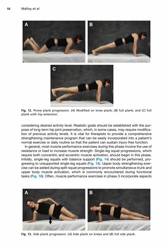
Fig. 12. Prone plank progression. (A) Modified on knee plank; (B) full plank; and (C) fullplank with hip extension.
Malloy et al10
considering desired activity level. Realistic goals should be established with the pur-pose of long-term hip joint preservation, which, in some cases, may require modifica-tion of previous activity levels. It is vital for therapists to provide a comprehensivestrengthening maintenance program that can be easily incorporated into a patient’snormal exercise or daily routine so that the patient can sustain injury-free function.In general, most muscle performance exercises during this phase involve the use of
resistance or load to increase muscle strength. Single-leg squat progressions, whichrequire both concentric and eccentric muscle activation, should begin in this phase.Initially, single-leg squats with balance support (Fig. 14) should be performed, pro-gressing to unsupported single-leg squats (Fig. 15). Upper body strengthening exer-cise can be added during split-squat progressions to promote simultaneous trunk andupper body muscle activation, which is commonly encountered during functionaltasks (Fig. 16). Often, muscle performance exercises in phase 3 incorporate aspects
Fig. 13. Side plank progression. (A) Side plank on knees and (B) full side plank.

Fig. 14. Splitbalance squat. (FromMalloyM,WoodR,MalloyP.Rehabilitationofnon-operativehip conditions. In: Nho S, LeunigM, LarsonM, et al, editors. Hip arthroscopy and hip joint pres-ervation surgery. New York: Springer Science1 Business Media, 2015; with permission.)
Fig. 15. Single-leg squat. (FromMalloyM,Wood R, Malloy P. Rehabilitation of non-operativehip conditions. In: Nho S, LeunigM, LarsonM, et al, editors. Hip arthroscopy andhip joint pres-ervation surgery. New York: Springer Science1 Business Media, 2015; with permission.)
11

Fig. 16. Split squat with trunk rotation.
Malloy et al12
of neuromuscular control, such as coordination and sequencing. A single-leg squatexercise with a unilateral row requires appropriate muscle performance andsegmental movement to maintain a neutral pelvic and trunk position during the move-ment (Fig. 17). In general, hip arthroscopy patients can be progressed during thisphase as tolerated. Therapists are encouraged to use creativity when developingstrengthening programs that uniquely meet the individual functional demands of thepatient during this phase.
Phase 4 Muscle Performance and Stability Exercises
Muscle performance during the final phase of rehabilitation should focus on powerdevelopment. Power is expressed as the product of force and velocity.26 Muscle po-wer output is dependent, however, on muscle length and type of activation performed.Although a full explanation of the mechanical variable of power is outside the scope ofthis article, clinicians should consider a few factors to improve power development forhigh-level activities in patients after hip arthroscopy. Muscles can produce the mostpower when a large force is produced at an intermediate velocity.26 For example,when a person moves a heavy load that requires large force production at a relativelyconstant nonmaximal velocity, a large amount of power is produced during the move-ment as the muscles shorten. Conversely, a large amount of power can be produced ifa large load is moved slowly as the muscle lengthens, as during an eccentric muscleactivation phase of an activity. Therefore, muscle performance during this phase mustfocus on altering the variables of velocity and load to achieve the greatest amount ofpower output. In addition, therapists must consider whether the movement involves aconcentric (shortening) or eccentric (lengthening) contraction of a muscle to appropri-ately alter the variables involved in power production.
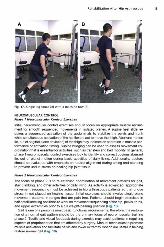
Fig. 17. Single leg squat (A) with a machine row (B).
Rehabilitation After Hip Arthroscopy 13
NEUROMUSCULAR CONTROLPhase 1 Neuromuscular Control Exercises
Initial neuromuscular control exercises should focus on appropriate muscle recruit-ment for smooth sequenced movements in isolated planes. A supine heel slide re-quires a sequenced activation of the abdominals to stabilize the pelvis and trunkwhile simultaneous activation of the hip flexors act to move the thigh. Aberrant motion(ie, out of sagittal plane deviation) of the thigh may indicate an alteration in muscle per-formance or activation timing. Supine bridging can be used to assess movement co-ordination that is essential for activities, such as transfers and bed mobility. In general,phase 1 neuromuscular control exercises look to identify and correct obvious aberrant(ie, out of plane) motion during basic activities of daily living. Additionally, postureshould be evaluated with emphasis on neutral alignment during sitting and standingto prevent undue stress on healing hip joint tissue.
Phase 2 Neuromuscular Control Exercises
The focus of phase 2 is to re-establish coordination of movement patterns for gait,stair climbing, and other activities of daily living. As activity is advanced, appropriatemovement sequencing must be achieved in hip arthroscopy patients so that unduestress in not placed on healing tissue. Initial exercises should involve single-planemovement patterns in ranges that are pain-free. Patients should begin exercises inhalf or tall kneeling positions to work onmovement sequencing of the hip, pelvis, trunk,and upper extremities prior to a full weight-bearing position (Fig. 18).Gait is one of a person’s most basic functional requirements; therefore, the restora-
tion of a normal gait pattern should be the primary focus of neuromuscular trainingphase 2. Tactile and visual feedback during exercise may assist patients in regainingaspects of proprioception that are affected by surgery. Manual techniques to promotemuscle activation and facilitate pelvic and lower extremity motion are useful in helpingrestore normal gait (Fig. 19).
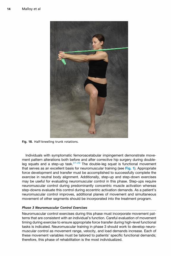
Fig. 18. Half-kneeling trunk rotations.
Malloy et al14
Individuals with symptomatic femoroacetabular impingement demonstrate move-ment pattern alterations both before and after corrective hip surgery during double-leg squats and a step-up task.27–29 The double-leg squat is functional movementthat serves as an excellent basis for neuromuscular training (see Fig. 1). Appropriateforce development and transfer must be accomplished to successfully complete theexercise in neutral body alignment. Additionally, step-up and step-down exercisesmay be useful for evaluating neuromuscular control in this phase. Step-ups requireneuromuscular control during predominantly concentric muscle activation whereasstep-downs evaluate this control during eccentric activation demands. As a patient’sneuromuscular control improves, additional planes of movement and simultaneousmovement of other segments should be incorporated into the treatment program.
Phase 3 Neuromuscular Control Exercises
Neuromuscular control exercises during this phase must incorporate movement pat-terns that are consistent with an individual’s function. Careful evaluation of movementtiming during exercise to ensure appropriate force transfer during high-level functionaltasks is indicated. Neuromuscular training in phase 3 should work to develop neuro-muscular control as movement range, velocity, and load demands increase. Each ofthese movement variables must be tailored to patients’ specific functional demands;therefore, this phase of rehabilitation is the most individualized.

Fig. 19. Manual resisted pelvic facilitation to promote pelvic and lower extremity flexionduring gait.
Rehabilitation After Hip Arthroscopy 15
Exercise progressions during this phase blend aspects of muscle performance andneuromuscular control. Many progressions involve maintaining stability of onesegment while another is moved. An example of this occurs during a side plank wherethe top leg can be flexed and extended to mimic a running type movement pattern. Awall lean with rapid high knee exercise can be used to sequence the rapid hip flexionneeded to run. This exercise can be progressed to a high-box step-up to increaseconcentric demand on the stance leg during the movement.Double-leg squat exercises can be progressed to incorporate simultaneous upper
extremity pressing to assist with force transfer between the upper body and lowerbody. Single-leg stance with rapid stepping with a resistance band around patients’ankles promotes both stability and neuromuscular control of each limb simulta-neously, which is often required in higher-level functions (Fig. 20). Diagonal choppingexercises should be performed to facilitate upper body control over a stable lower ex-tremity base.Initial plyometric movements can be initiated during the later parts of this phase.
Initially, small range-of-motion rapid movements, such as quick steps onto a box inthe forward and lateral directions are useful in preparing a patient for larger motionhopping or jumping exercises. Progression to modified broad jumps, lateral hops,and single-leg hops can be advanced as tolerated during this phase to assist in neuro-muscular control of both the concentric and eccentric activation phases of explosive

Fig. 20. (A) Resistance band standing and (B) stepping.
Malloy et al16
movement. Any increase or change in symptoms may indicate that the patient doesnot have the foundational muscle strength, endurance, or neuromuscular control forthe demand of the exercise; therefore, training should be modified until symptomssubside.If running is a goal after hip arthroscopy, appropriate running progression exercises
should be initiated during this phase.30 Patients must demonstrate an adequate de-gree of muscle strength, endurance, and activation patterning to prevent irritation sec-ondary to overuse. Previous investigators have recommended that patients passassessment that incorporates repeated double-leg squats, step-down, and manualhip abductor strength test prior to the initiation of a running program.2 Other aspectsof previously published return-to-sport tests, such as resisted single-leg squats andlateral agility, may be useful to evaluate sustained movement control and muscleendurance prior to initiating a return-to-running program.4,13 Patients should be moni-tored closely during a return-to-run progression and it is recommended that the move-ment variable of speed should be progressed last.30
Phase 4 Neuromuscular Control Exercises
Neuromuscular control exercises in this phase involve high demand training that mustfocus on speed, agility, power, and skill. The movement patterns are ones performedin sports and occupations that require a high degree of manual labor. Therefore, oftenan individual’s functional demands do not require progression into this final phase ofrehabilitation. Because the intensity of the activity performed in this phase increasesconsiderably, it is important to gradually introduce exercises. Initially, variables shouldbe manipulated one at a time to avoid soft tissue irritation secondary to overload.2
Functional testing may be useful during this phase to monitor progress to help deter-mine when an unrestricted return to high-level activity is appropriate.31
Sport-specific and high-demand activities require a great deal of control; therefore,all aspects of movement must be incorporated into a rehabilitation program.
Rehabilitation After Hip Arthroscopy 17
High-velocity and low-velocity movements under load should be advanced to developmuscle strength and power throughout the necessary range of motion.2,30 Olympic lift-ing exercises are useful to improve the rate of force development and movementsequencing for high-level activities. Plyometric exercises, such as countermovementjumps or box jumps, can assist with rate of force development and enhance the use ofthe stretch-shortening cycle.2,30 Agility exercises, such as cutting, sprinting, anddecelerating, should be progressed slowly to the level of sport-specific demand.13
Skills can be improved through repetition because this assists in patterning the neuro-muscular system to improve movement efficiency. The physician, physical therapists,athletic trainers, and coaches must clearly communicate as an athlete transitions topractice and competition. Previous investigators have advised incorporating restdays as athletes return to sports to prevent irritation or reinjury.2
SUMMARY
Adequate control of movement is essential for patients to return to unrestricted func-tion after hip arthroscopic surgery. Mobility, muscle performance and stability, andneuromuscular control are vital aspects commonly addressed in rehabilitation tohelp re-establish control of movement for function. Initial joint protection is a hallmarkfor all patients after hip arthroscopy to prevent intra-articular and extra-articular softtissue irritation. Initial mobility exercises should focus on restoration of motion, withthese exercises progressed to restore terminal ranges for patients’ desired function.Muscle performance and stability exercises should begin with submaximal muscle ac-tivations and be transitioned to exercises that involve cocontraction to promote stabil-ity for activity of daily living function. Muscle performance and stability exercisesshould be progressed to incorporate increasing loads to advance demand forhigher-level function. Initial neuromuscular control exercises initially should targetaberrant movements that may lead to undue stress on healing tissues. As a patientprogresses, neuromuscular control exercises are advanced to re-establish coordina-tion and timing of movement for higher-level functions. It is essential to tailor the ex-ercises of each phase to patients’ specific demands to prevent soft tissue injuryassociated with overuse or overload. Each phase of rehabilitation should be closelymonitored so that patients are not advanced too quickly, which can lead to setbacksand delays in return to normal function.
REFERENCES
1. Bozic KJ, Chan V, Valone FH, et al. Trends in hip arthroscopy utilization in theunited states. J Arthroplasty 2013;28(8):140–3.
2. Edelstein J, Ranawat A, Enseki KR, et al. Post-operative guidelines following hiparthroscopy. Curr Rev Musculoskelet Med 2012;5(1):15–23.
3. Malloy P, Malloy M, Draovitch P. Guidelines and pitfalls for the rehabilitationfollowing hip arthroscopy. Curr Rev Musculoskelet Med 2013;6(3):235–41.
4. Wahoff M, Dischiavi S, Hodge J, et al. Rehabilitation after labral repair and fem-oroacetabular decompression: criteria-based progression through the return tosport phase. Int J Sports Phys Ther 2014;9(6):813.
5. Cheatham SW, Kolber MJ. Rehabilitation after hip arthroscopy and labral repair ina high school football athlete. Int J Sports Phys Ther 2012;7(2):173–84.
6. Cheatham SW, Enseki KR, Kolber MJ. Post-operative rehabilitation after hiparthroscopy: a search for the evidence. J Sport Rehabil 2014;24(4):413–8.

Malloy et al18
7. Cheatham SW, Kolber MJ. Rehabilitation after hip arthroscopy and labral repair ina high school football athlete: a 3.6 year follow-up with insight into potential riskfactors. Int J Sports Phys Ther 2015;10(4):530.
8. Grzybowski JS, Malloy P, Stegemann C, et al. Rehabilitation following hiparthroscopy-A systematic review. Front Surg 2015;2:21.
9. LeBeau RT, Nho SJ. The use of manual therapy Post–Hip arthroscopy when anexercise-based therapy approach has failed: a case report. J Orthop SportsPhys Ther 2014;44(9):712–21.
10. Enseki KR, Kohlrieser D. Rehabilitation following hip arthroscopy: an evolvingprocess. Int J Sports Phys Ther 2014;9(6):765.
11. Spencer-Gardner L, Eischen JJ, Levy BA, et al. A comprehensive five-phaserehabilitation programme after hip arthroscopy for femoroacetabular impinge-ment. Knee Surg Sports Traumatol Arthrosc 2014;22(4):848–59.
12. Enseki KR, Martin RL, Draovitch P, et al. The hip joint: arthroscopic proceduresand postoperative rehabilitation. J Orthop Sports Phys Ther 2006;36(7):516–25.
13. Wahoff M, Ryan M. Rehabilitation after hip femoroacetabular impingementarthroscopy. Clin Sports Med 2011;30(2):463–82.
14. Buckwalter JA, Brown TD. Joint injury, repair, and remodeling: roles in post-traumatic osteoarthritis. Clin Orthop 2004;423:7–16.
15. Neumann DA. Biomechanical analysis of selected principles of hip joint protec-tion. Arthritis Rheum 1989;2(4):146–55.
16. Philippon MJ, Ejnisman L, Ellis HB, et al. Outcomes 2 to 5 years following hiparthroscopy for femoroacetabular impingement in the patient aged 11 to 16years. Arthroscopy 2012;28(9):1255–61.
17. Byrd JT, Jones KS. Adhesive capsulitis of the hip. Arthroscopy 2006;22(1):89–94.
18. Willimon SC, Briggs KK, Philippon MJ. Intra-articular adhesions following hiparthroscopy: a risk factor analysis. Knee Surg Sports Traumatol Arthrosc 2014;22(4):822–5.
19. Freeman S, Mascia A, McGill S. Arthrogenic neuromusculature inhibition: a foun-dational investigation of existence in the hip joint. Clin Biomech 2013;28(2):171–7.
20. Philippon MJ, Decker MJ, Giphart JE, et al. Rehabilitation exercise progressionfor the gluteus medius muscle with consideration for iliopsoas tendinitis: anin vivo electromyography study. Am J Sports Med 2011;39(8):1777–85.
21. Lewis CL, Sahrmann SA, Moran DW. Anterior hip joint force increases with hipextension, decreased gluteal force, or decreased iliopsoas force. J Biomech2007;40(16):3725–31.
22. Anderson FC, Pandy MG. Individual muscle contributions to support in normalwalking. Gait Posture 2003;17(2):159–69.
23. van der Krogt MM, Delp SL, Schwartz MH. How robust is human gait to muscleweakness? Gait Posture 2012;36(1):113–9.
24. Ekstrom RA, Donatelli RA, Carp KC. Electromyographic analysis of core trunk,hip, and thigh muscles during 9 rehabilitation exercises. J Orthop Sports PhysTher 2007;37(12):754–62.
25. Escamilla RF, Lewis C, Bell D, et al. Core muscle activation during swiss ball andtraditional abdominal exercises. J Orthop Sports Phys Ther 2010;40(5):265–76.
26. Knuttgen HG, Kraemer WJ. Terminology and measurement in exercise perfor-mance. J Strength Cond Res 1987;1(1):1–10.
27. Lamontagne M, Kennedy MJ, Beaule PE. The effect of cam FAI on hip and pelvicmotion during maximum squat. Clin Orthop 2009;467(3):645–50.

Rehabilitation After Hip Arthroscopy 19
28. Lamontagne M, Brisson N, Kennedy MJ, et al. Preoperative and postoperativelower-extremity joint and pelvic kinematics during maximal squatting of patientswith cam femoro-acetabular impingement. J Bone Joint Surg Am 2011;93(Suppl 2):40–5.
29. Rylander J, Shu B, Favre J, et al. Functional testing provides unique insights intothe pathomechanics of femoroacetabular impingement and an objective basis forevaluating treatment outcome. J Orthop Res 2013;31(9):1461–8.
30. Draovitch P, Maschi RA, Hettler J. Return to sport following hip injury. Curr RevMusculoskelet Med 2012;5(1):9–14.
31. Manske R, Reiman M. Functional performance testing for power and return tosports. Sports Health 2013;5(3):244–50.